Review

Exploiting the Indole Scaffold to Design Compounds Binding to Different Pharmacological Targets †

1
Department of Pharmacy, University of Pisa, Via Bonanno Pisano, 6, 56126 Pisa, Italy
2
Department of Pharmacy, University of Naples “Federico II”, Via D. Montesano, 49, 80131 Naples, Italy
*
Authors to whom correspondence should be addressed.
The authors wish to dedicate this review to the memory of Professor Barbara Cosimelli, our dear friend and colleague, who was a key participant in the research discussed herein.
Molecules 2020, 25(10), 2331; https://doi.org/10.3390/molecules25102331
Submission received: 20 April 2020 / Revised: 14 May 2020 / Accepted: 15 May 2020 / Published: 16 May 2020
(This article belongs to the Special Issue Indole and Its Bioisosteric Replacements in Medicinal Chemistry)

Abstract

:
Several indole derivatives have been disclosed by our research groups that have been collaborating for nearly 25 years. The results of our investigations led to a variety of molecules binding selectively to different pharmacological targets, specifically the type A γ-aminobutyric acid (GABAA) chloride channel, the translocator protein (TSPO), the murine double minute 2 (MDM2) protein, the A2B adenosine receptor (A2B AR) and the Kelch-like ECH-associated protein 1 (Keap1). Herein, we describe how these works were conceived and carried out thanks to the versatility of indole nucleus to be exploited in the design and synthesis of drug-like molecules.

1. Introduction

During the last 25 years our research groups have been engaged in preparing and testing several indole derivatives as ligands for some pharmacologically relevant targets: namely, the type A γ-aminobutyric acid chloride channel, the translocator protein, the murine double minute 2 protein, the A2B adenosine receptor and the Kelch-like ECH-associated protein 1. We chose indole as a privileged scaffold owing to its recognized ability of being exploited to obtain drug-like molecules [1]. In virtue of its chemical reactivity, this heterocycle is amenable to be readily modified in order to introduce multiple decorations, so to obtain a multitude of indole-based compounds. Indole is widely distributed among biologically active molecules, either natural (many alkaloids, tryptophan, plant hormones) and synthetic, acting on a huge number of therapeutic targets [1]. The present review summarizes our studies taking indole as the polar star of our medicinal chemistry strategies.

2. Indole Derivatives as Ligands of the Benzodiazepine Receptor
Many drugs structurally related to diazepam bind to a site known for a long time as the benzodiazepine receptor (BzR). Although this term was changed in 1998 to “benzodiazepine binding site” [2] the acronym BzR has been widely used for decades and it is still usually employed by the scientific community. This binding site is located within the transmembrane type A γ-aminobutyric acid (GABAA) chloride channel, one of the most important members of the family of pentameric ligand-gated ion channels [2]. When this channel is activated by interaction with the neurotransmitter GABA, the flow of chloride into the cell increases and produces hyperpolarisation. Mammals express a number of GABAA isoforms localized in the CNS which are composed by five subunits: two β3, one γ2 and two α among six types (α1, α2, α3, α4, α5, α6). Such a pentameric organization (Figure 1) originates the following six GABAA subtypes: α1β3γ2, α2β3γ2, α3β3γ2, α4β3γ2, α5β3γ2 and α6β3γ2 [3,4,5]. The above subtypes can be also named by specifying the α subunit which imparts specific physiological and pharmacological properties to the GABAA complex [6,7,8,9,10]. The α1 subtype, the dominant one, is present in both the cortex and cerebellum and mediates sedation; the α2 and α3 subtypes are found mainly in the cortex and the hippocampus and are involved in anxiolytic and myorelaxant effects; the α5 subtype is largely expressed in the hippocampus and is associated with cognition processes like learning and memorising; the α4 and α6 subtypes, less investigated, are known as benzodiazepine-insensitive binding sites because they do not bind diazepam but recognize several non-benzodiazepine ligands.
Following the introduction in the clinical usage of chlordiazepoxide in 1960, a huge number of chemically heterogeneous classes of compounds have been reported in literature as BzR ligands [11]. This binding site is located at the α/γ subunits’ interface, distinct from the GABA binding sites situated at α/β interfaces (Figure 1).
The pharmacological actions of BzR ligands range from full agonism (associated with anxiolytic, anticonvulsant, sedative-hypnotic, and myorelaxant effects) to antagonism (implying the ability to reverse sedation caused by agonists) and to inverse agonism (characterized by anxiogenic, somnolytic and proconvulsant effects) [12,13].
In 1987 Schofield and coworkers reported the sequence and the functional expression of the GABAA channel [14]. Subsequently to this pioneering work, further knowledge about the subunits composing the GABAA isoforms was achieved [15,16,17]. Since 2000, molecular genetics experiments and measurements of binding and efficacy of BzR ligands at each of the GABAA isoforms [18,19,20,21] paved the way for the search of compounds provided by affinity- and/or efficacy-based selectivity for α1, α23, α5 BzR subtypes [22].
Currently, the many BzR ligands available as drugs are agonists endowed with the above- mentioned depressant effects (ascribed to their action on the α1 subtype) and the antagonist flumazenil, employed as antidote to treat benzodiazepine overdose. Given the therapeutic potential of BzR ligands, this class of compounds has been one of the most intriguing and challenging field of medicinal chemistry research.
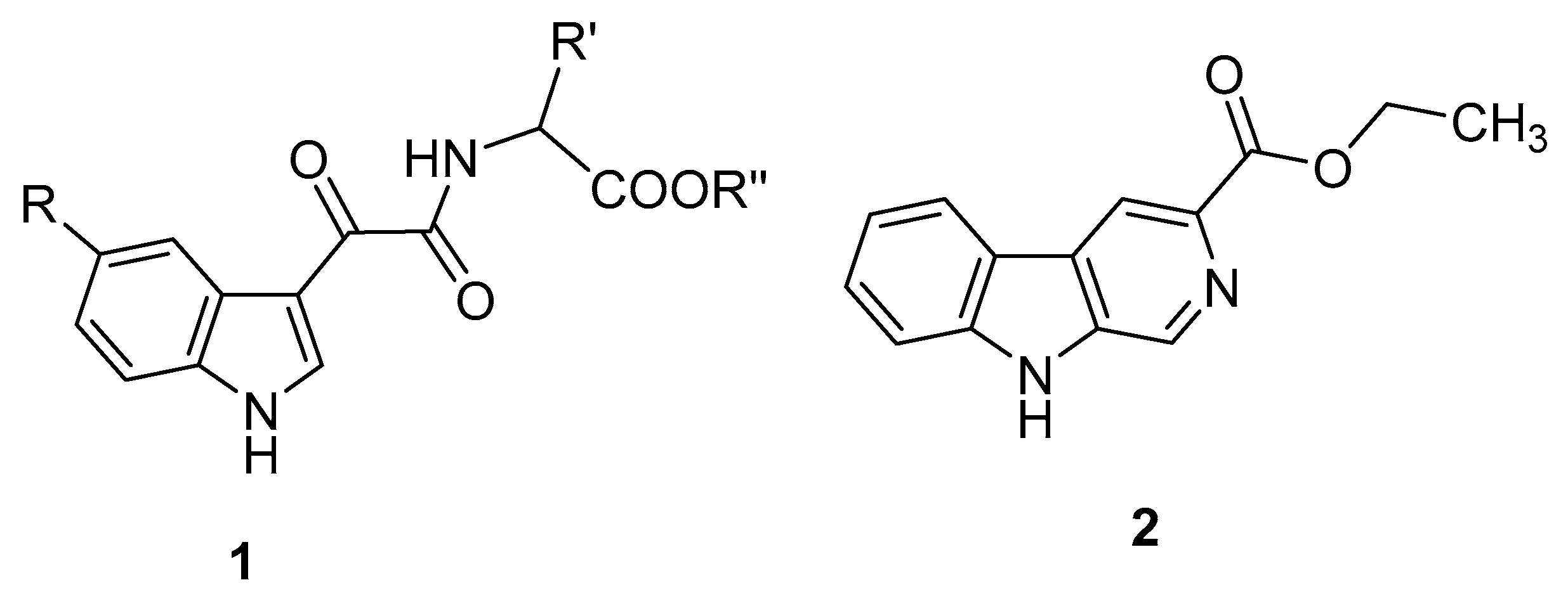
Our studies of indole derivatives as BzR ligands began in 1985 with the synthesis and the evaluation of the binding affinities of racemic N-(indol-3-ylglyoxylyl)amino acid derivatives (1) [23]. Compounds of series 1 were designed taking the 3-ethoxycarbonyl-β-carboline (2), reported to bind with nanomolar affinity to the BzR [24], as a reference structure.
Molecules 25 02331 i001
In series 1, the groups at the 5 position of the indole nucleus (R) were H, Cl, Br, OCH3, NO2, while the R’ substituents of the aminoacid moiety were hydrogen, alkyl, benzyl and indolylmethyl, some of which bearing small groups on their benzene rings. R’’ was a hydrogen in the subset of carboxylic acids and a methyl or an ethyl in the subset of esters.
At that time, the endpoint in our biological experiments was the ability of the tested compound to displace a radioligand (generally [3H]flunitrazepam) from neuronal membranes obtained by the cortex of bovine brain. In the light of the knowledge about the GABAA isoforms acquired several years after our initial studies, such binding data correlated mainly with the α1 BzR subtype.
Most of the compounds of series 1 exhibited affinity values in the micromolar-submillimolar range. The esters were more potent than the corresponding acids. In the ester subset two compounds were endowed by submicromolar affinity, both bearing a methyl group as R’, while the R substituent was a chlorine or a nitro group.
In a subsequent paper [25], we described the synthesis and the binding affinity for BzR of optically active forms of some N-(indol-3-ylglyoxylyl)aminoacids of series 1. Expectedly, the esters performed better than the corresponding acids. The two compounds provided with the highest affinity as racemic mixtures in the previous work [23] did not show appreciable differences in affinity when tested as pure optical isomers, thus suggesting that the α-methyl side chain does not occupies a sterically hindered cleft within the BzR.
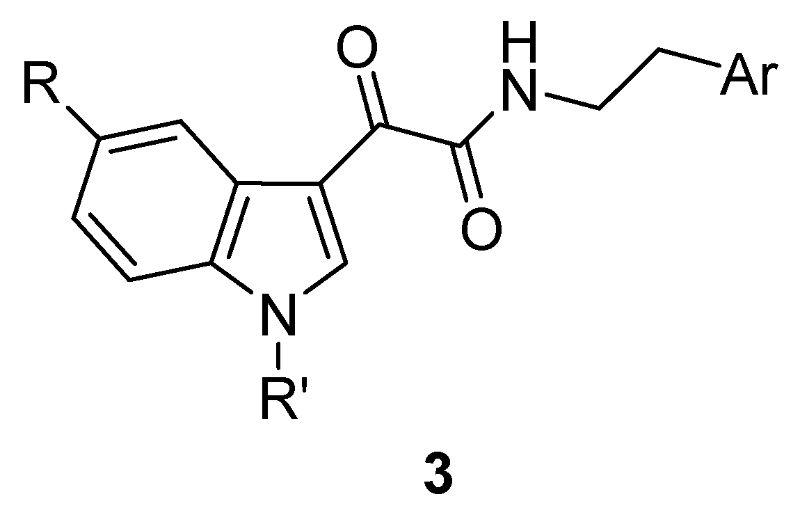
In 1992 we reported a series of N-(indol-3-ylglyoxylyl)-β-arylethylamines (3) as BzR ligands [26]. In this series, R was H, Cl, Br, NO2; R’ was H or CH3; the β-arylethyl side chain derived from tryptamine, tyramine, dopamine, α-phenylethlylamins bearing various substituents on the phenyl ring.
Molecules 25 02331 i002
All the 1-methyl derivatives (R’ = CH3) were inactive, suggesting that the NH of the indole nucleus is engaged in a H-bond with the BzR or, alternatively, is sterically repelled by the binding site. The best ligands of the new series showed Ki values of 85 nM (R = H, R’ = H, Ar = m-methoxyphenyl) and 90 nM (R = H, R’ = H, Ar = p-methoxyphenyl). Surprisingly, when the hydrogen at the 5-position of the indole scaffold of the two above compounds was replaced by a nitro group affinity decreased to a considerable exent (Ki > 10 μM). The remaining compounds showed Ki values in the micromolar range.
In order to highlight the role of the indole NH fragment in the interaction with the BzR, we prepared a number of benzofurane and benzothiophene derivatives of general formula 4 and, respectively, 5 in which the above fragment was replaced by an oxygen or, respectively, a sulphur [27].
Molecules 25 02331 i003
The new compounds were much worse in terms of potency compared with the corresponding isosteric indoles reported in our previous papers [23,26], clearly indicating that the indole NH donates a H-bond to an acceptor heteroatom of the BzR.
With the aim of finding indole derivatives with improved affinity for the BzR, three subsets of indolylglyoxylamides (general formulae 6, 7 and 8) were shortly after synthesized and tested [28]. In the three subsets R = H, Cl, NO2 while R’ = H, p-F, p-Cl, p-OCH3, m,p-(OCH3)2.
Molecules 25 02331 i004
While the anilides 7 and the γ-phenylpropylamides 8 showed poor affinity, several benzylamides 6 displayed nanomolar potency. The scarce performance of indoles 7 and 8 was ascribed to steric repulsive interactions taking place between their side chain phenyl rings and the boundaries of the BzR. The excellent binding data of some benzylamides 6 suggested that their phenyl ring was correctly oriented to find room into a hydrophobic region of the BzR. The best performing ligand of series 6 (Ki 11 nM) had R = NO2 and R’ = m,p-(OCH3)2. The structure-affinity relationships in series 6 were characterized by interdependent effects of the R and R’ substituents on potency. Particularly, affinity of the 5-Cl/NO2 derivatives was improved by hydroxyl/methoxy substituents on the side chain phenyl ring, while affinity of the 5-H derivatives was increased by halogens on the same phenyl ring. Actually, such a pattern of interdependent effects of the substituents at the 5-position of the indole scaffold had already been observed in series 3.
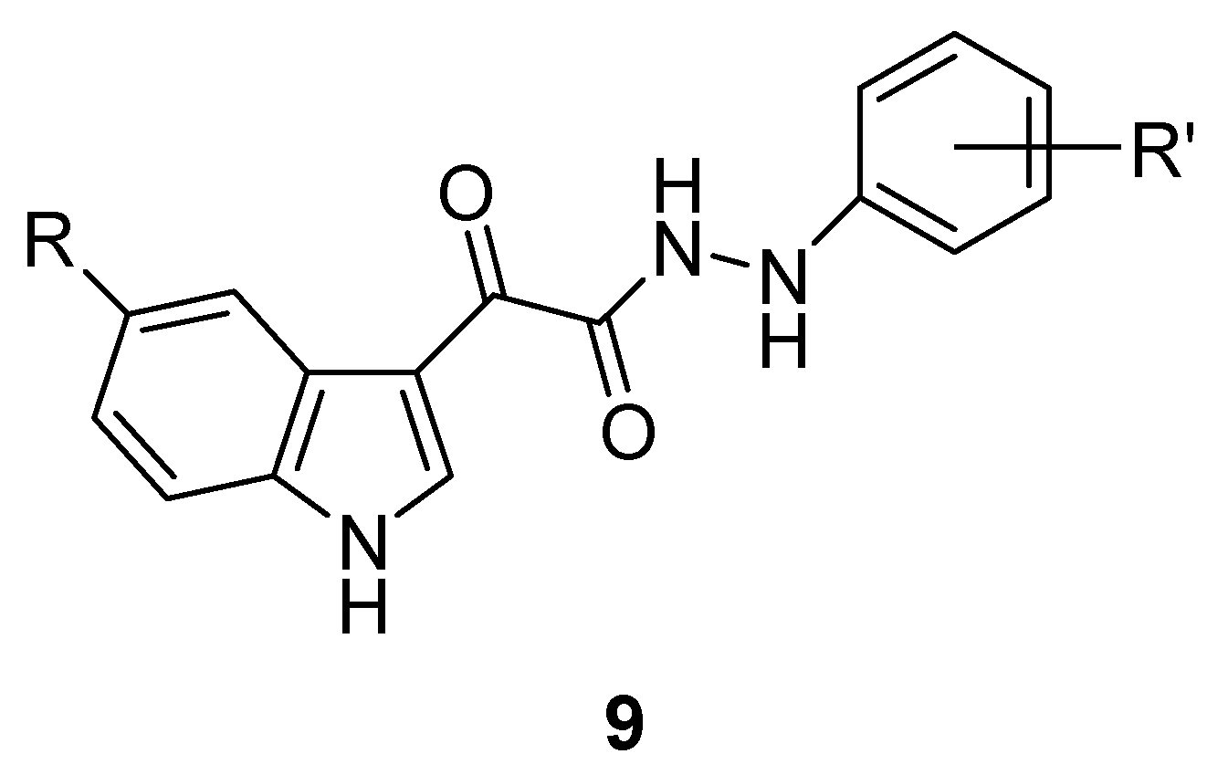
In order to identify indole derivatives as BzR ligands characterized by good water solubility, we prepared a number of N’-phenylindol-3-ylglyoxylhydrazides (9) [29]. In this series we inserted the following substituents on the parent structure: R = H, Cl, NO2 and R’ = H, p-F, p-Cl, p-NO2, m- or p-CH3, p-OH, p-OCH3.
Molecules 25 02331 i005
Affinity of compounds 9 was restricted to the 5-H derivatives, whereas the 5-Cl/NO2 derivatives were all devoid of affinity. Again, the structure-affinity relationships of these two subsets of compounds were divergent. In an attempt to explain the reasons underlying such findings, we searched the Cambridge Structural Database [30] looking for differences in the conformational properties of N’-arylhydrazides and N-arylamides. We realized that benzylamides 6 can adopt a transoid conformation about the C-N-C-Ar torsion angle (values comprised between −150° and +150°). Such a staggered conformation is forbidden to hydrazides 9 which are forced into a gauche disposition about the corresponding C-N-N-Ar torsion angle (values comprised between −60° and −120° or between +60° and +120°).
The above data led us to hypothesize that our indole derivatives might bind to BzR by adopting one out of two conformations and orientations within the binding cavity depending on the substituent at the 5-position of the indole nucleus. On the basis of this conjecture, we began to consider the 5-Cl/NO2 and the 5-H indole derivatives as different “families” of BzR ligands, each displaying peculiar structure-affinity relationships. In our speculations we were aided by the pharmacophore/topological model proposed by Cook and coworkers [31] defined by the same authors as “comprehensive” because it holds for agonists, antagonists and inverse agonists. This model includes the following interaction sites: (i) a H-bond acceptor (A2), (ii) a H-bond donor (H1), (iii) a bifunctional H-bond donor/acceptor (H2/A3), and (iv) four lipophilic pockets (L1, L2, L3, and LDi). The boundaries of the binding site are defined in terms of sterically forbidden sites (S1, S2, and S3). Regarding the efficacy profile, the only safe statement was that filling of the L3 pocket (occupied by the diazepam pendant phenyl ring) was mandatory for agonism. The two putative binding modes (named A and B) of our indole derivatives are depicted in Figure 2 in the framework of Cook’s model. The binding mode A of the 5-Cl/NO2 indoles requires a staggered conformation of the side chain and gives rise to the following interactions: (i) the indole NH is H-bond to the A2 site; (ii) the C=O1 and C=O2 are H-bound to the H2 and H1 sites, respectively; (iii) the CH2, the phenyl and the fused benzene ring fill the L1, L2, and LDi pockets, respectively. The binding mode B is accessible only to 5-H indoles because the sterically forbidden S2 site, closely facing the 5-position of the indole nucleus, cannot host substituents larger than a hydrogen. Such a binding mode, compatible with a folded conformation of the side chain, is characterized by the following interactions: (i) C=O1 and C=O2 are H-bound to the H2 and H1 sites, respectively; (ii) the lipophilic L1 and L2 pockets are filled by the pyrrole and, respectively, the benzene moieties of indole; (iii) the indole NH donates a H-bond to a heteroatom belonging to the S1 site. It is worth noticing that each of the two postulated binding modes benefits from three H-bonds with the BzR, consistently with the similar affinities displayed by the best performing 5-Cl/NO2 and 5-H indoles derivatives [29].
Some years later, we prepared and tested several N-(indol-3-ylglyoxyl)arylalkylamides 1015 characterized by conformationally or geometrically constrained side chains, most of which featured a chiral center [32]. We reasoned that such properties of the new side chains might increase the chances of conferring affinity-based selectivity for any of the BzR subtypes.
Molecules 25 02331 i006
In series 1015 the R substituents were H, Cl or NO2. In series 10 and 11 the R’ substituents on the phenyl ring were H, p-CH3, p-OCH3, m,p-(OCH3)2 and p-NO2. All of the S isomers (11 and 15) lacked affinity, likewise the optically inactive compounds 13. Some of the R isomers displayed Ki values in the micromolar/nanomolar range. The structure-affinity relationships of the R isomers confirmed our hypothesis about the different binding modes of the 5-Cl/NO2 derivatives and of the 5-H derivatives. Specifically, in series 10 and 14 the 5-Cl/NO2 were significantly more potent than the 5-H derivatives. Conversely, in series 12 affinity was appreciable (Ki 123 nM) only if R = H. The most potent compound described in this study belonged to series 10 (R = NO2, R’ = H, Ki 17 nM). An overlay of molecular models of a few indole derivatives representative of the four series investigated showed that the inactive ones projected portions of their side chains into the sterically forbidden S1 subsite of Cook’s model. A subset of compounds were tested for their ability to displace [3H]flumazenil from recombinant rat α1, α3 and α5 BzR subtypes. All of them displayed high affinity for α1β3γ2 receptors and moderate to good selectivity for α3 and α5 subtypes.
A number of N-(heteroarylmethyl)indol-3-ylglyoxylamides 16 were subsequently investigated [33] to probe the H-bonding properties previously ascribed to the S1 subsite of the BzR [29] (see binding mode B in Figure 2). The new compounds had R = H or NO2 at the 5-position of the indole moiety and several heterocycles on the side chain (Het).
Molecules 25 02331 i007
The 5-NO2 derivatives bearing a 2-pyrrolyl, a 2-furyl, a 4-methyl-2-furyl, a 3-pyrrolyl or a 3-furyl ring in the side chain exhibited affinities in the nanomolar range (Ki values comprised between 13 nM and 33 nM) comparable with the most active compounds in the benzylamide series 6. All the 5-NO2 derivatives bearing in the side chain a 2-thienyl, a 1-methyl-2-pyrrolyl, a 2-indolyl or a 2-imidazolyl were scarcely potent (Ki values in the micromolar range) or practically devoid of affinity. These data were explained as follows: the side chains of the most potent 5-NO2 indoles make H-bonds at the S1 site (Figure 3); the less potent or inactive NO2 indoles cannot make the same H-bonds if the heterocycle lacks a H-bond donor fragment (2-thienyl, 1-methy-2-pyrrolyl), is exceedingly bulky (2-indolyl) or is hydrophilic (2-imidazolyl).
All the 5-H derivatives of series 16 showed significant lower affinities (Ki in the submicromolar-micromolar range), probably because they are not able to engage H-bonds with their side chains in the binding mode B.
Four of the most potent compounds of series 16 were tested for their ability to displace [3H]flumazenil from recombinant rat α1, α2 and α5 GABAA/BzR pure isoforms. All of them showed binding selectivity for the α1 subtype over the α2 and α5 subtypes.
In continuing our research of indole derivatives endowed with selective affinity for BzR subtypes, we came back to indol-3-ylglyoxylamides of series 6 by inserting lipophilic substituents at the 4′-position of the side phenyl ring, some of which were characterized by considerable steric bulk (i.e., Br, CH3, C2H5, C≡CH, C≡C-CH3, C≡CSi(CH3)3 and C≡C-CH2Si(CH3)3) [34]. In addition to the novel benzylamide derivatives, in the same paper we also disclosed indole derivatives in which the benzyl moiety was replaced by alkyl groups (i.e., (CH2)3CH3, (CH2)4CH3, CH(CH3)2, CH(CH3)CH2CH3, C(CH3)3 and CH2CH(CH3)2). Unfortunately, the above structural changes did not improve affinity for the BzR. One of the new compounds (R = NO2 and R’ = p-CH3) exhibited nanomolar potency at the rat recombinant α1 BzR subtype (Ki 31 nM) with no appreciable affinity for the α2 and the α5 subtypes, displaying a full agonist efficacy profile and a zolpidem-like sedative-hypnotic activity in vivo.
According to the Cook’s research group, the shapes of the BzR subtypes are very similar, with the exception of α1 and α5 subtypes that seem to be slightly larger at the lipophilic pockets, called LDi and L2 regions, respectively [35]. The above steric differences have been exploited to obtain ligands that bind selectively to either α1 or α5 subtypes [22,36], but have also hampered the identification of affinity-based α2 and/or α3 selective ligands [37]. Investigators realized that a more fruitful strategy to obtain non-sedating anxiolytic agents or non-sedating cognition enhancers would be identifying compounds binding to all four subtypes, but preferentially activating only the targeted subtypes. The search of efficacy-based α2 and/or α3 selective ligands has indeed yielded better results [38]. Following this approach, we selected some of our indole derivatives of series 6, 10 and 14 and tested their affinity and efficacy for the rat recombinant α1, α2 and α5 subtypes [39]. Efficacy was evaluated by measuring the 36Cl uptake in transfected human embryonic kidney cells stably expressing the three rat subtypes upon treatment with the tested compound. The results of this work led to the identification of two N-(indol-3-ylglyoxyl)benzylamides showing α2 selective efficacy in vitro and anxioselective effects in vivo according to the mouse light/dark box test [40], namely one from series 6 (R = H and R’ = p-F) and one from series 10 (R = NO2 and R’ = p-CH3). Docking calculations of the two anxioselective indole derivatives, using a homology-built model of α1 BzR provided by Cromer Bet al. [41], were carried out by means of 10 ns molecular dynamics. Although homology building approaches should regard with cautions, our in silico simulations showed that the selected 5-NO2 and the 5-H indole derivatives adopt two different orientations and conformations within the BzR, corresponding to mode A and, respectively, to mode B conjectured in our previous papers [29,32,33,34]. Most of the interactions (H-bonds, lipophilic pockets, sterically forbidden regions) observed in the docking complexes were in a reasonable agreement with those hypothesized using the Cook’s pharmacophore/topological model (Figure 2).

3. Indole Derivatives as Ligands of the Translocator Protein
In 1977 Braestrup and Squires discovered an alternative binding site for diazepam in rat kidneys and called it peripheral benzodiazepine receptor (PBR) to distinguish it from the BzR of the GABAA ion channel [42]. Subsequently, wide studies on this protein allowed a deeper knowledge about its structure, tissue distribution and subcellular localization as well as physio-pathological functions. In virtue of these new findings, in 2006 Papadopoulos and collaborators proposed a new name for PBR, that is translocator protein (TSPO) [43].
TSPO is a transmembrane protein distributed in many tissues, mainly in those involved in steroid biosynthesis, including kidney, testis, liver and lung; in the CNS, it is mainly expressed in glial cells and, at lower levels, in neurons [43]. TSPO is located at the contact site between the inner and outer mitochondrial membrane, in strict association with other proteins that make up the mitochondrial permeability transition pore (MPTP): the voltage dependent anion channel, the adenine nucleotide transporter and the steroidogenesis regulatory protein. TSPO is involved in many biological processes, including mitochondrial respiration, cell proliferation and differentiation, induction of apoptosis. Moreover, TSPO plays a crucial role in steroid biosynthesis [44,45,46], specifically in the translocation of cholesterol from cytoplasm to inner mitochondria, which represents the rate-limiting step of steroidogenesis. As next step, cholesterol is converted by cytochrome P450 side chain cleavage (P450scc) into pregnenolone, the precursor of all steroid hormones [44,45,46].
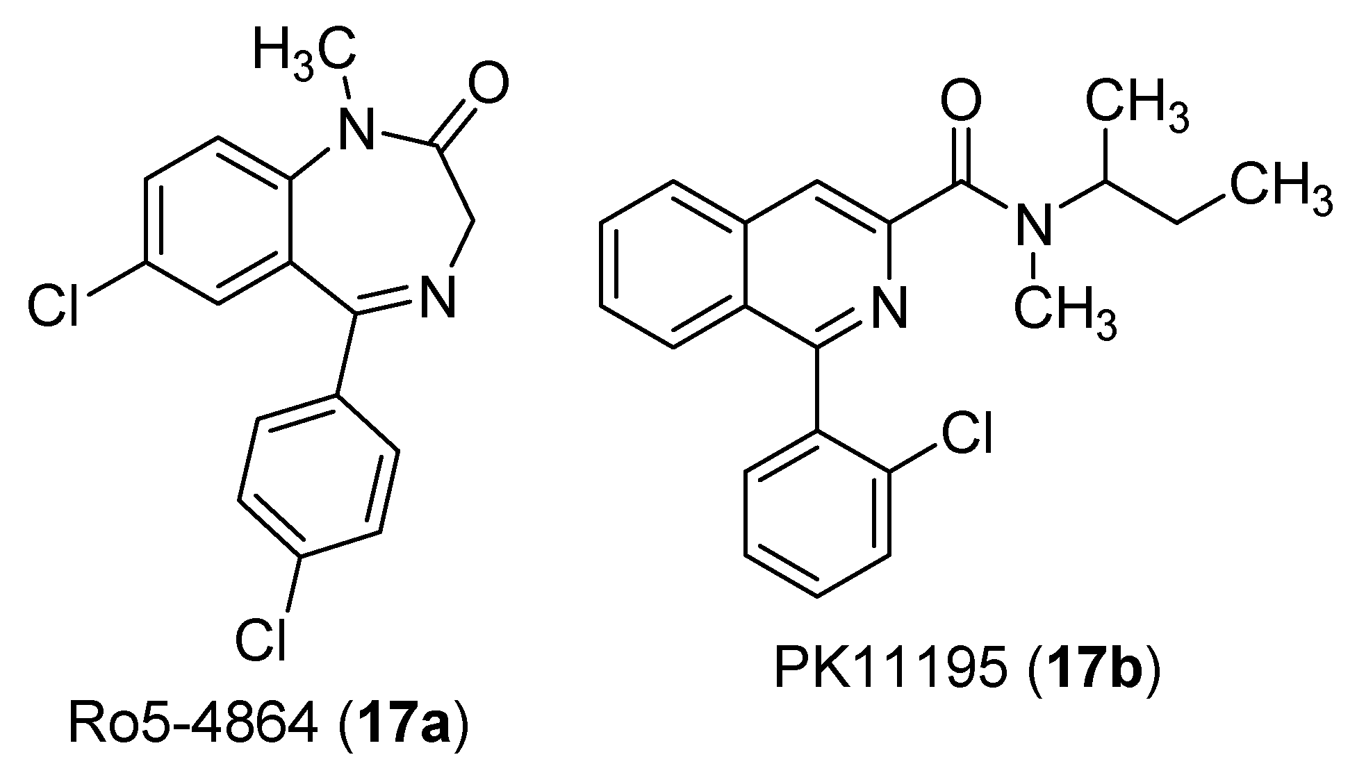
The basal expression of TSPO is altered in different pathological conditions: an up-regulation occurs in brain injury and pathologies involving neuroinflammation (e.g., neurodegenerative diseases, gliomas) and in certain tumors; a down-regulation is often observed in correlation with anxiety disorders and post-traumatic stress [47,48,49]. In addition to cholesterol [50], TSPO binds with high affinity to structurally different synthetic compounds [51], such as Ro5−4864 (17a) [52] and PK11195 (17b) [53], both widely employed as reference ligands.
Molecules 25 02331 i008
TSPO ligands may act as positive allosteric modulators of their target protein. Consequently, such ligands promote pregnenolone formation and increase the levels of endogenous steroids which, in turn, produce several physiological effects and result beneficial in pathological conditions of the CNS including inflammatory, neurological and psychiatric disorders [48,54]. In the last years, TSPO ligands are emerging as promising anxiolytics with favorable safety and tolerability and limited unwanted effects, like sedation, with respect to benzodiazepines [55,56,57]. Such pharmacological properties are ascribed to their ability to stimulate the synthesis of neurosteroids in the CNS, such as pregnenolone and allopregnanolone, which act as positive allosteric modulators of the GABAA chloride channel [55,56,57]. Examples of these non-sedating anxiolytic agents are emapunil (17c) [58] and etifoxine (17d) [59], the latter dually binding to TSPO and to a site on the β-subunit of the GABAA channel.
Molecules 25 02331 i009
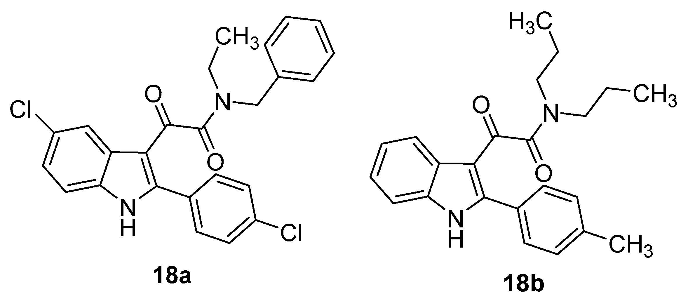
In 2004 and 2008 our research groups described the synthesis and the biological evaluation of a series of N,N-dialkyl-2-phenylindol-3-ylglyoxylamides (PIGAs, 18) [60,61] designed as conformationally constrained analogues of 2-arylindoleacetamides 19 reported by Kozikowsky et al. as TSPO ligands [62]. In the general formula 18, R1 and R2 were symmetric or asymmetric alkyl (linear and branched) or benzyl chains, R3 = H, CH3, F, Cl, NO2, CF3; R4 = H, OCH3, F, Cl, NO2; R5 = H, CH3, Cl.
Molecules 25 02331 i010
Most of the PIGAs showed affinity values for TSPO in the low nanomolar/subnanomolar range and full selectivity over the BzR. Noticeably, the most performant compounds exhibited a gain in affinity of one order of magnitude with respect to the indoleacetamide counterparts 19. The structure-affinity relationships within this class were rationalized by means of a pharmacophore/topological model made by three lipophilic pockets (L1, L3 and L4, hosting the 2-susbtituted phenyl group and, respectively, the substituents R1, and R2 on the amide nitrogen) and a H-bond donor group H1 interacting with the amide carbonyl oxygen (Figure 4).
PIGA ligands were further characterized for their efficacy measured as ability to improve the synthesis of pregnenolone in rat C6 glioma cells. A considerable number of them resulted stimulators of steroid biosynthesis more active than PK11195 and Ro5-4864 [60,61].
The best-performing PIGAs, in terms of affinity for TSPO and pregnenolone production, were evaluated in rats for their anxiolytic properties by means of the elevated plus-maze test [61,63,64]. In this assay, compounds 18a,b exhibited non-sedating anxiolytic properties. Results from investigations about the mechanism of their anxiolytic activity indicated that it involves the stimulation of endogenous neurosteroid production, which in turn determines a positive modulation of the GABAA chloride channel permeability [63].
Molecules 25 02331 i011
More recently, we investigated two novel series of PIGAs 18 featuring polar R3 groups (OH, NH2, COOH) on the 2-phenyl moiety or different 2-aryl substituents (Ar = 3-thienyl, p-biphenyl, 2-naphthyl) [65]. The 2-naphthyl derivatives exhibited the highest affinity values, thus confirming the crucial role of the ligand-receptor interaction involving the L1 pocket. In the same paper we reported a docking model of interaction between TSPO and three selected PIGAs based on a 3D structure of the target protein complexed to PK11195 [66,67]. This model was fairly consistent with the pharmacophore/topological scheme depicted in Figure 4, with the exception of the H1 site.
In two subsequent studies, a set of highly steroidogenic PIGA ligands demonstrated promising pharmacological activities [68,69]. Particularly, a number of these compounds were found to promote the oxidative metabolism of human astrocytes and prevent the oxidative damage and the inflammatory response in C6 glioma cells. The observed effects were completely counteracted by the co-treatment with D,L-aminoglutethimide, an inhibitor of P450scc involved in steroid biosynthesis, supporting the hypothesis that the PIGA-mediated protective mechanisms are mainly related to steroid production [68,69]. According to these results, PIGAs can be regarded as potential new therapeutic tools for the treatment of inflammatory-based neurodegenerative diseases characterized by astrocyte loss [68,69].
TSPO has been reported as a marker to reveal the onset of diseases related to its expression [47,49]. In virtue of the high affinity and selectivity of PIGAs for TSPO, we exploited the 2-phenylindol-3-ylglyoxylamide scaffold to develop specific TSPO molecular probes. In detail, reversible and irreversible fluorescent probes featuring the 7-nitrobenz-2-oxa-1,3-diazol-4-yl group (18c,d) were synthesized, characterized for their optical properties and tested in spectroscopy experiments to evaluate their ability to specifically label the mitochondrial localization of TSPO in Drosophila S2, rat C6 and human U87MG glioma cells [70,71,72]. These molecular probes emerged as useful tools to study the physiological role and the expressions levels of TSPO, especially the irreversible probes, whose lasting signal is maintained even after multiple washes, allowing a detection that is less affected by unspecific signal [71,72].
Molecules 25 02331 i012
Shortly after, we synthesized N,N-di-n-propyl-(N1-[11C]methyl-2-(4′-nitrophenyl)indol-3-yl) glyoxylamide (18e) as a high affinity radiolabelled probe of TSPO [73]. The corresponding unlabeled compound was selected from a small library of PIGAs thanks to its optimal combination of high TSPO binding affinity and moderate lipophilicity (calculated logP = 3.9) to ensure adequate brain entry and low non-specific binding [73]. Compound 18e was evaluated with positron emission tomography in monkey after being administered by intravenous injection. This probe readily entered monkey brain and gave a high proportion of specific and reversible TSPO binding, auguring well for its future application in humans [73].
Molecules 25 02331 i013
In the last years, our attention has focused on the lack of correlation between binding affinity and steroidogenic efficacy of TSPO ligands [74]. This represents a problem affecting the identification of effective lead compounds by a traditional affinity-based drug discovery strategy as well as the interpretation of pharmacological data [75]. Our efforts took advantage from studies showing that the biological effectiveness of a certain molecule cannot directly be deduced by its affinity for the target, but it may rather be related to the period for which it interacts with its target, defined as “residence time” (RT) [75]. This kinetic parameter corresponds to the reciprocal of the dissociation rate (Koff) of the ligand-target complex. Based on these findings, we recently investigated whether RT could be employed to estimate the steroidogenic efficacy of a TSPO ligand. For this purpose, we selected a set of representative PIGAs showing different combinations of TSPO affinity and steroidogenic properties in vitro [76]. Then, a kinetic radioligand binding assay was set up with rat kidney membrane homogenates and validated for determination of RT values using [3H]PK11195. Our experiments showed a direct correlation between the efficacy of TSPO ligands and their RT values [76,77]. These findings were further supported by two studies in which we retrospectively assessed the relationship between RT and the steroidogenic activities of emapunil and etifoxine [78,79].
Subsequently, computational studies were performed to get insights into the different kinetics of PIGAs. Specifically, the unbinding paths of three representative compounds were studied by enhanced-sampling molecular dynamics simulations, revealing that subtle structural differences between PIGAs produce relevant effects on the unbinding energetics, involving mainly the LP1, TM2, and TM5 domains of TSPO. This study accounted for the molecular basis of the efficacy of TSPO ligands [80].

4. Indole Derivatives as Dual Ligands of the Translocator Protein and the Murine Double Minute 2 Protein
The pathogenesis of malignant gliomas involves the aberration of several signaling pathways, and a number of targets have been identified for therapeutic approaches, including growth factor ligands, receptors and intracellular downstream effectors [81]. As these deregulated intracellular signaling pathways are points of convergence from different stimuli, the concept of multi-target therapy is currently considered a rational approach to develop innovative and more efficient therapies [82,83,84].
In glioblastoma multiforme (GBM), a particularly aggressive form of brain malignancy, the p53 protein and TSPO, both acting as apoptosis inducers, represent two attractive intracellular targets [85]. Indeed, the loss of the ability of cells to undergo programmed cell death is a common step in cancer. A crucial step in the regulation of apoptosis is an increase of mitochondrial outer membrane permeability (mediated by the opening of the MPTP) and the release of specific transcription factors [86,87].
As reported in the previous chapter, TSPO is an important constitutive protein of the MPTP, holding a major regulatory significance in apoptosis [88]. Actually, a ligand from the class of PIGAs (18, R1 = R2 = n-butyl, R = Cl, R4 = Cl) had shown moderate antiproliferative and pro-apoptotic activity by enhancing the MPTP opening in rat C6 glioma cells [89].
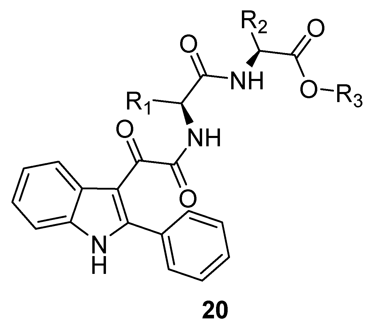
The tumour suppressor protein p53 promotes apoptosis by interacting with members of the protective Bcl2-family proteins which in turn mediate the release of cytochrome c. P53 is a transcription factor that controls cellular response to stress by inducing cell cycle arrest or apoptosis [90,91]. The murine double minute 2 protein (MDM2) downregulates p53 activity by binding to the transactivation domain of p53. In response to stress, phosphorylation of p53 decreases the affinity of this protein for MDM2. A number of human tumors are associated with inhibition of the p53 pathway and consequent uncontrolled cell proliferation. Disruption of the p53-MDM2 interaction is therefore a therapeutic goal for the treatment of cancer [92]. The MDM2-p53 complex is stabilized mainly by a strong hydrophobic interaction between a region of MDM2 and the Phe19, Trp23, and Leu26 residues of p53. A synthetic molecule displaying three lipophilic groups in an orientation that mimics the presentation of the side chains of the above aminoacids can occupy the MDM2 cleft and thereby inhibit the p53-MDM2 interaction [92]. Based on these findings, computational methods were applied on our in-house library of indole-based TSPO ligands to identify those suitable to undergo appropriate decorations in order to inhibit the p53-MDM2 interaction and to maintain TSPO affinity. Following this approach, we synthesized a series of 2-phenylindol-3-yl-glyoxylamides (20), bearing on the glyoxylyl bridge a number of dipeptide moieties (L-Leu-L-Phe, L-Phe-L-Leu, L-Val-L-Leu, L-Leu-L-Val, L-Ile-L-Val, L-Ile-L-Ile and L-Val-L-Ile) capped as methyl or ethyl esters [93,94]. An immune−enzymatic assay on native human MDM2/p53 complex was performed to determine the ability (expressed as IC50 values) of the new compounds to bind MDM2 and disrupt the MDM2/p53 complex; affinity to TSPO was evaluated by competition binding assays employing the radioligand [3H]-PK11195 and expressed as Ki values. The strategy resulted successful as all the new compounds revealed to disrupt the p53-MDM2 complex and bind to TSPO at nanomolar concentrations.
Molecules 25 02331 i014
The compound from series 20 (R1 = CH2C6H5, R2 = CH2CH(CH3)2, R3 = CH3), showing the highest ability to dissociate the p53-MDM2 complex (IC50 = 4.3 nM) and the highest affinity for TSPO (Ki = 87 nM), was selected for further biological studies, giving the following results: (i) reactivation of the p53 function and inhibition of the GBM cell growth, triggering subsequent apoptosis; (ii) no efficacy on a GBM cell line expressing mutant p53, supporting the involvement of this protein in the observed effect; (iii) reduction of viability of glioma cancer stem cells (CSCs), which are less sensitive to anticancer agents and responsible for GBM recurrence [95]. These effects were significantly stronger than those elicited by the p53 activator nutlin-3 and the TSPO ligand PK11195 [91], thanks to the synergism resulting from the simultaneous activation of both targets [93,94]. Finally, cell viability assays performed on non-tumor human mesenchymal stem cells (MSCs) showed that the antiproliferative effect of the selected indole derivative was preferentially directed toward tumor cells. All these findings confirmed that dual targeting MDM2-p53 and TSPO is a valuable anticancer strategy against GBM, where the downstream p53 signaling is not mutated.
Anticancer drugs binding reversibly to their targets may have several limitations in sustaining a therapeutic effect over time, thereby favoring the activation of alternative signaling pathways that escape drug action and cause resistance. Research in the field of oncology has recently been focused on the synthesis and development of new irreversible and long-lasting drugs [96]. As a continuation of our studies on 2-phenylindol-3-ylglyoxylyldipeptides 20, we synthesized 21, bearing a chemo-reactive isothiocyanate group at the 5-position of the indole nucleus. This compound, thanks to its ability to form covalent bonds with electrophilic groups, displayed a potent long-lasting binding affinity for TSPO and a prolonged inhibition of the MDM2-p53 complex [97]. Furthermore, 21 caused GBM cell death by arresting the cell cycle and inducing apoptosis; both effects were greater and more long-lasting than those of the reversible analogues of series 20. The observed apoptotic effects were irreversible so that the cells were not able to regain proliferative activity after drug wash-out [97].
Molecules 25 02331 i015
Compound 21 has been very recently employed in a study aimed to highlight the role played by the p53-MDM2 complex in osteoblast generation from MSCs [98]. The long-lasting MDM2-p53 dissociation determined by 21 enhances the MSC differentiation into osteoblasts through a pathway involving the G protein-coupled receptors kinase 2 and the A2B adenosine receptor.

5. Indole Derivatives as Allosteric Modulators of the Human Adenosine A2B Receptor

Adenosine plays a key role in a variety of physiological and pathological processes by interacting with specific receptors. Four different subclasses of adenosine receptors (ARs) have been identified to date, A1, A2A, A2B, and A3, all belonging to the superfamily of G-protein-coupled receptors [99,100]. Activation of ARs by adenosine or a synthetic agonist determines different intracellular events starting with inhibition (A1 and A3) or stimulation (A2A and A2B) of adenylate cyclase. Additional molecular mechanisms coupled to occupation of ARs by agonists are stimulation of phospholipase C (A1, A2B, and A3), activation of potassium channels, and inhibition of calcium channels (A1) [101].
Being ubiquitously distributed in tissues and organs of mammalians, ARs have been considered attractive targets for the development of agonist- and antagonist-based therapies against a wide range of pathologies, including CNS disorders, cardiac arrhythmia, ischemic injuries, asthma, renal failure and inflammatory diseases [102].
In the course of our researches on BzR ligands (discussed in the second chapter), we prepared and tested a number of [1,2,4]triazino [4,3-a]benzimidazoles (TBI, 22) as geometrically constrained analogues of indole derivatives (23) [103,104].
Molecules 25 02331 i016
The most potent TBIs (R = C6H5, C6H4-p-OCH3, 2-furyl, 2-thienyl) displayed Ki values at the BzR (obtained from bovine cerebral cortex) ranging from 13 nM to 56 nM.
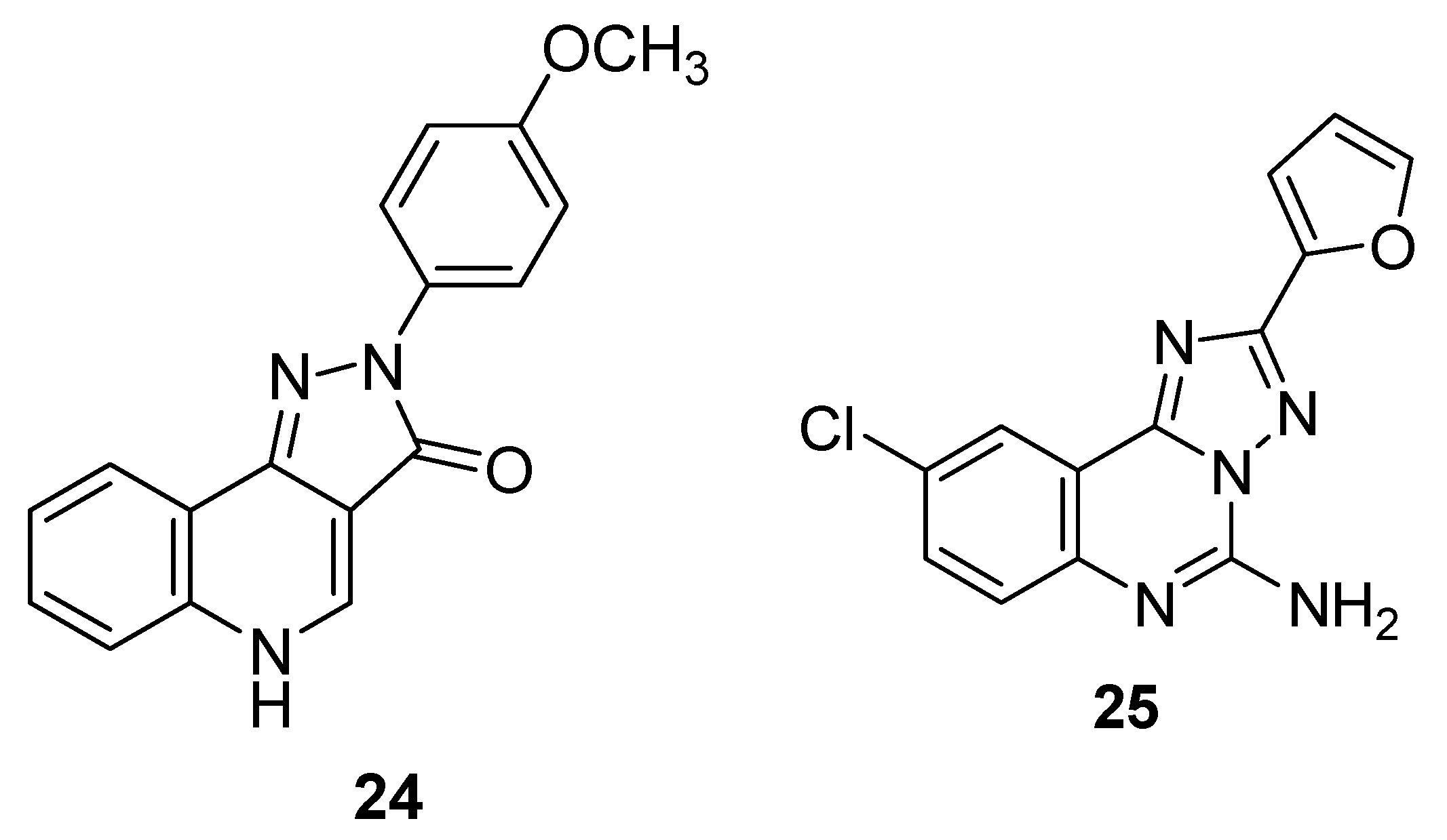
It is worth noting that the structures of certain BzR ligands are similar to those of several antagonists of the A1 AR. Compare, as an example, the BzR agonist CGS-9895 (24) [105] with the triazoloquinazoline derivative CGS-15943 (25) identified as the first non-xantine antagonist of the A1, A2A and A3 ARs [106].
Molecules 25 02331 i017
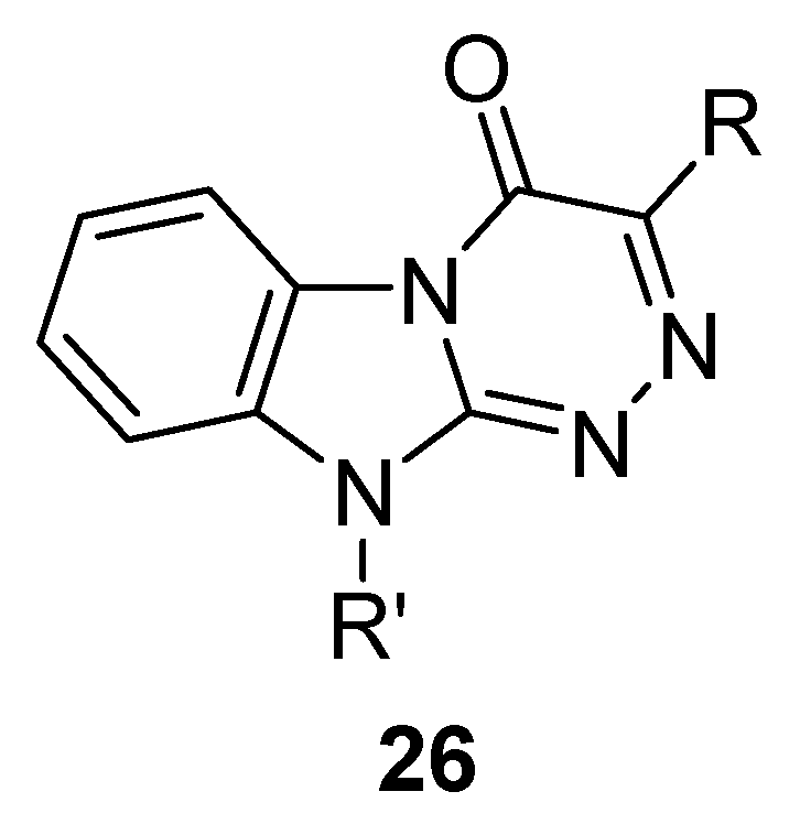
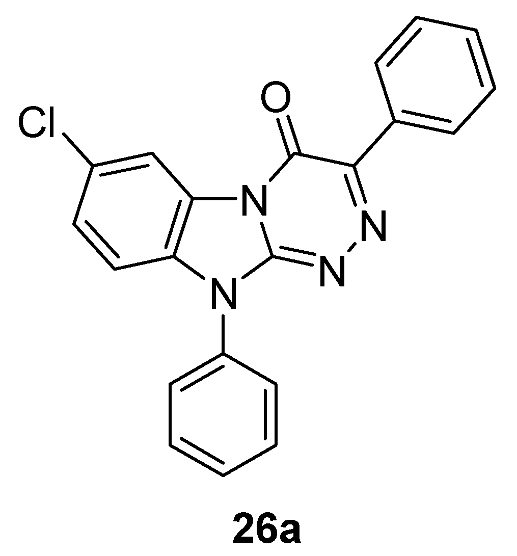
In the light of the above consideration, we prepared some novel TBIs (26) purposely designed as potential A1 AR antagonists through insertion of substituents not only at position 3 (R) but also at position 10 (R’) of the tricyclic system [107].
Molecules 25 02331 i018
Among the new TBI derivatives, the most potent (Ki 83 nM) and selective one at the human A1 AR had R = R’ = phenyl [108].
In 2012 we disclosed a TBI derivative 26a provided with high potency (IC50 of 3 nM) and selectivity for the human A2B AR [109]. For a long time, this receptor has been less characterized compared with the other AR subtypes, partly due to the scarcity of specific ligands [110].
Molecules 25 02331 i019
The therapeutic potential of agonists and antagonists of the A2B AR is remarkable. Particularly, selective agonists of this receptor have been reported to reduce inflammation after ventilator-induced lung injury [111] and to modulate myocardial adaptation to ischemia [112]. Selective A2B AR antagonists have been regarded as candidates for the treatment of cancer [113,114], colitis [115,116] and asthma [117,118,119].
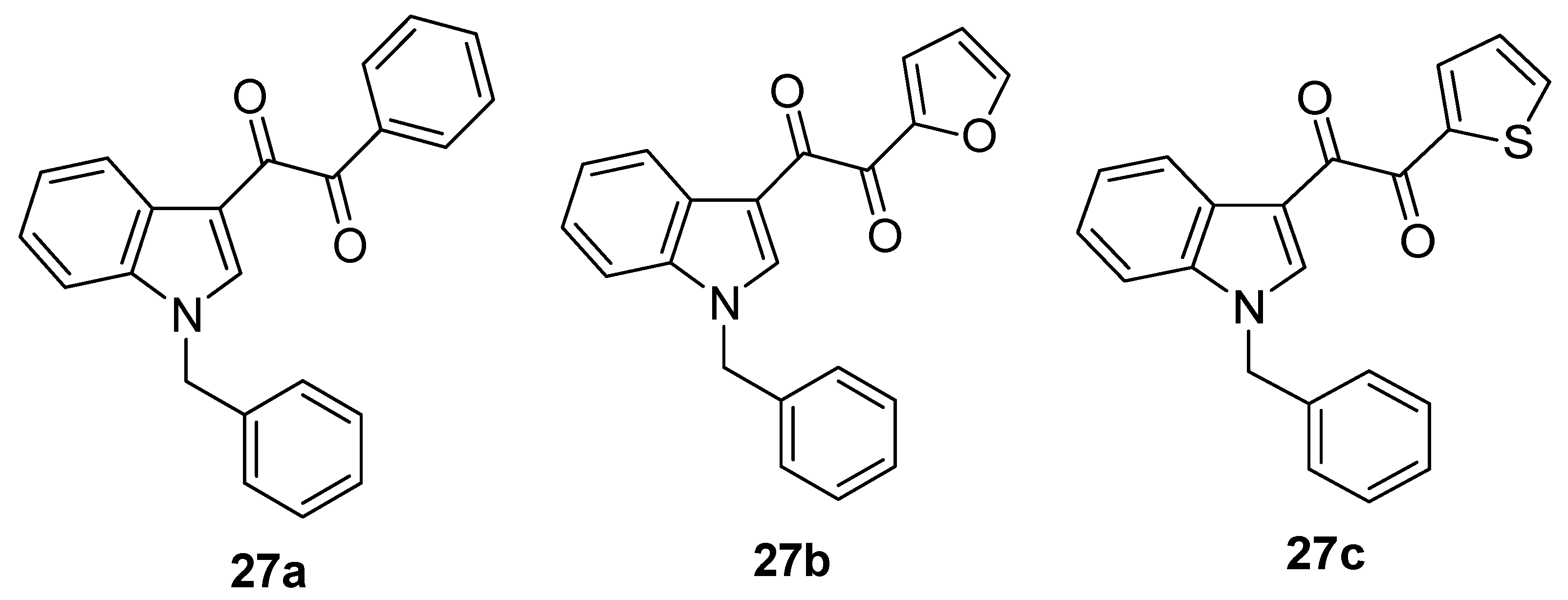
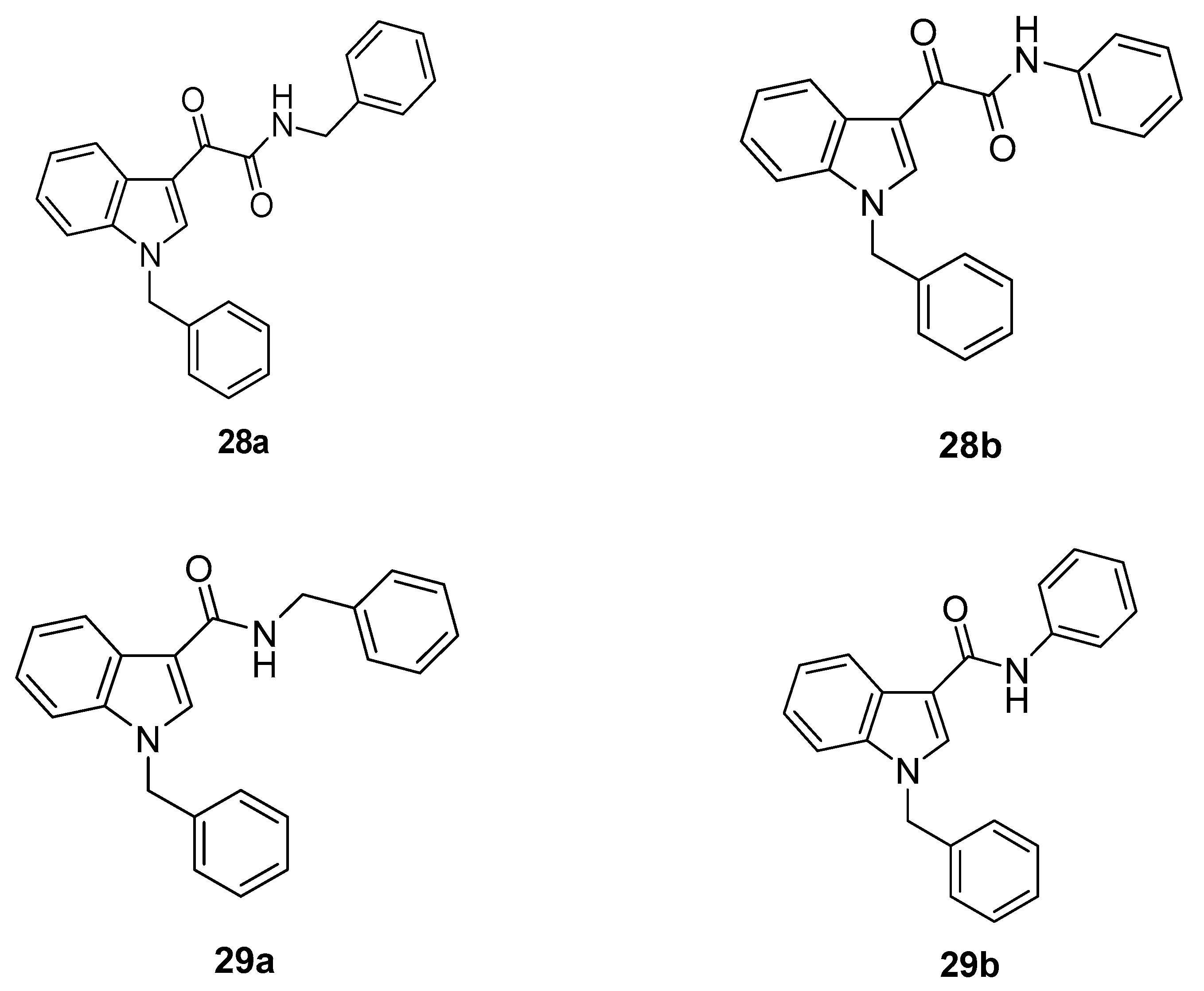
Continuing our searches of novel lead compounds binding to ARs, we synthesized five indole derivatives 27ac, 28a,b featuring a diketo moiety as a linker designed as open chain analogues of the TBI 26a. Additionally, we purchased two indole derivatives 29a,b characterized by an amide linker [120].
Molecules 25 02331 i020Molecules 25 02331 i021
The affinity of indoles 2729 for the human A1, A2A, and A3 AR expressed in CHO cells was determined by measuring their ability to displace specific radioligands from the above receptors. Compounds 27a and 27b exhibited submicromolar affinities for the A1 AR (Ki values of 161 nM and, respectively, 343 nM), whereas the remaining five indoles showed no appreciable affinity for the three AR subtypes. Functional experiments showed that 27a and 27b behaved at the A1 AR as antagonists.
To evaluate the pharmacological effect resulting from interaction between 2729 and the human A2B AR, we measured to what extent our compounds modified the levels of cAMP in CHO expressing only this receptor. None of the compounds increased the cAMP levels at the concentration of 10 μM, clearly showing a lack of A2B AR agonist activity. However, when the experiments were repeated in the presence of 5′-(N-ethylcarboxamido)adenosine (NECA), which acts as unspecific agonist of the ARs, 27a and 28a,b potentiated its agonist effects, suggesting that these three compounds interact with the A2B AR as positive modulators. Conversely, 27b,c and 29a,b potently counteracted the NECA-mediated increase in cAMP, indicating that they act as negative modulators of the A2B AR.
We were very satisfied by these unexpected preliminary results (a typical case of serendipity) as our indole derivatives 2729 are so far the only A2B AR allosteric modulators reported in literature.
In a subsequent paper, the pharmacology of the new indole derivatives at the A2B AR was characterized in more detail [121]. The potencies of compounds 2729 in modulating the activity of A2B AR agonists were determined by assessing the effects of different concentrations of each of them on cAMP accumulation induced by an EC50 concentration of NECA (100 nM). The resulting concentration-response curves indicated that 27a and 28a,b exhibit similar submicromolar potencies at A2B AR, with EC50 values between 250 nM and 446 nM.
The concentration-response curves of 27b and 29a,b, obtained under the same conditions, fitted a two-site equation model, suggesting that these compounds recognize two sites of the A2B AR with different affinities. The potency values obtained for the high and low affinity states of the receptor were in the subnanomolar/nanomolar and micromolar range, respectively. Conversely, the concentration-response curve of 27c fitted a one-site equation model, revealing that this compound recognizes a unique site of the A2B AR with nanomolar affinity.
Concentration-response curves in which the cAMP was measured by varying the concentration of the tested compound as well as the concentration of NECA gave us further information about their mechanisms of action. Particularly, from these curves we could infer that 27a and 28a,b enhance the efficacy of the agonist without affecting its potency, while 27b,c and 29a,b decrease either the efficacy and potency of the agonist. Several studies report that agonist efficacy and potency are not necessarily both modified by allosteric modulators [122,123]. A plausible hypothesis is that 27a and 28a,b affects specific conformational states of A2B AR so as to improve the functional coupling to the intracellular signaling system without altering the conformation of the orthosteric site. Probably, 27b,c and 29a,b affects the agonist potency by shifting the receptor conformational states toward the resting ones; at the same time, they decrease the agonist affinity by deforming the conformation of the orthosteric site.
In virtue of their indirect mechanism of receptor modulation, allosteric modulators of G-protein-coupled receptors offer therapeutic advantages compared to agonists and antagonists. Particularly, they tune pharmacological responses only when and where the endogenous agonist is present in the specific tissue. Given the role played by A2B AR in several physiological and pathological processes, discussed previously in this chapter, the positive and negative allosteric modulators of this receptor represent promising tools to identify novel druggable compounds.
Shortly after we reported the therapeutic potential of the indole derivatives acting as allosteric enhancers of A2B AR agonists in the treatment of bone-related diseases (e.g., osteoporosis, rheumatoid arthritis, osteogenesis imperfecta, multiple myeloma, fracture mal-union) [124]. Particularly, we demonstrated that compound 28b potentiates the effects of either adenosine and synthetic A2B AR agonists in mediating osteoblast differentiation in vitro. In detail, by treating the MCSs with 28b we observed an increase in the expression of osteoblast-related genes (runx2 and osterix) and osteoblast marker proteins (phosphatase alkaline and osteocalcin) associated with a stimulation of osteoblast mineralization.

6. Indole Derivatives as Ligands of the Kelch-like ECH-Associated Protein 1
Very recently, we have published a paper [125] disclosing novel indole derivatives binding to a pharmacological target playing a key role in cellular oxidative stress, namely the Kelch-like ECH-associated protein 1 (Keap1) [126]. Oxidative stress [127] is associated with an excess of reactive oxygen species (ROS), such as superoxide anion (O2·), hydrogen peroxide (H2O2), hydroxyl radical (OH·). ROS are potentially cytotoxic as they damage DNA, RNA, enzymes and cellular membranes; they are generated from molecular oxygen during physiological processes (e.g., oxidative phosphorylation) or pathological events (e.g., inflammatory responses that protect our body from foreign pathogens).
The cells reduce oxidative stress through radical scavengers (those best known are vitamins C, E and K) and antioxidant enzymes, both inactivating the ROS. The antioxidant enzymes include superoxide dismutase, catalase, heme oxygenase-1, glutathione S-transferase, NADPH:quinone oxidoreductase 1 and transketolase [128,129,130].
The expression of the above enzymes is regulated by the so called Keap1-Nrf2-ARE system [131], whose mechanism can be briefly schematized as follows. Under physiological conditions, the activity of the nuclear factor erythroid 2-related factor 2 (Nrf2) [132], a transcription factor, is inhibited by a strong interaction with Keap1. When the ROS exceed a safety threshold concentration, they disrupt the Keap1-Nrf2 complex by oxidizing a group of cysteine residues belonging to a specific domain of Keap1. This event triggers the release of Nrf2, allowing it to act as a transcriptional activator of genes that contain an enhancer sequence known as antioxidant response element (ARE) [133].
Inhibitors of the Keap1-Nrf2 interaction (KNI) are considered a promising new class of anti-inflammatory agents to treat diseases involving chronic oxidative stress, such as diabetes, cancer and neurodegenerative disorders [134].
The Keap1 binding cavity hosting the KNI inhibitors can be divided into six subpockets (P1-P6) [135]. P1 and P2 contain protonated arginine residues (Arg483, Arg415, Arg380), which give rise to strong electrostatic interactions with electron-rich parts of their ligands: salt bridges with carboxylate groups, H-bonds with nitro oxygens or azole nitrogens, cation-π contacts with aromatic rings. In the above mentioned six subpockets there are also lipophilic aminoacids.
Most of the KNI inhibitors reported in literature are chemically heterogeneous small molecules featuring a planar or quasi-planar scaffold which bear at least one aromatic ring and/or a weak acidic group involved in the abovementioned electrostatic interactions [136].
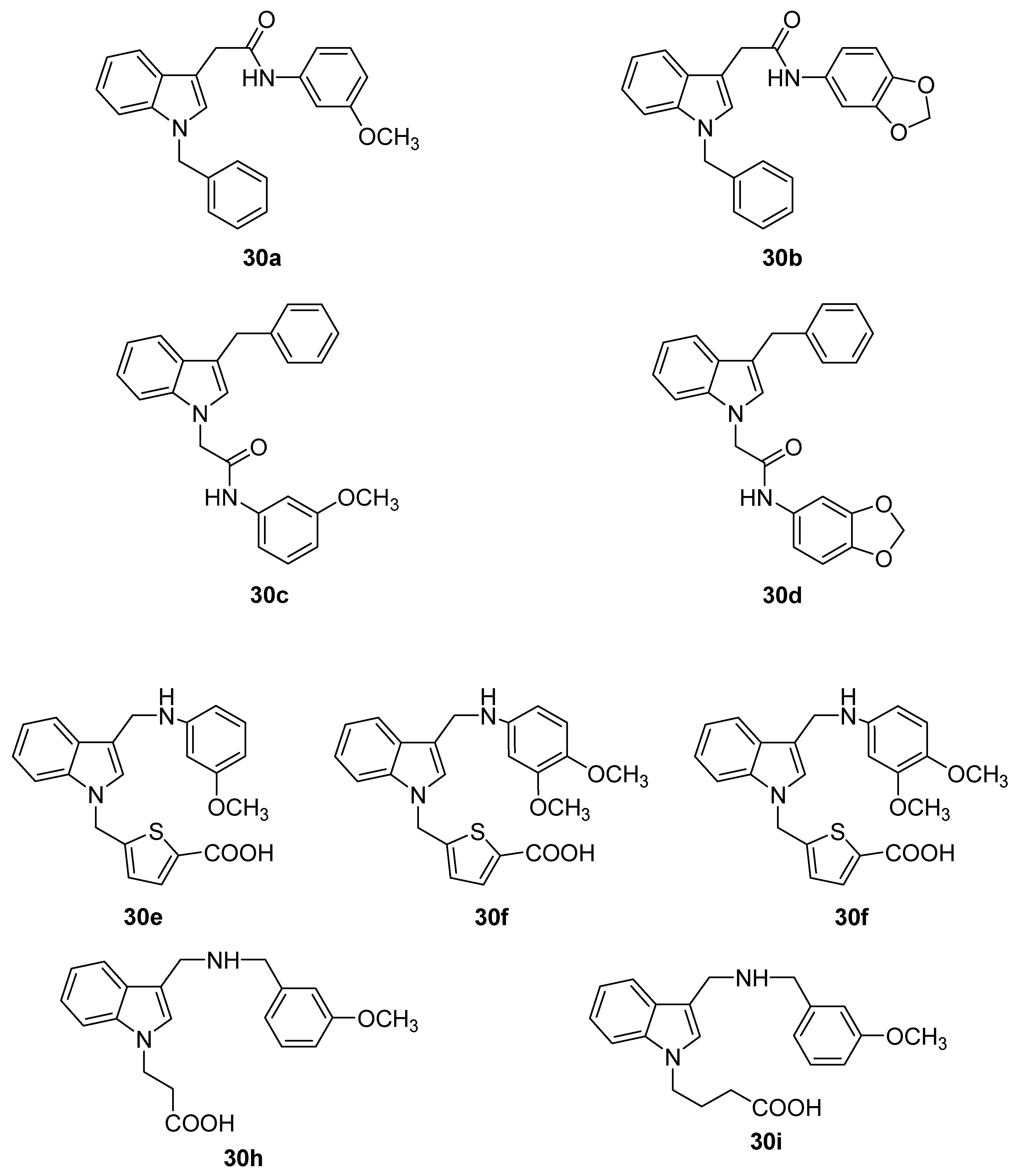
Our experience with indole as scaffold, led us to believe that it would be feasible to design indole derivatives acting as KNI inhibitors. The design of the compounds to be tested was mainly guided by the 3D structures of some KNI inhibitors co-crystallized with Keap1 [135,136,137]. With the help of molecular modelling and docking approaches, we selected nine indole derivatives 30ai, among which 30ad were synthesized, whereas 30ei were purchased.
Molecules 25 02331 i022
Based on their acid-basic properties, these compounds can be divided into three groups: (a) non-ionizable (30ad); (b) acidic (30e,f); ampholytic (30gi). The presence of methoxy group(s) or a methylendioxy moiety in the structures of 30ai was regarded as a chance for our compounds to strength potential cation-π interactions and/or engage H-bonds with the target protein. The thiophene ring featured by 30eg confers conformational rigidity and represents an electron-rich ring potentially able to interact with arginine residues. Compounds 30ai were evaluated for their ability to inhibit the Keap1-Nrf2 interaction through a cell-based luciferase reporter assay [138]. Nearly all of them were tested at the concentration of 10 μM; 30g was tested at the concentration of 5 μM owing to its limited solubility in phosphate buffer. t-Butylhydroxyquinone (t-BHQ) was employed as a positive control [139] at the concentration of 50 μM, a value which gave in our experiments the maximum luciferase activity.
Compounds 30e,f and 30g, characterized by a 5-carboxythien-2-yl substituent, increased luciferase activity by 152%, 263% and 486%, respectively; their activities were higher by 3.2, 5.5 and 10-fold, respectively, than that exhibited by t-BHQ (48% increase). The remaining compounds displayed activities below 50%. A western blot analysis confirmed that 30e,f and 30g increase the expression of Nrf2 and of two enzymes encoded by its downstream target ARE genes, namely NADPH:quinone oxidoreductase 1 and transketolase. The same three best performing compounds showed to be non-cytotoxic when tested on human peripheral blood lymphocytes.
Docking simulations of the interaction between 30g and Keap1, using available 3D structures of this protein [140], allowed us to explain the outstanding activity of this thiophene-contaning compound. The carboxylate group of 30g makes a salt bridge with the Arg483 protonated side chain and a charge-reinforced H-bond with the Ser508 hydroxy group; the thiophene ring establishes a cation-π interaction with the Arg415 positively charged side chain. Such a cation-π interaction cannot be established by the less active compounds 30h,i which bear a carboxylic group but lack an aromatic ring attached to the indole nitrogen. Furthermore, 30h,i are much more flexible than 30e,f and 30g.
The (m-methoxy)benzylaminomethyl substituent of 30g establishes hydrophobic interactions with Val512 and Leu472 side chains and a H-bond between the m-methoxy oxygen and the Leu472 backbone NH. The protonated nitrogen of 30g is not involved in any type of electrostatic interaction. This suggests that the higher activity of 30g with respect to those of 30e,f may be ascribed to the different length of the linker between the indole nucleus and the m-methoxy moiety. The indole ring of 30g contributes to the binding affinity through weak hydrophobic interactions with the Ala556 methyl group and the Arg415 dimethylene fragment.

7. Conclusions

The works described in this review confirm how useful and versatile indole can be as a molecular scaffold in designing drug-like molecules.

Author Contributions

All authors have read and agreed to the published version of the manuscript.

Funding

This paper was supported by University of Pisa and University of Naples “Federico II”.

Conflicts of Interest

The authors declare no conflict of interest

References and Notes

  1. Fraga, C.A.M.; Barreiro, E.J.; de Sa Alves, F.R. From nature to drug discovery: The indole scaffold as a ‘privileged structure’. Mini Rev. Med. Chem. 2009, 9, 782–793. [Google Scholar]
  2. Barnard, E.A.; Skolnick, P.; Olsen, R.W.; Mohler, H.; Sieghart, W.; Biggio, G.; Braestrup, C.; Bateson, A.N.; Langer, S.Z. International Union of Pharmacology. XV. Subtypes of γ-aminobutyric acidA receptors: Classification on the basis of subunit structure and receptor function. Pharmacol. Rev. 1998, 50, 291–313. [Google Scholar] [PubMed]
  3. Mohler, H. GABA(A) Receptor diversity and pharmacology. Cell Tissue Res. 2006, 326, 505–516. [Google Scholar] [CrossRef] [PubMed] [Green Version]
  4. Sieghart, W.; Sperk, G. Subunit composition, distribution and function of GABA(A) receptor subtypes. Cell Tissue Res. 2006, 326, 795–816. [Google Scholar] [CrossRef]
  5. Sieghart, W.; Savic, M.M. International Union of Basic and Clinical Pharmacology. CVI: GABAA receptor subtype- and function-selective ligands: Key issues in translation to humans. Pharmacol. Rev. 2018, 70, 836–878. [Google Scholar] [CrossRef] [Green Version]
  6. Sieghart, W.; Sperk, G. Subunit composition, distribution and function of GABA(A) receptor subtypes. Curr. Top. Med. Chem. 2002, 2, 795–816. [Google Scholar] [CrossRef]
  7. Mohler, H.; Fritschy, J.M.; Rudolph, U. A New benzodiazepine pharmacology. J. Pharmacol. Exp. Ther. 2002, 300, 2–8. [Google Scholar] [CrossRef] [Green Version]
  8. Whiting, P.J. GABA-A receptor subtypes in the brain: A paradigm for CNS drug discovery? Drug Discovery Today 2003, 8, 445–450. [Google Scholar] [CrossRef]
  9. Wafford, K.A.; Thompson, S.A.; Thomas, D.; Sikela, J.; Wilcox, A.S.; Whiting, P.J. Functional characterization of human γ-aminobutyric acidA receptors containing the α4 subunit. Mol. Pharmacol. 1996, 50, 670–678. [Google Scholar]
  10. Accardi, M.V.; Brown, P.M.; Miraucourt, L.S.; Orser, B.A.; Bowie, D. α6-Containing GABAA receptors are the principal mediators of inhibitory synapse strengthening by insulin in cerebellar granular cells. J. Neurosci. 2015, 35, 9676–9688. [Google Scholar] [CrossRef] [Green Version]
  11. Sigel, E. Mapping of the benzodiazepine recognition site on GABAA receptors. Curr. Top. Med. Chem. 2002, 2, 833–839. [Google Scholar] [CrossRef] [PubMed]
  12. Teuber, L.; Watjens, F.; Jensen, L.H. Ligands for the benzodiazepine binding site-a survey. Curr. Pharm. Des. 1999, 5, 317–343. [Google Scholar] [PubMed]
  13. Gardner, C.R. Interpretation of behavioral effects of benzodiazepine receptor ligands. Drugs Future 1989, 14, 51–67. [Google Scholar]
  14. Schofield, P.R.; Darlison, M.G.; Fijita, N.; Burt, D.R.; Sthepenson, F.A.; Rodriguez, H.; Rhee, L.M.; Ramachandran, J.; Reale, V.; Glencorse, T.A.; et al. Sequence and functional expression of the GABAA receptor shows a ligand-gated receptor super-family. Nature 1987, 328, 221–227. [Google Scholar] [CrossRef] [PubMed]
  15. Levitan, E.S.; Schofield, P.R.; Burt, D.R.; Rhee, L.M.; Wisden, W.; Kohler, M.; Fujita, N.; Rodriguez, H.; Stephenson, A.; Darlison, M.G.; et al. Structural and functional basis for GABAA receptor heterogeneity. Nature 1988, 335, 76–79. [Google Scholar] [CrossRef] [PubMed]
  16. Olsen, R.W.; Tobin, A.J. Molecular biology of GABAA receptor. FASEB J. 1990, 4, 1469–1480. [Google Scholar] [CrossRef]
  17. Burt, D.; Kamatachi, G. GABAA receptor subtypes: From pharmacology to molecular biology. FASEB J. 1991, 5, 2916–2923. [Google Scholar] [CrossRef]
  18. McKernan, R.M.; Rosahl, T.W.; Reynolds, D.S.; Sur, C.; Wafford, K.A.; Atack, J.R.; Farrar, S.; Myers, J.; Cook, G.; Ferris, P.; et al. Sedative but not anxiolytic properties of benzodiazepines are mediated by the GABAA receptor α1 subtype. Nat. Neurosci. 2000, 3, 587–592. [Google Scholar] [CrossRef]
  19. Rudolph, U.; Möhler, H. Analysis of GABAA receptor function and dissection of the pharmacology of benzodiazepines and general anesthetics through mouse genetics. Annu. Rev. Pharmacol. Toxicol. 2004, 44, 475–498. [Google Scholar] [CrossRef]
  20. Lüscher, B.; Keller, C.A. Regulation of GABAA receptor trafficking, channel activity, and functional plasticity of inhibitory synapses. Pharmacol. Ther. 2004, 102, 195–221. [Google Scholar] [CrossRef]
  21. Rudolph, U.; Möhler, H. GABA-based therapeutic approaches: GABAA receptor subtype functions. Curr. Opin. Pharmacol. 2006, 6, 18–23. [Google Scholar] [CrossRef]
  22. Da Settimo, F.; Taliani, S.; Trincavelli, M.L.; Montali, M.; Martini, C. GABAA/Bz receptor subtypes as targets for selective drugs. Curr. Med. Chem. 2007, 14, 2680–2701. [Google Scholar] [CrossRef] [PubMed]
  23. Martini, C.; Gervasio, T.; Lucacchini, A.; Da Settimo, A.; Primofiore, G.; Marini, A.M. Specific inhibition of benzodiazepine receptor binding by some N-(indol-3-ylglyoxylyl)amino acid derivatives. J. Med. Chem. 1985, 28, 506–509. [Google Scholar] [CrossRef] [PubMed]
  24. Braestrup, C.; Nielsen, M.C.; Olsen, C.E. Urinary and brain β-carboline carboxylates as potent inhibitors of brain benzodiazepine receptors. Proc. Natl. Acad. Sci. USA 1980, 77, 2288–2292. [Google Scholar] [CrossRef] [PubMed] [Green Version]
  25. Primofiore, G.; Marini, A.M.; Da Settimo, F.; Martini, C.; Bardellini, A.; Giannaccini, G.; Lucacchini, A. Specific inhibition of benzodiazepine receptor binding by some N-(indol-3-ylglyoxylyl)amino acid derivatives: Stereoselective interactions. J. Med. Chem. 1989, 32, 2514–2518. [Google Scholar] [CrossRef] [PubMed]
  26. Bianucci, A.M.; Da Settimo, A.; Da Settimo, F.; Primofiore, G.; Martini, C.; Giannaccini, G.; Lucacchini, A. Benzodiazepine receptor affinity and interaction of some N-(indol-3-ylglyoxylyl)amine derivatives. J. Med. Chem. 1992, 35, 2214–2220. [Google Scholar] [CrossRef]
  27. Da Settimo, A.; Lucacchini, A.; Marini, A.M.; Martini, C.; Primofiore, G.; Senatore, G.; Taliani, S. Isosteric replacement of the indole nucleus by benzothiophene and benzofuran in a series of indolylglyoxylylamine derivatives with partial agonist activity at the benzodiazepine receptor. Eur. J. Med. Chem. 1996, 31, 951–956. [Google Scholar] [CrossRef]
  28. Da Settimo, A.; Primofiore, G.; Da Settimo, F.; Marini, A.M.; Novellino, E.; Greco, G.; Martini, C.; Giannaccini, G.; Lucacchini, A. Synthesis, structure-activity relationships, and molecular modeling studies of N-(indol-3-ylglyoxylyl)benzylamine derivatives acting at the benzodiazepine receptor. J. Med. Chem. 1996, 39, 5083–5091. [Google Scholar] [CrossRef]
  29. Da Settimo, A.; Primofiore, G.; Da Settimo, F.; Marini, A.M.; Novellino, E.; Greco, G.; Gesi, M.; Martini, C.; Giannaccini, G.; Lucacchini, A. N’-Phenylindol-3-ylglyoxylohydrazide derivatives: Synthesis, structure-activity relationships, molecular modeling studies and pharmacological action on brain benzodiazepine receptors. J. Med. Chem. 1998, 41, 3821–3830. [Google Scholar] [CrossRef]
  30. Groom, C.R.; Bruno, I.J.; Lightfoo, M.P.; Ward, S.C. The Cambridge Structural Database. Acta Cryst. 2016, B72, 171–179. [Google Scholar] [CrossRef]
  31. Zhang, W.; Koehler, K.F.; Zhang, P.; Cook, J.M. Development of a comprehensive pharmacophore model for the benzodiazepine receptor. Drug. Des. Discov. 1995, 12, 193–248. [Google Scholar] [PubMed]
  32. Primofiore, G.; Da Settimo, F.; Taliani, S.; Marini, A.M.; Novellino, E.; Greco, G.; Lavecchia, A.; Besnard, F.; Trincavelli, L.; Costa, B.; et al. Novel N-(arylalkyl)indol-3-ylglyoxylamides targeted as ligands of the benzodiazepine receptor: Synthesis, biological evaluation, and molecular modeling analysis of the structure-activity relationships. J. Med. Chem. 2001, 44, 2286–2297. [Google Scholar] [CrossRef] [PubMed]
  33. Primofiore, G.; Da Settimo, F.; Marini, A.M.; Taliani, S.; La Motta, C.; Simorini, F.; Novellino, E.; Greco, G.; Cosimelli, B.; Ehlardo, M.; et al. Refinement of the benzodiazepine receptor site topology by structure-activity relationships of new N-(heteroarylmethyl)indol-3-ylglyoxylamides. J. Med. Chem. 2006, 49, 2489–2495. [Google Scholar] [CrossRef] [PubMed]
  34. Primofiore, G.; Taliani, S.; Da Settimo, F.; Marini, A.M.; La Motta, C.; Simorini, F.; Patrizi, M.P.; Sergianni, V.; Novellino, E.; Greco, G.; et al. Novel N-substituted indol-3-ylglyoxylamides probing the LDi and L1/L2 lipophilic regions of the benzodiazepine receptor site in search for subtype-selective ligands. J. Med. Chem. 2007, 50, 1627–1634. [Google Scholar] [CrossRef]
  35. Huang, Q.; He, X.; Ma, C.; Liu, R.; Yu, S.; Dayer, C.A.; Wenger, G.R.; McKernan, R.; Cook, J.M. Pharmacophore/receptor models for GABAA/BzR subtypes (α1β3γ2, α5β3γ2, and α6β3γ2) via a comprehensive ligand-mapping approach. J. Med. Chem. 2000, 43, 71–95. [Google Scholar] [CrossRef]
  36. He, X.; Huang, Q.; Ma, C.; Yu, S.; McKernan, R.; Cook, J.M. Pharmacophore/receptor models for GABAA/BzR α2β3γ2, α3β3γ2, and α4β3γ2 recombinant subtypes. Included volume analysis and comparison to α1β3γ2, α5β3γ2, and α6β3γ2. Drug Des. Discov. 2000, 17, 131–171. [Google Scholar]
  37. Yu, S.; He, X.; Ma, C.; McKernan, R.; Cook, J.M. Studies in search of α2 Selective ligands for GABAA/BzR receptor subtypes. Part I. Evidence for the conservation of pharmacophoric descriptors for DS subtypes. Med. Chem. Res. 1999, 9, 186–202. [Google Scholar]
  38. Atack, J.R. GABAA receptor subtype-selective modulators. I. α23-Selective agonists as non-sedating anxiolytics. Curr. Top. Med. Chem. 2011, 11, 1176–1202. [Google Scholar] [CrossRef]
  39. Taliani, S.; Cosimelli, B.; Da Settimo, F.; Marini, A.M.; La Motta, C.; Simorini, F.; Salerno, S.; Novellino, E.; Greco, G.; Cosconati, S.; et al. Identification of anxiolytic/nonsedative agents among indol-3-ylglyoxylamides acting as functionally selective agonists at the γ-aminobutyric acid-A (GABAA) α2 benzodiazepine receptor. J. Med. Chem. 2009, 52, 5798–5806. [Google Scholar] [CrossRef]
  40. Costall, B.; Jones, B.J.; Kelly, M.E.; Naylor, R.J.; Tomlins, D.M. Exploration of mice in a black and white test box: Validation as a model of anxiety. Pharmacol. Biochem. Behav. 1989, 32, 777–785. [Google Scholar] [CrossRef]
  41. Cromer, B.A.; Morton, C.J.; Parker, M.W. Anxiety over GABA(A) receptor structure relieved by AChBP. Trends Biochem. Sci. 2002, 27, 280–287. [Google Scholar] [CrossRef]
  42. Squires, R.F.; Braestrup, C. Benzodiazepine receptors in rat brain. Nature 1977, 266, 732–734. [Google Scholar] [CrossRef] [PubMed]
  43. Papadopoulos, V.; Baraldi, M.; Guilarte, T.R.; Knudsen, T.B.; Lacapere, J.J.; Lindemann, P.; Norenberg, M.D.; Nutt, D.; Weizman, A.; Zhang, M.R.; et al. Translocator protein (18 kDa): New nomenclature for the peripheral-type benzodiazepine receptor based on its structure and molecular function. Trends Pharmacol. Sci. 2006, 27, 402–409. [Google Scholar] [CrossRef] [PubMed]
  44. Papadopoulos, V.; Aghazadeh, Y.; Fan, J.; Campioli, E.; Zirkin, B.; Midzak, A. Translocator protein-mediated pharmacology of cholesterol transport and steroidogenesis. Mol. Cell. Endocrinol. 2015, 408, 90–98. [Google Scholar] [CrossRef] [PubMed] [Green Version]
  45. Fan, J.; Papadopoulos, V. Evolutionary origin of the mitochondrial cholesterol transport machinery reveals a universal mechanism of steroid hormone biosynthesis in animals. PLoS ONE 2013, 8, e76701. [Google Scholar] [CrossRef] [Green Version]
  46. Costa, B.; Da Pozzo, E.; Martini, C. Translocator protein and steroidogenesis. Biochem. J. 2018, 475, 901–904. [Google Scholar] [CrossRef] [PubMed]
  47. Liu, G.J.; Middleton, R.J.; Hatty, C.R.; Kam, W.W.; Chan, R.; Pham, T.; Harrison-Brown, M.; Dodson, E.; Veale, K.; Banati, R.B. The 18 kDa translocator protein, microglia and neuroinflammation. Brain Pathol. 2014, 24, 631–653. [Google Scholar] [CrossRef] [Green Version]
  48. Rupprecht, R.; Papadopoulos, V.; Rammes, G.; Baghai, T.C.; Fan, J.; Akula, N.; Groyer, G.; Adams, D.; Schumacher, M. Translocator protein (18 kDa) (TSPO) as a therapeutic target for neurological and psychiatric disorders. Nat. Rev. Drug Discov. 2010, 9, 971–988. [Google Scholar] [CrossRef]
  49. Da Pozzo, E.; Tremolanti, C.; Costa, B.; Giacomelli, C.; Milenkovic, V.M.; Bader, S.; Wetzel, C.H.; Rupprecht, R.; Taliani, S.; Da Settimo, F.; et al. Microglial pro-inflammatory and anti-inflammatory phenotypes are modulated by translocator protein activation. Int. J. Mol. Sci. 2019, 20, 4467. [Google Scholar] [CrossRef] [Green Version]
  50. Li, H.; Papadopoulos, V. Peripheral-type benzodiazepine receptor function in cholesterol transport. Identification of a putative cholesterol recognition/interaction amino acid sequence and consensus pattern. Endocrinology 1998, 139, 4991–4997. [Google Scholar] [CrossRef]
  51. Taliani, S.; Pugliesi, I.; Da Settimo, F. Structural requirements to obtain highly potent and selective 18 kDa translocator protein (TSPO) ligands. Curr. Top. Med. Chem. 2011, 11, 860–886. [Google Scholar] [CrossRef] [PubMed]
  52. Farges, R.; Joseph-Liauzun, E.; Shire, D.; Caput, D.; Le Fur, G.; Ferrara, P. Site-directed mutagenesis of the peripheral benzodiazepine receptor: Identification of amino acids implicated in the binding site of Ro5-4864. Mol. Pharmacol. 1994, 46, 1160–1167. [Google Scholar] [PubMed]
  53. Le Fur, G.; Guilloux, F.; Rufat, P.; Benavides, J.; Uzan, A.; Renault, C.; Dubroeucq, M.C.; Gueremy, C. Peripheral benzodiazepine binding sites: Effect of PK11195, 1-(2-chlorophenyl)-N-methyl-(1-methylpropyl)-3-isoquinolinecarboxamide. II. In vivo studies. Life Sci. 1983, 32, 1849–1856. [Google Scholar] [CrossRef]
  54. Da Pozzo, E.; Giacomelli, C.; Barresi, E.; Costa, B.; Taliani, S.; Da Settimo, F.; Martini, C. Targeting the 18-kDa translocator protein: Recent perspectives for neuroprotection. Biochem. Soc. Trans. 2015, 43, 559–565. [Google Scholar] [CrossRef]
  55. Taliani, S.; Da Settimo, F.; Da Pozzo, E.; Chelli, B.; Martini, C. Translocator protein ligands as promising therapeutic tools for anxiety disorders. Curr. Med. Chem. 2009, 16, 3359–3380. [Google Scholar] [CrossRef]
  56. Rupprecht, R.; Rammes, G.; Eser, D.; Baghai, T.C.; Schüle, C.; Nothdurfter, C.; Troxler, T.; Gentsch, C.; Kalkman, H.O.; Chaperon, F.; et al. Translocator protein (18 kD) as target for anxiolytics without benzodiazepine-like side effects. Science 2009, 325, 490–493. [Google Scholar] [CrossRef] [Green Version]
  57. Costa, B.; Da Pozzo, E.; Martini, C. Translocator protein as a promising target for novel anxiolytics. Curr. Top. Med. Chem. 2012, 12, 270–285. [Google Scholar] [CrossRef]
  58. Kita, A.; Kinoshita, T.; Kohayakawa, H.; Furukawa, K.; Akaike, A. Lack of tolerance to anxiolysis and withdrawal symptoms in mice repeatedly treated with AC-5216, a selective TSPO ligand. Prog. Neuro-Psychopharmacol. Biol. Psychiatry 2009, 33, 1040–1045. [Google Scholar] [CrossRef]
  59. Nguyen, N.; Fakra, E.; Pradel, V.; Jouve, E.; Alquier, C.; Le Guern, M.E.; Micallef, J.; Blin, O. Efficacy of etifoxine compared to lorazepam monotherapy in the treatment of patients with adjustment disorders with anxiety: A doubleblind controlled study in general practice. Hum. Psychopharmacol. 2006, 21, 139–149. [Google Scholar] [CrossRef]
  60. Primofiore, G.; Da Settimo, F.; Taliani, S.; Simorini, F.; Patrizi, M.P.; Novellino, E.; Greco, G.; Abignente, E.; Costa, B.; Chelli, B.; et al. N,N-Dialkyl-2-phenylindol-3-ylglyoxylamides. a new class of potent and selective ligands at the peripheral benzodiazepine receptor. J. Med. Chem. 2004, 47, 1852–1855. [Google Scholar] [CrossRef]
  61. Da Settimo, F.; Simorini, F.; Taliani, S.; La Motta, C.; Marini, A.M.; Salerno, S.; Bellandi, M.; Novellino, E.; Greco, G.; Cosimelli, B.; et al. Anxiolytic-like effects of N,N-dialkyl-2-phenylindol-3-ylglyoxylamides by modulation of translocator protein promoting neurosteroid biosynthesis. J. Med. Chem. 2008, 51, 5798–5806. [Google Scholar] [CrossRef] [PubMed]
  62. Kozikowski, A.P.; Ma, D.; Brewer, J.; Sun, S.; Costa, E.; Romeo, E.; Guidotti, A. Chemistry, binding affinity, and behavioral properties of a new class of “antineophobic” mitochondrial DBI receptor complex (mDRC) ligands. J. Med. Chem. 1993, 36, 2908–2920. [Google Scholar] [CrossRef] [PubMed]
  63. Costa, B.; Da Pozzo, E.; Chelli, B.; Simola, N.; Morelli, M.; Luisi, M.; Maccheroni, M.; Taliani, S.; Simorini, F.; Da Settimo, F.; et al. Anxiolytic properties of a 2-phenylindolglyoxylamide TSPO ligand: Stimulation of in vitro neurosteroid production affecting GABAA receptor activity. Psychoneuroendocrinology 2011, 36, 463–472. [Google Scholar] [CrossRef] [PubMed]
  64. Simorini, F.; Marini, A.M.; Taliani, S.; La Motta, C.; Salerno, S.; Pugliesi, I.; Da Settimo, F. Medicinal chemistry of indolylglyoxylamide TSPO high affinity ligands with anxiolytic-like effects. Curr. Top. Med. Chem. 2012, 12, 333–351. [Google Scholar] [CrossRef]
  65. Barresi, E.; Bruno, A.; Taliani, S.; Cosconati, S.; Da Pozzo, E.; Salerno, S.; Simorini, F.; Daniele, S.; Giacomelli, C.; Marini, A.M.; et al. Deepening the topology of the translocator protein binding site by novel N,N-dialkyl-2-arylindol-3-ylglyoxylamides. J. Med. Chem. 2015, 58, 6081–6092. [Google Scholar] [CrossRef]
  66. Jaremko, Ł.; Jaremko, M.; Giller, K.; Becker, S.; Zweckstetter, M. Structure of the mitochondrial translocator protein in complex with a diagnostic ligand. Science 2014, 343, 1363–1366. [Google Scholar] [CrossRef] [Green Version]
  67. This code univocally identifies the quoted protein. PDB code: 2MGY.
  68. Da Pozzo, E.; Giacomelli, C.; Costa, B.; Cavallini, C.; Taliani, S.; Barresi, E.; Da Settimo, F.; Martini, C. TSPO PIGA ligands promote neurosteroidogenesis and human astrocyte well-being. Int. J. Mol. Sci. 2016, 17, 1028. [Google Scholar] [CrossRef]
  69. Santoro, A.; Mattace Raso, G.; Taliani, S.; Da Pozzo, E.; Simorini, F.; Costa, B.; Martini, C.; Laneri, S.; Sacchi, A.; Cosimelli, B.; et al. TSPO-ligands prevent oxidative damage and inflammatory response in C6 glioma cells by neurosteroid synthesis. Eur. J. Pharm. Sci. 2016, 88, 124–131. [Google Scholar] [CrossRef]
  70. Taliani, S.; Simorini, F.; Sergianni, V.; La Motta, C.; Da Settimo, F.; Cosimelli, B.; Abignente, E.; Greco, G.; Novellino, E.; Rossi, L.; et al. New fluorescent 2-phenylindolglyoxylamide derivatives as probes targeting the peripheral type benzodiazepine receptor: Design, synthesis, and biological evaluation. J. Med. Chem. 2007, 50, 404–407. [Google Scholar] [CrossRef]
  71. Taliani, S.; Da Pozzo, E.; Bellandi, M.; Bendinelli, S.; Pugliesi, I.; Simorini, F.; La Motta, C.; Salerno, S.; Marini, A.M.; Da Settimo, F.; et al. Novel irreversible fluorescent probes targeting the 18 kDa translocator protein: Synthesis and biological characterization. J. Med. Chem. 2010, 53, 4085–4093. [Google Scholar] [CrossRef]
  72. Lin, R.; Angelin, A.; Da Settimo, F.; Martini, C.; Taliani, S.; Zhu, S.; Wallace, D.C. Genetic analysis of dTSPO, an outer mitochondrial membrane protein, reveals its functions in apoptosis, longevity, and Ab42-induced neurodegeneration. Aging Cell 2014, 13, 507–518. [Google Scholar] [CrossRef] [PubMed]
  73. Pike, V.W.; Taliani, S.; Lohith, T.G.; Owen, D.R.; Pugliesi, I.; Da Pozzo, E.; Hong, J.; Zoghbi, S.S.; Gunn, R.N.; Parker, C.A.; et al. Evaluation of novel N1-methyl-2-phenylindol-3-ylglyoxylamides as a new chemotype of 18 kDa translocator protein-selective ligand suitable for the development of positron emission tomography radioligands. J. Med. Chem. 2011, 54, 366–373. [Google Scholar] [CrossRef] [PubMed] [Green Version]
  74. Scarf, A.M.; Auman, K.M.; Kassiou, M. Is there any correlation between binding and functional effects at the translocator protein (TSPO) (18 kDa)? Curr. Mol. Med. 2012, 12, 387–397. [Google Scholar] [PubMed]
  75. Copeland, R.A.; Pompliano, D.L.; Meek, T.D. Drug-target residence time and its implications for lead optimization. Nat. Rev. Drug Discov. 2006, 5, 730–739. [Google Scholar] [CrossRef]
  76. Costa, B.; Da Pozzo, E.; Giacomelli, C.; Barresi, E.; Taliani, S.; Da Settimo, F.; Martini, C. TSPO ligand residence time: A new parameter to predict compound neurosteroidogenic efficacy. Sci. Rep. 2016, 6, 18164. [Google Scholar] [CrossRef] [Green Version]
  77. Costa, B.; Taliani, S.; Da Pozzo, E.; Barresi, E.; Robello, M.; Cavallini, C.; Cosconati, S.; Da Settimo, F.; Novellino, E.; Martini, C. Residence time, a new parameter to predict neurosteroidogenic efficacy of translocator protein (TSPO) ligands: The case study of N,N-dialkyl-2-arylindol-3-ylglyoxylamides. ChemMedChem 2017, 12, 1275–1278. [Google Scholar] [CrossRef] [Green Version]
  78. Costa, B.; Da Pozzo, E.; Cavallini, C.; Taliani, S.; Da Settimo, F.; Martini, C. Long residence time at the neurosteroidogenic 18 kDa translocator protein characterizes the anxiolytic ligand XBD173. ACS Chem. Neurosci. 2016, 7, 1041–1046. [Google Scholar] [CrossRef]
  79. Costa, B.; Cavallini, C.; Da Pozzo, E.; Taliani, S.; Da Settimo, F.; Martini, C. The anxiolytic etifoxine binds to TSPO Ro5−4864 binding site with long residence time showing a high neurosteroidogenic activity. ACS Chem. Neurosci. 2017, 8, 1448–1454. [Google Scholar] [CrossRef]
  80. Bruno, A.; Barresi, E.; Simola, N.; Da Pozzo, E.; Costa, B.; Novellino, E.; Da Settimo, F.; Martini, C.; Taliani, S.; Cosconati, S. Unbinding of Translocator Protein 18 kDa (TSPO) Ligands: From in Vitro Residence Time to in Vivo Efficacy via in Silico Simulations. ACS Chem. Neurosci. 2019, 10, 3805–3814. [Google Scholar] [CrossRef]
  81. O’Boyle, N.M.; Meegan, M.J. Designed multiple ligands for cancer therapy. Curr. Med. Chem. 2001, 18, 4722–4737. [Google Scholar]
  82. Petrelli, A.; Giordano, S. From single- to multi-target drugs in cancer therapy: When aspecificity becomes an advantage. Curr. Med. Chem. 2008, 15, 422–432. [Google Scholar] [PubMed]
  83. Petrelli, A.; Valabrega, G. Multitarget drugs: The present and the future of cancer therapy. Expert Opin. Pharmacother. 2009, 10, 589–600. [Google Scholar] [CrossRef] [PubMed]
  84. Amelio, I.; Lisitsa, A.; Knight, R.A.; Melino, G.; Antonov, A.V. Polypharmacology of Approved Anticancer Drugs. Curr. Drug Targets 2017, 18, 534–543. [Google Scholar] [CrossRef] [PubMed]
  85. Griguer, C.E.; Oliva, C.R. Bioenergetics pathways and therapeutic resistance in gliomas: Emerging role of mitochondria. Curr. Pharm. Des. 2011, 17, 2421–2427. [Google Scholar] [CrossRef] [PubMed]
  86. Green, D.R.; Walczak, H. Apoptosis therapy: Driving cancers down the road to ruin. Nat. Med. 2013, 19, 131–133. [Google Scholar] [CrossRef] [PubMed]
  87. Fulda, S.; Galluzzi, L.; Kroemer, G. Targeting mitochondria for cancer therapy. Nat. Rev. Drug Discov. 2010, 9, 447–464. [Google Scholar] [CrossRef]
  88. Austin, C.J.; Kahlert, J.; Kassiou, M.; Rendina, L.M. The translocator protein (TSPO): A novel target for cancer chemotherapy. Int. J. Biochem. Cell Biol. 2013, 45, 1212–1216. [Google Scholar] [CrossRef]
  89. Chelli, B.; Rossi, L.; Da Pozzo, E.; Costa, B.; Spinetti, F.; Rechichi, M.; Salvetti, A.; Lena, A.; Simorini, F.; Vanacore, R.; et al. PIGA (N,N-di-n-butyl-5-chloro-2-(4-chlorophenyl)indol-3-ylglyoxylamide), a new mitochondrial benzodiazepine-receptor ligand, induces apoptosis in C6 glioma cells. ChemBioChem 2005, 6, 1082–1088. [Google Scholar] [CrossRef]
  90. England, B.; Huang, T.; Karsy, M. Current understanding of the role and targeting of tumor suppressor p53 in glioblastoma multiforme. Tumor Biol. 2013, 34, 2063–2074. [Google Scholar] [CrossRef]
  91. Villalonga-Planells, R.; Coll-Mullet, L.; Martinez-Soler, F.; Castano, E.; Acebes, J.J.; Gimenéz-Bonafé, P.; Gil, J.; Tortosa, A. Activation of p53 by nutlin-3a induces apoptosis and cellular senescence in human glioblastoma multiforme. PLoS ONE 2011, 6, e18588. [Google Scholar] [CrossRef] [Green Version]
  92. Vassilev, L.T.; Vu, B.T.; Graves, B.; Carvajal, D.; Podlaski, F.; Filipovic, Z.; Kong, N.; Kammlott, U.; Lukacs, C.; Klein, C.; et al. In vivo activation of the p53 pathway by small-moleculea ntagonists of MDM2. Science 2004, 303, 844–848. [Google Scholar] [CrossRef] [PubMed] [Green Version]
  93. Daniele, S.; Taliani, S.; Da Pozzo, E.; Giacomelli, C.; Costa, B.; Trincavelli, M.L.; Rossi, L.; La Pietra, V.; Barresi, E.; Carotenuto, A.; et al. Apoptosis therapy in cancer: The first single-molecule coactivating p53 and the translocator protein in glioblastoma. Sci. Rep. 2014, 4, 4749. [Google Scholar] [CrossRef] [PubMed] [Green Version]
  94. Daniele, S.; La Pietra, V.; Barresi, E.; Di Maro, S.; Da Pozzo, E.; Robello, M.; La Motta, C.; Cosconati, S.; Taliani, S.; Marinelli, L.; et al. Lead optimization of 2-phenylindolylglyoxylyldipeptide murine double minute (MDM)2/translocator protein (TSPO) dual inhibitors for the treatment of gliomas. J. Med. Chem. 2016, 59, 4526–4538. [Google Scholar] [CrossRef] [PubMed] [Green Version]
  95. Smalley, M.; Piggott, L.; Clarkson, R. Breast cancer stem cells: Obstacles to therapy. Cancer Lett. 2013, 338, 57–62. [Google Scholar] [CrossRef]
  96. Tjin Tham Sjin, R.; Lee, K.; Walter, A.O.; Dubrovskiy, A.; Sheets, M.; Martin, T.S.; Labenski, M.T.; Zhu, Z.; Tester, R.; Karp, R.; et al. In vitro and in vivo characterization of irreversible mutant-selective EGFR inhibitors that are wild-type sparing. Mol. Cancer Ther. 2014, 13, 1468–1479. [Google Scholar] [CrossRef] [Green Version]
  97. Daniele, S.; Barresi, E.; Zappelli, E.; Marinelli, L.; Novellino, E.; Da Settimo, F.; Taliani, S.; Trincavelli, M.L.; Martini, C. Long lasting MDM2/translocator protein modulator: A new strategy for irreversible apoptosis of human glioblastoma cells. Oncotarget 2016, 7, 7866–7884. [Google Scholar] [CrossRef] [Green Version]
  98. Daniele, S.; Giacomelli, C.; Pietrobono, D.; Barresi, E.; Piccarducci, R.; La Pietra, V.; Taliani, S.; Da Settimo, F.; Marinelli, L.; Novellino, E.; et al. Long lasting inhibition of Mdm2-p53 interaction potentiates mesenchymal stem cell differentiation into osteoblasts. Biochim. Biophys. Acta Mol. Cell Res. 2019, 1866, 737–749. [Google Scholar] [CrossRef]
  99. Fredholm, B.B.; IJzerman, A.P.; Jacobson, J.K.A.; Linden, K.N.; Muller, C.E. International Union of Basic and Clinical Pharmacology. LXXXI. Nomenclature and classification of adenosine receptors-an update. Pharmacol. Rev. 2011, 63, 1–34. [Google Scholar] [CrossRef]
  100. Muller, C.E.; Jacobson, K.A. Recent developments in adenosine receptor ligands and their potential as novel drugs. Biochim. Biophys. Acta 2011, 1808, 1290–1308. [Google Scholar] [CrossRef] [Green Version]
  101. Schulte, G.; Fredholm, B.B. Signalling from adenosine receptors to mitogen-activated protein kinases. Cell. Signal. 2003, 15, 813–827. [Google Scholar] [CrossRef]
  102. Jacobson, K.A.; Gao, Z.G. Adenosine receptors as therapeutic targets. Nat. Rev. Drugs Discov. 2006, 5, 247–264. [Google Scholar] [CrossRef] [PubMed] [Green Version]
  103. Primofiore, G.; Da Settimo, F.; Taliani, S.; Marini, A.M.; La Motta, C.; Novellino, E.; Greco, G.; Gesi, M.; Trincavelli, L.; Martini, C. 3-Aryl-[1,2,4]triazino [4,3-a]benzimidazol-4(10H)-ones: Tricyclic heteroaromatic derivatives as a new class of benzodiazepine receptor ligands. J. Med. Chem. 2000, 43, 96–102. [Google Scholar] [CrossRef] [PubMed]
  104. Primofiore, G.; Da Settimo, F.; Taliani, S.; Marini, A.M.; Simorini, F.; Novellino, E.; Greco, G.; Trincavelli, L.; Martini, C. Geometrically constrained analogues of N-benzylindolylglyoxylylamides: [1,2,4]triazino[4,3-a]benzimidazol-4(10H)-one derivatives as potential new ligands at the benzodiazepine receptor. Arch. Pharm. 2003, 336, 413–421. [Google Scholar] [CrossRef] [PubMed]
  105. Yokoyama, N.; Ritter, B.; Neubert, A.D. 2-Arylpyrazolo[4,3-c]quinolin-3-ones: A novel agonist, a partial agonist and an antagonist of benzodiazepines. J. Med. Chem. 1982, 25, 337–339. [Google Scholar] [CrossRef]
  106. Francis, J.E.; Cash, W.D.; Psychoyos, S.; Ghai, G.; Friedmann, R.C.; Atkins, C.; Warren, V.; Furness, P.; Hyun, J.L.; Stone, G.A.; et al. Structure-activity profile of novel triazoloquinazoline adenosine antagonists. J. Med. Chem. 1988, 31, 1014–1020. [Google Scholar] [CrossRef]
  107. Da Settimo, F.; Primofiore, G.; Taliani, S.; Marini, A.M.; La Motta, C.; Novellino, E.; Greco, G.; Lavecchia, A.; Trincavelli, L.; Claudia, M. 3-Aryl[1,2,4]triazino[4,3-a]benzimidazol-4(10H)-ones: A new class of selective A1 adenosine receptor antagonists. J. Med. Chem. 2001, 44, 316–327. [Google Scholar] [CrossRef]
  108. Da Settimo, F.; Primofiore, G.; Taliani, S.; La Motta, C.; Novellino, E.; Greco, G.; Lavecchia, A.; Cosimelli, B.; Iadanza, M.; Klotz, K.N.; et al. A1 adenosine receptor antagonists, 3-aryl[1,2,4]triazino[4,3-a]benzimidazol-4-(10H)-ones (ATBIs) and N-alkyl and N-acyl-(7-substituted-2-phenylimidazo[1,2-a][1,3,5]triazin-4-yl)amines (ITAs): Different recognition of bovine and human binding sites. Drug Dev. Res. 2004, 63, 1–7. [Google Scholar] [CrossRef]
  109. Taliani, S.; Pugliesi, I.; Barresi, E.; Simorini, F.; Salerno, S.; La Motta, C.; Marini, A.M.; Cosimelli, B.; Cosconati, S.; Di Maro, S.; et al. Aryl-[1,2,4]triazino[4,3-a]benzimidazol-4(10H)-one: A novel template for the design of highly selective A2B adenosine receptor antagonists. J. Med. Chem. 2012, 55, 1490–1499. [Google Scholar] [CrossRef]
  110. Kalla, R.V.; Zablocki, J.; Tabrizi, M.A.; Baraldi, P.G. Recent developments in A2B adenosine receptor ligands. Handb. Exp. Pharmacol. 2009, 193, 99–122. [Google Scholar]
  111. Eckle, T.; Grenz, A.; Laucher, S.; Eltzschig, H.K. A2B adenosine receptor signalling attenuates acute lung injury by enhancing alveolar fluid clearance in mice. J. Clin. Investig. 2008, 118, 3301–3315. [Google Scholar]
  112. Eckle, T.; Hartmann, K.; Bonney, S.; Reithel, S.; Mittelbronn, M.; Walker, L.A.; Lowes, B.D.; Han, J.; Borchers, C.H.; Buttrick, P.M.; et al. Adora2b-elicited Per2 stabilization promotes a HIF-dependent metabolic switch crucial for myocardial adaptation to ischemia. Nat. Med. 2012, 18, 774–782. [Google Scholar] [CrossRef] [PubMed] [Green Version]
  113. Ryzhov, S.; Novitskiy, S.V.; Zaynagetdinov, R.; Goldstein, A.E.; Carbone, D.P.; Biaggioni, I.; Dikov, M.M.; Feoktistov, I. Host A2B adenosine receptors promote carcinoma growth. Neoplasia 2008, 10, 987–995. [Google Scholar] [CrossRef] [PubMed] [Green Version]
  114. Gao, Z.-G.; Jacobson, K.A. A2B Adenosine Receptor and Cancer. Int. J. Mol. Sci. 2019, 20, 5139. [Google Scholar] [CrossRef] [PubMed] [Green Version]
  115. Kolachala, V.; Asamoah, V.; Wang, L.; Obertone, T.S.; Ziegler, T.R.; Merlin, D.; Sitaraman, S.V. TNF-alpha upregulates adenosine 2b (A2b) receptor expression and signaling in intestinal epithelial cells: A basis for A2bR overexpression in colitis. Cell. Mol. Life Sci. 2005, 62, 2647–2657. [Google Scholar] [CrossRef] [PubMed]
  116. Kolachala, V.; Ruble, B.; Vijay-Kumar, M.; Wang, L.; Mwangi, S.; Figler, H.; Figler, R.; Srinivasan, S.; Gewirtz, A.; Linden, J.; et al. Blockade of adenosine A2B receptors ameliorates murine colitis. Br. J. Pharmacol. 2008, 155, 127–137. [Google Scholar] [CrossRef] [PubMed]
  117. Zablocki, J.; Elzein, E.; Kalla, R.V. A2B adenosine receptor antagonists and their potential indications. Expert Opin. Ther. Pat. 2006, 16, 1347–1357. [Google Scholar] [CrossRef]
  118. Kalla, R.V.; Zablocki, J. Progress in the discovery of selective, high affinity A2B adenosine receptor antagonists as clinical candidates. Purinergic Signal. 2009, 5, 21–29. [Google Scholar] [CrossRef] [Green Version]
  119. Chandrasekaran, B.; Samarneh, S.; Jaber, A.M.Y.; Kassab, G.; Agrawal, N. Therapeutic Potentials of A2B Adenosine Receptor Ligands: Current Status and Perspectives. Curr. Pharm. Des. 2019, 25, 2741–2771. [Google Scholar] [CrossRef]
  120. Taliani, S.; Trincavelli, M.L.; Cosimelli, B.; Laneri, S.; Severi, E.; Barresi, E.; Pugliesi, I.; Daniele, S.; Giacomelli, C.; Greco, G.; et al. Modulation of A2B adenosine receptor by 1-benzyl-3-ketoindole derivatives. Eur. J. Med. Chem. 2013, 69, 331–337. [Google Scholar] [CrossRef]
  121. Trincavelli, M.L.; Giacomelli, C.; Daniele, S.; Taliani, S.; Cosimelli, B.; Laneri, S.; Severi, E.; Barresi, E.; Pugliesi, I.; Greco, G.; et al. Allosteric modulators of human A2B adenosine receptor. Biochim. Biophys. Acta 2014, 1840, 1194–1203. [Google Scholar] [CrossRef]
  122. Keov, P.; Sexton, P.M.; Christopoulos, A. Allosteric modulation of G protein-coupled receptors: A pharmacological perspective. Neuropharmacology 2011, 60, 24–35. [Google Scholar] [CrossRef] [PubMed]
  123. Gao, Z.G.; Jacobson, K.A. Allosteric modulation and functional selectivity of G protein coupled receptors. Drug Discov. Today 2013, 10, 237–243. [Google Scholar] [CrossRef] [PubMed] [Green Version]
  124. Trincavelli, M.L.; Daniele, S.; Giacomelli, C.; Taliani, S.; Da Settimo, F.; Cosimelli, B.; Greco, G.; Novellino, E.; Martini, C. Osteoblast differentiation and survival: A role for A2B adenosine receptor. Biochim. Biophys. Acta 2014, 1843, 2957–2966. [Google Scholar] [CrossRef] [Green Version]
  125. Cosimelli, B.; Greco, G.; Laneri, S.; Novellino, E.; Sacchi, A.; Amendola, G.; Cosconati, S.; Bortolozzi, R.; Viola, G. Identification of novel indole derivatives acting as inhibitors of the Keap1-Nrf2 interaction. J. Enzym. Inhib. Med. Chem. 2019, 34, 1152–1157. [Google Scholar] [CrossRef] [Green Version]
  126. Itoh, K.; Wakabayashi, N.; Katoh, Y.; Ishii, T.; Igarashi, K.; Engel, J.D.; Yamamoto, M. Keap1 represses nuclear activation of antioxidant responsive elements by Nrf2 through binding to the amino-terminal Neh2 domain. Genes Dev. 1999, 13, 76–86. [Google Scholar] [CrossRef] [Green Version]
  127. Burton, G.J.; Jauniaux, E. Oxidative stress. Best. Pract. Res. Clin. Obstet. Gynaecol. 2011, 25, 287–299. [Google Scholar] [CrossRef] [Green Version]
  128. Dinkova-Kostova, A.T.; Talalay, P. Direct and indirect antioxidant properties of inducers of cytoprotective proteins. Mol. Nutr. Food Res. 2008, 52, S128–S138. [Google Scholar] [CrossRef]
  129. Zhu, H.; Itoh, K.; Yamamoto, M.; Zweir, J.L.; Yunbo, L. Role of Nrf2 signaling in regulation of antioxidants and phase 2 enzymes in cardiac fibroblasts: Protection against reactive oxygen and nitrogen species-induced cell injury. FEBS Lett. 2005, 579, 3029–3036. [Google Scholar] [CrossRef] [Green Version]
  130. Xu, I.M.; Lai, R.K.; Lin, S.H.; Tse, A.P.; Chiu, D.K.; Koh, H.Y.; Law, C.T.; Wong, C.M.; Cai, Z.; Wong, C.C.; et al. Transketolase counteracts oxidative stress to drive cancer development. Proc. Natl. Acad. Sci. USA 2016, 113, E725–E734. [Google Scholar] [CrossRef] [Green Version]
  131. Nguyen, T.; Sherratt, P.J.; Pickett, C.B. Regulatory mechanisms controlling gene expression mediated by the antioxidant response element. Annu. Rev. Pharmacol. Toxicol. 2003, 43, 233–260. [Google Scholar] [CrossRef]
  132. Moi, P.; Chan, K.; Asunis, I.; Cao, A.; Kan, Y.W. Isolation of NF-E2 related factor 2 (Nrf2), a NF-E2-like basic leucine zipper transcriptional activator that binds to the tandem NF-E2/AP1 repeat of the β-globin locus control region. Proc. Natl. Acad. Sci. USA 1994, 91, 9926–9930. [Google Scholar] [CrossRef] [Green Version]
  133. Rushmore, T.H.; Morton, M.R.; Pickett, C.B. The antioxidant responsive element. Activation by oxidative stress and identification of the DNA consensus sequence required for functional activity. J. Biol. Chem. 1991, 266, 11632–11639. [Google Scholar]
  134. Magesh, S.; Chen, Y.; Hu, L. Small molecule modulators of Keap1−Nrf2−ARE pathway as potential preventive and therapeutic agents. Med. Res. Rev. 2012, 32, 687–726. [Google Scholar] [CrossRef] [Green Version]
  135. Jiang, Z.Y.; Lu, M.C.; You, Q.D. Discovery and development of Kelch-like ECH-associated protein 1. Nuclear factor erythroid 2-related factor 2 (Keap1:Nrf2) protein-protein interaction inhibitors: Achievements, challenges, and future directions. J. Med. Chem. 2016, 59, 10837–10858. [Google Scholar] [CrossRef]
  136. Marcotte, D.; Zeng, W.; Hus, J.C.; McKenzie, A.; Hession, C.; Jin, P.; Bergeron, C.; Lugovskoy, A.; Enyedy, I.; Hernan, C.; et al. Small molecules inhibit the interaction of Nrf2 and the Keap1 Kelch domain through a non-covalent mechanism. Bioorg. Med. Chem. 2013, 21, 4011–4019. [Google Scholar] [CrossRef]
  137. Zhuang, C.; Narayanapillai, S.; Zhang, W.; Sham, Y.Y.; Xing, C. Rapid identification of Keap1-Nrf2 small-molecule inhibitors through structure-based virtual screening and hit-based substructure search. J. Med. Chem. 2014, 57, 1121–1126. [Google Scholar] [CrossRef]
  138. Wang, X.J.; Hayes, J.D.; Wolf, C.R. Generation of a stable antioxidant response element-driven reporter gene cell line and its use to show redox-dependent activation of Nrf2 by cancer chemotherapeutic agents. Cancer Res. 2006, 66, 10983–10994. [Google Scholar] [CrossRef] [Green Version]
  139. Smirnova, N.A.; Haskew-Layton, R.E.; Basso, M.; Hushpulian, D.M.; Payappilly, J.B.; Speer, R.E.; Ahn, Y.H.; Rakhman, I.; Cole, P.A.; Pinto, J.T.; et al. Development of Neh2-luciferase reporter and its application for high throughput screening and real-time monitoring of Nrf2 activators. Chem. Biol. 2011, 18, 752–765. [Google Scholar] [CrossRef] [Green Version]
  140. This code univocally identifies the quoted protein. PDB codes: 3VNG, 3WNK, 4IFN, 4IQK, 4L7B, AL7D, 4N1B, 4L7D and 4XMB.
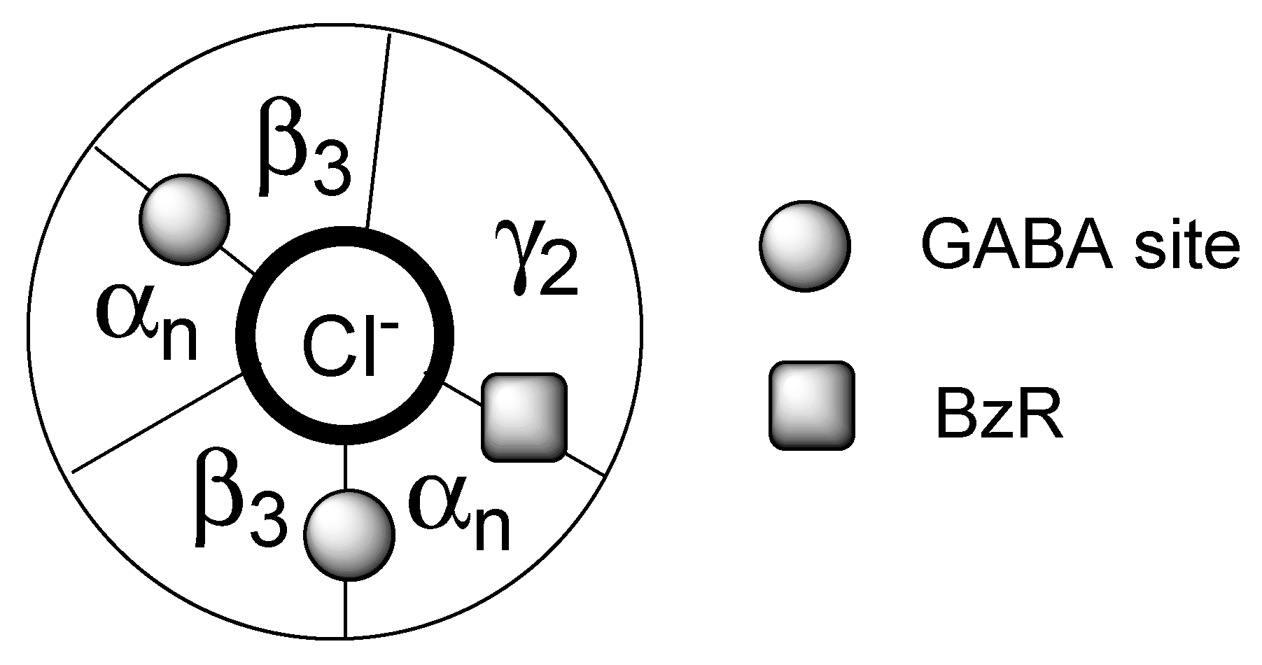
Figure 1. Schematic representation of the organization of the five protein subunits composing the major GABAA complex isoforms. There are two β3 subunits, one γ2 subunits and two among six α subunits (where n varies from 1 to 6). The circle in bold corresponds to the chloride channel. The binding sites of the neurotransmitter GABA and of the BzR ligands are evidenced by filled circles and, respectively, a square.
Molecules 25 02331 g001
Figure 2. The binding modes A and B hypothesized for the 5-Cl/NO2 indoles and, respectively of the 5-H indoles oriented in the framework of the Cook’s pharmacophore/topological model [31]. Z is a CH2 in benzylamides 6 or a NH in hydrazides 9.
Molecules 25 02331 g002
Figure 3. Putative binding mode A of some of the most potent ligands of series 16 hypothesized to interact with a hydroxy group located at the S1 subsite of the BzR.
Molecules 25 02331 g003
Figure 4. Pharmacophore/topological model of interaction between PIGAs and TSPO.
Molecules 25 02331 g004