
The History of Nanoscience and Nanotechnology: From Chemical–Physical Applications to Nanomedicine

 ,
, 
 ,
,  and
and
Abstract
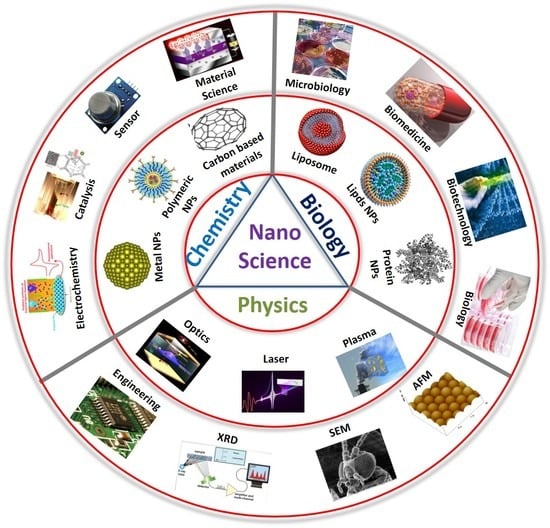
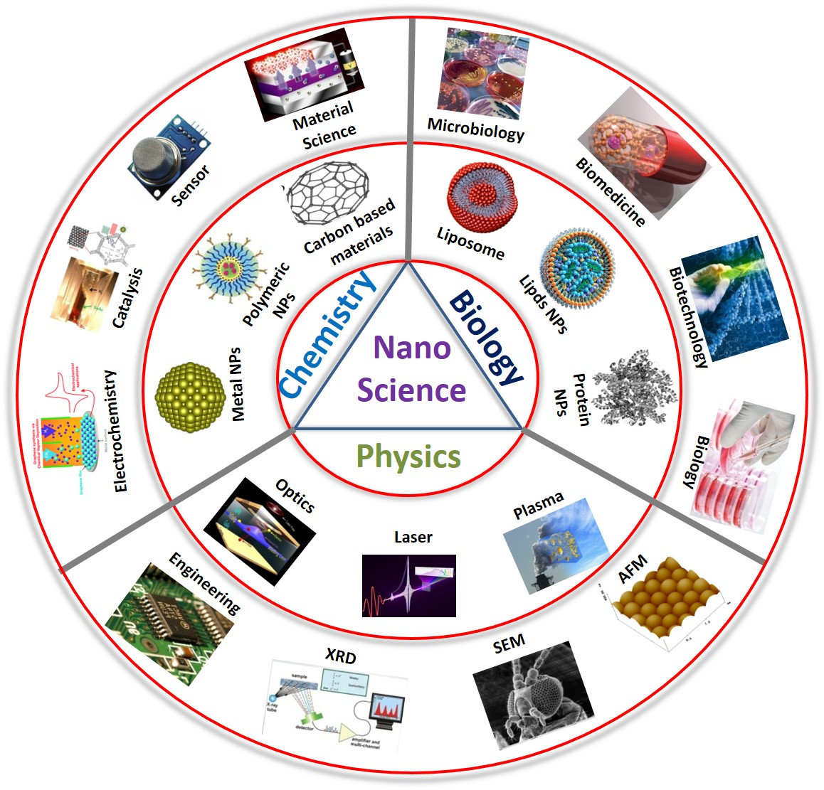
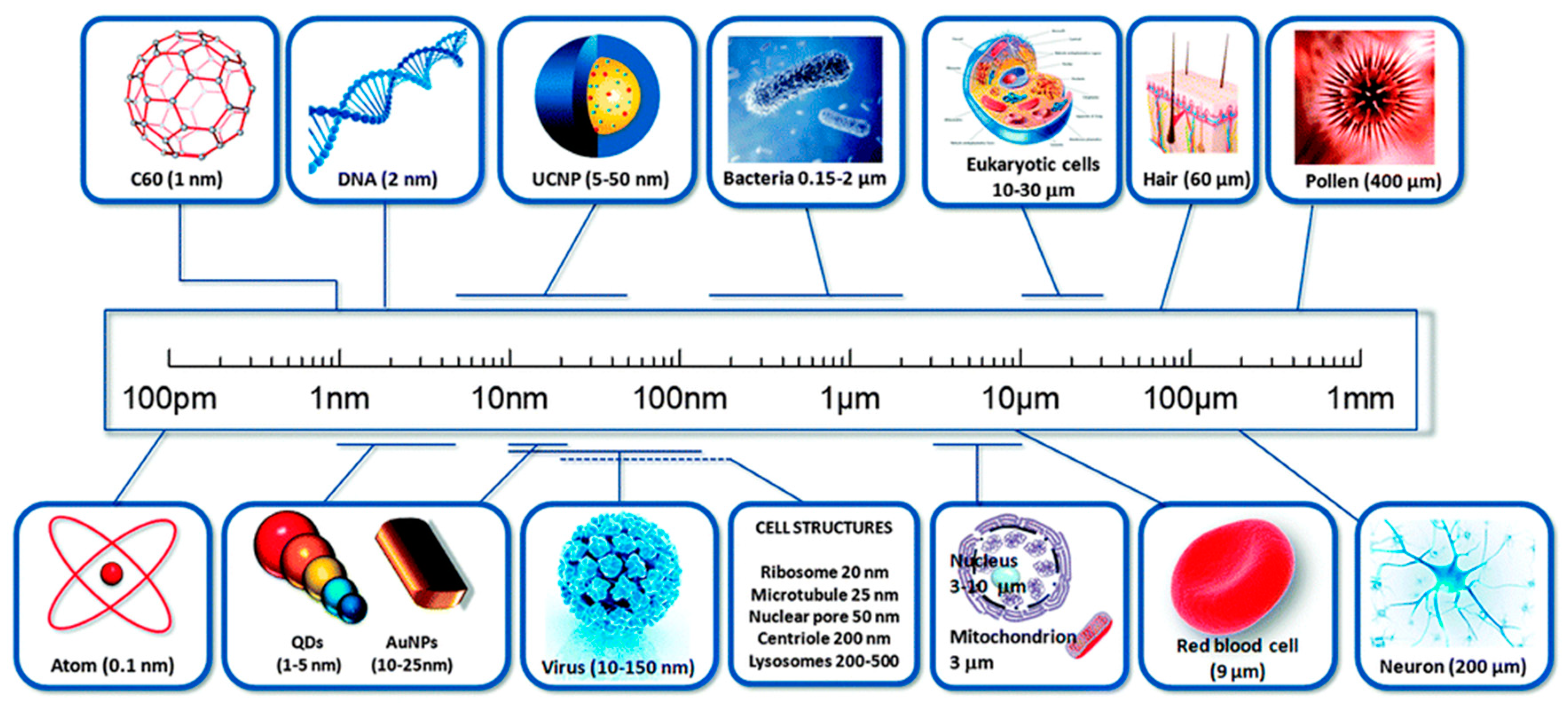
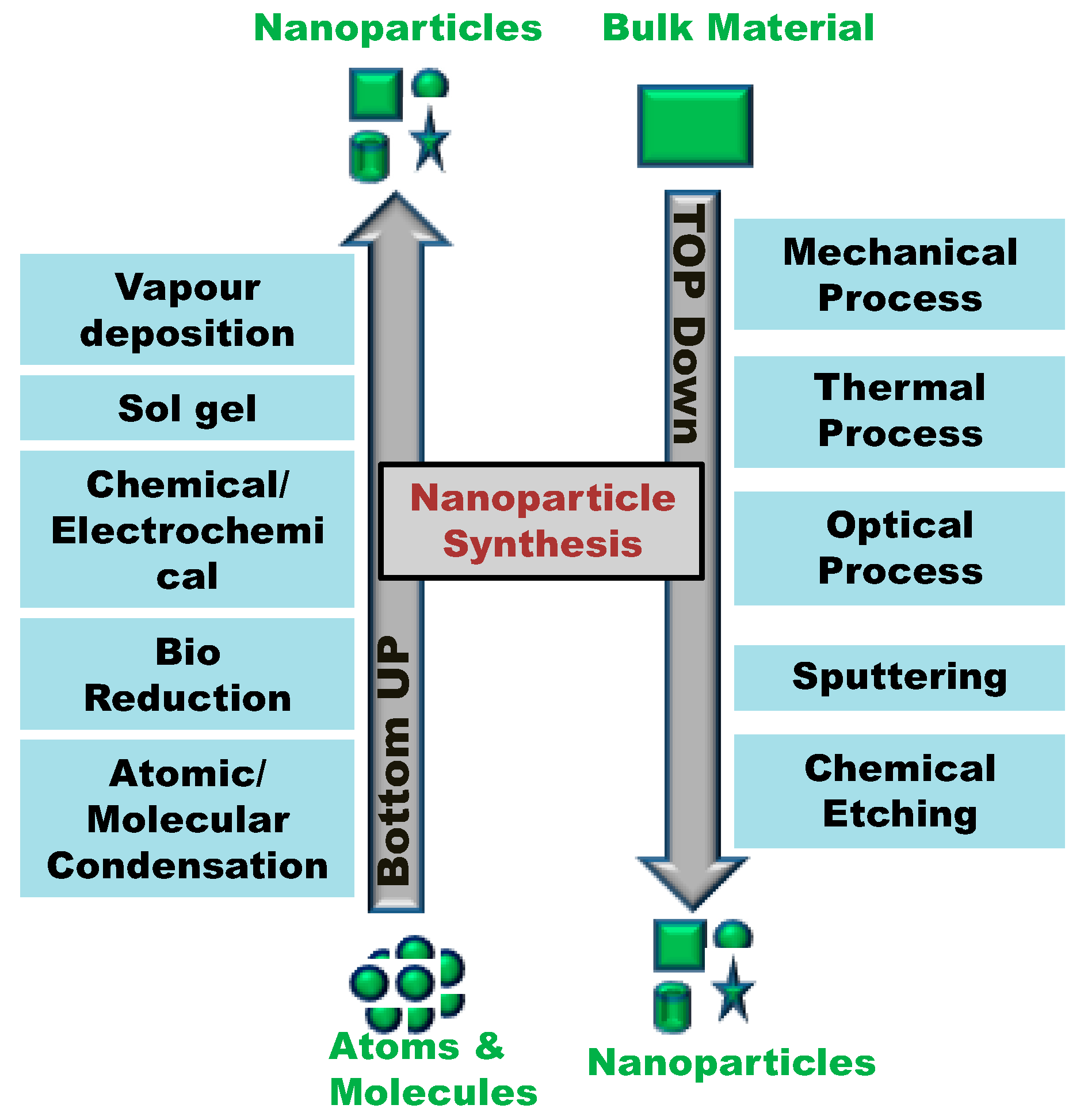
1. Definition of Nanoscience and Nanotechnology
2. The Imaginative Pioneers of Nanotechnology
3. History of Nanotechnology
4. Modern Era of Nanotechnology
5. Conclusions
Author Contributions
Funding
Acknowledgments
Conflicts of Interest
References
- Mansoori, G.; Fauzi Soelaiman, T. Nanotechnology—An Introduction for the Standards Community. J. ASTM Int. 2005, 2, 1–22. [Google Scholar]
- Gnach, A.; Lipinski, T.; Bednarkiewicz, A.; Rybka, J.; Capobianco, J.A. Upconverting nanoparticles: Assessing the toxicity. Chem. Soc. Rev. 2015, 44, 1561–1584. [Google Scholar] [CrossRef] [PubMed]
- National Nanotechnology Initiative (NNI). Available online: www.nano.gov (accessed on 22 July 2019).
- Allhoff, F. On the Autonomy and Justification of Nanoethics. Nanoethics 2007, 1, 185–210. [Google Scholar] [CrossRef]
- Feynman, R.P. There’s plenty of room at the bottom. Eng. Sci. 1960, 23, 22–36. [Google Scholar]
- Taniguchi, N.; Arakawa, C.; Kobayashi, T. On the basic concept of nano-technology. In Proceedings of the International Conference on Production Engineering, Tokyo, Japan, 26–29 August 1974. [Google Scholar]
- Iqbal, P.; Preece, J.A.; Mendes, P.M. Nanotechnology: The “Top-Down” and “Bottom-Up” Approaches. In Supramolecular Chemistry; John Wiley & Sons, Ltd.: Chichester, UK, 2012. [Google Scholar]
- Drexler, E.K. Engines of Creation: The Coming Era of Nanotechnology; Anchor Press: Garden City, NY, USA, 1986. [Google Scholar]
- Drexler, E.K.; Peterson, C.; Pergamit, G. Unbounding the Future: The Nanotechnology Revolution; William Morrow and Company, Inc.: New York, NY, USA, 1991. [Google Scholar] [CrossRef][Green Version]
- The British Museum. Available online: www.britishmuseum.org/research/collection_online/collection_object_details.aspx?objobjec=61219&partId=1 (accessed on 22 July 2019).
- Barber, D.J.; Freestone, I.C. An investigation of the origin of the colour of the Lycurgus Cup by analytical transmission electron microscopy. Archaeometry 1990, 32, 33–45. [Google Scholar] [CrossRef]
- Freestone, I.; Meeks, N.; Sax, M.; Higgitt, C. The Lycurgus Cup—A Roman nanotechnology. Gold Bull. 2007, 40, 270–277. [Google Scholar] [CrossRef]
- Wagner, F.E.; Haslbeck, S.; Stievano, L.; Calogero, S.; Pankhurst, Q.A.; Martinek, K.-P. Before striking gold in gold-ruby glass. Nature 2000, 407, 691–692. [Google Scholar] [CrossRef]
- The New York Times. Available online: www.nytimes.com/imagepages/2005/02/21/science/20050222_NANO1_GRAPHIC.html (accessed on 22 July 2019).
- Pradell, T.; Climent-Font, A.; Molera, J.; Zucchiatti, A.; Ynsa, M.D.; Roura, P.; Crespo, D. Metallic and nonmetallic shine in luster: An elastic ion backscattering study. J. Appl. Phys. 2007, 101, 103518. [Google Scholar] [CrossRef]
- Poole, C.P.; Owens, F.J. Introduction to Nanotechnology; John Wiley & Sons: New York, NY, USA, 2003. [Google Scholar]
- Reibold, M.; Paufler, P.; Levin, A.A.; Kochmann, W.; Pätzke, N.; Meyer, D.C. Materials: Carbon nanotubes in an ancient Damascus sabre. Nature 2006, 444, 286. [Google Scholar] [CrossRef]
- Faraday, M. The Bakerian Lecture: Experimental Relations of Gold (and Other Metals) to Light. Philos. Trans. R. Soc. Lond. 1857, 147, 145–181. [Google Scholar]
- Binnig, G.; Rohrer, H.; Gerber, C.; Weibel, E. Tunneling through a controllable vacuum gap. Appl. Phys. Lett. 1982, 40, 178. [Google Scholar] [CrossRef]
- Binnig, G.; Rohrer, H.; Gerber, C.; Weibel, E. Surface Studies by Scanning Tunneling Microscopy. Phys. Rev. Lett. 1982, 49, 57–61. [Google Scholar] [CrossRef]
- Binnig, G.; Rohrer, H.; Gerber, C.; Weibel, E. 7 × 7 Reconstruction on Si(111) Resolved in Real Space. Phys. Rev. Lett. 1983, 50, 120–123. [Google Scholar] [CrossRef]
- Institute of Physics Polish Academy of Sciences. Available online: http://info.ifpan.edu.pl/~wawro/subframes/Surfaces.htm (accessed on 22 July 2019).
- Eigler, D.M.; Schweizer, E.K. Positioning single atoms with a scanning tunnelling microscope. Nature 1990, 344, 524–526. [Google Scholar] [CrossRef]
- Binnig, G.; Quate, C.F.; Gerber, C. Atomic Force Microscope. Phys. Rev. Lett. 1986, 56, 930–933. [Google Scholar] [CrossRef]
- Binnig, G. Atomic Force Microscope and Method for Imaging Surfaces with Atomic Resolution. U.S. Patent 4724318A, 16 October 1990. [Google Scholar]
- Kroto, H.W.; Heath, J.R.; O’Brien, S.C.; Curl, R.F.; Smalley, R.E. C60: Buckminsterfullerene. Nature 1985, 318, 162–163. [Google Scholar] [CrossRef]
- Iijima, S. Helical microtubules of graphitic carbon. Nature 1991, 354, 56–58. [Google Scholar] [CrossRef]
- Xu, X.; Ray, R.; Gu, Y.; Ploehn, H.J.; Gearheart, L.; Raker, K.; Scrivens, W. A Electrophoretic Analysis and Purification of Fluorescent Single-Walled Carbon Nanotube Fragments. J. Am. Chem. Soc. 2004, 126, 12736–12737. [Google Scholar] [CrossRef]
- Baker, S.N.; Baker, G.A. Luminescent carbon nanodots: Emergent nanolights. Angew. Chem. Int. Ed. Engl. 2010, 49, 6726–6744. [Google Scholar] [CrossRef]
- Esteves da Silva, J.C.G.; Gonçalves, H.M.R. Analytical and bioanalytical applications of carbon dots. TrAC Trends Anal. Chem. 2011, 30, 1327–1336. [Google Scholar] [CrossRef]
- Yang, S.-T.; Cao, L.; Luo, P.G.; Lu, F.; Wang, X.; Wang, H.; Meziani, M.J.; Liu, Y.; Qi, G.; Sun, Y.-P. Carbon Dots for Optical Imaging in Vivo. J. Am. Chem. Soc. 2009, 131, 11308–11309. [Google Scholar] [CrossRef] [PubMed]
- Yang, S.-T.; Wang, X.; Wang, H.; Lu, F.; Luo, P.G.; Cao, L.; Meziani, M.J.; Liu, J.-H.; Liu, Y.; Chen, M.; et al. Carbon Dots as Nontoxic and High-Performance Fluorescence Imaging Agents. J. Phys. Chem. C 2009, 113, 18110–18114. [Google Scholar] [CrossRef] [PubMed]
- Cao, L.; Wang, X.; Meziani, M.J.; Lu, F.; Wang, H.; Luo, P.G.; Lin, Y.; Harruff, B.A.; Veca, L.M.; Murray, D.; et al. Carbon Dots for Multiphoton Bioimaging. J. Am. Chem. Soc. 2007, 129, 11318–11319. [Google Scholar] [CrossRef] [PubMed]
- Li, Q.; Ohulchanskyy, T.Y.; Liu, R.; Koynov, K.; Wu, D.; Best, A.; Kumar, R.; Bonoiu, A.; Prasad, P.N. Photoluminescent Carbon Dots as Biocompatible Nanoprobes for Targeting Cancer Cells in Vitro. J. Phys. Chem. C 2010, 114, 12062–12068. [Google Scholar] [CrossRef]
- Bayda, S.; Hadla, M.; Palazzolo, S.; Kumar, V.; Caligiuri, I.; Ambrosi, E.; Pontoglio, E.; Agostini, M.; Tuccinardi, T.; Benedetti, A.; et al. Bottom-up synthesis of carbon nanoparticles with higher doxorubicin efficacy. J. Control. Release 2017, 248, 144–152. [Google Scholar] [CrossRef]
- Wang, X.; Cao, L.; Lu, F.; Meziani, M.J.; Li, H.; Qi, G.; Zhou, B.; Harruff, B.A.; Kermarrec, F.; Sun, Y.-P. Photoinduced electron transfers with carbon dots. Chem. Commun. 2009, 25, 3774–3776. [Google Scholar] [CrossRef]
- Li, Y.; Hu, Y.; Zhao, Y.; Shi, G.; Deng, L.; Hou, Y.; Qu, L. An Electrochemical Avenue to Green-Luminescent Graphene Quantum Dots as Potential Electron-Acceptors for Photovoltaics. Adv. Mater. 2011, 23, 776–780. [Google Scholar] [CrossRef]
- Zhou, L.; Lin, Y.; Huang, Z.; Ren, J.; Qu, X. Carbon nanodots as fluorescence probes for rapid, sensitive, and label-free detection of Hg2+ and biothiols in complex matrices. Chem. Commun. 2012, 48, 1147–1149. [Google Scholar] [CrossRef]
- Liu, L.; Li, Y.; Zhan, L.; Liu, Y.; Huang, C. One-step synthesis of fluorescent hydroxyls-coated carbon dots with hydrothermal reaction and its application to optical sensing of metal ions. Sci. China Chem. 2011, 54, 1342–1347. [Google Scholar] [CrossRef]
- Kinnear, C.; Moore, T.L.; Rodriguez-Lorenzo, L.; Rothen-Rutishauser, B.; Petri-Fink, A. Form Follows Function: Nanoparticle Shape and Its Implications for Nanomedicine. Chem. Rev. 2017, 117, 11476–11521. [Google Scholar] [CrossRef]
- Weissig, V.; Pettinger, T.K.; Murdock, N. Nanopharmaceuticals (part 1): Products on the market. Int. J. Nanomed. 2014, 9, 4357–4373. [Google Scholar] [CrossRef] [PubMed]
- Rothemund, P.W.K. Folding DNA to create nanoscale shapes and patterns. Nature 2006, 440, 297–302. [Google Scholar] [CrossRef] [PubMed]
- Seeman, N.C. Nucleic acid junctions and lattices. J. Theor. Biol. 1982, 99, 237–247. [Google Scholar] [CrossRef]
- Kumar, V.; Bayda, S.; Hadla, M.; Caligiuri, I.; Russo Spena, C.; Palazzolo, S.; Kempter, S.; Corona, G.; Toffoli, G.; Rizzolio, F. Enhanced Chemotherapeutic Behavior of Open-Caged DNA@Doxorubicin Nanostructures for Cancer Cells. J. Cell. Physiol. 2016, 231, 106–110. [Google Scholar] [CrossRef]
- Kumar, V.; Palazzolo, S.; Bayda, S.; Corona, G.; Toffoli, G.; Rizzolio, F. DNA Nanotechnology for Cancer Therapy. Theranostics 2016, 6, 710–725. [Google Scholar] [CrossRef]
- Palazzolo, S.; Hadla, M.; Spena, C.R.; Bayda, S.; Kumar, V.; Lo Re, F.; Adeel, M.; Caligiuri, I.; Romano, F.; Corona, G.; et al. Proof-of-Concept Multistage Biomimetic Liposomal DNA Origami Nanosystem for the Remote Loading of Doxorubicin. ACS Med. Chem. Lett. 2019, 10, 517–521. [Google Scholar] [CrossRef]
- Palazzolo, S.; Hadla, M.; Russo Spena, C.; Caligiuri, I.; Rotondo, R.; Adeel, M.; Kumar, V.; Corona, G.; Canzonieri, V.; Toffoli, G.; et al. An Effective Multi-Stage Liposomal DNA Origami Nanosystem for In Vivo Cancer Therapy. Cancers 2019, 11, 1997. [Google Scholar] [CrossRef]
- Lee, P.Y.; Wong, K.K.Y. Nanomedicine: A new frontier in cancer therapeutics. Curr. Drug Deliv. 2011, 8, 245–253. [Google Scholar]
- Yuan, Y.; Gu, Z.; Yao, C.; Luo, D.; Yang, D. Nucleic Acid–Based Functional Nanomaterials as Advanced Cancer Therapeutics. Small 2019, 15, 1900172. [Google Scholar] [CrossRef]
- Cordani, M.; Somoza, Á. Targeting autophagy using metallic nanoparticles: A promising strategy for cancer treatment. Cell. Mol. Life Sci. 2019, 76, 1215–1242. [Google Scholar] [CrossRef]
- Sharma, N.; Sharma, M.; Sajid Jamal, Q.M.; Kamal, M.A.; Akhtar, S. Nanoinformatics and biomolecular nanomodeling: A novel move en route for effective cancer treatment. Environ. Sci. Pollut. Res. Int. 2019, 1–15. [Google Scholar] [CrossRef] [PubMed]
- Hulla, J.; Sahu, S.; Hayes, A. Nanotechnology. Hum. Exp. Toxicol. 2015, 34, 1318–1321. [Google Scholar] [CrossRef] [PubMed]
- Sciau, P. Nanoparticles in Ancient Materials: The Metallic Lustre Decorations of Medieval Ceramics. In The Delivery of Nanoparticles; InTech: Rijeka, Croatia, 2012. [Google Scholar] [CrossRef]
- Mie, G. Beiträge zur Optik trüber Medien, speziell kolloidaler Metallösungen. Ann. Phys. 1908, 330, 377–445. [Google Scholar] [CrossRef]
- Synge, E.H. XXXVIII. A suggested method for extending microscopic resolution into the ultra-microscopic region. Lond. Edinb. Dublin Philos. Mag. J. Sci. 1928, 6, 356–362. [Google Scholar] [CrossRef]
- Knoll, M.; Ruska, E. Beitrag zur geometrischen Elektronenoptik. I. Ann. Phys. 1932, 404, 607–640. [Google Scholar] [CrossRef]
- Knoll, M.; Ruska, E. Beitrag zur geometrischen Elektronenoptik. II. Ann. Phys. 1932, 404, 641–661. [Google Scholar] [CrossRef]
- Müller, E.W. Experimente zur Theorie der Elektronenemission unter dem Einfluß starker Felder. Phys. Z. 1936, 37, 838–841. [Google Scholar]
- Shockley, W. Circuit Element Utilizing Semiconductive Material. U.S. Patent 2569347A, 25 September 1951. [Google Scholar]
- Müller, E.W. Das Feldionenmikroskop. Z. Phys. 1951, 131, 136–142. [Google Scholar] [CrossRef]
- Müller, E.W.; Bahadur, K. Field Ionization of Gases at a Metal Surface and the Resolution of the Field Ion Microscope. Phys. Rev. 1956, 102, 624–631. [Google Scholar] [CrossRef]
- Watson, J.D.; Crick, F.H.C. Molecular Structure of Nucleic Acids: A Structure for Deoxyribose Nucleic Acid. Nature 1953, 171, 737–738. [Google Scholar] [CrossRef]
- Von Hippel, A. Molecular Engineering. Science 1956, 123, 315–316. [Google Scholar] [CrossRef] [PubMed]
- Esaki, L. New Phenomenon in Narrow Germanium p-n Junctions. Phys. Rev. 1958, 109, 603–604. [Google Scholar] [CrossRef]
- Plank, C.J.; Rosinski, E.J. Catalytic Cracking of Hydrocarbons with a Crystalline Zeolite Catalyst Composite. U.S. Patent 3140249A, 7 July 1964. [Google Scholar]
- Papell, S.S. Low Viscosity Magnetic Fluid Obtained by the Colloidal Suspension of Magnetic Particles. U.S. Patent 3215572A, 2 November 1965. [Google Scholar]
- Moore, G.E. Cramming more components onto integrated circuits. Electronics 1965, 38, 114–117. [Google Scholar] [CrossRef]
- Osawa, E. Superaromaticity. Kagaku Kyoto 1970, 25, 854–863. [Google Scholar]
- Aviram, A.; Ratner, M.A. Molecular rectifiers. Chem. Phys. Lett. 1974, 29, 277–283. [Google Scholar] [CrossRef]
- Jeanmaire, D.L.; Van Duyne, R.P. Surface raman spectroelectrochemistry. J. Electroanal. Chem. Interfacial Electrochem. 1977, 84, 1–20. [Google Scholar] [CrossRef]
- Sagiv, J. Organized monolayers by adsorption. 1. Formation and structure of oleophobic mixed monolayers on solid surfaces. J. Am. Chem. Soc. 1980, 102, 92–98. [Google Scholar] [CrossRef]
- Binnig, G.; Rohrer, H. Scanning Tunneling Microscope. U.S. Patent 4343993A, 10 August 1982. [Google Scholar]
- Ekimov, A.; Onushchenko, A. Quantum size effect in the optical-spectra of semiconductor micro-crystals. Sov. Phys. Semicond. 1982, 16, 775–778. [Google Scholar]
- Drexler, E.K. Molecular engineering: An approach to the development of general capabilities for molecular manipulation. Proc. Natl. Acad. Sci. USA 1981, 78, 5275–5278. [Google Scholar] [CrossRef]
- Pinheiro, A.V.; Han, D.; Shih, W.M.; Yan, H. Challenges and opportunities for structural DNA nanotechnology. Nat. Nanotechnol. 2011, 6, 763–772. [Google Scholar] [CrossRef]
- Rossetti, R.; Nakahara, S.; Brus, L.E. Quantum size effects in the redox potentials, resonance Raman spectra, and electronic spectra of CdS crystallites in aqueous solution. J. Chem. Phys. 1983, 79, 1086. [Google Scholar] [CrossRef]
- Steigerwald, M.L.; Alivisatos, A.P.; Gibson, J.M.; Harris, T.D.; Kortan, R.; Muller, A.J.; Thayer, A.M.; Duncan, T.M.; Douglass, D.C.; Brus, L.E. Surface derivatization and isolation of semiconductor cluster molecules. J. Am. Chem. Soc. 1988, 110, 3046–3050. [Google Scholar] [CrossRef]
- Averin, D.V.; Likharev, K.K. Coulomb blockade of single-electron tunneling, and coherent oscillations in small tunnel junctions. J. Low Temp. Phys. 1986, 62, 345–373. [Google Scholar] [CrossRef]
- Kresge, C.T.; Leonowicz, M.E.; Roth, W.J.; Vartuli, J.C.; Beck, J.S. Ordered mesoporous molecular sieves synthesized by a liquid-crystal template mechanism. Nature 1992, 359, 710–712. [Google Scholar] [CrossRef]
- Beck, J.S.; Vartuli, J.C.; Roth, W.J.; Leonowicz, M.E.; Kresge, C.T.; Schmitt, K.D.; Chu, C.T.W.; Olson, D.H.; Sheppard, E.W.; McCullen, S.B.; et al. A new family of mesoporous molecular sieves prepared with liquid crystal templates. J. Am. Chem. Soc. 1992, 114, 10834–10843. [Google Scholar] [CrossRef]
- Iijima, S.; Ichihashi, T. Single-shell carbon nanotubes of 1-nm diameter. Nature 1993, 363, 603–605. [Google Scholar] [CrossRef]
- Bethune, D.S.; Klang, C.H.; de Vries, M.S.; Gorman, G.; Savoy, R.; Vazquez, J.; Beyers, R. Cobalt-catalysed growth of carbon nanotubes with single-atomic-layer walls. Nature 1993, 363, 605–607. [Google Scholar] [CrossRef]
- Mirkin, C.A.; Letsinger, R.L.; Mucic, R.C.; Storhoff, J.J. A DNA-based method for rationally assembling nanoparticles into macroscopic materials. Nature 1996, 382, 607–609. [Google Scholar] [CrossRef]
- Zyvex Technologies. Available online: www.zyvex.com (accessed on 22 July 2019).
- Tans, S.J.; Verschueren, A.R.M.; Dekker, C. Room-temperature transistor based on a single carbon nanotube. Nature 1998, 393, 49–52. [Google Scholar] [CrossRef]
- Piner, R.D.; Zhu, J.; Xu, F.; Hong, S.; Mirkin, C.A. “Dip-Pen” Nanolithography. Science 1999, 283, 661–663. [Google Scholar] [CrossRef]
- Hersam, M.C.; Guisinger, N.P.; Lyding, J.W. Isolating, imaging, and electrically characterizing individual organic molecules on the Si(100) surface with the scanning tunneling microscope. J. Vac. Sci. Technol. A Vac. Surf. Films 2000, 18, 1349. [Google Scholar] [CrossRef]
- Lok, C. Nanotechnology: Small wonders. Nature 2010, 467, 18–21. [Google Scholar] [CrossRef] [PubMed][Green Version]
- Montemagno, C.D. Nanomachines: A Roadmap for Realizing the Vision. J. Nanoparticle Res. 2001, 3, 1–3. [Google Scholar] [CrossRef]
- Williams, K.A.; Veenhuizen, P.T.M.; de la Torre, B.G.; Eritja, R.; Dekker, C. Nanotechnology: Carbon nanotubes with DNA recognition. Nature 2002, 420, 761. [Google Scholar] [CrossRef] [PubMed]
- 21st Century Nanotechnology Research and Development Act. Available online: https://www.congress.gov/bill/108th-congress/senate-bill/189 (accessed on 22 July 2019).
- Loo, C.; Lin, A.; Hirsch, L.; Lee, M.-H.; Barton, J.; Halas, N.; West, J.; Drezek, R. Nanoshell-Enabled Photonics-Based Imaging and Therapy of Cancer. Technol. Cancer Res. Treat. 2004, 3, 33–40. [Google Scholar] [CrossRef]
- Hirsch, L.R.; Stafford, R.J.; Bankson, J.A.; Sershen, S.R.; Rivera, B.; Price, R.E.; Hazle, J.D.; Halas, N.J.; West, J.L. Nanoshell-mediated near-infrared thermal therapy of tumors under magnetic resonance guidance. Proc. Natl. Acad. Sci. USA 2003, 100, 13549–13554. [Google Scholar] [CrossRef]
- Novoselov, K.S.; Geim, A.K.; Morozov, S.V.; Jiang, D.; Zhang, Y.; Dubonos, S.V.; Grigorieva, I.V.; Firsov, A.A. Electric Field Effect in Atomically Thin Carbon Films. Science 2004, 306, 666–669. [Google Scholar] [CrossRef]
- Shirai, Y.; Osgood, A.J.; Zhao, Y.; Kelly, K.F.; Tour, J.M. Directional Control in Thermally Driven Single-Molecule Nanocars. Nano Lett. 2005, 5, 2330–2334. [Google Scholar] [CrossRef]
- Morin, J.-F.; Shirai, Y.; Tour, J.M. En Route to a Motorized Nanocar. Org. Lett. 2006, 8, 1713–1716. [Google Scholar] [CrossRef]
- Du, G.; Moulin, E.; Jouault, N.; Buhler, E.; Giuseppone, N. Muscle-like Supramolecular Polymers: Integrated Motion from Thousands of Molecular Machines. Angew. Chem. 2012, 124, 12672–12676. [Google Scholar] [CrossRef]
- Sanders, J.K.M.; Jackson, S.E. The discovery and development of the green fluorescent protein, GFP. Chem. Soc. Rev. 2009, 38, 2821. [Google Scholar] [CrossRef] [PubMed]
- Zheng, J.; Birktoft, J.J.; Chen, Y.; Wang, T.; Sha, R.; Constantinou, P.E.; Ginell, S.L.; Mao, C.; Seeman, N.C. From molecular to macroscopic via the rational design of a self-assembled 3D DNA crystal. Nature 2009, 461, 74–77. [Google Scholar] [CrossRef] [PubMed]
- Knoll, A.W.; Pires, D.; Coulembier, O.; Dubois, P.; Hedrick, J.L.; Frommer, J.; Duerig, U. Probe-Based 3-D Nanolithography Using Self-Amplified Depolymerization Polymers. Adv. Mater. 2010, 22, 3361–3365. [Google Scholar] [CrossRef] [PubMed]
- Lafferentz, L.; Ample, F.; Yu, H.; Hecht, S.; Joachim, C.; Grill, L. Conductance of a Single Conjugated Polymer as a Continuous Function of Its Length. Science 2009, 323, 1193–1197. [Google Scholar] [CrossRef]
- Richards, V. 2016 Nobel Prize in Chemistry: Molecular machines. Nat. Chem. 2016, 8, 1090. [Google Scholar] [CrossRef]
- Nobel Foundation. Nobel Prize in Physics 2017: Gravitational Waves. Available online: www.sciencedaily.com/releases/2017/10/171003095828.htm (accessed on 22 July 2019).
- Petersen, P.; Tikhomirov, G.; Qian, L. Information-based autonomous reconfiguration in systems of interacting DNA nanostructures. Nat. Commun. 2018, 9, 5362. [Google Scholar] [CrossRef]
- Oran, D.; Rodriques, S.G.; Gao, R.; Asano, S.; Skylar-Scott, M.A.; Chen, F.; Tillberg, P.W.; Marblestone, A.H.; Boyden, E.S. 3D nanofabrication by volumetric deposition and controlled shrinkage of patterned scaffolds. Science 2018, 362, 1281–1285. [Google Scholar] [CrossRef]


| Year | Event | References |
|---|---|---|
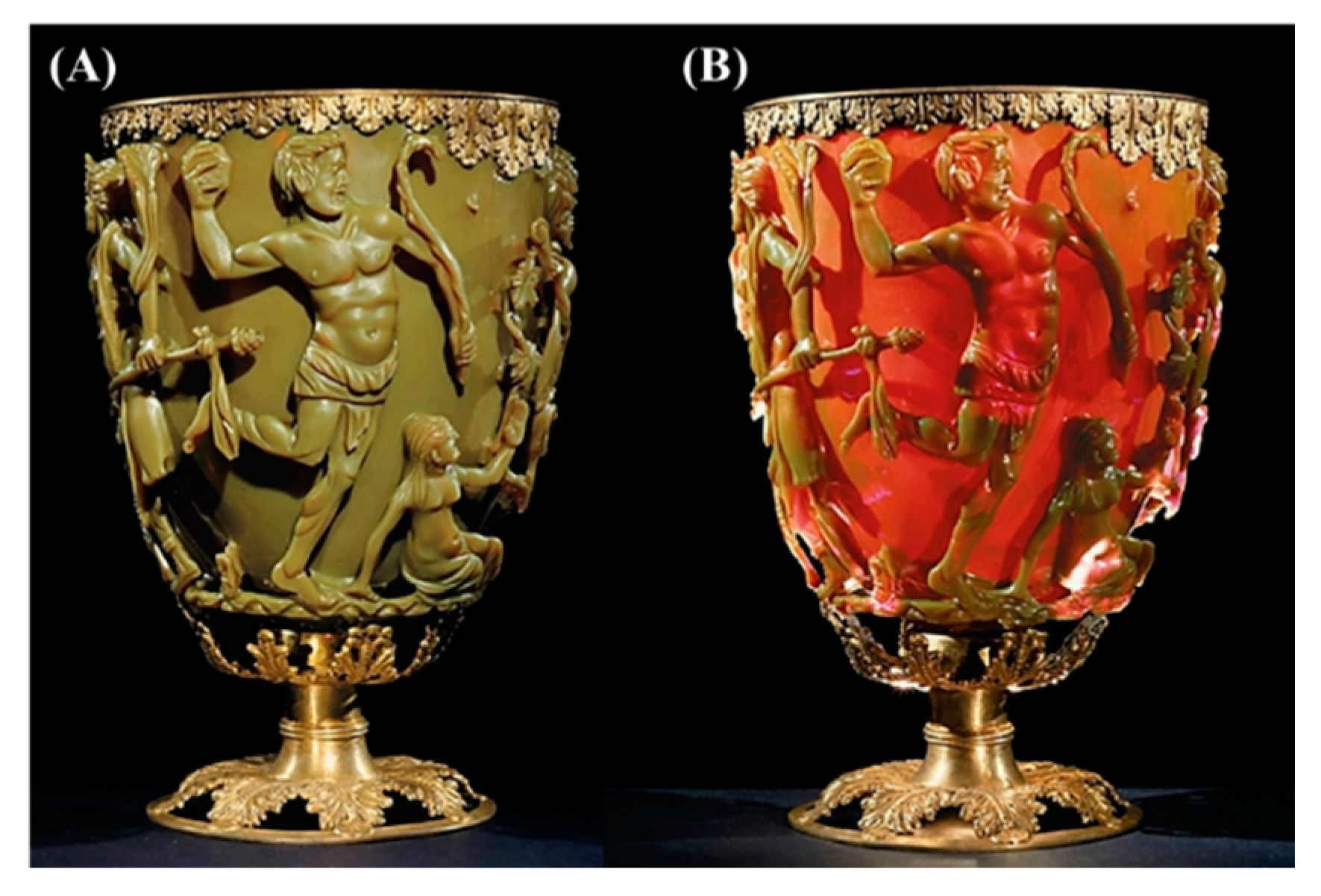
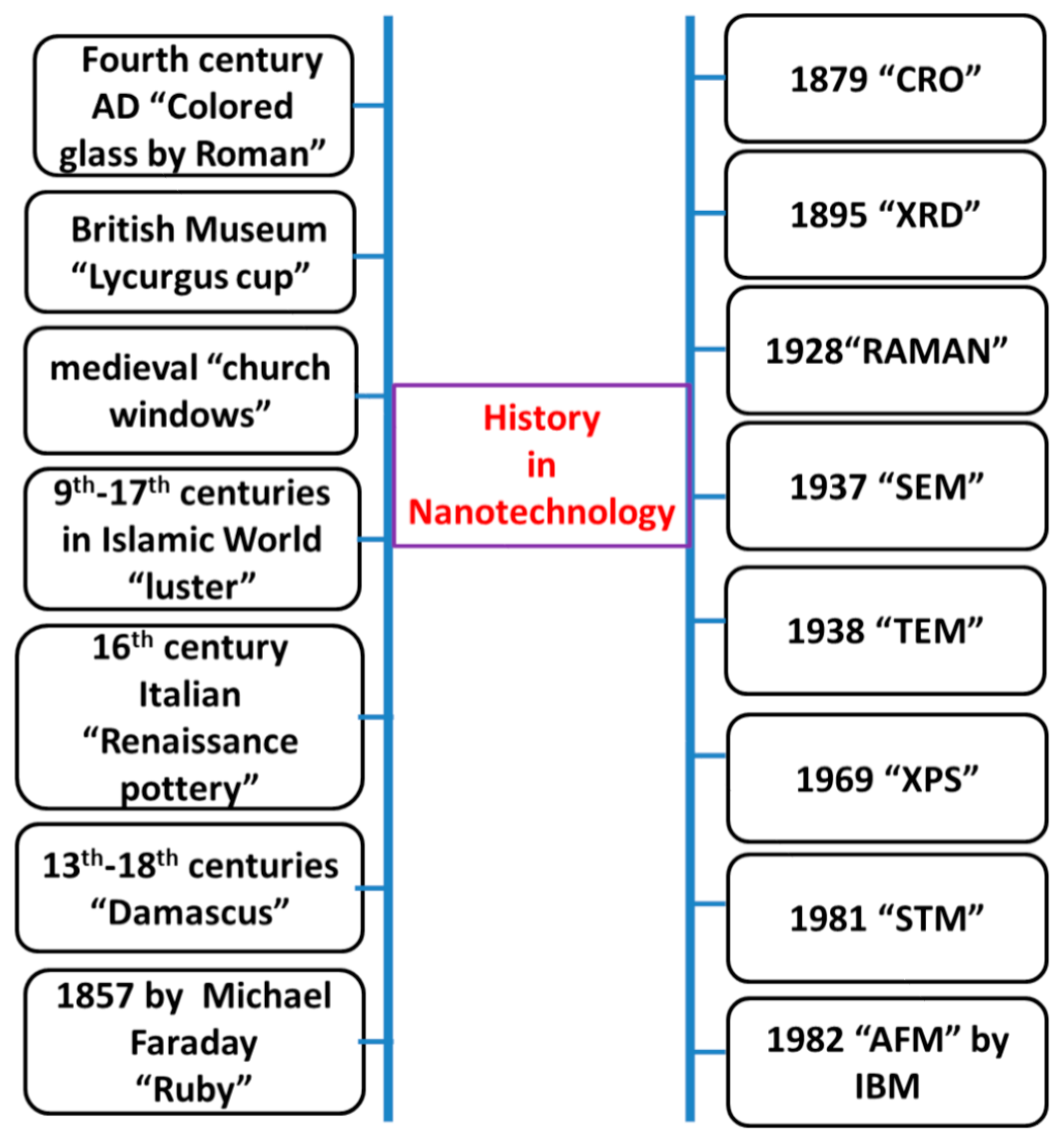
| 4th Century | Lycurgus Cup (Colored glass). | [12] |
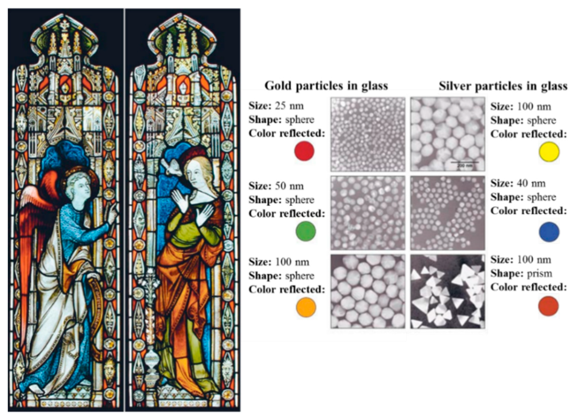
| 500–1450 | Cathedrals (Stained glasses windows). | [53] |
| 1450–1600 | Deruta Pottery (Iridescent/metallic clusters). | [53] |
| 1857 | Michael Faraday (Synthesis of colloidal ruby gold nanoparticles). | [18] |
| 1908 | Gustav Mie (Light scattering nanoparticles). | [54] |
| 1928 | Edward Synge (Near-field optical microscope). | [55] |
| 1931 | Max Knoll and Ernst Ruska (invention of transmission electron microscope (TEM)). | [56,57] |
| 1936 | Erwin Müller (Invention of field electron microscope). | [58] |
| 1947 | William Shockley, Walter Brattain and John Bardeen (Discovery of the semiconductor transistor). | [59] |
| 1951 | Erwin Müller (Invention of field-ion microscope, first to see atoms on the surface). | [60,61] |
| 1953 | James Watson and Francis Crick (Discovery of DNA). | [62] |
| 1956 | Arthur Von Hippel (Molecular Engineering). | [63] |
| 1958 | Leo Esaki (Electron tunneling). | [64] |
| 1959 | Richard Feynman (There’s Plenty of Room at the Bottom). | [5] |
| 1960 | Charles Plank and Edward Rosinski (Zeolites and catalysis). | [65] |
| 1963 | Stephen Papell (Invention of Ferrofluids). | [66] |
| 1965 | Gordon E. Moore (Moore’s Law). | [67] |
| 1970 | Eiji Osawa (Predicted the existence of C60 in the form of icosahedron). | [68] |
| 1974 | Norio Taniguchi (First use of the term “Nanotechnology”). | [6] |
| 1974 | Mark A. Ratner and Arieh Aviram (Molecular electronics). | [69] |
| 1977 | Richard P. Van Duyne (Discovery of Surface Enhanced Raman Spectroscopy (SERS)). | [70] |
| 1980 | Jacop Sagiv (Discovery of Self-Assembly Monolayers (SAMs)). | [71] |
| 1981 | Gerd Binnig and Heinrich Rohrer (Invention of Scanning Tunneling Microscope (STM)). | [72] |
| 1981 | Alexey Ekimov (Discovery of nanocrystalline Quantum Dots in a glass matrix). | [73] |
| 1981 | Eric Drexler (Molecular Engineering). | [74] |
| 1982 | Nadrian Seeman (Development of the concept of DNA Nanotechnology). | [43,75] |
| 1983 | Louis Brus (Discovery of colloidal Quantum Dots). | [76,77] |
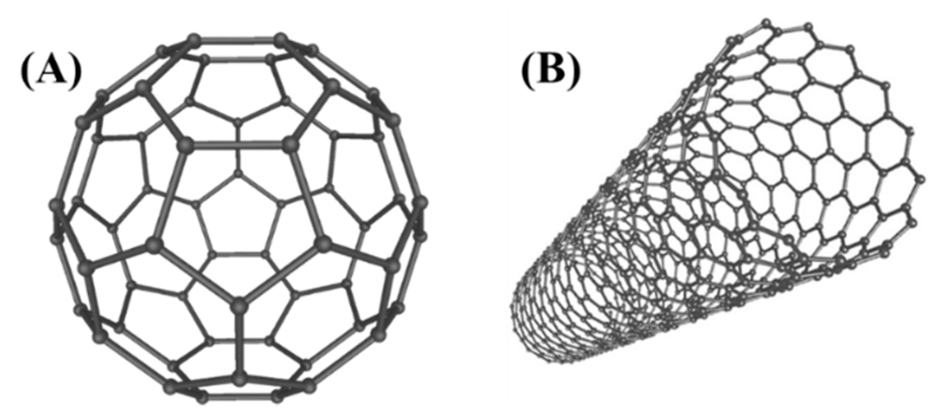
| 1985 | Richard Smalley, Robert Curl and Harold Kroto (Discovery of Buckminsterfullerenes C60). | [26] |
| 1986 | Gerd Binnig, Christoph Gerber and Calvin F. Quate (Invention of Atomic Force Microscope (AFM). | [24] |
| 1987 | Dimitri Averin and Konstantin Likharev (Single-Electron Tunneling (SET) transistor). | [78] |
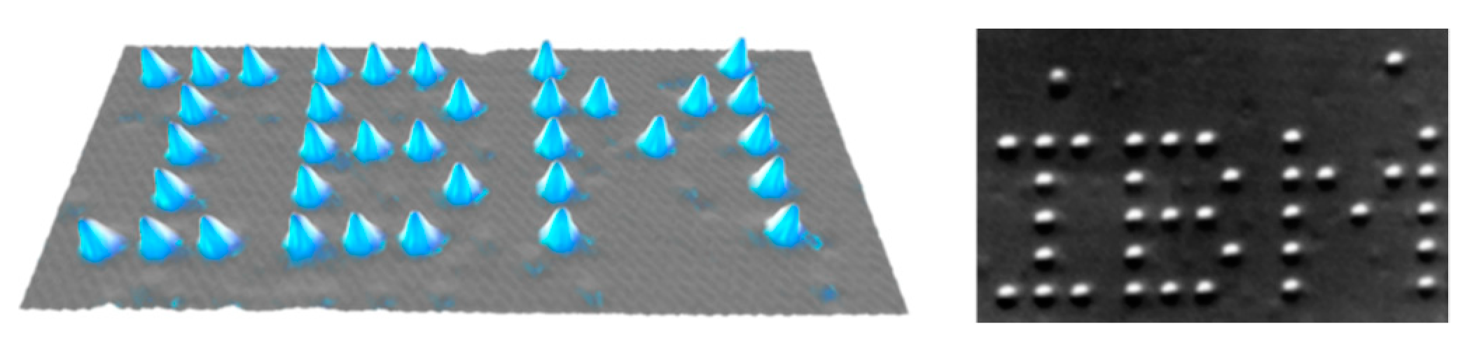
| 1990 | Donald Eigler and Erhard Schweizer (Arranged of individual Xenon atoms to form the letters IBM). | [23] |
| 1991 | Sumio Iijima (Discovery of Multi-wall Carbon nanotubes). | [27] |
| 1992 | Charles T. Kresge (Discovery of mesoporous silica MCM-41). | [79,80] |
| 1993 | Sumio Iijima and Donald Bethune (Discovery of Single-wall Carbon nanotubes). | [81,82] |
| 1996 | Chad Mirkin and Robert Letsinger (SAM of DNA+gold colloids). | [83] |
| 1997 | Zyvex (First nanotechnology company founded). | [84] |
| 1998 | Cees Dekker (Creation of a Transistor using carbon nanotubes). | [85] |
| 1999 | Chad Mirkin (Development of Dip-pen Nanolithography (DPN)). | [86] |
| 2000 | Mark Hersam and Joseph Lyding (Feedback-Controlled Lithography (FCL). | [87] |
| 2000 | President Bill Clinton announces US National Nanotechnology Initiative (NNI). | [88] |
| 2001 | Carlo Montemagno (Molecular nanomachines: molecular motor (rotor) with nanoscale silicon devices). | [89] |
| 2002 | Cees Dekker (Carbon nanotubes functionalized with DNA). | [90] |
| 2003 | President George W. Bush signed into law the 21st Century Nanotechnology Research and Development Act. | [91] |
| 2003 | Naomi Halas (Development of gold nanoshells). | [92,93] |
| 2004 | Andre Geim and Konstantin Novoselov (Discovery of graphene). | [94] |
| 2004 | Xu et al. (Discovery of Fluorescent Carbon dots). | [28] |
| 2005 | James Tour (Nanocar with turning buckyball wheels). | [95,96] |
| 2006 | Paul Rothemund (DNA origami). | [42] |
| 2007 | J. Fraser Stoddart (artificial molecular machines: pH-triggered muscle-like). | [97] |
| 2008 | Osamu Shimomura, Martin Chalfie and Roger Y. Tsien (Nobel Prize in Chemistry for the discovery and development of the green fluorescent protein, GFP). | [98] |
| 2009 | Nadrian Seeman (DNA structures fold into 3D rhombohedral crystals). | [99] |
| 2010 | IBM (Development of an ultra-fast lithography to create 3D nanoscale textured surface). | [100] |
| 2011 | Leonhard Grill (scanning tunneling microscope (STM) describes the electronic and mechanical properties of individual molecules and the polymer chains). | [101] |
| 2016 | Jean-Pierre Sauvage, Sir J. Fraser Stoddart and Bernard L. Feringa (Nobel Prize in Chemistry for the design and synthesis of molecular machines). | [102] |
| 2017 | Nobel Prize in Physics 2017: Gravitational waves. | [103] |
| 2018 | World’s smallest tic-tac-toe game board made with DNA. | [104] |
| 2018 | Shrinking objects to the nanoscale. | [105] |
© 2019 by the authors. Licensee MDPI, Basel, Switzerland. This article is an open access article distributed under the terms and conditions of the Creative Commons Attribution (CC BY) license (http://creativecommons.org/licenses/by/4.0/).
Share and Cite
Bayda, S.; Adeel, M.; Tuccinardi, T.; Cordani, M.; Rizzolio, F. The History of Nanoscience and Nanotechnology: From Chemical–Physical Applications to Nanomedicine. Molecules 2020, 25, 112. https://doi.org/10.3390/molecules25010112
Bayda S, Adeel M, Tuccinardi T, Cordani M, Rizzolio F. The History of Nanoscience and Nanotechnology: From Chemical–Physical Applications to Nanomedicine. Molecules. 2020; 25(1):112. https://doi.org/10.3390/molecules25010112
Chicago/Turabian StyleBayda, Samer, Muhammad Adeel, Tiziano Tuccinardi, Marco Cordani, and Flavio Rizzolio. 2020. "The History of Nanoscience and Nanotechnology: From Chemical–Physical Applications to Nanomedicine" Molecules 25, no. 1: 112. https://doi.org/10.3390/molecules25010112
APA StyleBayda, S., Adeel, M., Tuccinardi, T., Cordani, M., & Rizzolio, F. (2020). The History of Nanoscience and Nanotechnology: From Chemical–Physical Applications to Nanomedicine. Molecules, 25(1), 112. https://doi.org/10.3390/molecules25010112