Article

New Caffeoylquinic Acid Derivatives and Flavanone Glycoside from the Flowers of Chrysanthemum morifolium and Their Bioactivities

1
College of Food and Biological Engineering, Zhengzhou University of Light Industry, Zhengzhou 450000, China
2
State Key Laboratory of Bioactive Substance and Function of Natural Medicines, Institute of Materia Medica, Chinese Academy of Medical Sciences and Peking Union Medical College, Beijing 100050, China
3
Technology Center, China Tobacco Henan Industrial Co. Ltd., Zhengzhou 450000, China
*
Authors to whom correspondence should be addressed.
Molecules 2019, 24(5), 850; https://doi.org/10.3390/molecules24050850
Submission received: 13 January 2019 / Revised: 20 February 2019 / Accepted: 23 February 2019 / Published: 28 February 2019

Abstract

:
The Chrysanthemum morifolium flower is widely used in China and Japan as a food, beverage, and medicine for many diseases. In our work, two new caffeoylquinic acid derivatives (1, 2), a new flavanone glycoside (3), and six reported flavanones (49) were isolated and identified from the flowers of C. morifolium. The chemical structures of all isolates were elucidated by the analysis of comprehensive spectroscopic data as well as by comparison with previously reported data. The isolated constituents 18 were evaluated for their neuroprotective activity, and compounds 3 and 4 displayed neuroprotective effects against hydrogen peroxide-induced neurotoxicity in human neuroblastoma SH-SY5Y cells.

Graphical Abstract

1. Introduction

The flowers of Chrysanthemum morifolium Ramat., Flos Chrysanthemi, which are cultivated in Zhejiang Province as ‘Hangbaiju′ (HJ), have been widely and traditionally consumed in China as a medicinal and edible cognate for about 2000 years. As a well-known herb medicine, HJ is used for its efficacy in dispersing cold, eliminating heat, and improving liver function [1]. Simultaneously, tea made from HJ with hot or boiling water is a popular beverage that cools fever and heightens eyesight [2]. The biological activities of HJ have been exhaustively reported and include cardiovascular protection [3], anti-oxidation [4,5,6], anti-human immune deficiency viruses activity [7,8], anti-inflammatory activity [9], vasorelaxant activity [10], neuroprotective activity [11], anti-cancer activity [12,13], hepatoprotective effects [14,15], aldose reductase inhibition [16,17], and anti-mutagenesis activity [18]. Because of its diversiform and appealing pharmacological activities, HJ has been the subject of intense investigation. Previous research hasshown that HJ containes a wide variety of chemical compounds, including flavonoids and their corresponding glucosides [6,7,8], caffeoylquinic acids [4,5], sesquiterpenes [19], triterpenes [9], and unsaturated fatty acids [20]. Among them, caffeoylquinic acids, flavonoids, and their corresponding glycosides were considered to be the main bioactive ingredients. As such, it is imperative that efforts are exerted to develop more new bioactive compounds from this flower. We herewith report the isolation and elucidation of two new caffeoylquinic acid derivatives (1, 2), a new flavanone glycoside (3), and six reported flavanones (49) (Figure 1). The structures of the new compounds were identified by analyzing spectroscopic data (1D and 2D NMR, MS, IR, ECD, ORD, and UV). The isolated compounds 18 were also examined for their neuroprotective and hepatoprotective properties. The isolation, structure determination, and bioactivity of these compounds are described in this paper.

2. Results and Discussion

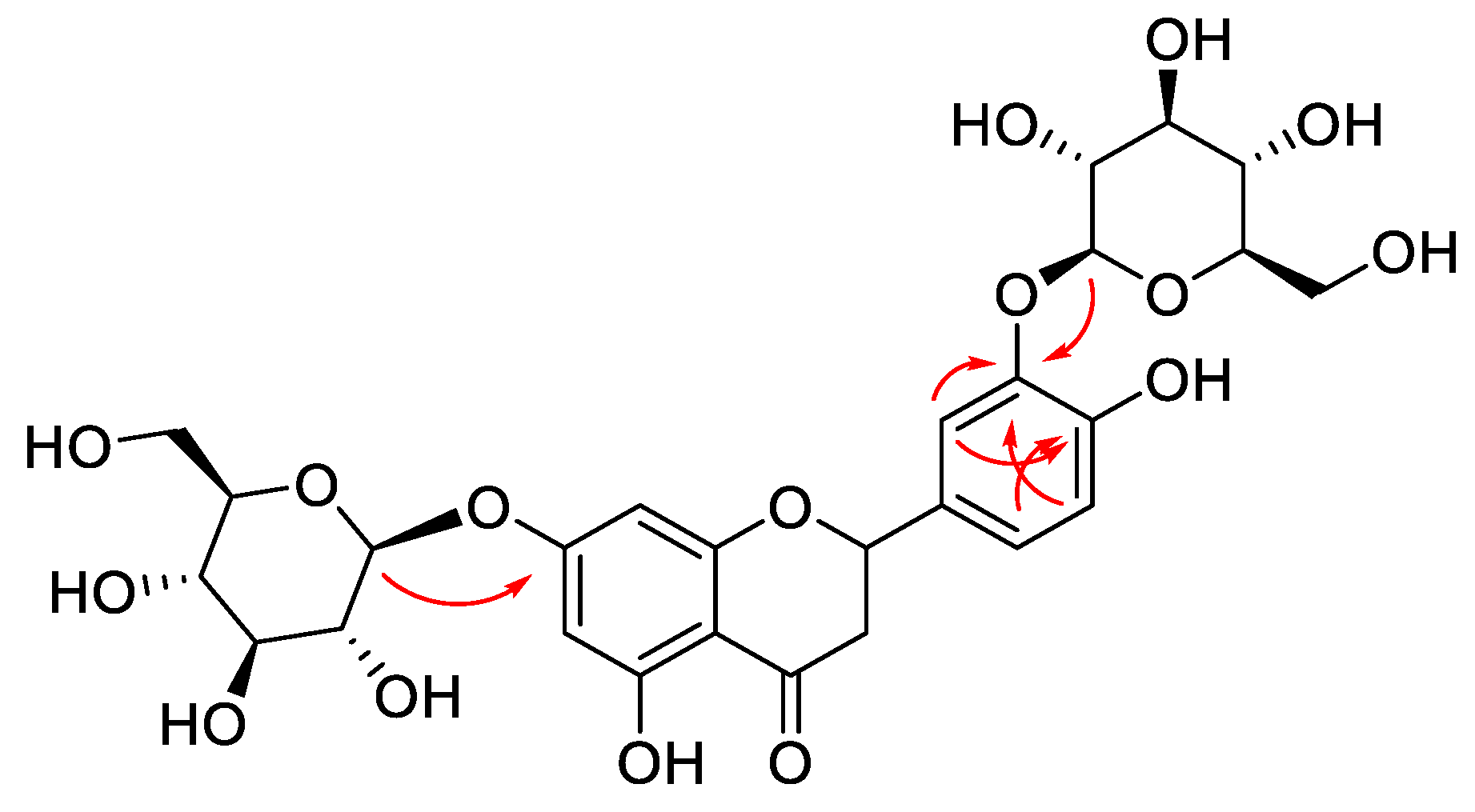
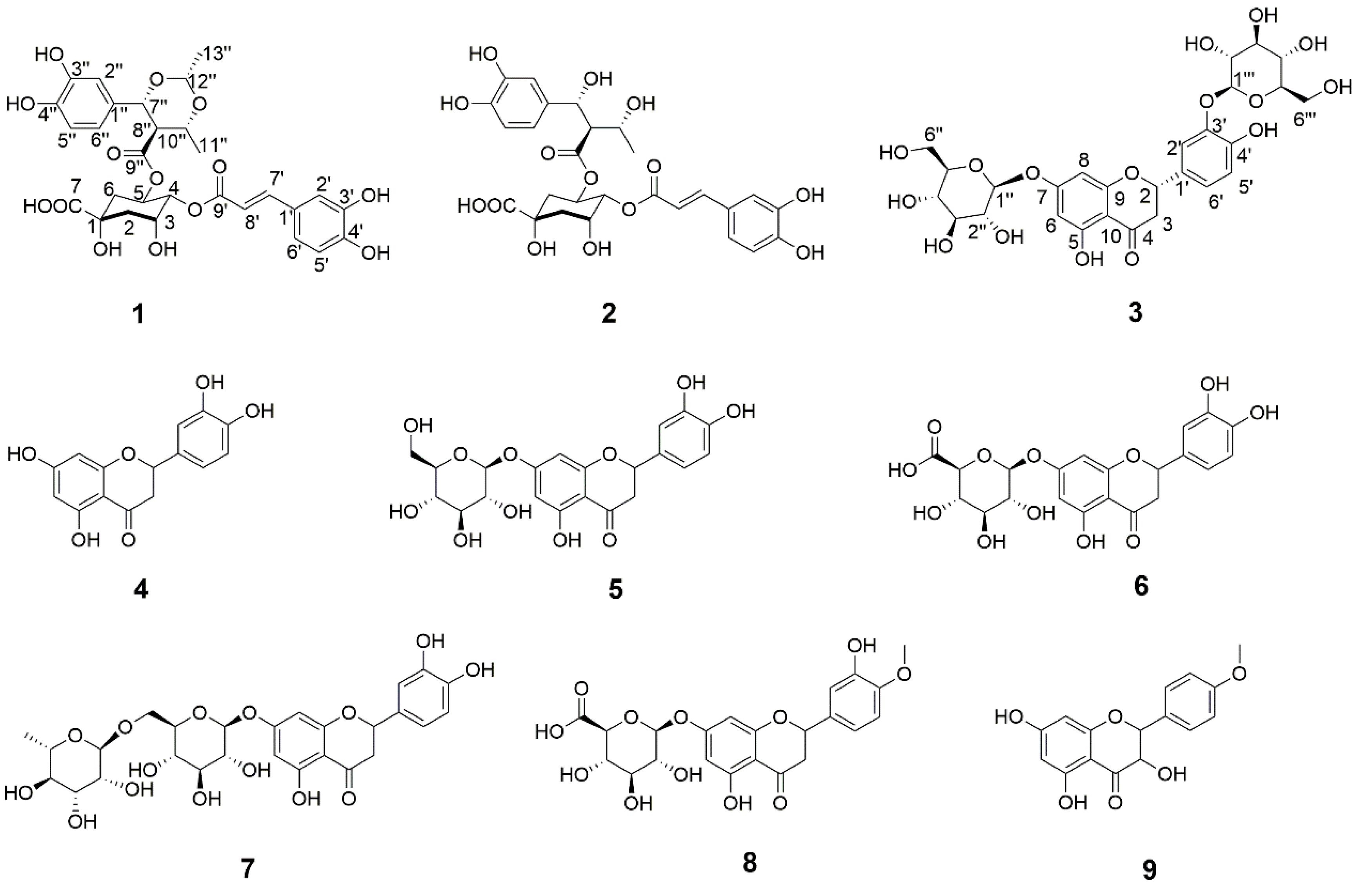
2.1. Structural Elucidation

Here, an 80% ethanol extract of C. morifolium flowers cv. HJ was fractionated sequentially with petroleum ether, EtOAc, and n-butanol, and the n-butanol fraction was successively subjected to column chromatography with HP-20 and SP-700 macroporous absorption resins, Sephadex LH-20, and purified by semi-preparative RP–HPLC to afford three new compounds (13) and six previously known compounds (4–9) (Figure 1). By comparing their NMR and MS data with values reported in the literature, the known isolates were identified as eriodictyol (4) [21], eriodictyol 7-O-β-d-glucopyranoside (5) [22], eriodictyol 7-O-β-d-glucuronide (6) [22], eriodictyol 7-O-β-d-rutinoside (7) [23], hesperetin 7-O-β-d-glucuronide (8) [22], dehydrokaempferide (9) [24].
Compound 1, a white, amorphous power, possesses a molecular formula of C29H32O14, which was determined by HRESIMS (m/z 627.1704 [M + Na]+, calculated for C29H32NaO14, 627.1684) and 13C-NMR data, corresponding to 14 degrees of unsaturation. The IR spectrum displayed characteristic absorptions of hydroxy (3365 cm−1), carbonyl (1711 cm−1), and aromatic ring (1522 and 1445 cm−1) groups. The 1H- NMR data (Table 1) of 1 showed signals readily corresponding to two sets of ABX systems at δH 7.03 (1H, d, J = 2.0 Hz, H-2′′), 6.78 (1H, d, J = 8.0 Hz, H-5′), 6.94 (1H, dd, J = 8.0, 2.0 Hz, H-6′) and 6.76 (1H, d, J = 2.0 Hz, H-2′′), 6.67 (1H, d, J = 8.0 Hz, H-5′′), 6.58 (1H, dd, J = 8.0, 2.0 Hz, H-6′′), belonging to two tri-substituted benzene rings, and two typical trans olefinic protons at δH 7.58 (1H, d, J = 16.0 Hz, H-7′) and 6.24 (1H, d, J = 16.0 Hz, H-8′). The proton spin system of H-7′/H-8′ in 1H-1H COSY (Figure 2a) spectra was observed, in addition to HMBC correlations (Figure 2a) from H-7′ to C-1′, C-2′, C-6′, and C-9′ and from H-8′ to C-1′ and C-9′. Taken together, the above NMR data suggested the existence of a caffeoyl motif in the structure. A quinic acid moiety was revealed via 1H NMR resonances of three oxymethine protons at δH 5.44 (m, H-5), 4.90 (m, H-4), and 4.28 (m, H-3), together with two sets of sp3 methylene protons at δH 2.06 (m, 2H) and δH 1.70 (2H, m) for H2-2 and H2-6, respectively, as shown in Table 1. In combination with the HSQC correlations, these resonances corresponded to three oxygenated methine carbons at δC 69.4 (C-5), 76.3 (C-4), 70.3 (C-3), as well as to two sp3 methylene carbons at δC 40.2 (C-6) and 38.2 (C-2). In addition, there were an oxygenated tertiary carbon at δC 75.2 (C-1) and a carbonyl at δC 175.9 (C-7) in the 13C-NMR spectrum, which are characteristic of a quinic acid unit. The assignments for the quinic acid nucleus were confirmed by analysis of the 1H-1H COSY cross peaks H-2ax/H-3/H-4/H-5/H-6ax and HMBC correlation from H-2ax to C-7 (175.9). The HMBC correlation between δH 4.90 (H-4) and 168.2 (C-9′) suggested the caffeoyl group was esterified to 4-OH of the quinic acid. Besides the signals of a caffeoyl group and a quinic acid, additional 13 carbon resonances were observed. After analyzing the 1H, 13C, and HSQC data, the remaining signals were attributed to one carbonyl groups (δC 171.8), six aromatic carbons (one ABX system, δC 131.7, 115.2, 146.6, 146.7, 116.0, and 119.8), three oxygenated methane groups (δC 84.3, 75.4, 100.2), one methylene group (δC 57.2), and two secondary methyls (δC 20.3, 21.2). The assignments of substitution pattern and planar structure of compound 1 were further confirmed by analysis of the 1H-1H COSY and HMBC correlations. The 1H-1H COSY cross peaks H-7′′/H-8′′/H-10′′/ H3-11′′ together with the HMBC correlations from H-7′′, H-8′′, and C-9′′ established the structural fragment C-7′′–C-8′′–(C-9′′)–C-10′′−C-11′′. The 1H-1H COSY cross peaks H-12′′/H3-13′′ and HMBC correlations from H-12′′ to C-7′′ and C-10′′ enabled the establishment of a 2,6-dimethyl-5-carboxyl-1,3-dioxane ring. The HMBC correlations from H-7′′ to C-1′′, C-2′′, and C-6′′ and from H-8′′ to C-1′′ indicated the connectivity C-7′′–C-1′′. Moreover, the HMBC correlations from H-5 to C-9′′ confirmed C-9′′ is connected with C-3 via an ester bond. Thus, the planar structure of 1 is shown in Figure 1. The relative configuration of 1 was determined by the coupling constant and ROESY data (Figure 2b). The J7′′, 8′′ and J8′′, 10′′ values of 10.0 Hz [25] together with the ROESY correlations from H-10′′ to H-7′′ and H-12′′ suggested that H-7′′, H-10′′, and H-12′′ are cis-orientated. Attempts to determine the absolute configuration of compound 1 were unsuccessful due to the insufficient amount of sample. Thus, the structure of 1 was identified as (rel)-(7′′R,8′′R,10′′R,12′′R)-5-O-[4-(3, 4-dihydroxyphenyl)-2,6-dimethyl-1,3-dioxane-5-carboxyl]-4-O-caffeoylquinic acid. Determined by HRESIMS (m/z 601.1537 [M + Na]+, calculated for C27H30NaO14, 601.1528), compound 2, a white, amorphous power, possesses the molecular formula of C27H30O14, corresponding to 13 degrees of unsaturation. The UV spectra (λmax 330, 288 nm) was highly similar to that of 1 (λmax 331, 288 nm), which indicated that compound 2 may be a caffeoylquinic acid derivative as well. The detailed analyses of 1D NMR (Table 1) and HSQC spectra suggested the presence of 3 carbonyls, 12 aromatic carbons, 2 olefinic carbons, 1 secondary methyl, 2 methylenes, 6 oxygenated methines, and 1 oxygenated quaternary carbon. Compared with those of 1, these characteristic signals imply that one quinic moiety and one caffeoyl group exist in the structure of 2. The assignments of substitution pattern and planar structure of compound 2 were further confirmed by analysis of the 1H-1H COSY and HMBC correlations (Figure 3a). The 1H-1H COSY cross peaks H-7′′/H-8′′/H-10′′/ H3-11′′ together with the HMBC correlations from H-7′′, H-8′′, and H-10′′ established the structural fragment C-7′′–C-8′′–(C-9′′)–C-10′′−C-11′′. The HMBC correlations from H-7′′ to C-1′′, C-2′′, and C-6′′ and from H-8′′ to C-1′′ indicated the connectivity C-7′′–C-1′′. The HMBC correlations from H-2′′ to C-7′′ and from C-4′′ and H-6′′ to C-4′′ along with that from H-5′′ to C-3′′ suggested the presence of a 1, 3, 4-trisubstituted phenyl unit. Moreover, the HMBC correlations from H-3 to C-9′′ confirmed C-9′′ is connected with C-3 via an ester bond, whereas the HMBC cross-peak of H-4 with its ester carbonyl carbon C-9′ indicated the attachment of the caffeoyl group at C-4. On the basis of these results, the planar structure of 2 was assigned as shown in Figure 1. In the ROESY spectra (Figure 3b), the correlation from H-7′′ to H-10′′, together with the coupling constant of H-7′′ and H-8′′, confirmed that H-7′′ and H-8′′ are trans-orientated, and H-7′′ and H-10′′ are cofacial. Consequently, 2 was established as (rel)-(7′′R,8′′R,10′′R,12′′R)-5-O-[8-(1′-hydroxyethyl)]-dihydrocaffeoyl-4-O-caffeoylquinic aicd.
The molecular formula of compound 3, a white, amorphous solid, was assigned to be C27H32O16, as deduced from HRESIMS (m/z 635.1554 [M + H]+ and calculated for C27H32NaO16, 635.1583) and 13C-NMR spectra. The IR spectrum showed absorption bands at 1580, 1519, and 1441 cm−1 (benzyl) and strong absorption bands at 3328 and 1635 cm−1 (hydroxyl, carbonyl, respectively). The 1H-NMR data showed resonances at δH 5.50 (1H, m, H-2), 3.32 (1H, m, H-3), and 2.75 (1H, dd, J = 17.0, 2.0 Hz, H-3) (Table 2). These evidence, together with characteristic UV absorption bands at 283, 333 nm, suggested the presence of a flavanone skeleton [26]. The 1H-NMR data showed one ABX spin system aromatic proton signals at δH 7.38 (1H, d, J = 2.0 Hz, H-2′), 7.03 (1H, dd, J = 8.0, 2.0 Hz, H-6′), 6.85 (1H, d, J = 2.0 Hz, H-5′), attributed to the B ring of the flavanone skeleton, two aromatic doublets at δH 6.17 (1H, d, J = 2.0 Hz, H-8) and 6.14 (1H, d, J = 2.0 Hz, H-6), assignable to the A ring, as well as well as two phenolic hydroxyl groups at δH 12.06 and 8.84. In addition, protons of two β-glucose moieties, for which the anomeric protons resonated at δH 4.97 (1H, d, J = 7.5 Hz, H-1′′), 4.73 (1H, d, J = 7.0 Hz, H-1′′′), respectively, were found in the high-field region. The 13C-NMR spectrum exhibited 27 carbon signals, corresponding to a flavanone skeleton bearing two hydroxyl units and two sugar moieties. The locations of two glucose units were determined at C-7 and C-3′ on the basis of the cross-peak from the anomeric protons H-1″ to C-7, and H-1″′ to C-3′ in the HMBC spectra (Figure 4). The coupling constant (J = 7.5 Hz, H-1′′ and H-1′′′) of two anomeric protons indicated two β-glycosidic linkages. The d-configuration of the glucose moieties was determined via GC analysis of its chiral derivatives after acid hydrolysis. Moreover, the stereochemistry at C-2 was established as S due to the presence of a negative Cotton effect at 287 nm and a positive Cotton effect at 342 nm in the circular dichroism (CD) spectrum (see Supplementary Materials) [27]. Accordingly, the structure of 3 was identified as (2S)-eriodictyol 7,3′-di-O-β-d-glucopyranoside.

2.2. Biological Activities

2.2.1. Hepatoprotective Activity

Compounds 18 were tested for their hepatoprotective activities against N-acetyl-p-aminophenol (APAP)-induced injury in HepG2 cells using bicyclol as a positive control in the MTT method. All compounds were inactive at a concentration of 10 μM (Figure 5).

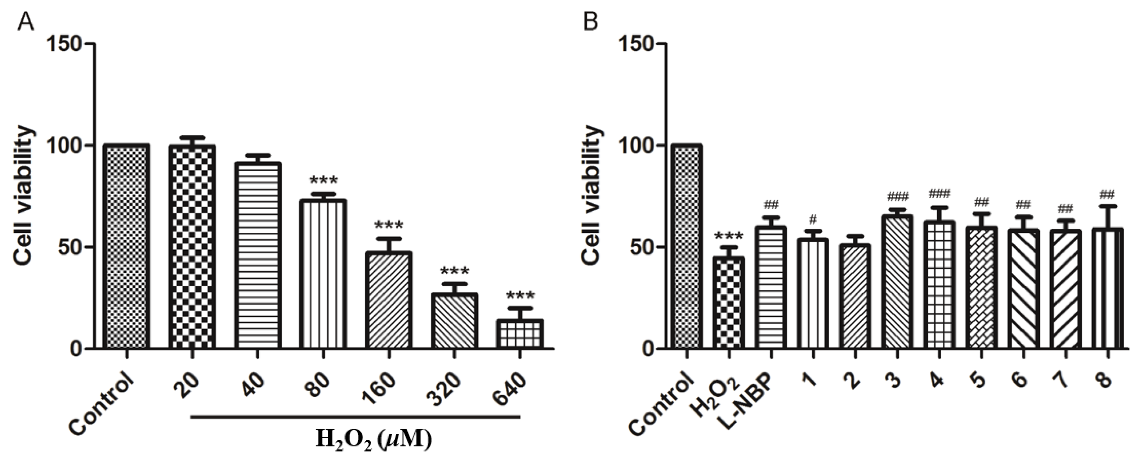
2.2.2. Neuroprotective Activity

The isolated compounds 18 were tested for their neuroprotective effect against H2O2-induced cell toxicity in SH-SY5Y cells. Compounds 3 and 4 exhibited a moderate neuroprotective effect at a concentration of 10 μM against SH-SY5Y cell damage with cell viability of 65.08% and 62.24%, respectively, compared with that measured in the presence of l-NBP (l-3-n-butylphthalide) corresponding to 59.74%. Other compounds displayed mild activities, ranging from 57.19% to 59.57% in cell viability at 10 μM (Figure 6).

3. Materials and Methods

3.1. General Experimental Procedures

Data of optical rotations, UV, and ECD spectra were obtained using Jasco P2000, JASCO V-650, and Jasco J-815 spectrophotometers (Jasco Corporation, Tokyo, Japan), respectively. IR spectra were measured with a Nicolet 5700 spectrometer (Thermo Nicolet Corporation, Madison, SD, USA). GC was performed with an Agilent 7890A instrument (Agilent Technologies, Waldbronn, Germany). The 1D- and 2D-NMR spectra were obtained at 500 or 600 MHz for 1H and at 125 or 150 MHz for 13C, using Bruker 600 and 500 MHz spectrometers (Bruker Corporation, Karlsruhe, Germany). HRESIMS data were acquired with an Agilent 1100 series LC/MSD ion trap mass spectrometer (Agilent Technologies, Waldbronn, Germany). Column chromatography was performed using macroporous resin (Diaion HP-20 and SP-700, from Mitsubishi Chemical Corp., Tokyo, Japan and Sephadex LH-20 columns Pharmacia Fine Chemicals, Uppsala, Sweden. Preparative HPLC was carried out with a Shimadzu LC-6AD instrument with an SPD-20A detector Shimadzu Corp., Tokyo, Japan, using a YMC-Pack ODS-A column (250 mm × 20 mm, 5 μm; YMC Corp., Kyoto, Japan). HPLC-DAD analysis was performed using an Agilent 1200 series system (Agilent Technologies, Waldbronn, Germany) with an Apollo C18 column (250 mm × 4.6 mm, 5 μm; Alltech Corp., Lexington, KY, USA).

3.2. Plant Materials

The dried flower of C. morifolium Ramat. was collected in Tongxiang, Zhejiang province, China, in September 2014. The plant was identified by Professor Lin Ma Institute of Materia Medica, Chinese Academy of Medical Sciences, Beijing.

3.3. Extraction and Isolation

Dried flowers of C. morifolium Ramat. (100 kg) were extracted three times with 80% EtOH (3 × 150 L) under reflux for 3 h. The EtOH solution was evaporated under reduced pressure, and then the dark brown residue (6.4 kg) was suspended in H2O (10 L) and fractionated sequentially using petroleum ether (5 × 10 L), EtOAc (5 × 10 L), and n-BuOH (6 × 10 L). The n-BuOH-soluble fraction (2355 g) was subjected to chromatography on an HP-20 macroporous absorption resin column (200 cm × 15 cm i.d.) and eluted successively with 0, 15, 30, 50, 75, and 95% ethanol (50 L each). The 30% ethanol solution (480 g) was fractionated by column chromatography (200 cm × 10 cm i.d.) on the adsorptive macroporous resin SP-700, eluting with 10, 15, 20, 25, 30, 50, and 95% EtOH (30 L each) to afford seven fractions (fractions A–G).
Fraction D (80 g) was separated on a Sephadex LH-20 column (120 cm × 8 cm i.d.), eluting with a MeOH/H2O mixture (0–100%, with 10% stepwise increase of MeOH, 10 L each) to give fractions D1–D10. Fraction D5 was further chromatographed on a Sephadex LH-20 column (150 cm × 5 cm i.d.) with MeOH/H2O (0, 10, 20, 30, 40, 50, 60, 70, 85, and 100%, v/v, 2 L each) to generate fractions D5-1–D5-23. All of these sub-fractions were further chromatographed on Sephadex LH-20 columns and purified by semi-preparative HPLC. Fraction D5-7 afforded compounds 1 (2 mg) and 2 (5 mg), using MeOH/H2O/HOAc (30:70:0.1, v/v) as the mobile phase at 3 mL/min. Fraction D7 was fractionated by semi-preparative HPLC using MeOH/H2O/HOAc (40:60:0.1, v/v) at 3 mL/min to produce compounds 3 (18 mg), 4 (6 mg), and 5 (13 mg). Compounds 6 (9 mg), 7 (11 mg), 8 (8 mg), and 9 (6 mg) were obtained from fraction D6 using MeOH/H2O/HOAC (40:60:0.1, v/v) as the mobile phase at 3 mL/min.

3.4. Characterization

Compound 1: White amorphous powder; [ α ] D 20 +146 (c 0.1, MeOH); UV (MeOH) λmax (log ε) 330 (4.15), 288 (3.89) nm; IR (KBr) νmax 3365, 1711, 1603, 1522, 1495, 1375, 1264, 978, 817 cm−1; NMR data see Table 1; HRESIMS m/z 627.1704 [M + Na]+ (calculated for 627.1684, C29H32NaO14).
Compound 2: White amorphous powder; [ α ] D 20 +125 (c 0.1, MeOH); UV (MeOH) λmax (log ε) 331 (3.45), 288 (3.19) nm; IR (KBr) νmax 3378, 1711, 1605, 1523, 1449, 1377, 1282, 1077, 981, 818 cm−1; NMR data see Table 1; HRESIMS m/z 601.1537 [M + Na]+ (calculated for 601.1528, C27H30NaO14).
Compound 3: White amorphous powder; [ α ] D 20 ‒116 (c 0.1, MeOH); UV (MeOH) λmax (log ε) 333 (sh), 283 (3.79) nm; IR (KBr) νmax 3329, 2905, 1648, 1580, 1519, 1441, 1400, 1371, 1297, 1203, 1084, 973, 907cm−1; NMR data see Table 1; HRESIMS m/z 635.1554 [M + Na]+ (calculated for 635.1583, C27H32NaO16).

3.5. Hydrolysis of Compound 3 and GC Analysis

Compounds (4 mg) was hydrolyzed with 2 M HCl (3 mL) at 80 °C for 5 h. The sugars gained from the hydrolysates were analyzed by GC on the basis of the reported method [28].

3.6. Hepatoprotective Activity Assay

HepG2 cells were cultured in DMEM with 10% FCS, penicillin (100 U/mL), and streptomycin (100 μg/mL) at 37 °C (5% CO2, 100% relative humidity). These cells were digested using 0.25% trypsin and then seeded into 96-well plates. After incubation for 24 h, the cells were treated with the isolated compounds (10 μM) and APAP (8 mM) and incubated for 48 h. A volume of 100 μL of MTT solution (0.5 mg/mL) was then added into each well. After incubation for 4 h at 37 ℃, the cells were finally lysed with 150 μL DMSO. Finally, the absorbance was measured at 570 nm with a microplate reader, using bicyclol as the positive control. The results were expressed as percentage of cell viability (%), hypothesizing the viability of control cells as 100%.

3.7. Neuroprotective Activity Assay

Human neuroblastoma SH-SY5Y cells were maintained in DMEM supplemented with 10% FBS, 100 U/mL penicillin, and 100 µg/mL streptomycin at 37 ℃ under a water-saturated atmosphere of 5% CO2. The cells were seeded in 96-well culture plates (1 × 10⁴ cells/well) in 100 μL for 18 h, then incubated with the isolated compounds (10 μL, 10 μM) for 4 h. In order to induce an oxidative stress, 100 μL freshly prepared culture medium with 200 μM H2O2 (IC50 = 128.5 μM) was added to the cells and incubated with the compounds at 37 °C for 24 h. Then, 10 μL MTT solution (5 mg/mL) was added into each well. After incubation for 4 h at 37 °C, the cells were finally lysed with 150 μL DMSO. The absorbance was measured at 570 nm, using l-NBP (l-3-n-butylphthalide) as the positive control. The results were expressed as percentage of cell viability (%).

4. Conclusions

In summary, two new caffeoylquinic acid derivatives (1,2), a new flavanone glycoside (3), and six reported flavanone glycosides (49), were isolated and characterized from the dried flowering head of C. morifolium Ramat. The in vitro evaluation of neuroprotective activity suggested that compounds 3 and 4 could improve cell viability at a concentration of 10 μM. This paper not only enriches the chemical diversity of C. morifolium compounds but also provides useful clues on the neuroprotective agents of C. morifolium Ramat cv. “HJ”, which will contribute to the development and application of this functional food. Further investigations should be implemented to examine the neuroprotective activity in vivo and the required isolates’ concentrations and provide real biological significance in the future.

Supplementary Materials

NMR, HR-ESI-MS, IR, UV, and CD spectra of the new compounds (13) in this article are available in the online version.

Author Contributions

P.-F.Y. and C.-Y.H. performed the isolation, structure elucidation of the constituents, and prepared the manuscript; Z.-F.C., Q.-S.Y., S.-C.Z., and Y.-F.F. participated in the measurement of the spectra and also in the preparation of the manuscript; Y.-N.Y. contributed to the interpretation of the spectra. The whole research was devised by D.-B.M. and P.-C.Z. All authors approved the final version of the manuscript.

Funding

This work was supported by the Chinese Academy of Medical Sciences (CAMS) Initiative for Innovative Medicine (No. 2016-I2M-1-010), National Natural Science Foundation of China (21476216), and Doctoral Scientific Research Foundation of Zhengzhou University of Light Industry (13501050063).

Acknowledgments

The authors are thankful to the Institute of Materia Medica, Chinese Academy of Medical Sciences and Peking Union Medical College, for the measurement of hepatoprotective and neuroprotective activities.

Conflicts of Interest

All authors declared no conflicts of interest.

References

  1. Chinese Pharmacopeia Commission. Pharmacopeia of the People′s Republic of China; China Medical Science Publisher: Beijing, China, 2010; Part 1; p. 292. [Google Scholar]
  2. Ma, R.L.; Huang, C.L.; Zhang, X.H.; Wu, Z.Y. Advancement on the study of tea chrysanthemum. North. Hortic. 2009, 8, 151–154. [Google Scholar]
  3. Peng, Y.R.; Shi, L.; Luo, Y.H.; Ding, Y.F. Protective effect of total flavones from chrysanthemum on isoprenaline-induced myocardial ischemia in rats. Lishizhen Med. Mater. Med. Res. 2006, 17, 1131–1132. [Google Scholar]
  4. Kim, H.J.; Lee, Y.S. Identification of new dicaffeoylquinic acids from Chrysanthemum morifolium and their antioxidant activities. Planta Med. 2005, 71, 871–876. [Google Scholar] [CrossRef] [PubMed]
  5. Duh, P.D.; Yen, G.C. Antioxidative activity of three herbal water extracts. Food Chem. 1997, 60, 639–645. [Google Scholar] [CrossRef]
  6. He, D.X.; Ru, X.C.; Wen, L.; Wen, Y.C.; Jiang, H.D.; Bruce, I.C.; Jin, J.; Ma, X.; Xia, Q. Total flavonoids of Flos Chrysanthemi protectarterial endothelial cells against oxidative stress. J. Ethnopharmacol. 2012, 139, 68–73. [Google Scholar] [CrossRef] [PubMed]
  7. Lee, J.S.; Kim, H.J.; Lee, Y.S. A new anti-HIV flavonoid glucuronide from Chrysanthemum morifolium. Planta Med. 2003, 69, 859–861. [Google Scholar] [PubMed]
  8. Hu, C.Q.; Chen, K.; Shi, Q.; Kilkuskie, R.E.; Cheng, Y.C.; Lee, K.H. Anti-AIDS agents, 10. Acacetin-7-O-β-D-galactopyranoside, an anti HIV principle from Chrysanthemum morifolium and a structure- activity correlation with some related flavonoids. J. Nat. Prod. 1994, 57, 42–51. [Google Scholar] [CrossRef] [PubMed]
  9. Ukiya, M.; Akihisa, T.; Yasukawa, K.; Kasahara, Y.; Kimura, Y.; Koike, K.; Nikaido, T.; Takido, M. Constituents of compositae plants. 2. Triterpene diol, triols, and their 3-O-fatty acid esters from edible chrysanthemum flower extract and their anti-inflammatory effects. J. Agric. Food Chem. 2001, 49, 3187–3197. [Google Scholar] [CrossRef] [PubMed]
  10. Jiang, H.D.; Wang, L.F.; Zhou, X.M.; Xia, Q. Vasorelaxant effects and underlying mechanism of EtOAc extract from Chrysanthemum morifolium in rat thoracic aorta. Chin. J. Pathophysiol. 2005, 21, 334–338. [Google Scholar]
  11. Kim, I.S.; Koppula, S.; Park, P.J.; Kim, E.H.; Kim, C.J.; Choi, W.S.; Lee, K.H.; Choi, D.K. Chrysanthemum morifolium Ramat (CM) extract protects human neuroblastoma SH-SY5Y cells against MPP+-induced cytotoxicity. J. Ethnopharmacol. 2009, 126, 447–454. [Google Scholar] [CrossRef] [PubMed]
  12. Ukiya, M.; Akihisa, T.; Tokuda, H.; Suzuki, H.; Mukainaka, T.; Ichiishi, E.; Yasukawa, K.; Kasahara, Y.; Nishino, H. Constituents of Compositae plants III. Anti-tumor promoting effects and cytotoxic activity against human cancer cell lines of triterpene diols and triols from edible chrysanthemum flowers. Cancer Lett. 2002, 177, 7–12. [Google Scholar] [CrossRef]
  13. Xie, Y.Y.; Yuan, D.; Yang, J.Y.; Wang, L.H.; Wu, C.F. Cytotoxic activity of flavonoids from Flos Chrysanthemum on human colon cancer Colon 205 cells. J. Asian Nat. Prod. Res. 2009, 11, 771–778. [Google Scholar] [CrossRef] [PubMed]
  14. Kang, W.Y.; Huang, X.; Lian, T.T.; Xu, Q.T. Protective Effect of Dendranthema morifolium on CCl4- induced Liver Injury in Mice. Nat. Prod. Res. Dev. 2012, 24, 1634–1636. [Google Scholar]
  15. Wang, P.; Pan, X.; Chen, G. Increased exposure of vitamin A by Chrysanthemum morifolium Ramat extract in rat was not via induction of CYP1A1, CYP1A2 and CYP2B1. J. Food Sci. 2012, 77, 121–127. [Google Scholar] [CrossRef] [PubMed]
  16. Matsuda, H.; Morikawa, T.; Toguchida, I.; Yoshikawa, M. Structural requirements of flavonoids and related compounds for aldose reductase inhibitory activity. Chem. Pharm. Bull. 2002, 50, 788–795. [Google Scholar] [CrossRef] [PubMed]
  17. Terashima, S.; Shimizu, M.; Horie, S.; Morita, N. Studies on aldose reductase inhibitors from natural products. IV. Constituents and aldose reductase inhibitory effect of Chrysanthemum morifolium, Bixa orellana and Ipomoea batatus. Chem. Pharm. Bull. 1991, 39, 3346–3347. [Google Scholar] [CrossRef] [PubMed]
  18. Miyazawa, M.; Hisama, M. Antimutagenic activity of flavonoids from Chrysanthemum morifolium. Biosci. Biotechnol. Biochem. 2003, 67, 2091–2099. [Google Scholar] [CrossRef] [PubMed]
  19. Hu, L.H.; Chen, Z.L. Sesquiterpenoid alcohols from Chrysanthemum morifolium. Phytochemistry 1997, 44, 1287–1290. [Google Scholar]
  20. Tsao, R.; Attygalle, A.B.; Schroeder, F.C.; Marvin, C.H.; McGarvey, B.D. Isobutylamides of unsaturated fatty acids from Chrysanthemum morifolium associated with host-plant resistance against the western Flower Thrips. J. Nat. Prod. 2003, 66, 1229–1231. [Google Scholar] [CrossRef] [PubMed]
  21. Wang, X.G.; Shen, L.T.; Zeng, Y.Y.; Tian, Y.Q.; Xu, H.H. Flavonoids from Ficus sarmentosa var. henryi. Chin. Tradit. Herb. Drugs 2010, 41, 526–529. [Google Scholar]
  22. Sun, Y.; Ma, X.B.; Liu, J.X. Compounds from fraction with cardiovascular activity of Chrysanthemum indicum. China J. Chin. Mater. Med. 2012, 37, 61–65. [Google Scholar]
  23. Zhou, D.N.; Ruan, J.L.; Cai, Y.L. Flavonoids from Aerial Parts of Arachniodes exilis. Chin. Pharm. J. 2008, 43, 1218–1220. [Google Scholar]
  24. Shang, H.Q.; Qin, M.J.; Wu, J.R. Constituents of Rhizomes of Iris tectorum. Chin. J. Nat. Med. 2007, 5, 312–314. [Google Scholar]
  25. Abate, A.; Brenna, E.; Costantini, A.; Fuganti, C.; Gatti, F.G.; Malpezzi, L.; Serra, S. Enzymatic approach to enantiomerically pure 5-alken-2,4-diols and 4-hydrocy-5-alken-2-ones: Application to the synthesis of chiral synthons. J. Org. Chem. 2006, 71, 5228–5240. [Google Scholar] [CrossRef] [PubMed]
  26. Lin, J.H.; Chiou, Y.N.; Lin, Y.L. Phenolic Glycosides from Viscum angulatum. J. Nat. Prod. 2002, 65, 638–640. [Google Scholar] [CrossRef] [PubMed]
  27. Mizuno, M.; Kato, M.; Iinuma, M.; Tanaka, T.; Kimura, A.; Ohashi, H.; Sakai, H. Acylated luteolin glucosides from Salix gilgiana. Phytochemistry 1987, 26, 2418–2420. [Google Scholar] [CrossRef]
  28. Yang, Y.N.; Huang, X.Y.; Feng, Z.M.; Jiang, J.S.; Zhang, P.C. New butyrolactone type lignans from Arctii Fructus and their anti-inflammatory activities. J. Agric. Food Chem. 2015, 63, 7958–7966. [Google Scholar] [CrossRef] [PubMed]
Sample Availability: Samples of the compounds 13 are available from the authors.
Figure 1. Chemical structures of compounds 19.
Molecules 24 00850 g001
Figure 2. (a) 1H-1H COSY and key HMBC correlations of compound 1; (b) selected ROESY correlations of compound 1.
Molecules 24 00850 g002
Figure 3. (a) 1H-1H COSY and key HMBC correlations of Compound 1; (b) selected ROESY correlations of compound.
Molecules 24 00850 g003
Figure 4. Key HMBC correlations of compound 3.
Molecules 24 00850 g004
Figure 5. Hepatoprotective effects of compounds 18 against N-acetyl-p-aminophenol (APAP)-induced HepG2 cell injury.
Molecules 24 00850 g005
Figure 6. (A) Toxicity of different H2O2 concentrations in SH-SY5Y cells; (B) neuroprotective effect of compounds 18 against H2O2-induced toxicity in SH-SY5Y cells.
Molecules 24 00850 g006
Table 1. NMR spectroscopic data for compounds 1, 2 in methanol-d4 (δ in ppm, J in Hz).
1 a2 b
PositionδHδCδHδC
175.275.1
22.06, m38.22.01, m
2.19, m
38.4
34.28, m70.34.24, m70.1
44.90, m76.35.01, dd (3.0, 10.0)75.7
55.44, m69.45.29, m69.8
61.70, m40.21.89, m39.9
7175.9175.2
1′127.7127.7
2′7.03, d (2.0)115.17.07, d (2.0)115.2
3′146.8146.8
4′149.8149.7
5′6.78, d (8.0)116.56.78, d (8.0)115.9
6′6.94, dd (2.0, 8.0)123.16.97, dd (2.0, 8.0)123.2
7′7.58, d (16.0)147.87.63, d (16.0)147.6
8′6.24, d (16.0)114.96.27, d (16.0)114.9
9′168.2168.4
1′′131.7134.3
2′′6.76, d (2.0)115.26.66, d (2.0)115.4
3′′146.6146.2
4′′146.7146.5
5′′6.67, d (8.0)116.06.66, d (8.0)116.5
6′′6.58, dd (2.0, 8.0)119.86.81, dd (2.0, 8.0)120.2
7′′4.56, d (10.0)81.84.77, d (10.0)76.7
8′′2.38, dd (10.0, 10.0)57.22.72, dd (8.5, 10.0)62.4
9′′171.8171.2
10′′3.93, dq (6.0, 10.0)75.44.14, dq (6.0, 8.5)70.3
11′′1.10, d (6.0)20.31.14, d (6.0)21.7
12′′4.90, m100.2
13′′1.29, d (5.0)21.2
a 1H and 13C-NMR were measured at 500 and 150 MHz. b 1H and 13C-NMR were measured at 500 and 125 MHz.
Table 2. NMR spectroscopic data for compound 3 in DMSO-d6 (500 MHz for 1H NMR, 125 MHz for 13C NMR, δ in ppm, J in Hz).
33
PositionδHδCPositionδHδC
25.50, m78.86′7.03, dd (2.0, 8.0)121.9
33.32, m
2.75, dd (2.0, 17.0)
42.01′′4.97, d (7.5)99.6
4197.32′′3.2–3.4, m73.1
5162.83′′3.2–3.4, m76.1
66.14, d (2.0)96.64′′3.2–3.4, m69.5
7165.45′′3.2–3.4, m77.1
86.17, d (2.0)95.56′′3.67, m
3.47, m
60.6
9163.01′′′4.73, d (7.0)101.9
10103.32′′′3.2–3.4, m73.4
1′129.33′′′3.2–3.4, m76.4
2′7.38, d (2.0)115.44′′′3.2–3.4, m69.9
3′145.25′′′3.2–3.4, m77.3
4′147.36′′′3.67, m
3.47, m
60.8
5′6.85, d (8.0)115.8

