
Biosensors and Bioassays Based on Lipases, Principles and Applications, a Review


Abstract
1. Introduction
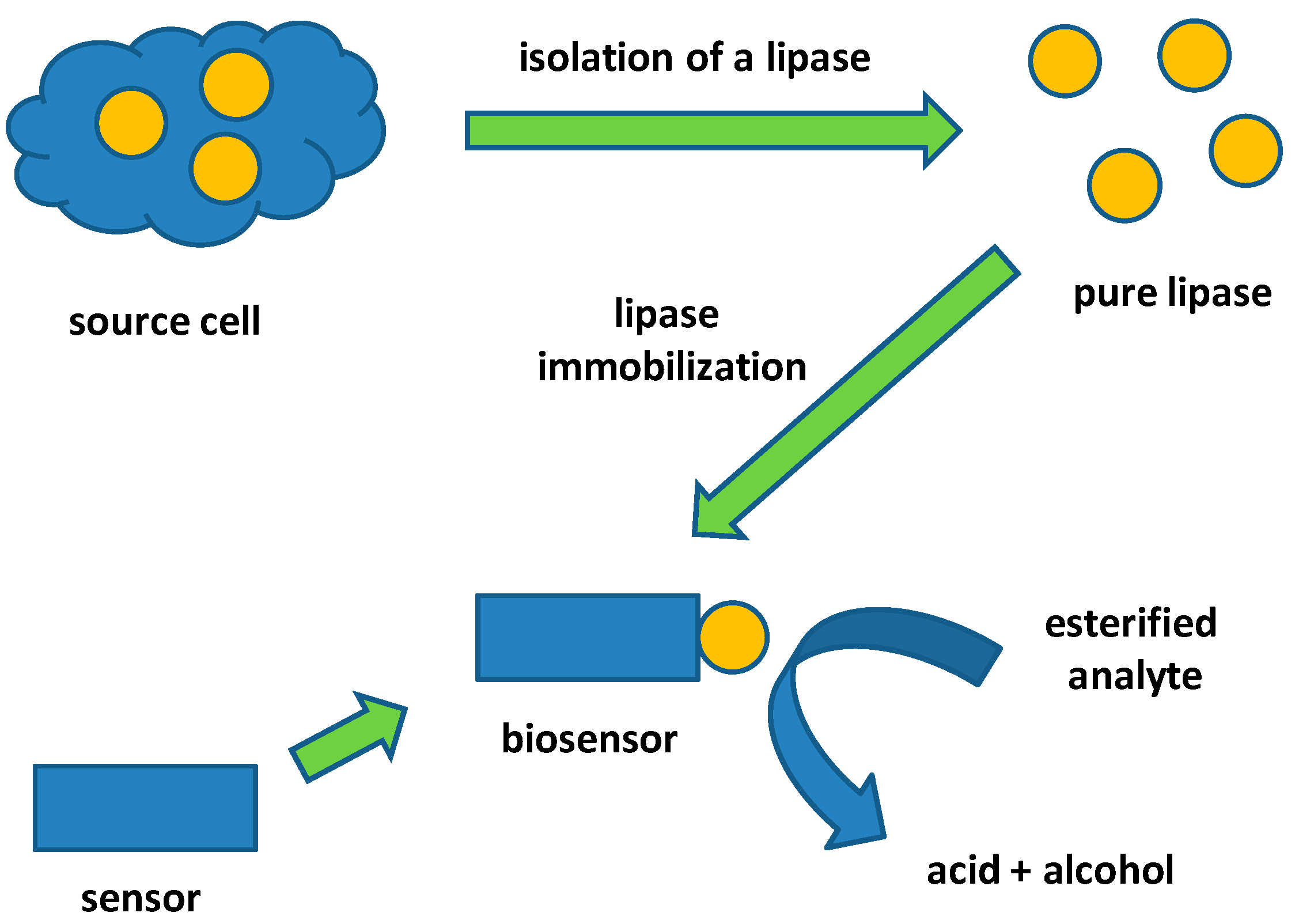
2. Lipases and Their Applicability
3. Standard Methods for Lipase Activity Assay
4. Electrochemical Lipase Biosensors and Bioassays
5. Optical Lipase Biosensors and Bioassays
6. Conclusions
Funding
Conflicts of Interest
References
- Pohanka, M. Cholinesterases in biorecognition and biosensor construction, a review. Anal. Lett. 2013, 46, 1849–1868. [Google Scholar] [CrossRef]
- Pohanka, M. Overview of piezoelectric biosensors, immunosensors and DNA sensors and their applications. Materials 2018, 11, 448. [Google Scholar] [CrossRef] [PubMed]
- Priyanka, P.; Kinsella, G.; Henehan, G.T.; Ryan, B.J. Isolation, purification and characterization of a novel solvent stable lipase from pseudomonas reinekei. Protein Expr. Purif. 2019, 153, 121–130. [Google Scholar] [CrossRef] [PubMed]
- Mehta, A.; Grover, C.; Gupta, R. Purification of lipase from aspergillus fumigatus using octyl sepharose column chromatography and its characterization. J. Basic Microbiol. 2018, 58, 857–866. [Google Scholar] [CrossRef] [PubMed]
- Zhou, Y.J.; Hu, C.L.; Wang, N.; Zhang, W.W.; Yu, X.Q. Purification of porcine pancreatic lipase by aqueous two-phase systems of polyethylene glycol and potassium phosphate. J. Chromatogr. B Analyt. Technol. Biomed. Life Sci. 2013, 926, 77–82. [Google Scholar] [CrossRef] [PubMed]
- Herrera-Lopez, E.J. Lipase and phospholipase biosensors: A review. Methods Mol. Biol. 2012, 861, 525–543. [Google Scholar] [PubMed]
- Sandoval, G.; Herrera-Lopez, E.J. Lipase, phospholipase, and esterase biosensors (review). Methods Mol. Biol. 2018, 1835, 391–425. [Google Scholar]
- Nguyen, H.H.; Lee, S.H.; Lee, U.J.; Fermin, C.D.; Kim, M. Immobilized enzymes in biosensor applications. Materials 2019, 12, 121. [Google Scholar] [CrossRef]
- Akoh, C.C.; Lee, G.C.; Liaw, Y.C.; Huang, T.H.; Shaw, J.F. Gdsl family of serine esterases/lipases. Prog. Lipid Res. 2004, 43, 534–552. [Google Scholar] [CrossRef]
- Doolittle, M.H.; Peterfy, M. Mechanisms of lipase maturation. Clin. Lipidol. 2010, 5, 71–85. [Google Scholar] [CrossRef]
- Pohanka, M. Cholinesterases, a target of pharmacology and toxicology. Biomed. Pap. Olomouc 2011, 155, 219–229. [Google Scholar] [CrossRef] [PubMed]
- Pohanka, M. Acetylcholinesterase inhibitors: A patent review (2008 - present). Expert Opin. Ther. Pat. 2012, 22, 871–886. [Google Scholar] [CrossRef] [PubMed]
- Pohanka, M. Biosensors containing acetylcholinesterase and butyrylcholinesterase as recognition tools for detection of various compounds. Chem. Pap. 2015, 69, 4–16. [Google Scholar] [CrossRef]
- Wong, H.; Schotz, M.C. The lipase gene family. J. Lipid Res. 2002, 43, 993–999. [Google Scholar] [CrossRef]
- Holmes, R.S.; Vandeberg, J.L.; Cox, L.A. Vertebrate hepatic lipase genes and proteins: A review supported by bioinformatic studies. Open Access Bioinformatics 2011, 22, 85–95. [Google Scholar] [CrossRef] [PubMed]
- Murthy, V.; Julien, P.; Gagne, C. Molecular pathobiology of the human lipoprotein lipase gene. Pharmacol. Ther. 1996, 70, 101–135. [Google Scholar] [CrossRef]
- Patel, R.N. Stereoselective biotransformations in synthesis of some pharmaceutical intermediates. Adv. Appl. Microbiol. 1997, 43, 91–140. [Google Scholar] [PubMed]
- Yamamoto, K.; Ueno, Y.; Otsubo, K.; Kawakami, K.; Komatsu, K. Production of s-(+)-ibuprofen from a nitrile compound by acinetobacter sp. Strain ak226. Appl. Environ. Microbiol. 1990, 56, 3125–3129. [Google Scholar]
- Matsumane, H.; Furui, M.; Shibatani, T.; Tosa, T. Production of optically active 3-phenylglycidic acid ester by the lipase from serratia marcescens in a hollow-fiber membrane reactor. J. Ferment. Bioeng. 1994, 78, 59–63. [Google Scholar] [CrossRef]
- Patel, R.N.; Banerjee, A.; Ko, R.Y.; Howell, J.M.; Li, W.S.; Comezoglu, F.T. Enzymic preparation of (3r-cis)-3-acetyloxy-4-phenyl-2-azetidinone: A taxol side-chain synthon. Biotechnol. Appl. Biochem. 1994, 20, 23–33. [Google Scholar]
- Acosta, A.; Filice, M.; Fernandez-Lorente, G.; Palomo, J.M.; Guisan, J.M. Kinetically controlled synthesis of monoglyceryl esters from chiral and prochiral acids methyl esters catalyzed by immobilized rhizomucor miehei lipase. Bioresour. Technol. 2011, 102, 507–512. [Google Scholar] [CrossRef] [PubMed]
- Kaewprapan, K.; Wongkongkatep, J.; Panbangred, W.; Phinyocheep, P.; Marie, E.; Durand, A.; Inprakhon, P. Lipase-catalyzed synthesis of hydrophobically modified dextrans: Activity and regioselectivity of lipase from candida rugosa. J. Biosci. Bioeng. 2011, 112, 124–129. [Google Scholar] [CrossRef] [PubMed]
- Lianghua, T.; Liming, X.; Min, S.; Huaying, G. Purification and application of a lipase from penicillium expansum ped-03. Appl. Biochem. Biotechnol. 2007, 142, 194–199. [Google Scholar] [CrossRef] [PubMed]
- Athenstaedt, K.; Daum, G. Tgl4p and tgl5p, two triacylglycerol lipases of the yeast saccharomyces cerevisiae are localized to lipid particles. J. Biol. Chem. 2005, 280, 37301–37309. [Google Scholar] [CrossRef] [PubMed]
- Maruyama, T.; Nakajima, M.; Kondo, H.; Kawasaki, K.; Seki, M.; Goto, M. Can lipases hydrolyze a peptide bond? Enzyme Microb. Technol. 2003, 32, 655–657. [Google Scholar] [CrossRef]
- Fernandez, J.; Mohedano, A.F.; Fernandez-Garcia, E.; Medina, M.; Nunez, M. Purification and characterization of an extracellular tributyrin esterase produced by a cheese isolate, micrococcus sp. Inia 528. Int. Dairy J. 2004, 14, 135–142. [Google Scholar] [CrossRef]
- Arreguin-Espinosa, R.; Arreguin, B.; Gonzalez, C. Purification and properties of a lipase from cephaloleia presignis (coleoptera, chrysomelidae). Biotechnol. Appl. Biochem. 2000, 31, 239–244. [Google Scholar] [CrossRef]
- Quiroga, A.D.; Lehner, R. Pharmacological intervention of liver triacylglycerol lipolysis: The good, the bad and the ugly. Biochem. Pharmacol. 2018, 155, 233–241. [Google Scholar] [CrossRef]
- Srivastava, G.; Apovian, C. Future pharmacotherapy for obesity: New anti-obesity drugs on the horizon. Curr. Obes. Rep. 2018, 7, 147–161. [Google Scholar] [CrossRef]
- Bialecka-Florjanczyk, E.; Fabiszewska, A.U.; Krzyczkowska, J.; Kurylowicz, A. Synthetic and natural lipase inhibitors. Mini Rev. Med. Chem. 2018, 18, 672–683. [Google Scholar] [CrossRef]
- Lunagariya, N.A.; Patel, N.K.; Jagtap, S.C.; Bhutani, K.K. Inhibitors of pancreatic lipase: State of the art and clinical perspectives. Excli. J. 2014, 13, 897–921. [Google Scholar] [PubMed]
- Kumari, A.; Gupta, R. Extracellular expression and characterization of thermostable lipases, lip8, lip14 and lip18, from yarrowia lipolytica. Biotechnol. Lett. 2012, 34, 1733–1739. [Google Scholar] [CrossRef] [PubMed]
- Deb, C.; Daniel, J.; Sirakova, T.D.; Abomoelak, B.; Dubey, V.S.; Kolattukudy, P.E. A novel lipase belonging to the hormone-sensitive lipase family induced under starvation to utilize stored triacylglycerol in mycobacterium tuberculosis. J. Biol. Chem. 2006, 281, 3866–3875. [Google Scholar] [CrossRef] [PubMed]
- Nishio, T.; Chikano, T.; Kamimura, M. Purification and some properties of lipase produced by pseudomonas fragi 22.39 b. Agric. Biol. Chem. 1987, 51, 181–186. [Google Scholar]
- Zhao, H.; Zheng, L.; Wang, X.; Liu, Y.; Xu, L.; Yan, Y. Cloning, expression and characterization of a new lipase from yarrowia lipolytica. Biotechnol. Lett. 2011, 33, 2445–2452. [Google Scholar] [CrossRef]
- Wilcox, M.D.; Brownlee, I.A.; Richardson, J.C.; Dettmar, P.W.; Pearson, J.P. The modulation of pancreatic lipase activity by alginates. Food Chem. 2014, 146, 479–484. [Google Scholar] [CrossRef] [PubMed]
- Dutta, S.; Ray, L. Production and characterization of an alkaline thermostable crude lipase from an isolated strain of bacillus cereus c(7). Appl. Biochem. Biotechnol. 2009, 159, 142–154. [Google Scholar] [CrossRef]
- Muderhwa, J.M.; Ratomahenina, R.; Pina, M.; Graille, J.; Galzy, P. Purification and properties of the lipases from rhodotorula pilimanae hedrick and burke. Appl. Microbiol. Biotechnol. 1986, 23, 348–354. [Google Scholar] [CrossRef]
- Edashige, Y.; Murakami, N.; Tsujita, T. Inhibitory effect of pectin from the segment membrane of citrus fruits on lipase activity. J. Nutr. Sci. Vitaminol. 2008, 54, 409–415. [Google Scholar] [CrossRef]
- Ebrahimpour, A.; Rahman, R.N.; Basri, M.; Salleh, A.B. High level expression and characterization of a novel thermostable, organic solvent tolerant, 1,3-regioselective lipase from geobacillus sp. Strain arm. Bioresour. Technol. 2011, 102, 6972–6981. [Google Scholar] [CrossRef]
- Rivera-Perez, C.; del Toro Mde, L.; Garcia-Carreno, F. Purification and characterization of an intracellular lipase from pleopods of whiteleg shrimp (litopenaeus vannamei). Comp. Biochem. Physiol. B Biochem. Mol. Biol. 2011, 158, 99–105. [Google Scholar] [CrossRef] [PubMed]
- Salah, R.B.; Mosbah, H.; Fendri, A.; Gargouri, A.; Gargouri, Y.; Mejdoub, H. Biochemical and molecular characterization of a lipase produced by rhizopus oryzae. FEMS Microbiol. Lett. 2006, 260, 241–248. [Google Scholar] [CrossRef] [PubMed]
- Sahebkar, A.; Simental-Mendia, L.E.; Reiner, Z.; Kovanen, P.T.; Simental-Mendia, M.; Bianconi, V.; Pirro, M. Effect of orlistat on plasma lipids and body weight: A systematic review and meta-analysis of 33 randomized controlled trials. Pharmacol. Res. 2017, 122, 53–65. [Google Scholar] [CrossRef]
- Guerrand, D. Lipases industrial applications: Focus on food and agroindustries. OCL 2017, 24, D403. [Google Scholar] [CrossRef]
- Hasan, F.; Shah, A.A.; Hameed, A. Inducstrial applications of microbial lipases. Enzyme Microb. Technol. 2006, 39, 235–251. [Google Scholar] [CrossRef]
- Andualeme, B.; Gessesse, A. Microbial lipases and their industrial applications: Review. Biotechnology 2012, 11, 100–118. [Google Scholar] [CrossRef]
- Stoytcheva, M.; Montero, G.; Zlatev, R.; Leon, J.A.; Gochev, V. Analytical methods for lipases activity determination: A review. Curr. Anal. Chem. 2012, 8, 400–407. [Google Scholar] [CrossRef]
- Gopinath, S.C.; Anbu, P.; Lakshmipriya, T.; Hilda, A. Strategies to characterize fungal lipases for applications in medicine and dairy industry. Biomed. Res. Int. 2013, 154549, 24. [Google Scholar] [CrossRef]
- Kumar, D.; Kumar, L.; Nagar, S.; Raina, C.; Parshad, R.; Gupta, V.K. Screening, isolation and production of lipase/esterase producing bacillus sp. Strain dvl2 and its potential evaluation in esterification and resolution reactions. Arch. Appl. Sci. Res. 2012, 4, 1763–1770. [Google Scholar]
- Plou, F.J.; Ferrer, M.; Nuero, O.M.; Calvo, M.V.; Alcalde, M.; Reyes, F.; Ballesteros, A. Analysis of tween 80 as an esterase/lipase substrate for lipolytic activity assay. Biotechnol. Tech. 1998, 12, 183–186. [Google Scholar] [CrossRef]
- Glogauer, A.; Martini, V.P.; Faoro, H.; Couto, G.H.; Muller-Santos, M.; Monteiro, R.A.; Mitchell, D.A.; de Souza, E.M.; Pedrosa, F.O.; Krieger, N. Identification and characterization of a new true lipase isolated through metagenomic approach. Microb. Cell Fact. 2011, 10, 1475–2859. [Google Scholar] [CrossRef] [PubMed]
- Walter, G.L.; McGraw, P.; Tvedten, H.W. Serum lipase determination in the dog: A comparison of a titrimetric method with an automated kinetic method. Vet. Clin. Pathol. 1992, 21, 23–27. [Google Scholar] [CrossRef] [PubMed]
- Jensen, R.G. Detection and determination of lipase (acylglycerol hydrolase) activity from various sources. Lipids 1983, 18, 650–657. [Google Scholar] [CrossRef] [PubMed]
- Kartal, F.; Kilinc, A.; Timur, S. Lipase biosensor for tributyrin and pesticide detection. Int. J. Environ. Anal. Chem. 2007, 87, 715–722. [Google Scholar] [CrossRef]
- Huang, X.R.; Li, Y.Z.; Yang, G.L.; Liu, L.L.; Qu, Y.; Zhang, W.J. A novel method for fabrication of a glass-electrode-based lipase sensor. Chin. Chem. Lett. 2001, 12, 453–456. [Google Scholar]
- Pijanowska, D.G.; Baraniecka, A.; Wiater, R.; Ginalska, G.; Lobarzewski, J.; Torbicz, W. The ph-detection of triglycerides. Sens. Actuator B-Chem. 2001, 78, 263–266. [Google Scholar] [CrossRef]
- Ma, B.K.; Cheong, L.Z.; Weng, X.C.; Tan, C.P.; Shen, C. Lipase@zif-8 nanoparticles-based biosensor for direct and sensitive detection of methyl parathion. Electrochim. Acta 2018, 283, 509–516. [Google Scholar] [CrossRef]
- Reedy, K.G.; Madhavi, G.; Swamy, B.E.K. Mobilized lipase enzymatic biosensor for the determination of chlorfenvinphos and malathion in contaminated water samples: A voltammetric study. J. Mol. Liq. 2014, 198, 181–186. [Google Scholar]
- Tchieno, F.M.M.; Tonle, I.K. P-nitrophenol determination and remediation: An overview. Rev. Anal. Chem. 2018, 37, 20170019. [Google Scholar] [CrossRef]
- Zehani, N.; Dzyadevych, S.V.; Kherrat, R.; Jaffrezic-Renault, N.J. Sensitive impedimetric biosensor for direct detection of diazinon based on lipases. Front. Chem. 2014, 2, 44. [Google Scholar] [CrossRef]
- Pohanka, M. Photography by cameras integrated in smartphones as a tool for analytical chemistry represented by an butyrylcholinesterase activity assay. Sensors 2015, 15, 13752–13762. [Google Scholar] [CrossRef] [PubMed]
- Pohanka, M. Small camera as a handheld colorimetric tool in the analytical chemistry. Chem. Pap. 2017, 71, 1553–1561. [Google Scholar] [CrossRef]
- Pliego, J.; Mateos, J.C.; Rodriguez, J.; Valero, F.; Baeza, M.; Femat, R.; Camacho, R.; Sandoval, G.; Herrera-Lopez, E.J. Monitoring lipase/esterase activity by stopped flow in a sequential injection analysis system using p-nitrophenyl butyrate. Sensors 2015, 15, 2798–2811. [Google Scholar] [CrossRef] [PubMed]
- Krieg, A.K.; Gauglitz, G. An optical sensor for the detection of human pancreatic lipase. Sens. Actuator B-Chem. 2014, 203, 663–669. [Google Scholar] [CrossRef]
- Pohanka, M.; Zakova, J.; Sedlacek, I. Digital camera-based lipase biosensor for the determination of paraoxon. Sens. Actuator B Chem. 2018, 273, 610–615. [Google Scholar] [CrossRef]
- Zheng, J.; Wei, W.; Lan, X.; Zhang, Y.; Wang, Z. Fluorescent microplate assay method for high-throughput detection of lipase transesterification activity. Anal. Biochem. 2018, 549, 26–28. [Google Scholar] [CrossRef]


| Substrate for Hydrolysis | Reaction Products | Origin of Lipase | References |
|---|---|---|---|
| (R,S)-ibuprofen + 1-propanol | (S)-ibuprofen n-propylester + (R)-ibuprofen | Penicillium expansum | [23] |
| 4-nitrophenyl palmitate + water | 4-nitrophenol + palmitate | Penicillium expansum | [23] |
| 1,2,3-trihexaicosanoylglycerol + water | 1,2-dihexaicosanoylglycerol + hexaicosanoate | Saccharomyces cerevisiae | [24] |
| trimyristin + water | dimyristin + myristate | Saccharomyces cerevisiae | [24] |
| tripalmitin + water | dipalmitin + palmitate | Saccharomyces cerevisiae | [24] |
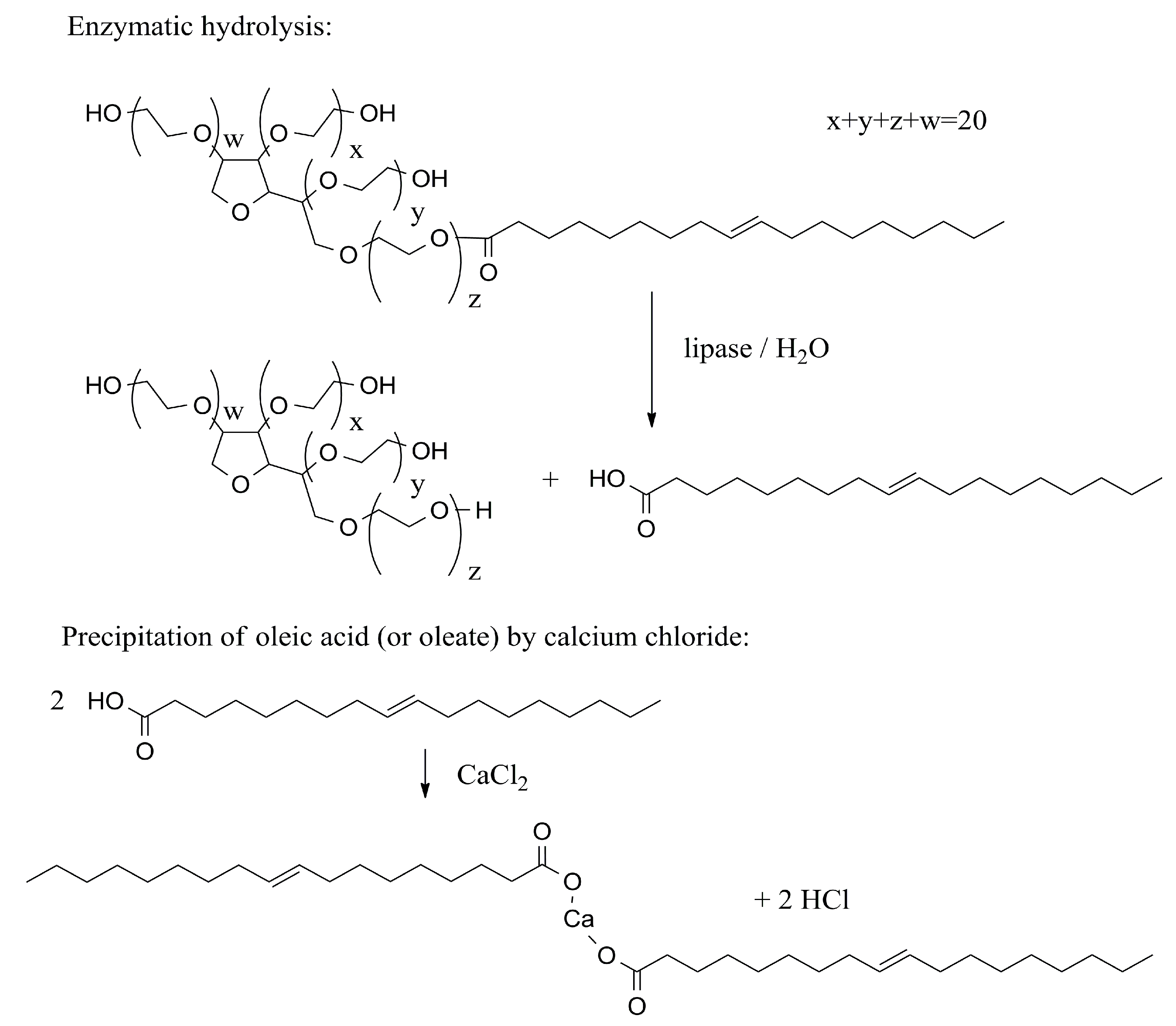
| tributyrin + water | dibutyrin + butyrate | Bacillus subtilis, Chromobacterium viscosum, Micrococcus sp., Rhizomucor miehei, R. oryzae, Fusarium solani, wild boar (Sus scrofa), humans Homo sapiens | [25,26] |
| α/β-naphthyl stearate + water | α/β-naphthol + stearate | Micrococcus sp. | [26] |
| β-naphthyl butyrate + water | β-naphthol + butyrate | Micrococcus sp. | [26] |
| β-naphthyl laureate + water | β-naphthol + laureate | Micrococcus sp. | [26] |
| 4-nitrophenyl esters (laurate, oleate, palmitate, propionate) + water | 4-nitrophenol + laurate, oleate, palmitate respective propionate | Cephaloeia presignis | [27] |
| α-naphthyl acetate + water | α-naphthol + acetate | Cephaloeia presignis | [27] |
| methyl esters (acetate, butyrate, palmitate, propionate, stearate) + water | methanol + acetate, butyrate, palmitate, propionate, respective stearate | Cephaloeia presignis | [27] |
| Inhibitor of Lipase | Origin of Lipase | References |
|---|---|---|
| Cd (II+) | Yarrowia lipolytica | [32] |
| Co (II+) | Mycobacterium tuberculosis | [33] |
| Fe (III+) | Pseudomonas fragi | [34] |
| Hg (II+), Ni (II+), Cu (II+), Zn (II+) | Yarrowia lipolytica | [35] |
| alginic acid | wild boar (Sus scrofa) | [36] |
| sodium dodecyl sulfate, n-dodecyltrimethylammonium bromide, sorbitan monooleate | Pseudomonas fragi | [34] |
| sodium cholate, sodium lauryl sulfate, p-hydroxymeruribenzoate, N-bromosuccinimide, acetylacetone, 4-dimethylaminobenzaldehyde | Rhodotorula mucilaginosa | [38] |
| galacturonic acid and pectin | wild boar (Sus scrofa) | [39] |
| 1-butanol, 1-propanol, 2-propanol, acetone, acetonitrile, benzyl alcohol, iso-amyl alcohol, iso-butanol, phenylmethylsulfonyl fluoride | Geobacillus sp. | [40] |
| Orlistat (tetrahydrolipstatin) | humans Homo sapiens, Rhizopus oryzae, whiteleg shrimp Litopenaeus vannamei | [41,42,43] |
| Origin of Used Lipase | Principle of Lipase Use in the Assay | Analyte | Limit of Detection | References |
|---|---|---|---|---|
| fungus Candida rugosa | lipase was immobilized on a glass pH electrode and converted tributyrin, which caused a decrease of pH; methyl-paraoxon stopped the reaction | methyl-parathion | 93 µmol/L | [54] |
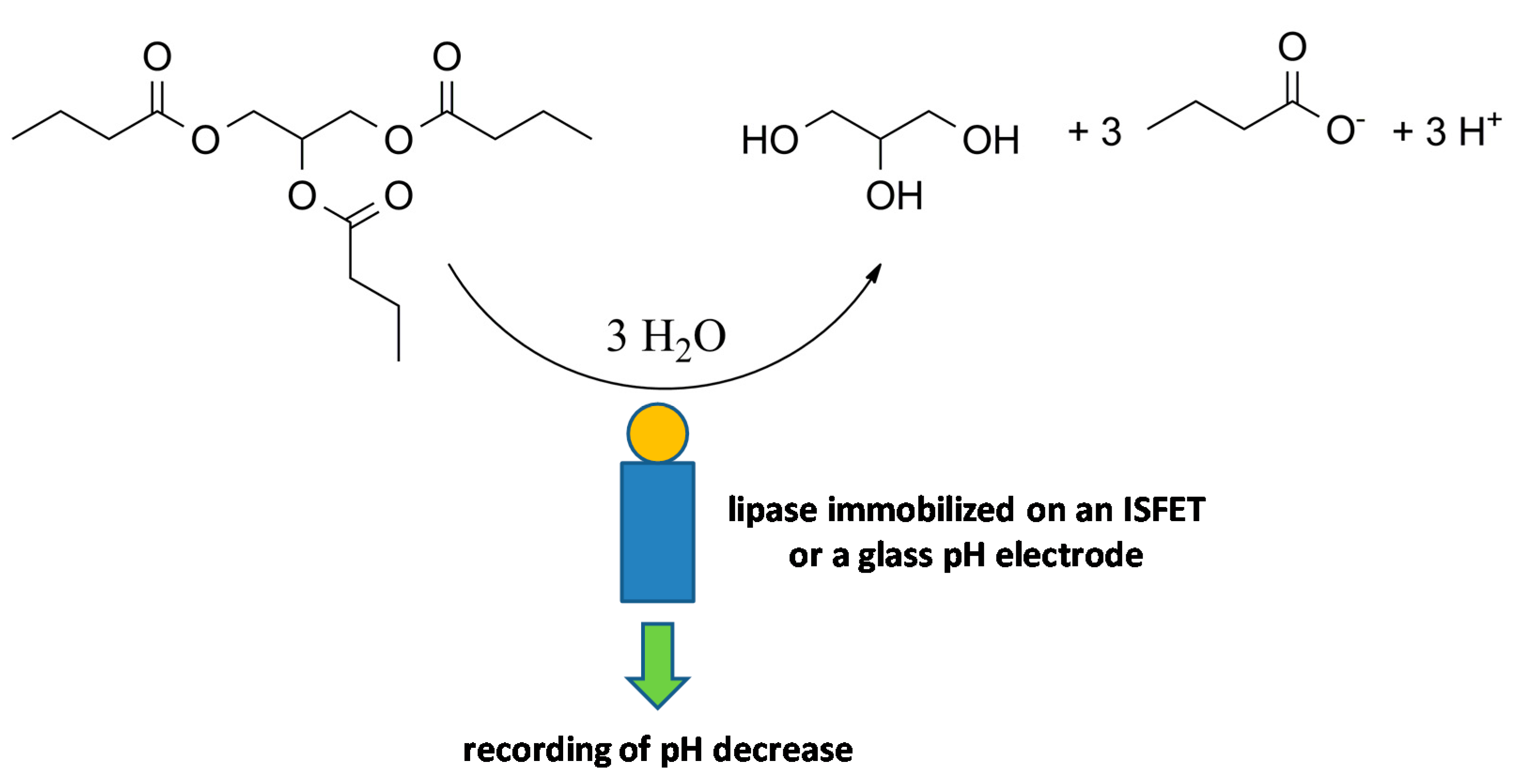
| porcine pancreate | lipase was immobilized on an ISFET and hydrolyzed triglycerides as an analyte, a change in pH was recorded | triacetin, tributyrin and triolein | around 1 mmol/L | [56] |
| bacterium Burkholderia cepacia | lipase was immobilized on zeolitic nanoparticles and then into chitosan on a glassy carbon electrode, pesticides like methyl parathion were hydrolyzed to p-nitrophenyl that was electrochemically oxidized in the next step | methyl parathion | 0.28 µmo/L | [57] |
| fungus Candida rugosa | lipase converted p-nitrophenyl acetate to p-nitrophenol and acetic acid, p-nitrophenol was oxidized and a current at 0.024 V was recorded, analytes inhibited lipase and stopped the reaction | chlorfenvinphos, malathion | 84.5 µmol/L for chlorfenvinphos and 282 µmol/L for malathion | [58] |
| fungus Candida rugosa and porcine pancreas | lipase converted diazinon to diethyl phosphorothioic acid and 2-isopropyl-4-methyl-6-hydroxypyrimidine. which caused a change in the impedance of the medium | diazinon | 10 nmol/L (fungal lipase), 100 nmol/L (porcine pancreas lipase) | [60] |
| Origin of Used Lipase | Principle of Lipase Use in the Assay | Analyte | Limit of Detection | References |
|---|---|---|---|---|
| fungus Candida antarctica and Yarrowia lipolytica | p-nitrophenyl butyrate hydrolysis to butyric acid and p-nitrophenol, coloration caused by p-nitrophenol was measured | lipase itself | 0.05 U/mL | [63] |
| human pancreatic lipase | flows through assay, lipase competed with another immobilized lipase for a fluorescent-dye-labeled antibody, a decrease of fluorescence was measured | lipase itself | 0.068 mg/L | [64] |
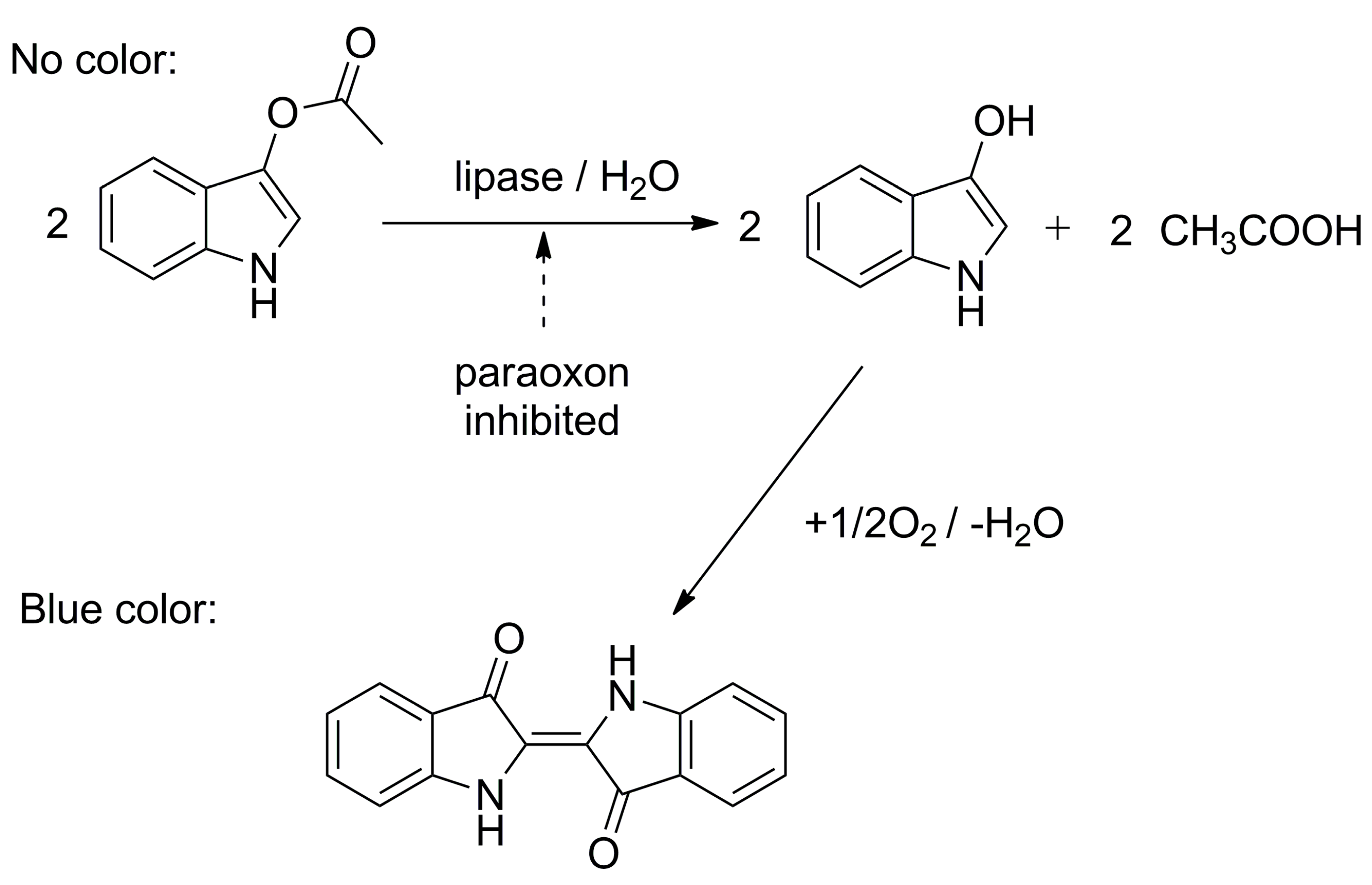
| Psychrobacter sp. | lipase hydrolyzed indoxyl acetate and blue indigo arose, paraoxon stopped the reaction, the intensity of coloration was measured by camera | paraoxon ethyl | 37 nmol/L | [65] |
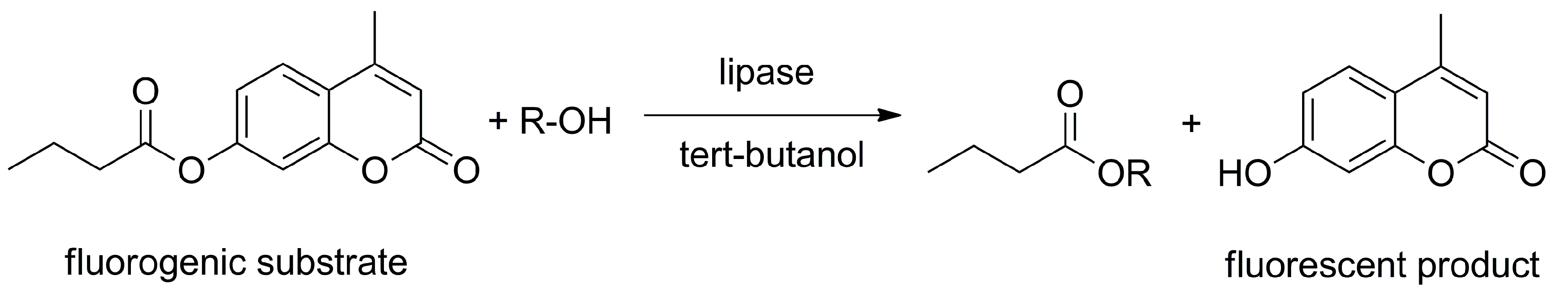
| fungus Candida antarctica, fungus Mucor miehei, fungus Thermomyces lanuginosus, and bacteria Pseudomonas cepacia and P. fluorescens | 4-methyl umbelliferone and methanol in tert-butanol were trans-esterified in the presence of lipase, production of 4-methylumbelliferone was measured fluorometrically | lipase itself | n/a | [66] |
| Analyte | Role of Lipase | Expected Application |
|---|---|---|
| triglycerides, pesticides, various esters | lipase converts the analyte and the reaction is measured | environmental control, agriculture, food industry etc. |
| pesticide or other toxic compounds | analyte inhibits lipase | environmental control, agriculture, military or police forces etc. |
| lipase itself | there is measured lipase activity in the sample | health care, providing healthcare outside hospitals, small medical institutions |
© 2019 by the author. Licensee MDPI, Basel, Switzerland. This article is an open access article distributed under the terms and conditions of the Creative Commons Attribution (CC BY) license (http://creativecommons.org/licenses/by/4.0/).
Share and Cite
Pohanka, M. Biosensors and Bioassays Based on Lipases, Principles and Applications, a Review. Molecules 2019, 24, 616. https://doi.org/10.3390/molecules24030616
Pohanka M. Biosensors and Bioassays Based on Lipases, Principles and Applications, a Review. Molecules. 2019; 24(3):616. https://doi.org/10.3390/molecules24030616
Chicago/Turabian StylePohanka, Miroslav. 2019. "Biosensors and Bioassays Based on Lipases, Principles and Applications, a Review" Molecules 24, no. 3: 616. https://doi.org/10.3390/molecules24030616
APA StylePohanka, M. (2019). Biosensors and Bioassays Based on Lipases, Principles and Applications, a Review. Molecules, 24(3), 616. https://doi.org/10.3390/molecules24030616