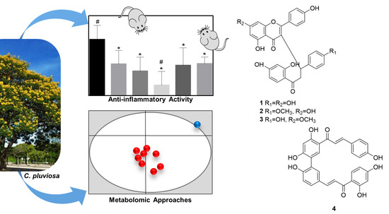
Anti-Inflammatory Derivatives with Dual Mechanism of Action from the Metabolomic Screening of Poincianella pluviosa

 , ,
, ,  ,
,
Abstract
:
1. Introduction
2. Results and Discussion
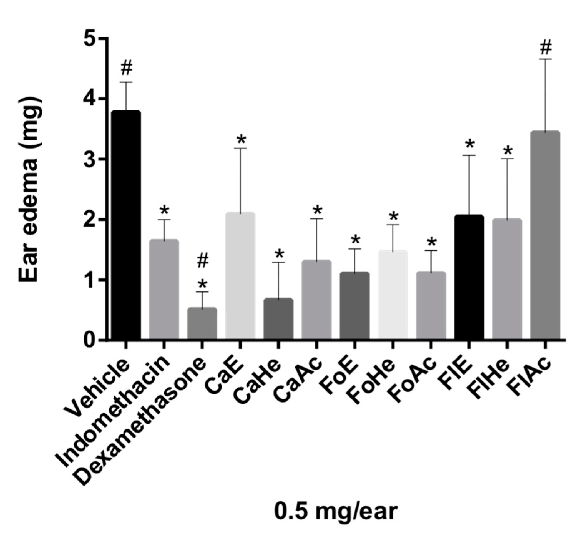
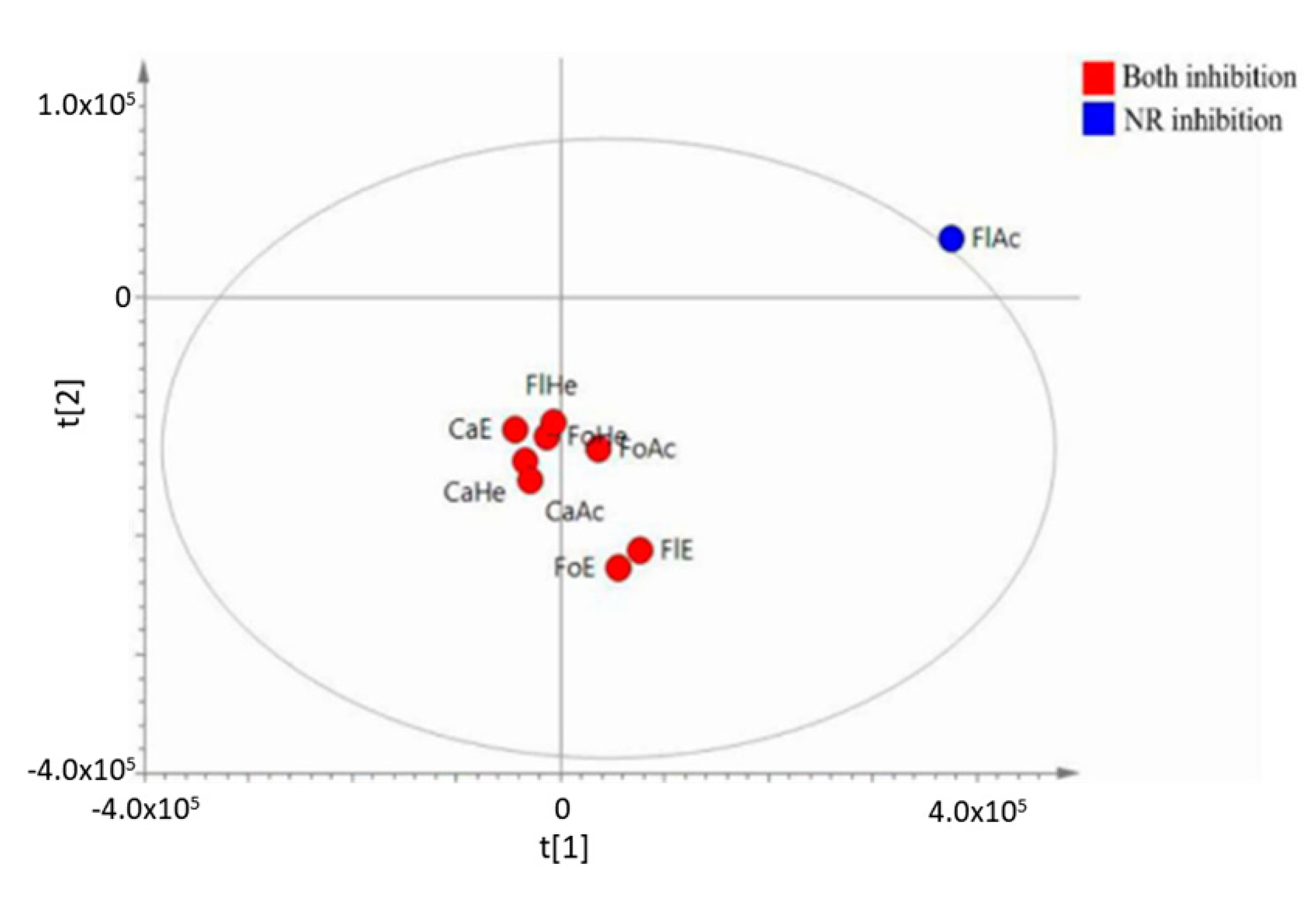
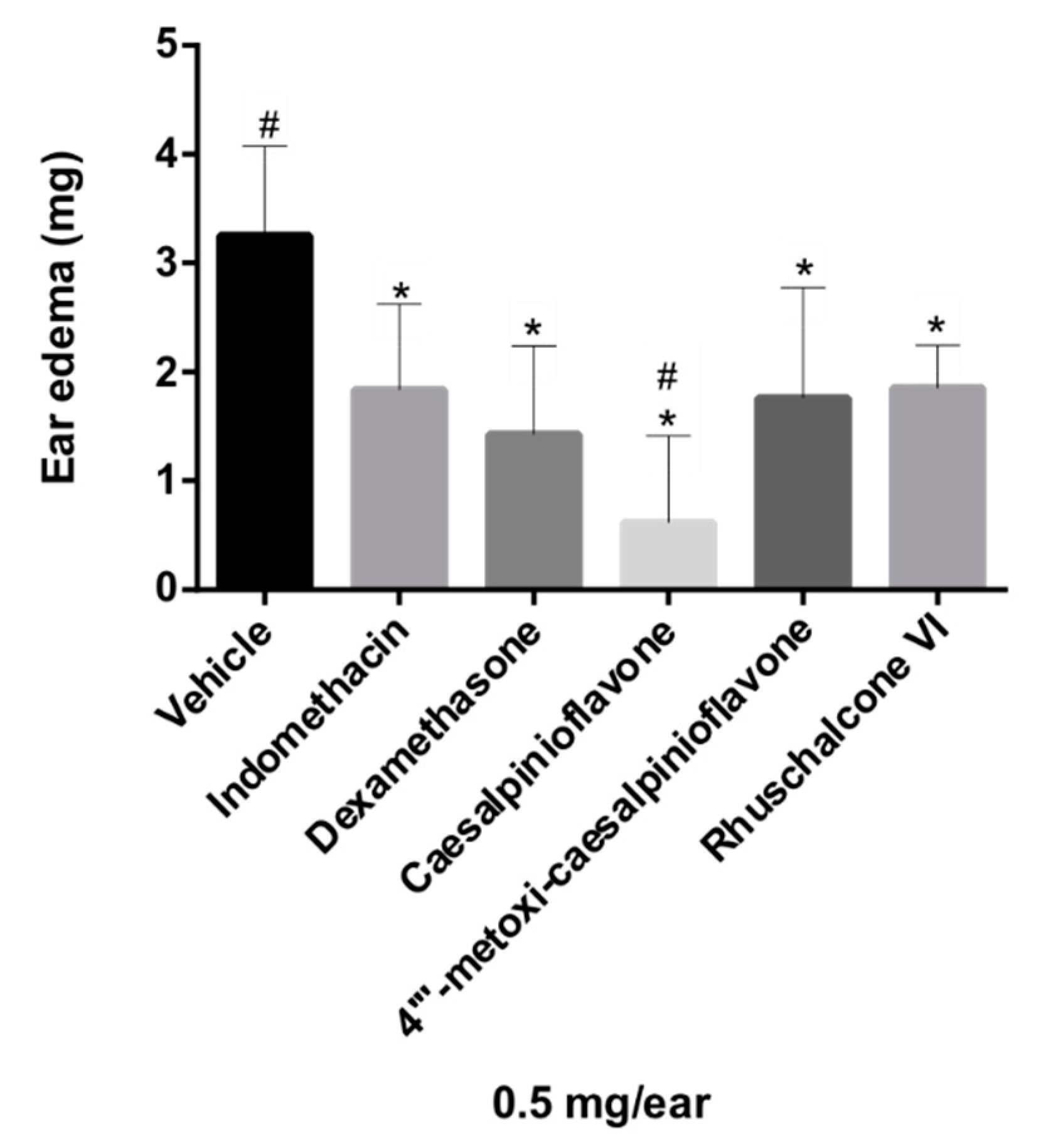
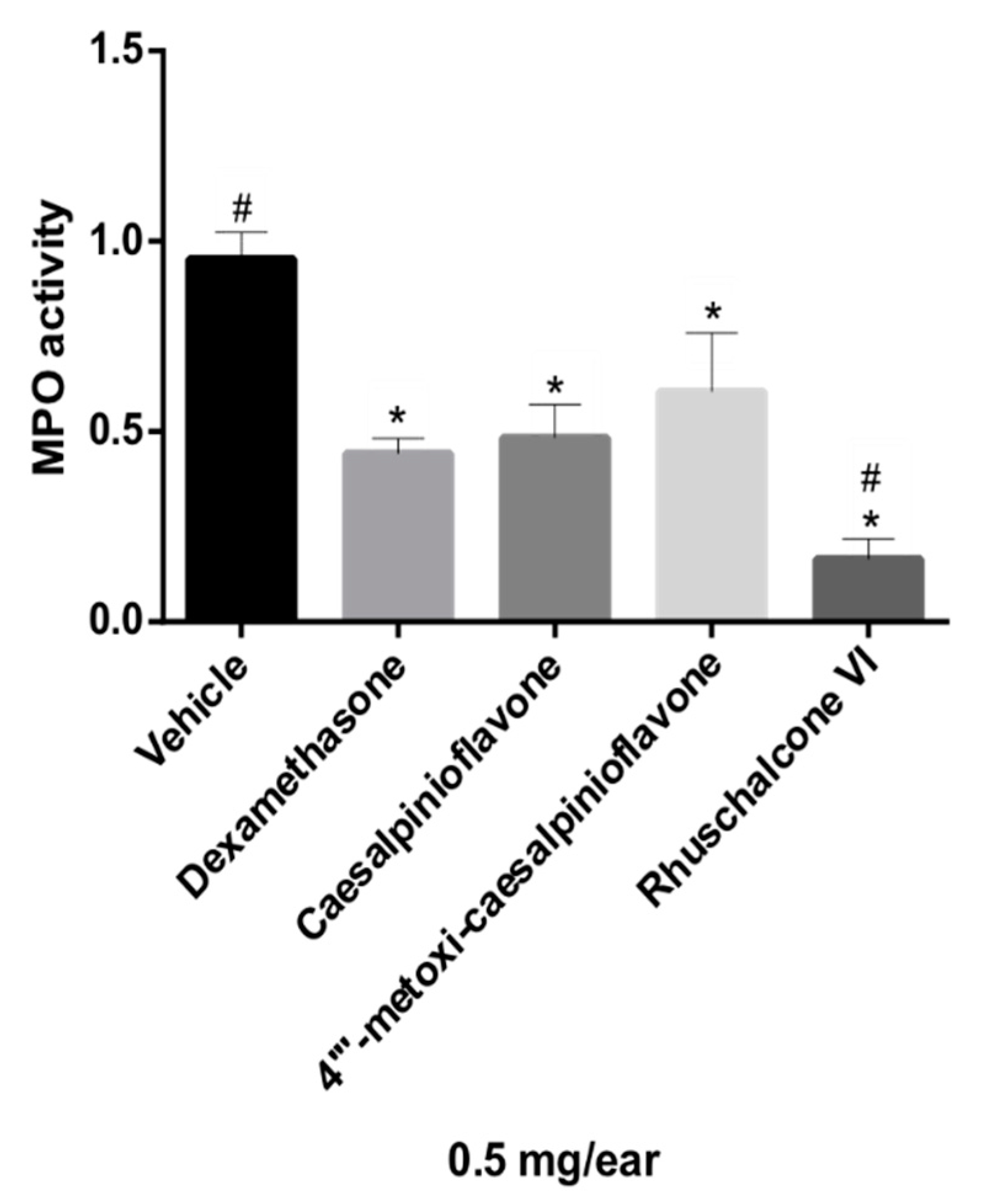
2.1. Anti-Inflammatory Activity Evaluation Analysis
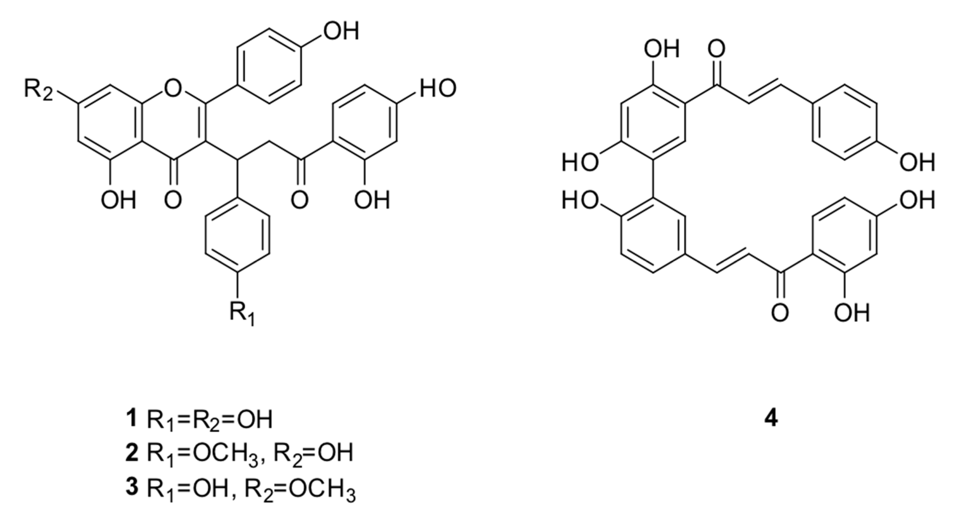
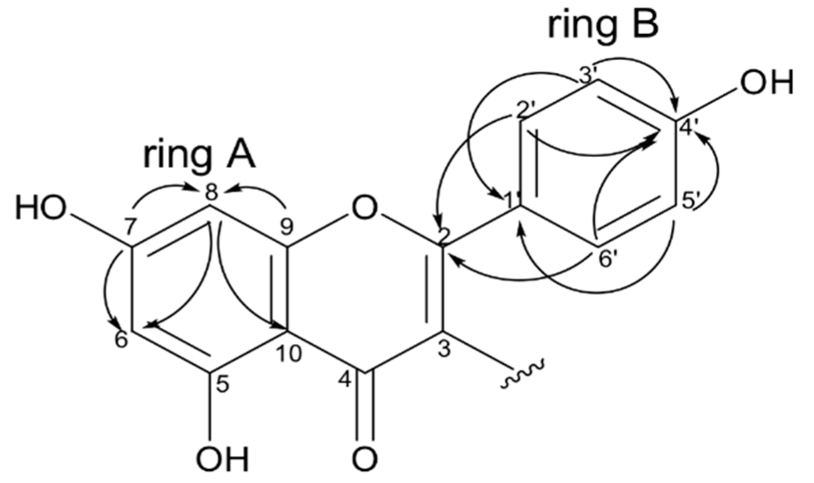
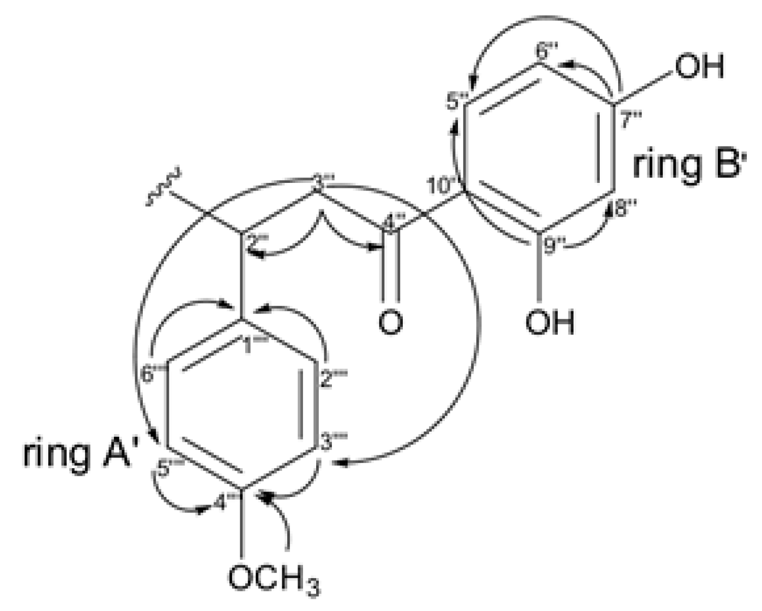
2.2. Chemical Characterization of Compounds 1–4
3. Materials and Methods
3.1. Equipments, Reagents and Solvents
3.2. Plant Material and Preparation of Extracts
3.3. Isolation of Compounds 1–4
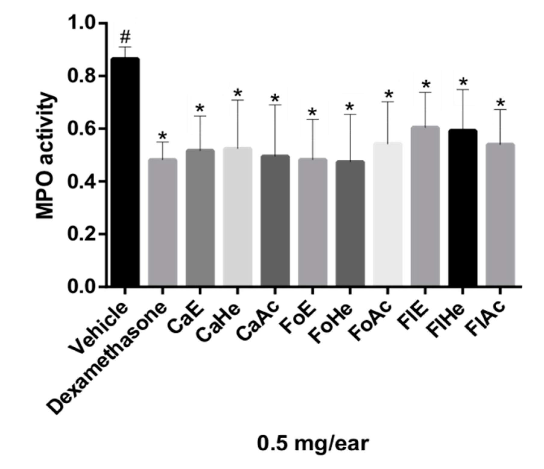
3.4. Anti-Inflammatory Bioassay
3.5. Procedure for Neutrophil Recruitment
3.6. Chemical and Metabolomics Analysis
4. Conclusions
Supplementary Materials
Author Contributions
Funding
Acknowledgments
Conflicts of Interest
References
- Ngo, L.T.; Okogun, J.I.; Folk, W.R. 21st Century natural product research and drug development and traditional medicines. Nat. Prod. Rep. 2013, 30, 584–592. [Google Scholar] [CrossRef]
- Newman, D.J.; Cragg, G.M. Natural products as sources of new drugs from 1981 to 2014. J. Nat. Prod. 2016, 79, 629–661. [Google Scholar] [CrossRef] [PubMed]
- Summer, L.W.; Lei, Z.; Nikolau, B.J.; Saito, K. Modern plant metabolomics: Advanced natural product gene discoveries, improved technologies, and future prospects. Nat. Prod. Rep. 2015, 32, 212–229. [Google Scholar] [CrossRef] [PubMed]
- Sampaio, B.L.; Edrada-Ebel, R.; Da Costa, F.B. Effect of the environment on the secondary metabolic profile of Tithonia diversifolia: A model for environmental metabolomics of plants. Sci. Rep. 2016, 6, 1–11. [Google Scholar] [CrossRef] [PubMed]
- Wolfender, J.L.; Marti, G.; Thomas, A.; Bertrand, S. Current approaches and challenges for the metabolite profiling of complex natural extracts. J. Chromatogr. A 2015, 1382, 136–164. [Google Scholar] [CrossRef] [PubMed]
- Chagas-Paula, D.A.; Oliveira, T.B.; Zhang, T.; Edrada-Ebel, R.; Da Costa, F.B. Prediction of anti-inflammatory plants and discovery of their biomarkers by machine learning algorithms and metabolomic studies. Planta Med. 2015, 81, 450–458. [Google Scholar] [CrossRef] [PubMed]
- Chagas-Paula, D.A.; Zhang, T.; Da Costa, F.B.; Edrada-Ebel, R. A metabolomic approach to target compounds from the asteraceae family for dual COX and 5-LOX inhibition. Metabolites 2015, 5, 404–430. [Google Scholar] [CrossRef]
- Parente, L. Pros and cons of selective inhibition of cyclooxygenase-2 versus dual lipoxygenase/cyclooxygenase inhibition: Is two better than One? J. Rheumatol. 2001, 28, 2375–2382. [Google Scholar]
- Meng, H.; Liu, Y.; Lai, L. Diverse ways of perturbing the human arachidonic acid metabolic network to control inflammation. Acc. Chem. Res. 2015, 48, 2242–2250. [Google Scholar] [CrossRef]
- Pallio, G.; Bitto, A.; Pizzino, G.; Galfo, F.; Irrera, N.; Minutoli, L.; Arcoraci, V.; Squadrito, G.; Macrì, S.; Squadrito, F.; et al. Use of a balanced dual cyclooxygenase-1/2 and 5-lypoxygenase inhibitor in experimental colitis. Eur. J. Pharmacol. 2016, 789, 152–162. [Google Scholar] [CrossRef]
- Stanbury, R.M.; Graham, E.M. Systemic corticosteroid therapy-side effects and their management. Br. J. Ophthalmol. 1998, 82, 704–708. [Google Scholar] [CrossRef] [PubMed]
- Newton, R. Anti-in flammatory glucocorticoids: Changing concepts. Eur. J. Pharmacol. 2013, 724, 1–6. [Google Scholar]
- Meirer, K.; Steinhilber, D.; Proschak, E. Inhibitors of the arachidonic acid cascade: Interfering with multiple pathways. Basic Clin. Pharmacol. Toxicol. 2014, 114, 83–91. [Google Scholar] [CrossRef]
- Koeberle, A.; Werz, O. Multi-target approach for natural products in inflammation. Drug Discov. Today 2015, 19, 1871–1882. [Google Scholar] [CrossRef] [PubMed]
- Chagas-Paula, D.A.; Oliveira, R.B.; Silva, V.C.; Gobbo-Neto, L.; Gasparoto, T.L.; Campanelli, A.P.; Faccioli, L.H.; Da Costa, F.B. Chlorogenic acids from Tithonia diversifolia demonstrate better anti-inflammatory effect than indomethacin and its sesquiterpene lactones. J. Ethnopharmacol. 2011, 136, 355–362. [Google Scholar] [CrossRef]
- Sosa, S.; Balick, M.J.; Arvigo, R.; Esposito, R.G.; Pizza, C.; Altinier, G.; Tubaro, A. Screening of the topical anti-inflammatory activity of some Central American plants. J. Ethnopharmacol. 2002, 81, 211–215. [Google Scholar] [CrossRef]
- Tubaro, A.; Dri, P.; Melato, M.; Mulas, G.; Bianchi, P.; Del Negro, P.; Della Loggia, R. In the croton oil ear test the effects of non steroidal antiinflammatory drugs (NSAIDs) are dependent on the dose of the irritant. Agents Actions 1986, 19, 371–373. [Google Scholar] [CrossRef]
- Zanin, J.L.B.; Carvalho, B.A.; Martineli, P.S.; Santos, M.H.; Lago, J.H.G.; Sartorelli, P.; Viegas, C.; Soares, M.G. The Genus Caesalpinia L. (Caesalpiniaceae): Phytochemical and Pharmacological Characteristics. Molecules 2012, 17, 7887–7902. [Google Scholar] [CrossRef]
- Carvalho, B.A.; Domingos, O.S.; Massoni, M.; Santos, M.H.; Ionta, M.; Lago, J.H.G.; Figueiredo, C.R.; Matsuo, A.L.; Soares, M.G. Essential oil from Caesalpinia peltophoroides flowers–chemical composition and in vitro cytotoxic evaluation. Nat. Prod. Commun. 2013, 8, 679–682. [Google Scholar] [CrossRef]
- Deharo, E.; Bourdy, G.; Quenevo, C.; Munoz, V.; Ruiz, G.; Sauvain, M. A search for natural bioactive compounds in Bolivia through a multidisciplinary approach. Part V. Evaluation of the antimalarial activity of plants used by the Tacana Indians. J. Ethnopharmacol. 2001, 77, 91–98. [Google Scholar] [CrossRef]
- Kayano, A.C.A.V.; Lopes, S.C.P.; Bueno, F.G.; Cabral, E.C.; Souza-Neiras, W.C.; Yamauchi, L.M.; Foglio, M.A.; Eberlin, M.N.; Mello, J.C.P.; Costa, F.T.M. In vitro and in vivo assessment of the anti- malarial activity of Caesalpinia pluviosa. Malar. J. 2011, 10, 1–11. [Google Scholar] [CrossRef] [PubMed]
- Takemura, T.; Sakuno, E.; Kamo, T.; Hiradate, S.; Fujii, Y. Screening of the growth-inhibitory effects of 168 plant species against Lettuce Seedlings. Am. J. Plant Sci. 2013, 4, 1095–1104. [Google Scholar] [CrossRef]
- Bueno, F.G.; Panizzona, G.P.; Mello, E.V.S.L.; Lechtenberg, M.; Petereit, F.; Mello, J.C.P.; Hensel, A. Hydrolyzable tannins from hydroalcoholic extract from Poincianella pluviosa stem bark and its wound-healing properties: Phytochemical investigations and influence on in vitro cell physiology of human keratinocytes and dermal fibroblasts. Fitoterapia 2014, 99, 252–260. [Google Scholar] [CrossRef] [PubMed]
- Bueno, F.G.; Moreira, E.A.; Morais, G.R.; Pacheco, I.A.; Baesso, M.L.; Leite-Mello, E.V.S.; Mello, J.C.P. Enhanced cutaneous wound healing in vivo by standardized crude extract of Poincianella pluviosa. PLoS ONE 2016, 1–13. [Google Scholar] [CrossRef] [PubMed]
- Zanin, J.L.B.; Massoni, M.; Santos, M.H.; Freitas, G.C.; Niero, E.L.O.; Schefer, R.R.; Lago, J.H.G.; Ionta, M.; Soares, M.G. Caesalpinioflavone, a new cytotoxic biflavonoid isolated from Caesalpinia pluviosa var. peltophoroides. J. Braz. Chem. Soc. 2015, 26, 804–809. [Google Scholar]
- Fachini-Queiroz, F.C.; Kummer, R.; Estevão-Silva, C.F.; Carvalho, M.D.B.; Cunha, J.C.; Grespan, R.; Bersani-Amado, C.A.; Cuman, R.K.N.J. Effects of thymol and carvacrol, constituents of Thymus vulgaris L. Essential Oil, on the inflammatory response. Evid Based Complementary Altern. Med. 2012, 2012, 1–10. [Google Scholar] [CrossRef]
- Medzhitov, R. Overview Essay Inflammation 2010: New Adventures of an Old Flame. Cell 2010, 140, 771–776. [Google Scholar] [CrossRef]
- Correa, L.B.; Pádua, T.A.; Seito, L.N.; Costa, T.E.M.M.; Silva, M.A.; Candéa, A.L.P.; Rosas, E.C.; Henriques, M.G. Anti-inflammatory effect of methyl gallate on experimental arthritis: Inhibition of neutrophil recruitment, production of inflammatory mediators, and activation of macrophages. J. Nat. Prod. 2016, 79, 1554–1566. [Google Scholar] [CrossRef]
- Zhong, L.; Hua, Y.; Ji, P.; Yao, W.; Zhang, W.; Li, J.; Wei, Y. Evaluation of the anti-inflammatory effects of volatile oils from processed products of Angelica sinensis radix by GC–MS-based metabolomics. J. Ethnopharmacol. 2016, 191, 195–205. [Google Scholar] [CrossRef]
- Katajamaa, M.; Oresic, M. Data processing for mass spectrometry-based metabolomics. J. Chromatogr A 2007, 1158, 318–328. [Google Scholar] [CrossRef]
- Golbraikh, A.; Tropsha, A. Beware of q2! J. Mol. Graph. Model. 2002, 20, 269–276. [Google Scholar] [CrossRef]
- Hawkins, D.M. The Problem of Overfitting. J. Chem. Inf. Comput. Sci. 2004, 44, 1–12. [Google Scholar] [CrossRef] [PubMed]
- Mahadevan, S.; Shah, S.L.; Marrie, T.J.; Slupsky, C.M. Analysis of Metabolomic Data Using Support Vector Machines. Anal. Chem. 2008, 80, 7562–7570. [Google Scholar] [CrossRef] [PubMed]
- Soh, J.L.P.; Wang, F.; Boersen, N.; Pinal, R.; Peck, G.E.; Carvajal, M.T. Utility of multivariate analysis in modeling the effects of raw material properties and operating parameters on granule and ribbon properties prepared in roller compaction. Drug Dev. Ind. Pharm. 2008, 34, 1022–1035. [Google Scholar] [CrossRef] [PubMed]
- Flores, Y.; Vila, J.; Almanza, G.R. Secondary metabolites from Caesalpinia pluviosa. Rev. Bol. Quim. 2006, 23, 1–8. [Google Scholar]
Sample Availability: Samples of the compounds, extract and fractions are available from the authors. |


| ID | VIP | m/z | Rt | Error (ppm) | Molecular Formula [M − H]− | Hits |
|---|---|---|---|---|---|---|
| 43 * | 1.8 | 329.030087 | 10.6 | −0.62 | C16H9O8 | 11 (phenolic compounds) |
| 31 * | 1.4 | 525.118560 | 16.8 | −2.71 | C30H21O9 | 22 (caesalpinioflavone, 1) |
| 28 * | 0.9 | 509.123606 | 18.5 | −2.24 | C30H21O8 | 22 (rhuschalcone VI, 4) |
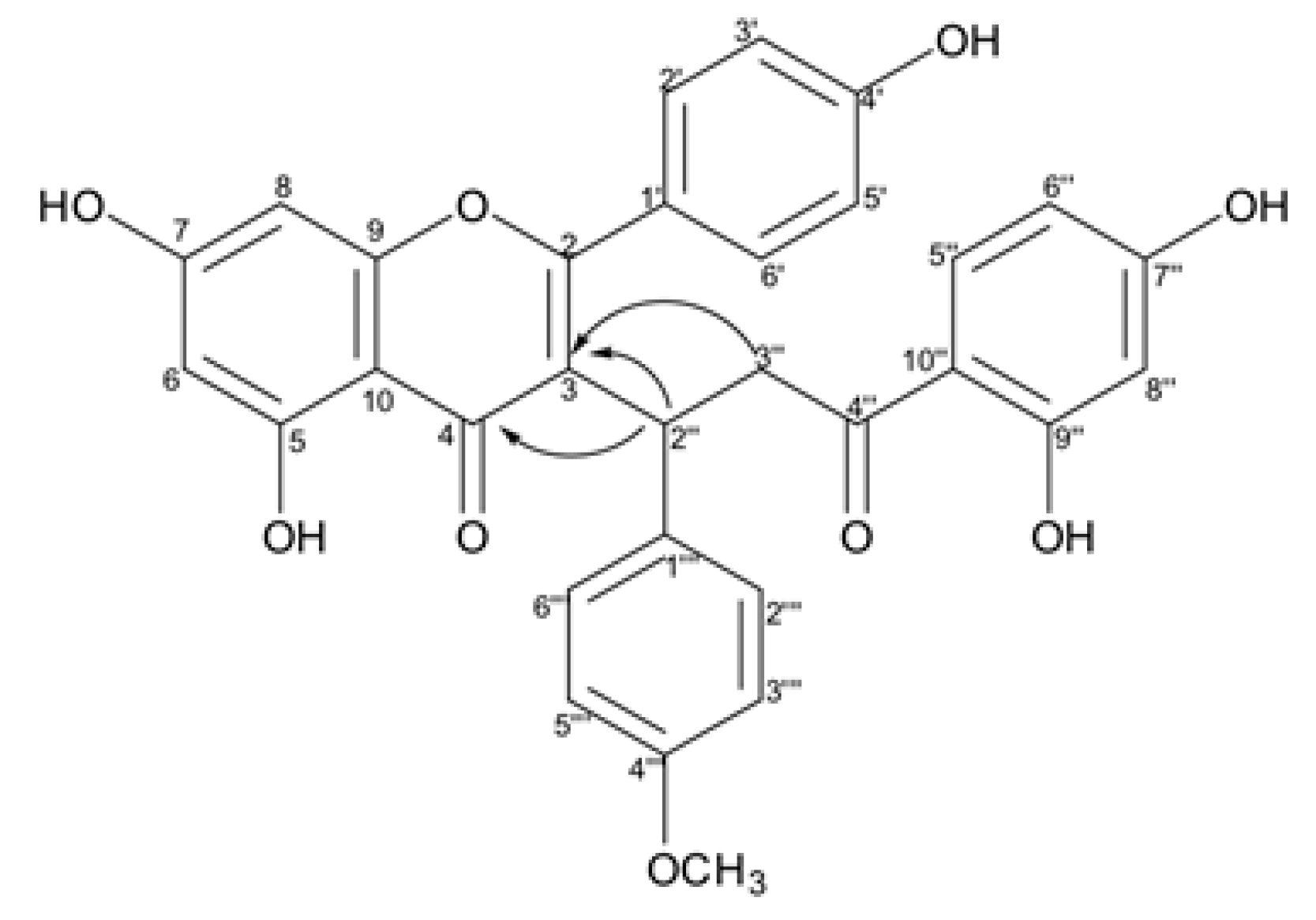
| 100 * | 1.5 | 539.134155 | 19.1 | −2.26 | C31H23O9 | 1 (7-methoxycaesalpinioflavone, 3) |
| 6 * | 1.3 | 539.134196 | 19.9 | −2.26 | C31H23O9 | 1 (4‴-methoxycaesalpinioflavone,2) |
| 231 * | 1.1 | 271.227588 | 24.1 | −1.43 | C16H31O3 | 17 (fatty acids) |
| 457 | 2.2 | 433.113306 | 10.0 | 0.40 | C21H26O3N2Br | 0 |
| 188 | 1.2 | 319.045785 | 10.6 | −0.50 | C15H11O8 | 13 (ethyl brevifolincarboxylate) |
| 491 | 2.6 | 395.161091 | 11.1 | −0.37 | C22H23O5N2 | 0 |
| 352 | 1.8 | 237.040059 | 11.1 | −1.31 | C11H9O6 | 18 (phenolic compounds) |
| 402 | 1.2 | 262.071942 | 11.3 | −0.59 | C13H12O5N | 0 |
| 502 | 2.1 | 425.171936 | 11.6 | 0.17 | C23H25O6N2 | 0 |
| 489 | 2.9 | 379.165825 | 12.0 | C22H23O4N2 | 0 | |
| 488 | 3.0 | 409.176757 | 12.4 | −0.26 | C23H25O5N2 | 0 |
| 508 | 1.7 | 354.098236 | 12.7 | −0.21 | C19H16O6N | 0 |
| 509 | 1.9 | 439.187347 | 12.8 | −0.26 | C24H27O6N2 | 0 |
| 309 | 1.2 | 571.088124 | 13.1 | −0.13 | C30H19O12 | 14 (phenolic compounds) |
| 190 | 3.8 | 285.040309 | 13.2 | −1.16 | C15H9O6 | 10 (phenolic compounds) |
| 403 | 1.2 | 391.066808 | 13.2 | −0.67 | C18H15O10 | 3 (phenolic compounds) |
| 512 | 1.2 | 402.249603 | 13.9 | −0.30 | C20H36O7N | 0 |
| 520 | 1.3 | 437.231079 | 14.4 | −0.17 | C21H38O7Cl | 0 |
| 486 | 2.1 | 543.129247 | 14.7 | −0.78 | C30H23O10 | 19 (phenolic compounds) |
| 301 | 1.8 | 269.045442 | 14.7 | 0.27 | C15H9O5 | 103 (phenolic compounds) |
| 504 | 1.2 | 655.442627 | 15.3 | −0.07 | C36H63O10 | 0 |
| 181 | 3.6 | 327.217286 | 15.4 | −1.37 | C18H31O5 | 37 (phenolic compounds) |
| 475 | 2.5 | 363.193962 | 15.4 | −1.20 | C18H32O5Cl | 0 |
| 494 | 1.7 | 329.066286 | 15.9 | −1.18 | C17H13O7 | 153 (phenolic compounds) |
| 392 | 6.9 | 271.061001 | 16.1 | −1.39 | C15H11O5 | 115 (phenolic compounds) |
| 76 | 1.8 | 329.233110 | 16.3 | −0.69 | C18H33O5 | 22 (fatty acids) |
| 528 | 1.2 | 323.199127 | 16.7 | −1.03 | C16H32O4Cl | 0 |
| 506 | 1.9 | 365.209715 | 17.2 | −0.85 | C18H34O5Cl | 0 |
| 505 | 2.0 | 403.233625 | 17.5 | −0.25 | C20H35O8 | 0 |
| 487 | 2.1 | 387.238416 | 18.7 | −1.02 | C20H35O7 | 1 (norcaperatic acid) |
| 490 | 1.8 | 309.082260 | 2.1 | −1.49 | C11H17O10 | 1 (glucopyranosyl) |
| 451 | 1.3 | 331.066886 | 2.3 | −0.55 | C13H15O10 | 12 (phenolic compounds) |
| 419 | 1.2 | 433.235646 | 21.2 | 2.85 | C23H33O6N2 | 0 |
| 205 | 6.6 | 611.408728 | 22.4 | −0.41 | C35H60O6Cl | 0 |
| 496 | 2.3 | 621.437117 | 22.4 | −0.29 | C36H61O8 | 11 (phenolic compounds) |
| 485 | 4.1 | 613.406587 | 22.6 | −0.58 | C32H57O9N2 | 0 |
| 534 | 1.4 | 734.517439 | 22.6 | 0.28 | C37H72O11N3 | 0 |
| 495 | 2.6 | 750.529089 | 24.0 | 1.30 | C40H82O2N3BrCl | 0 |
| 466 | 1.2 | 561.429542 | 25.8 | 0.74 | C32H62O5Cl | 0 |
| 282 | 1.4 | 748.513773 | 26.2 | C41H70O9N3 | 0 | |
| 517 | 1.8 | 830.114318 | 8.3 | 0.01 | C30H24O20N9 | 0 |
| 206 | 2.3 | 951.074661 | 8.4 | −1.21 | C42H23O23N4 | 0 |
| 359 | 1.2 | 275.019645 | 8.5 | −0.39 | C13H7O7 | 3 (xanthones) |
| 526 | 1.7 | 476.040832 | 8.6 | 0.62 | C19H14O10N3S | 0 |
| 492 | 2.6 | 337.092738 | 9.2 | −0.27 | C16H17O8 | 18 (phenolic compounds) |
| 185 | 2.6 | 300.998685 | 9.3 | −1.10 | C14H5O8 | 1 (ellagic acid) |
| 493 | 2.3 | 252.050914 | 9.6 | −1.96 | C11H10O6N | 0 |
| 180 | 2.8 | 463.087903 | 9.9 | −1.60 | C21H19O12 | 71 (phenolic compounds) |
| Position | δC, Type | δH, mult. (J in Hz) |
|---|---|---|
| 2 | 164.4, C | - |
| 3 | 119.4, C | - |
| 4 | 181.1, C | - |
| 5 | 161.9, C | - |
| 6 | 103.5, CH | 6.17 d (2.0) |
| 7 | 165.2, C | - |
| 8 | 94.1, CH | 6.26 d (2.0) |
| 9 | 157.7, C | - |
| 10 | 99.4, C | - |
| 1′ | 122.8, C | - |
| 2′/6′ | 130.7, CH | 7.08 d (8.7) |
| 3′/5′ | 115.7, CH | 6.81 d (8.7) |
| 4′ | 160.0, C | - |
| 2″ | 48.4, CH | 4.57 t (6.7) |
| 3a″ | 34.5, CH | 2.97 dd (13.9, 6.7) |
| 3b″ | 34.5, CH | 3.44 (m) |
| 4″ | 201.3, C | - |
| 5″ | 131.7, CH | 7.18 d (8.6) |
| 6″ | 108.2, CH | 6.15 dd (8.6, 2.2) |
| 7″ | 163.5, C | - |
| 8″ | 103.0, CH | 6.17 d (2.2) |
| 9″ | 164.4, C | - |
| 10″ | 113.1, C | - |
| 1‴ | 132.2, C | - |
| 2‴/6‴ | 114.0, CH | 6.73 d (8.6) |
| 3‴/5‴ | 130.4, CH | 6.98 d (8.6) |
| 4‴ | 158.1, C | - |
| 4‴-OCH3 | 55.4 | 3.66 s |
| OH | - | 11.87 s |
| OH | - | 12.66 s |
| Position | δC, Type | δH, mult. (J in Hz) |
|---|---|---|
| 2 | 164.6, C | - |
| 3 | 119.7, C | - |
| 4 | 181.4, C | - |
| 5 | 157.6, C | - |
| 6 | 92.7, CH | 6.57 d (2.0) |
| 7 | 165.9, C | - |
| 8 | 98.6, CH | 6.36 d (2.0) |
| 9 | 161.6, C | - |
| 10 | 104.6, C | - |
| 1′ | 122.7, C | - |
| 2′/6′ | 130.7, CH | 7.07 d (8.7) |
| 3′/5′ | 115.7, CH | 6.81 d (8.7) |
| 4′ | 160.1, C | - |
| 2″ | 48.6, CH | 4.55 t (6.9) |
| 3a″ | 34.5, CH | 2.98 dd (13.9, 7.14) |
| 3b″ | 34.5, CH | 3.35 (m) |
| 4″ | 201.2, C | - |
| 5″ | 131.7, CH | 7.17 d (8.6) |
| 6″ | 108.3, CH | 6.15 dd (9.4, 2.2) |
| 7″ | 163.5, C | - |
| 8″ | 103.1, CH | 6.16 d (2.2) |
| 9″ | 164.8, C | - |
| 10″ | 113.1, C | - |
| 1‴ | 130.3, C | - |
| 2‴/6‴ | 130.4, CH | 6.85 d (8.6) |
| 3‴/5‴ | 115.4, CH | 6.56 d (8.6) |
| 4‴ | 156.0, C | - |
| 7-OCH3 | 56.6 | 3.80 s |
| OH | - | 11.85 s |
| OH | - | 12.69 s |
© 2019 by the authors. Licensee MDPI, Basel, Switzerland. This article is an open access article distributed under the terms and conditions of the Creative Commons Attribution (CC BY) license (http://creativecommons.org/licenses/by/4.0/).
Share and Cite
Domingos, O.d.S.; Alcântara, B.G.V.; Santos, M.F.C.; Maiolini, T.C.S.; Dias, D.F.; Baldim, J.L.; Lago, J.H.G.; Soares, M.G.; Chagas-Paula, D.A. Anti-Inflammatory Derivatives with Dual Mechanism of Action from the Metabolomic Screening of Poincianella pluviosa. Molecules 2019, 24, 4375. https://doi.org/10.3390/molecules24234375
Domingos OdS, Alcântara BGV, Santos MFC, Maiolini TCS, Dias DF, Baldim JL, Lago JHG, Soares MG, Chagas-Paula DA. Anti-Inflammatory Derivatives with Dual Mechanism of Action from the Metabolomic Screening of Poincianella pluviosa. Molecules. 2019; 24(23):4375. https://doi.org/10.3390/molecules24234375
Chicago/Turabian StyleDomingos, Olívia da S., Bianca G. V. Alcântara, Mário F. C. Santos, Tatiane C. S. Maiolini, Danielle F. Dias, João L. Baldim, João Henrique G. Lago, Marisi G. Soares, and Daniela A. Chagas-Paula. 2019. "Anti-Inflammatory Derivatives with Dual Mechanism of Action from the Metabolomic Screening of Poincianella pluviosa" Molecules 24, no. 23: 4375. https://doi.org/10.3390/molecules24234375
APA StyleDomingos, O. d. S., Alcântara, B. G. V., Santos, M. F. C., Maiolini, T. C. S., Dias, D. F., Baldim, J. L., Lago, J. H. G., Soares, M. G., & Chagas-Paula, D. A. (2019). Anti-Inflammatory Derivatives with Dual Mechanism of Action from the Metabolomic Screening of Poincianella pluviosa. Molecules, 24(23), 4375. https://doi.org/10.3390/molecules24234375