Article

A Newly Discovered Phenylethanoid Glycoside from Stevia rebaudiana Bertoni Affects Insulin Secretion in Rat INS-1 Islet β Cells

1
School of Chinese Materia Medica, Beijing University of Chinese Medicine, The Key Unit of Exploring Effective Substances of Classical and Famous Prescription of SATCM, Beijing 102488, China
2
Institute of Medicinal Plant Development, Chinese Academy of Medical Sciences, Beijing 200293, China
3
Quality Standards, Key Laboratory of Beijing for Identification and Safety Evaluation of Chinese Medicine, Institute of Chinese Materia Medica, China Academy of Chinese Medical Sciences, Beijing 100700, China
*
Authors to whom correspondence should be addressed.
These authors contributed equally to this work.
Molecules 2019, 24(22), 4178; https://doi.org/10.3390/molecules24224178
Submission received: 13 October 2019 / Revised: 8 November 2019 / Accepted: 10 November 2019 / Published: 18 November 2019

Abstract

:
The tea-like beverage Stevia rebaudiana Bertoni (Stevia) is popular in China because it reduces blood glucose and has a sweet taste. In this work, a comprehensive quality assessment of Stevia led to the discovery of five phenylethanoid glycosides, namely steviophethanoside (1), cuchiloside (2), salidroside (3), icariside D (4), and tyrosol (5). Of them, compound 1 is a novel compound. Mass spectrometry and NMR spectroscopy were employed to confirm the absolute configuration. A hydrolytic step with 4 N TFA at 95 °C for 4 h was used to confirm the monosaccharides. In addition, Discovery Studio 4.0 was used to predict the ADME and toxicity activity of compound 1. The results suggested that compound 1 was biocompatible and had poor toxicity, which was verified by rat INS-1 islet β cells through an MTT assay. Meanwhile, a significant stimulatory effect on INS-1 cells was observed, which indicated a hypoglycemic effect of compound 1. This is the first report that describes a natural, novel, and hypoglycemic phenylethanoid glycoside in Stevia.

1. Introduction

Stevia rebaudiana Bertoni (Stevia), has been used as a non-cariogenic sweetener for many years [1]. Early in the development of sweeteners, several non-cariogenic sweeteners, such as steviol glycosides, stevioside, and rebaudioside (A to F), were isolated from Stevia. Steviol glycosides have been used to replace sucrose in the treatment of diabetes mellitus and obesity [2]. Recently, several biological activities such as the beneficial effects of blood glucose regulation, antioxidant, and renal protective properties have attracted attention [3,4,5]. It is reported that the crude extract of Stevia had the ability to decrease blood glucose [6]. In addition, stevioside may indirectly contribute to anti-hyperglycemic action [7]. The aqueous extracts of the plant have been extensively used in Paraguay, and the clinical efficacy of regulating blood glucose was confirmed in related studies [8,9].
Normally, blood glucose was decreased by promoting insulin secretion, inhibiting glucose absorption, and promoting glucose decomposition. Among them, insulin is an important and effective tool to control hyperglycemia. Stevia can decrease plasma glucagon in diabetic rats by the mechanism of an improvement of insulin and suppression of glucagon level [7]. Recently, our group discovered that the polyphenolic compounds of Stevia could prevent diabetes and its associated complications in the streptozotocin rat and mouse model [10,11]. Compared with Stevia leaves, polyphenolic compounds decreased blood glucose more significantly [4]. What is more, a PK/PD-DI investigation displayed that some of the phenolic components may be absorbed into blood immediately. More interestingly, those compounds had the highest blood–drug concentrations at the drug effect time [11].
Hence, our previous research systematically studied the chemical constituents of the phenolic components in Stevia leaves [12]. In this process, a new type compound, phenylethanoid glycosides, was found in the phenolic components of Stevia. Phenylethanoid glycosides are widely distributed in the medicinal kingdom of plants. They are a type of phenolic compounds characterized by a phenethyl alcohol structure. Some of them are phenylethanoid and phenylpropanoid hybrids forming ester bonds with sugars [13]. Those structures contribute a broad range of biological activities and guarantee safety for phenylethanoid glycosides, which provides great potential in pharmaceutical applications. It is well known that phenylethanoid glycosides have strong antioxidant effects [14,15]. Phenylethanoid glycosides also can retard the occurrence of diabetic complications [16]. In recent years, many natural products with antioxidant effects have been shown to have hypoglycemic effects. More recently, the natural epicatechin was confirmed to modulate insulin secretion from pancreatic β cells [17,18]. Furthermore, Quercetin and oleic acid have a contribution to the insulin secretory dysfunction [19]. Quercetin-3-Oleoyl and berberine are potential novel agonists of fatty acid receptor GPR40 [20,21]. Natural products seem to become the preferred choice to deal with the chronic metabolic disorde. This raised our attention to find the connection between diabetes and phenylethanoid glycosides, which exist in the phenolic extract of Stevia.
Since Stevia can decrease plasma glucagon through the mechanism of an improvement of insulin, phenylethanoid glycosides are probably involved in promoting insulin secretion. Generally, the islet contains four different endocrine cell types, which play a major role in control of metabolic fuel homeostasis. Except the dysfunction of α-cells, δ-cells, and the pancreatic-polypeptide secreting cells (PP cells), a dysregulation of insulin secretion from β cells directly leads to type II diabetes [22]. INS-1 cell line is a widely used cell line for evaluating insulin secretion and β cell function in vitro. It was established in the culture medium, which contains 2-mercaptoethanol (2-ME). With the elevation of the glucose concentration, INS-1 cell line showed a steady increase in insulin secretion. Currently, there is no evidence that phenylethanoid glycosides promote insulin secretion. Therefore, exploring its hypoglycemic effect becomes interesting and important. This paper describes, in detail, the isolation, structural elucidation, and the stimulatory effect of phenylethanoid glycosides on insulin secretion.

2. Results and Discussion

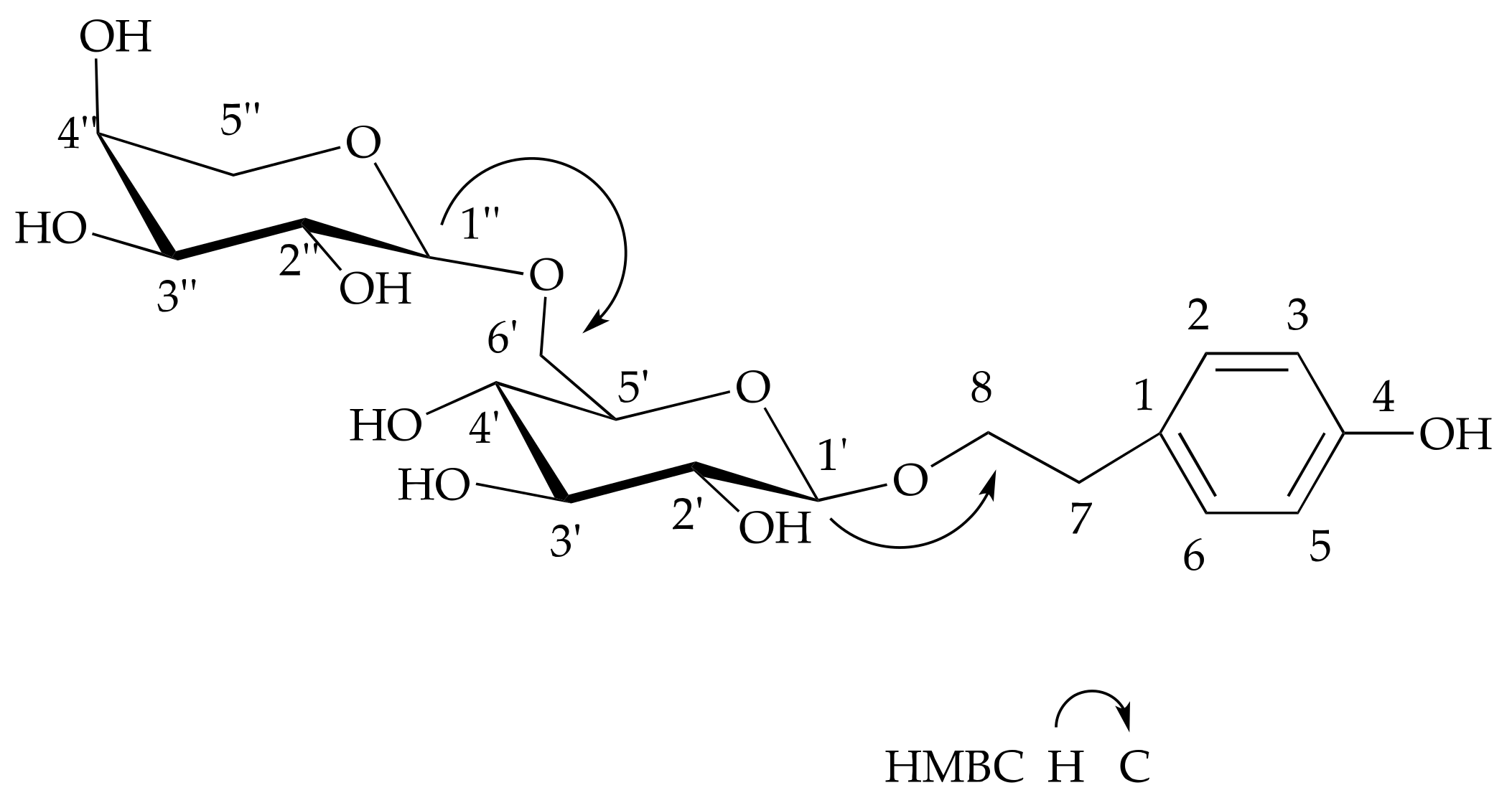
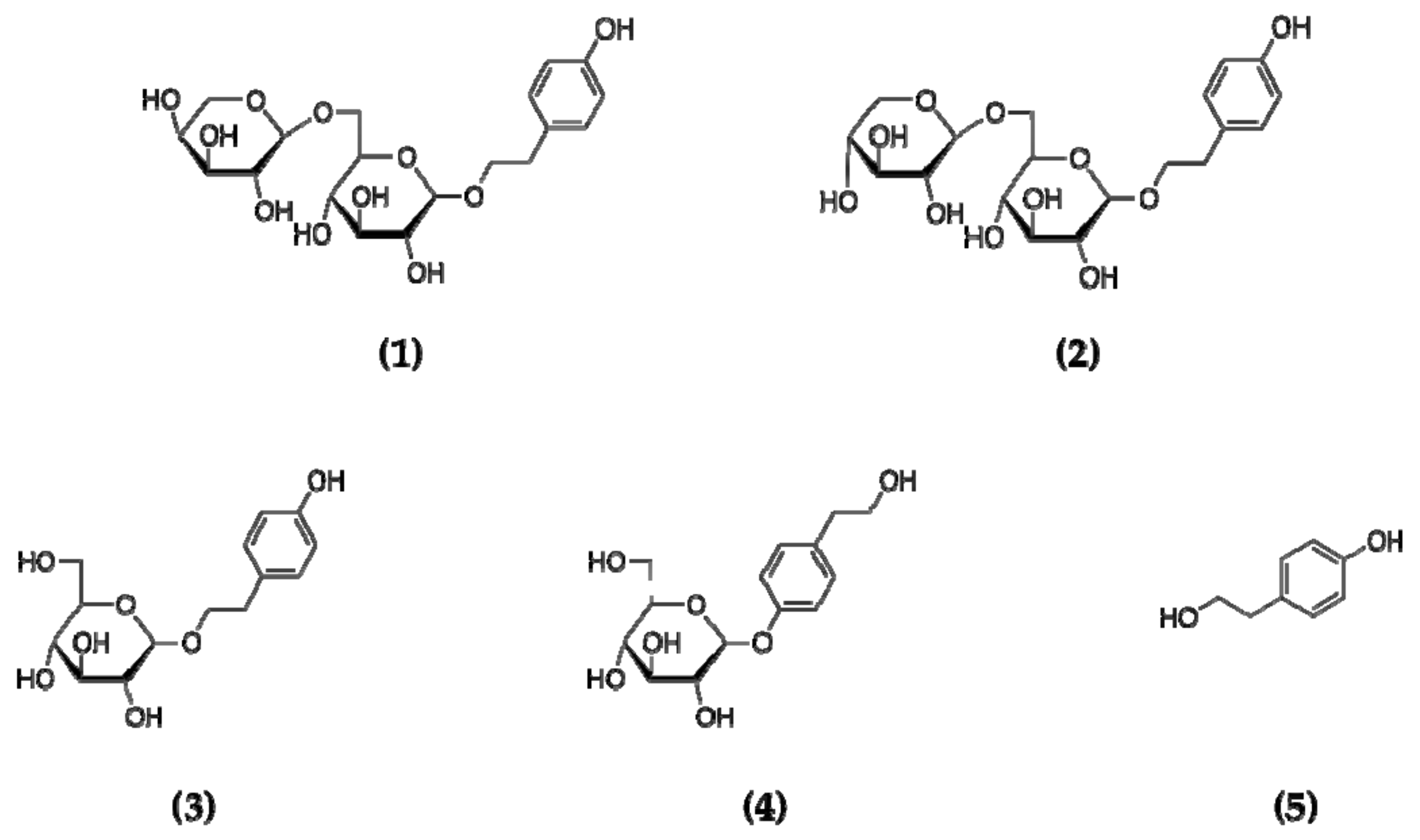
2.1. Structure Elucidation

The dried leaves of Stevia were extracted twice with 50% EtOH. A new phenylethanoid glycoside (1) and four known compounds (25) were isolated and purified via AB-8 and Diaion HP-20 macroporous adsorptive resin, Sephadex LH-20 gel chromatograpy, and semi-preparative HPLC.
Steviophethanoside (1) was isolated as white amorphous powder, which showed the molecular ion peak in the high-resolution mass spectrum at m/z 455.1515, corresponding to the molecular formula C19H28O11 (calculated for C19H28O11Na: 455.1529). The 1H-NMR data at δH 6.82 (2H, d, J = 8.0 Hz, H-3, H-5) and 7.19 (2H, d, J = 8.0 Hz, H-2, H-6) showed a p-disubstituted benzene ring, as evidenced by the 13C-NMR data: δC 118.3 (d, C-3, C-5), 133.2 (d, C-2, C-6), 133.5 (s, C-1), 156.8 (s, C-4). In addition, the aliphatic proton signals at δH 2.89 (2H, t, J = 7.0 Hz, H-7), 3.84 (1H, dd, J = 12.0, 7.0 Hz, H-8a), 4.09 (1H, dd, J = 12.0, 7.0 Hz, H-8b), as well as 13C-NMR data δC 37.2 (t, C-7) and 74.0 (t, C-8) indicated one phenylethanoid skeleton. These spectral data were similar to Tyrosol [23].
Apart from the tyrosol moiety, compound 1 possessed two sugar units, as evidenced by two anomeric protons at δH 4.43 (1H, d, J = 8.0 Hz, H-1’) and 4.33 (1H, d, J = 7.6 Hz, H-1’’). The glucose signals at δC 105. 1 (C-1’), 75.9 (C-2’), 78.5 (C-3’), 72.2 (C-4’), 77.9 (C-5’), and 71.1 (C-6’), as well as the signal of C-8 being shifted downfield by 11.2 ppm to δC 74.0 (t) in 1, were observed, which indicated the glucopyranosyl moiety being linked at C-8 in 1. This deduction was confirmed by the 1H detected heteronuclear multiple bond correlation (HMBC) from the anomeric proton signal at δH 4.43 (1H, d, J = 8.0 Hz, H-1’) to δC 37.2 (t, C-7). The coupling value (J = 8.0 Hz) of anomeric proton suggested the presence of a β-glucopyranosyl moiety [24].
Furthermore, the HMBC correlation observed from δH 3.78 (1H, dd, J = 11.7, 5.6 Hz, H-6’a) and δH 4.13 (1H, dd, J = 11.7, 5.6 Hz, H-6’b) to δC 106.5 (d, C-1’’) revealed that the other sugar unit was linked to C-6’. In addition, the 13C-NMR data δC 106.5 (C-1’’), 73.5 (C-2’’), 75.1 (C-3’’), 71.1 (C-4’’), and 69.1 (C-5’’) indicated that the other sugar unit was arabinopyranosy. The coupling constants (J 1’–2’= 8.0 Hz) confirmed the moiety to be α-arabinopyranosyl, which suggests glycosylation at C-8 of aglycone with an ara (1→6)-glcmoiety. The absolute configuration of the monosaccharides was determined to be D-glu and L-ara through GC analysis of the chiral derivatives of the monosaccharides in the hydrolysate. The key HMBC correlations in 1 are shown in Figure 1. On the basis of all results, the structure of this new compound was established as 4-hydroxyphenyl ethyl-8-O-[α-l-arabinopyranosyl-(1→6)]-β-d-glucopyranoside.
Compound 2 was isolated as white amorphous powder. It is soluble in water and methanol, and has a positive response of FeCl3 and molish reagent. UV (MeOH − H2O) λmax nm: 220.0, 279.1. ESI-MSn m/z: 455 [M + Na]+, 431 [M − H], 455 (323), 431(299, 191, 179, 161, 149, 143, 131). 1H-NMR (500 MHz, D2O): δH 2.89 (2H, t, J = 6.8 Hz, H2-7), 3.25 (1H, t, J = 8.3 Hz, H-2’), 3.29 (1H, dd, J = 10.0, 6.4 Hz, H-2’’), 3.31 (1H, dd, J = 9.5, 3.0 Hz, H-5’’ a), 3.43 (1H, t, J = 8.5 Hz, H-4’), 3.46 (1H, t, J = 9.5 Hz, H-3’’), 3.47 (1H, t, J = 9.0 Hz, H-3’), 3.58 (1H, dd, J = 9.5, 6.5 Hz, H-5’), 3.63 (1H, ddd, J = 15.0, 9.8, 5.3Hz, H-4’’), 3.84 (1H, dd, J = 11.8, 5.8 Hz, H-6’ a), 3.89 (1H, dt, J = 9.5, 7.3 Hz, H-8a), 3.96 (1H, dd, J = 11.5, 5.5 Hz, H-5’’ b), 4.09 (1H, dt, J = 10.0, 7.0 Hz, H-8b), 4.14 (1H, d, J = 12.0 Hz, H-6’ b), 4.45 (1H, d, J = 9.0 Hz, H-1’’), 4.47 (1H, d, J = 8.5 Hz, H-1’), 6.88 (2H, d, J = 8.0 Hz, H-3, 5), 7.23 (2H, d, J = 8.0 Hz, H-2, 6). 13C-NMR (125 MHz, D2O): δC 37.2 (t C-7), 68.0 (t, C-5’’), 71.4 (t, C-6’), 72.1 (d, C-4’’), 72.3 (d, C-4’), 74.0 (t, C-8), 75.8 (d, C-2’), 75.9 (d, C-2’’), 77.8 (d, C-5’), 78.5 (d, C-3’’), 78.5 (d, C-3’), 105.1 (d, C-1’), 106.4 (d, C-1’’), 118.3 (d, C-3, 5), 133.2 (d, C-2, 6), 133.4 (s, C-1), 156.8 (s, C-4). The spectral data of compound 2 are consistent with the cuchiloside [25].
Compound 3 was isolated as white amorphous powder. It is soluble in water and methanol, and has a positive response of FeCl3 and molish reagent. UV (MeOH − H2O) λmaxnm: 216.5, 273.2. ESI-MSn m/z: 323 [M + Na]+, 323 (291, 237, 230, 209, 193, 121). 1H-NMR (500 MHz, D2O): δH 2.89 (2H, t, J = 5.7 Hz, H2-7), 3.25 (1H, t, J = 8.5 Hz, H-2’), 3.38 (1H, t, J = 9.3 Hz, H-4’), 3.42 (1H, d, J = 10.5 Hz, H-3’) 3.47 (1H, t, J = 9.0 Hz, H-5’), 3.72 (1H, dd, J = 12.3, 5.7 Hz, H-6’a), 3.88 (1H, d, J = 14.5 Hz, H-8a), 3.91 (1H, d, J = 12.0 Hz, H-8b), 4.09 (1H, t, J = 8.3 Hz, H-6’ b), 4.46 (1H, d, J = 8.0 Hz, H-1’), 6.87 (2H, d, J = 8.0 Hz, H-3, 5), 7.23 (2H, d, J = 7.5 Hz, H-2, 6). 13C-NMR (125 MHz, D2O): δC 37.2 (t, C-7), 63.6 (t, C-6’), 72.5 (d, C-4’), 73.9 (t, C-8), 76.0 (d, C-2’), 78.6 (d, C-5’), 78.8 (d, C-3’), 105.1 (d, C-1’), 118.3 (d, C-3, 5), 133.2 (d, C-2, 6), 133.4 (s, C-1), 156.8 (s, C-4). The spectral data of compound 3 are consistent with the salidroside [26].
Compound 4 was isolated as white amorphous powder. It is soluble in water and methanol. UV (MeOH − H2O) λmaxnm: 218.9, 279.1. ESI-MSn m/z: 323 [M + Na]+, 323 (302, 266, 248, 230, 205, 185, 180, 156, 145, 121). 1H-NMR (500 MHz, D2O): δH 2.86 (2H, t, J = 6.3 Hz, H2-7), 3.53 (1H, t, J = 9.3 Hz, H-4’), 3.58 (1H, d, J = 9.0 Hz, H-2’), 3.62 (1H, d, J = 7.5 Hz, H-3’), 3.65 (1H, d, J = 9.0 Hz, H-5’), 3.78 (2H, dd, J = 12.0, 5.5 Hz, H2-8), 3.84 (1H, t, J = 6.5 Hz, H-6’a), 3.96 (1H, d, J = 12.5 Hz, H-6’ b), 5.13 (1H, d, J = 7.5 Hz, H-1’), 7.13 (2H, d, J = 8.0 Hz, H-2, 6), 7.31 (2H, d, J = 8.0 Hz, H-3, 5). 13C-NMR (125 MHz, D2O): δC 39.8 (t, C-7), 63.4 (t, C-6’), 65.4 (t, C-8), 72.3 (d, C-4’), 75.8 (d, C-2’), 78.4 (d, C-5’), 78.9 (d, C-3’), 103.2 (d, C-1’), 119.5 (d, C-3, 5), 133.1 (d, C-2, 6), 136.7 (s, C-1), 157.9 (s, C-4). The spectral data of compound 4 are consistent with the icariside D [27].
Compound 5 was isolated as white amorphous powder. It is soluble in water and methanol. UV (MeOH − H2O) λmaxnm: 221.2, 274.4. ESI-MSn m/z: 161 [M + Na]+, 161 (141, 133, 121, 107, 91, 65). 1H-NMR (500 MHz, D2O): δH 2.79 (2H, t, J = 6.3 Hz, H-2), 3.79 (2H, t, J = 8.5 Hz, H-1), 6.87 (2H, d, J = 7.5 Hz, H-5, 7), 7.20 (2H, d, J = 8.0 Hz, H-4, 8). 13C-NMR (125 MHz, D2O): δC 36.9 (t, C-2), 62.8 (t, C-1), 115.4 (d, C-5, 7), 130.3 (d, C-4, 8), 131.0 (s, C-3), 153.8 (s, C-6). The spectral data of compound 5 are consistent with tyrosol [23] (Figure 2).

2.2. Pharmacokinetic Properties

The predicted pharmacokinetic properties include HIA (human intestinal absorption), BBB (blood brain barrier), PPB (plasma protein binding), CYP2D6, aqueous solubility, and toxicity. Our results showed that compound 1 was found to be non-hepatotoxic (Hepatotoxicity level of 0) and non-inhibitors of CYP2D6 (CYP2D6 level of 0). In addition, the binding was below 90% to carrier proteins. It is well known that the liver plays a critical role in transforming and clearing chemicals, and it is susceptible to the toxicity from these agents. Chemicals that cause liver injury may also induce the injury of other organs (like kidney). In early studies, traditional Chinese medicine presenting with high security and low toxicity compounds even without toxicity, such as phenylethanoid glycosides, which existed in purified fraction of goldengermander, were confirmed to show a lack of hepatotoxicity [28]. Combined with the current results of pharmacokinetic properties, compound 1 could be considered as the one that had low hepatotoxicity, good bioavailability [29], and involved less drug–drug interaction. In addition, the results revealed that compound 1 had optimal aqueous solubility of 4 with undefined BBB level at 4 and very poor absorption (HIA levels of 3), which might be considered as more hydrogen bond donors and acceptors. The property of high solubility in water was probably due to the lower log p value of the compounds, which depicted lower lipophilicity [30].
TOPKAT provides the capability to rapidly and confidently evaluate the toxic effects of chemicals directly from the molecular structure. In this study, compound 1 was found to be non-mutagenic (NM), non-carcinogenic (NC), and non-skin irritant in the toxicity parameter. Besides, the calculated LD50 (oral administration to rats) value of compound 1 was 5.61 g/kg. According to the different categories defined by the GHS, the category of compound 1 is higher than 5, which means that compound 1 is non-toxic after deglutition [31]. Although the prediction suggests that compound 1 may involve skin sensitization, skin, and ocular irritancy, i.e., some kind of developmental toxicity potential, the toxic dose is currently unclear (Table 1).

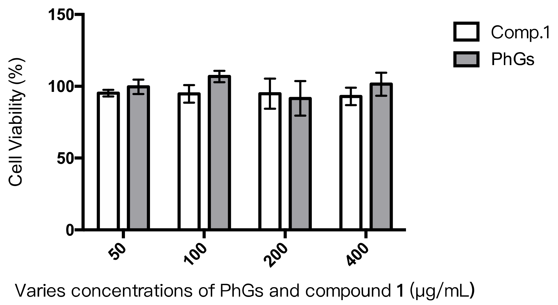
2.3. Cytotoxicity Assay

To investigate the cytotoxic effects of compound 1, an MTT assay was used. Regarding the similarity structure, phenylethanoid glycosides (PhGs) (including compound 1 and 2) were evaluated together. After 48 h of exposure, no significant decreases in cell viability was observed dealing with various concentrations of compound 1 and PhGs (Figure 3). This means that both compound 1 and PhGs were not toxic to the INS-1 cells. The result indicated that compound 1 and compound 2 could not exert harmful effects on rat INS-1 cells even at the higher concentration of 400 μg/mL. Although there is no significant difference between compound 1 and PhGs, PhGs presented a trend of more secure effects than single compound 1. Those results provided another proof of low toxicity.

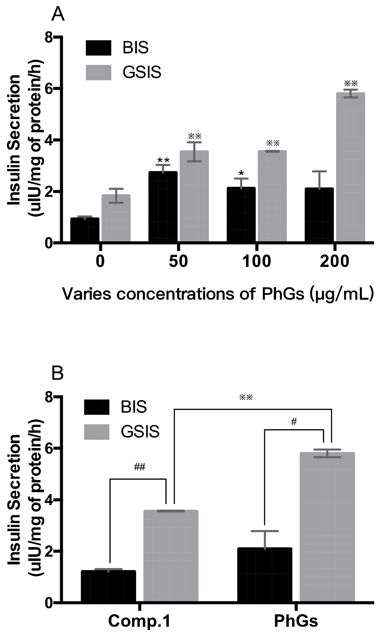
2.4. Insulin Secretion in Vitro

Shivanna found that when rats were fed with whole leaves powder and extracted polyphenols of Stevia, a reduction of blood glucose, alanine transaminase (ALT), aspartate transaminase (AST), and increment of insulin level were observed [4]. In this study, the results for the glucose-mediated insulin release from INS-1 cells are presented in Figure 4. Substimulatory glucose (2.8 mM, BIS) showed less insulin release (0.99 µIU/mg of protein/h) than that (1.99 µIU/mg of protein/h) at high glucose levels (16.7 mM, GSIS).
Compound 1 and PhGs were investigated in this study. The results of PhGs group exhibited dose-dependent insulin release effects on INS-1 cells after treatment with the drug (50, 100, and 200 µg/mL) for 1 h, and the corresponding values were 2.74 ± 0.22, 2.12 ± 0.27, and 2.10 ± 0.49 µIU/mg of protein/h on 2.8 mM glucose, 3.54 ± 0.26, 3.56 ± 0.01, and 5.81 ± 0.11 µIU/mg of protein/h on 16.7 mM glucose, respectively (Figure 4A). The rate of basic insulin secretion (BIS) was only slightly changed relative to the rate found in the glucose stimulated insulin secretion (GSIS). In a high-glucose environment, there was a significant increase of insulin secretion in 200 μg/mL compared to the other two concentrations. Although both compound 1 and PhGs group stimulated insulin secretion of INS-1 cells, a significant difference in the rates of the two groups were observed at the same dose (200 µg/mL) of 16.7 mM glucose. These results are in agreement with those expected from hypoglycemic agents. The effects of phenylethanoid glycoside on insulin secretion have previously been reported with type 2 diabetic db/db mice [32]. The results demonstrated that the investigation could be a starting point for an interesting investigation about the potential antidiabetic activity of the new compound. In early studies, the hypoglycaemic chemicals of Stevia rebaudiana Bertoni have been reported to be steviol and polyphenols, known as secretagogues and facilitate insulin release, protecting pancrease, etc. [33,34,35]. This paper demostrated that phenylethanoid glycosides could directly stimulate insulin secretion dose dependently. Those discoveries are meaningful for the investigation of hypoglycemic active component group of Stevia.

3. Materials and Methods

3.1. Material and Chemicals

Stevia were collected from Ganzhou Julong High-tech Industrial Co., Ltd. in Jiangxi province, China, and were naturally dried at room temperature. Samples were identified by Assistant Prof. Chunsheng Liu, Department of Botany, Beijing University of Chinese Medicine. A voucher specimen (No. 20111107) has been deposited in the Herbarium of the Department of Phytochemistry, Beijing University of Chinese Medicine. Column chromatography was performed on Diaion HP-20 (Mitsubishi Heavy Industries, Ltd, Tokyo, Japan); Sephadex LH-20 column (Fuji Silysia Chemical Co., Ltd, Osaka, Japan); and Chromatorex ODS (Fuji Silysia Chemical Co., Ltd, Osaka, Japan), respectively. TLC was carried on silica gel G precoated plates (Qingdao Haiyang Chemical Co., Qingdao, China) with EtOAc-HOAc-H2O (3:1:0.1). The spots were detected by spraying with 10% H2SO4 ethanol solution followed by heating.
Rat INS-1 islet β cells were generously presented by Prof. BaoLin Bian (China Academy of Chinese Medical Science, Beijing, China). INS-1 cell culture medium RPMI 1640, 0.25% Trypsin-EDTA (Ethylene diamine tetraacetate), FBS, DMSO, penicillin, streptomycin, and supplements were purchased from Gibco (Grand island, NewYork, NY, USA). MTT was purchased from Sigma-Aldrich (St. Louis, MO, USA). RIA kit purchased from HuayingBio (Shenzhen, China). Glucose and β-mercaptoethanol were purchased from BEIJING DINGGUO CHANGSHENG BIOTECHNOLOGY CO.LTD (Beijing, China). RIPA Lysis buffer (Item No. c1053) and protease inhibitor (Item No. p1265) were purchased from Applygen technologies Inc. (Beijing, China). Culture plates, culture flasks, tubes, and cell scrapers (Item No. 3010) were purchased from Corning (One Riverfront Plaza Corning, NewYork, NY, USA).

3.2. Isolation and Purification of Compounds 15

The phenolic extracts of Stevia were enriched from the plant as described previously [36]. Compounds 15 were isolated from the phenolic extracts. In detail, the phenolic extracts were dispersed with H2O and then subjected to macroporous resin Diaion HP-20 eluted with a step gradient of H2O/MeOH (1:0→0:1) to obtain eight fractions (Fr. A–Fr. H). Fr. B (55.7 g) was separated by a Sephadex LH-20 column (5 × 80 cm), eluted with MeOH, and then purified with a preparative high-performance liquid chromatography, which was performed on a CXTH 3000 chromatograph (Beijing Chuangxintongheng Co., Ltd. Beijing, China) equipped with UVD detector. A YMC C18 column (250 × 20 mm, 5 μm) was used at a flow rate of 8 mL/min to give compound 2 (30 mg), 3 (25 mg), and 4 (20 mg). Fr. D (44.6 g) was separated via reverse-phase chromatography over C-18 silica gel, eluted with MeOH − H2O (100:0→70:30 100:0→50:50), and then purified through preparative HPLC elution using a MeOH/H2O/HCOOH (15:75:0.1 20:80:0.1 25:75:0.1) system. Finally, compound 1 (50 mg) and 5 (15 mg) were obtained.

3.3. Structure Validation and Property Prediction

3.3.1. Identification of the Isolated Compounds

1H and 13C NMR spectra, as well as a DEPT spectra, were recorded in D2O with Bruker DRX-500 spectrometers operating at 500 MHz for 1H, and 125 MHz for 13C, respectively. Coupling constants were expressed in Hertz (Hz) and chemical shifts were given on a (ppm) scale with tetramethyl silane as internal standard. Positive ion ESI-MS and HR ESI-MS were recorded on an AutoSpec 3000 spectrometer (VG, Manchester, UK) with glycerol as the matrix. UV spectra were obtained on a UV 210A Shimadzu spectrometer. ESI-MS measurements were done by positive ionization with the capillary temperature set at 200 °C, Electrospray voltage 4.5 kV/cm; nitrogen was used as nebulizer gas at 20 arb. Collision-induced dissociation was optimized for each compound around 26–30%. The MS was recorded in the range of 80 to 1000 m/z.

3.3.2. Hydrolysis of Phenylethanoid Glycoside

The phenylethanoid glycoside (5 mg) was heated in 4 N TFA (trifluoroacetic acid, aqueous solution, 3 mL) at 95 °C for 4 h. The reaction mixture was extracted with CHCl3 (3 × 3 mL). Each remaining aqueous layer was concentrated to dryness to yield a residue that was subsequently dissolved in pyridine (2 mL), and L-cysteine methyl ester hydrochloride (2 mg) was added to the solution. The mixture was heated at 60 °C for 2 h, chlorotrimethylsilane (0.3 mL) was added, and the mixture was further heated at 60 °C for 2 h. After the reactions had been performed, the supernatant was analyzed by GC, which was performed on an Agilent Technology 7890 (Agilent Technologies, Palo Alto, CA, USA). The conditions were as follows: The column temperature was maintained at 80 °C for 5 min, then increased from 80 to 280 °C at a rate of 25 °C/min, and finally maintained at 280 °C for 5 min; the carrier gas was N2 (1.4 mL/min); the split ratio was 1/20; the injection temperature was 250 °C; and the injection volume was 1 μL. The absolute configurations of the monosaccharides were confirmed to be all D-glu and L-ara by comparison of the retention times of the monosaccharide derivatives with those of the standard samples D-glu (13.489 min) and L-ara (17.376 min).

3.3.3. ADMET Prediction

In this work, both ADMET (absorption, distribution, metabolism, excretion, and toxicity) and TOPKAT tests were performed using Discovery Studio® 4.0 (Accelrys, San Diego, CA, USA) [37,38]. The Parameter values from six parts including aqueous solubility, human intestinal absorption (HIA), hepatotoxicity, blood brain barrier (BBB) penetration, cytochrome P450 2D6 inhibition, and plasma protein binding (PPB) were evaluated through ADMET descriptors algorithm [39]. In toxicity prediction, DS TOPKAT was used to evaluate toxicities under small molecules. Compound 1 was selected in models section containing screening for developmental toxicity potentials (DTP), rodent carcinogenicity, rat chronic oral lowest observed adverse effect level, rat oral LD 50, Ames mutagenicity, skin sensitization (GPMT), skin irritancy, and ocular irritancy. Ultimately, all the parameters were analyzed in order to predict the new compound’s properties.

3.4. Cell Culture and Cytotoxicity Assay

The INS-1 cells were grown in RPMI 1640 medium supplemented with 10% FBS, 50 μM β-mercaptoethanol, 100 U/mL penicillin, and 100 μg/mL streptomycin at 37 °C in a humidified atmosphere of 5% CO2.
The potential cellular toxicity of compound 1 on INS-1 cells was assessed with the MTT method. It is based on the conversion of MTT into formazan crystals by living cells. In this paper, INS-1 (1 × 10⁵ cells per well) were cultured in a 96-well culture plate for 24 h at 37 °C in atmosphere of 5% CO2. Then, different concentrations of compound 1 were added for culture another 48 h. In parallel, different concentrations of phenylethanoid glycosides (PhGs), which contain compound 1 and compound 2, were added. At the end of the treatment, supernatants were removed, a 5 mg/mL MTT was added, and incubated for an additional 4 h. The purple formazan crystals that developed from tetrazolium within the cells by the action of mitochondrial succinate dehydrogenase were extracted into DMSO. The absorbance was measured at 490 nm using a Multiskan Go microplate spectrophotometer (Thermo Fisher scientific, Waltham, MA, USA). All measurements were performed three times.

3.5. Insulin Secretion Assay

Insulin secretion by the INS-1 cells was studied with a slight modification of the method described before [40]. In brief, 2 × 10⁵ of INS-1 cells per well were grown in RPMI 1640 medium, which was supplemented with 10% (v/v) fetal calf serum, penicillin (100 U/mL), and streptomycin (0.1 mg/mL), at 37 °C with 5% CO2 in a humidified atmosphere. Prior to the experiment, the cells were washed three times with phosphate-buffered saline and then incubated in Krebs–Ringer bicarbonate HEPES buffer (KRBH), which contained 115 mM NaCl, 5 mM KCl, 1 mM MgCl2, 24 mM NaHCO3, 2.5mM CaCl2, and 10 mM HEPES. In order to measure insulin secretion, half-confluent cells in a 12-well cell plate were incubated for 2 h at 37 °C in the KRBH buffer. After that, different compounds with 2.8 mM (substimulatory concentration) or 16.7 mM (high concentration) glucose were added in the treatment groups for another 1 h; PhGs was added at various concentrations ranging from 100 to 400 μg/mL. Control experiments (with no compounds) were performed using KRBH buffer containing 2.8 mM and 16.7 mM glucose. Both medium and cell debris were separated, collected, and centrifuged at 4 °C, 700 g, 5 min. Total proteins per well were measured through cell debris to corrected insulin. Insulin secretion was assessed by radio-immunoassay (RIA) as the concentration of insulin in both the culture medium and the cell debris.

3.6. Statistical Analysis

Values are given as the mean ± SEM, and n values refer to the number of independent experiments. In the case of representative experiments, values are the mean ± SD. The significance of differences was assessed by t test for unpaired data.

4. Conclusions

A new phenylethanoid glycoside, 4-hydroxyphenyl ethyl-8-O-[α- l-arabinopyranosyl-(1→6)]-β-d-glucopyranoside, and four phenylethanolyl glycosides were isolated from the leaves of Stevia. The new compound 1 showed moderate properties for its low toxicity on both DS prediction and cytotoxicity assay. In addition, a significant stimulatory effect was observed on the INS-1 cell line. In summary, this is the first report that describes a natural, novel phenylethanoid glycoside in Stevia. The research is a starting point for further investigation into the potential anti-diabetic activity of steviophethanoside, as well as the investigation of hypoglycemic active component group of Stevia.

5. Patents

Ren-bing Shi has a pending patent application “Preparation, activity, application and quality control of a new compound”, CN 104447900 A.

Author Contributions

R.-B.S. conceived, designed, and supervised the study. B.-L.B., G.-M.S., Y.-Y.J., and Y.-L.Z. supervised the study. N.-L.Z., L.-F.L., J.K., and P.P. participated in the chemical assays, analyzed the data, and wrote the manuscript. J.H. and X.-L.W. participated in ADMET Prediction and insulin secretion assays, analyzed the data, and wrote the manuscript. All authors approved and reviewed the final manuscript.

Funding

This research was financially by the innovative research team of the Beijing University of Chinese Medicine of China (Grant no. 2011-CXTD-12), the Twelfth Five-Year National Science and Technology Support Program (Grant no. 2012BAI29B06), and fundamental research funds of Beijing University of Chinese Medicine (2016-JYB-XS060).

Conflicts of Interest

The authors declare no conflict of interest.

References

  1. Ferrazzano, G.F.; Cantile, T.; Alcidi, B.; Coda, M.; Ingenito, A.; Zarrelli, A.; Di Fabio, G.; Pollio, A. Is Stevia rebaudiana Bertoni a non cariogenic sweetener? A Review. Molecules 2015, 21, 38. [Google Scholar] [CrossRef] [PubMed]
  2. Pol, J.; Hohnova, B.; Hyotylainen, T. Characterisation of Stevia rebaudiana by comprehensive two-dimensional liquid chromatography time-of-flight mass spectrometry. J. Chromatogr. A 2007, 1150, 85–92. [Google Scholar] [CrossRef] [PubMed]
  3. Tateo, F.; Escobar Sanchez, M.L.; Bononi, M.; Lubian, E. Stevioside content of Stevia rebaudiana (Bertoni) Bertoni grown in East Paraguay. Ital. J. Food Sci. 1999, 11, 265–269. [Google Scholar]
  4. Shivanna, N.; Naika, M.; Khanum, F.; Kaul, V.K. Antioxidant, anti-diabetic and renal protective properties of Stevia rebaudiana. J. Diabetes Complicat. 2013, 27, 103–113. [Google Scholar] [CrossRef] [PubMed]
  5. Chan, P.; Tomlinson, B.; Chen, Y.J.; Liu, J.C.; Hsieh, M.H.; Cheng, J.T. A double-blind placebo-controlled study of the effectiveness and tolerability of oral stevioside in human hypertension. Brit. J. Clin. Pharmacol. 2000, 50, 215–220. [Google Scholar] [CrossRef] [PubMed]
  6. Kujur, R.S.; Singh, V.; Ram, M.; Yadava, H.N.; Singh, K.K.; Kumari, S.; Roy, B.K. Antidiabetic activity and phytochemical screening of crude extract of Stevia rebaudiana in alloxan-induced diabetic rats. Pharm. Res. 2010, 2, 258–263. [Google Scholar]
  7. Suanarunsawat, T.; Klongpanichapak, S.; Rungseesantivanon, S.; Chaiyabutr, N. Glycemic effect of stevioside and Stevia rebaudiana in streptozotocin- induced diabetic rats. East. J. Med. 2004, 9, 51–56. [Google Scholar]
  8. Curi, R.; Alvarez, M.; Bazotte, R.B.; Botion, L.M.; Godoy, J.L.; Bracht, A. Effect of Stevia rebaudiana on glucose tolerance in normal adult humans. Braz. J. Med. Boil. Res. 1986, 19, 771–774. [Google Scholar]
  9. Soejarto, D.D.; Kinghorn, A.D.; Farnsworth, N.R. Potential sweetening agents of plant origin. III. Organoleptic evaluation of Stevia leaf herbarium samples for sweetness. J. Nat. Prod. 1982, 45, 590. [Google Scholar] [CrossRef]
  10. Peng, P. Study on Preparation Technology and Quality Standard of Effective Parts of Stevia and Its Hypoglycemic Effect. Master’s Thesis, Beijing University of Chinese Medicine, Beijing, China, 2012. [Google Scholar]
  11. Zhu, N.L. The Drug System Characterization of stevia Based on Antidiabetic. Ph.D. Thesis, Beijing University of Chinese Medicine, Beijing, China, 2014. [Google Scholar]
  12. Li, J.; Hua, J.; Shi, R. A new acylated quercetin glycoside from the leaves of Stevia rebaudiana Bertoni. Nat. Prod. Res. 2009, 23, 1378–1383. [Google Scholar] [CrossRef]
  13. Martin, F.; Hay, A.E.; Corno, L.; Gupta, M.P.; Hostettmann, K. Iridoid glycosides from the stems of Pithecoctenium crucigerum (Bignoniaceae). Phytochemistry 2007, 68, 1307–1311. [Google Scholar] [CrossRef] [PubMed]
  14. Georgiev, M.; Alipieva, K.; Orhan, I.; Abrashev, R.; Denev, P.; Angelova, M. Antioxidant and cholinesterases inhibitory activities of Verbascum xanthophoeniceum Griseb. and its phenylethanoid glycosides. Food Chem. 2011, 128, 100–105. [Google Scholar] [CrossRef] [PubMed]
  15. Harput, U.S.; Genc, Y.; Saracoglu, I. Cytotoxic and antioxidative activities of Plantago lagopus L. and characterization of its bioactive compounds. Food Chem. Toxicol. 2012, 50, 1554–1559. [Google Scholar] [CrossRef] [PubMed]
  16. Kim, J.K.; Lee, Y.S.; Kim, S.H.; Bae, Y.S.; Lim, S.S. Inhibition of aldose reductase by phenylethanoid glycoside isolated from the seeds of Paulownia coreana. Biol. Pharm. Bull. 2011, 34, 160–163. [Google Scholar] [CrossRef] [PubMed]
  17. Yang, K.; Hashemi, Z.; Han, W.; Jin, A.; Yang, H.; Ozga, J.; Li, L.; Chan, C.B. Hydrolysis enhances bioavailability of proanthocyanidin-derived metabolites and improves beta-cell function in glucose intolerant rats. J. Nutr. Biochem. 2015, 26, 850–859. [Google Scholar] [CrossRef]
  18. Yang, K.; Chan, C.B. Epicatechin potentiation of glucose-stimulated insulin secretion in INS-1 cells is not dependent on its antioxidant activity. Acta Pharmacol. Sin. 2018, 39, 893–902. [Google Scholar] [CrossRef]
  19. Badolato, M.; Carullo, G.; Perri, M.; Cione, E.; Manetti, F.; Di Gioia, M.L.; Brizzi, A.; Caroleo, M.C.; Aiello, F. Quercetin/oleic acid-based G-protein-coupled receptor 40 ligands as new insulin secretion modulators. Future Med. Chem. 2017, 9, 1873–1885. [Google Scholar] [CrossRef]
  20. Rayasam, G.V.; Tulasi, V.K.; Sundaram, S.; Singh, W.; Kant, R.; Davis, J.A.; Saini, K.S.; Ray, A. Identification of berberine as a novel agonist of fatty acid receptor GPR40. Phytother. Res. 2010, 24, 1260–1263. [Google Scholar] [CrossRef]
  21. Carullo, G.; Perri, M.; Manetti, F.; Aiello, F.; Caroleo, M.C.; Cione, E. Quercetin-3-oleoyl derivatives as new GPR40 agonists: Molecular docking studies and functional evaluation. Bioorg. Med. Chem. Lett. 2019, 29, 1761–1764. [Google Scholar] [CrossRef]
  22. Clark, S.A.; Quaade, C.; Constandy, H.; Hansen, P.; Halban, P.; Ferber, S.; Newgard, C.B.; Normington, K. Novel insulinoma cell lines produced by iterative engineering of GLUT2, glucokinase, and human insulin expression. Diabetes 1997, 46, 958. [Google Scholar] [CrossRef]
  23. Miyase, T.; Ueno, A.; Takizawa, N.; Kobayashi, H.; Oguchi, H. Ionone and lignan glycosides from Epimedium diphyllum. Phytochemistry 1989, 28, 3483–3485. [Google Scholar] [CrossRef]
  24. Ma, S.J.; Mizutani, M.; Hiratake, J.; Sakata, K. Substrate specificity of β-primeverosidase, A key enzyme in aroma formation during oolong tea and black tea manufacturing. Biosci. Biotech. Bioch. 2001, 65, 2719–2729. [Google Scholar] [CrossRef] [PubMed]
  25. Bisset, N.G.; Choudhury, A.K.; Houghton, P.J. Phenolic glycosides from the fruit of strychnos nux-vomica. Phytochemistry 1989, 28, 1553–1554. [Google Scholar] [CrossRef]
  26. Saimaru, H.; Orihara, Y. Biosynthesis of acteoside in cultured cells of olea europaea. J. Nat. Med. 2010, 64, 139–145. [Google Scholar] [CrossRef]
  27. Owen, R.W.; Mier, W.; Giacosa, A.; Hull, W.E.; Spiegelhalder, B.; Bartsch, H. Identification of lignans as major components in the phenolic fraction of olive oil. Clin. Chem. 2000, 46, 976. [Google Scholar]
  28. Severina, P.; Brigida, D.A.; Monica, S.; Grazia, D.A.; Marialuisa, G.; Silvia, G.; Pietro, M.; Antonio, F. NMR-based metabolic profiling and in vitro antioxidant and hepatotoxic assessment of partially purified fractions from Golden germander (Teucrium polium L.) methanolic extract. J. Food Chem. 2012, 135, 1957–1967. [Google Scholar]
  29. Smith, D.A.; Di, L.; Kerns, E.H. The effect of plasma protein binding on in vivo efficacy: Misconceptions in drug discovery. Nat. Rev. Drug Discov. 2010, 9, 929–939. [Google Scholar] [CrossRef]
  30. Kujawski, J.; Popielarska, H.; Myka, A.; Drabińska, B.; Bernard, M.K. The log P parameter as a molecular descriptor in the computer-aided drug design—an overview. CMST 2012, 18, 81–88. [Google Scholar] [CrossRef]
  31. Szumilak, M.; Galdyszynska, M.; Dominska, K.; Bak, S., II; Merecz-Sadowska, A.; Stanczak, A.; Karwowski, B.T.; Piastowska-Ciesielska, A.W. Synthesis, biological activity and preliminary in Silico ADMET screening of polyamine conjugates with bicyclic systems. Molecules 2017, 22, 794. [Google Scholar] [CrossRef]
  32. Xiong, W.T.; Gu, L.; Wang, C.; Sun, H.X.; Liu, X. Anti-hyperglycemic and hypolipidemic effects of Cistanche tubulosa in type 2 diabetic db/db mice. J. Ethnopharmacol. 2013, 150, 935–945. [Google Scholar] [CrossRef]
  33. Jeppesen, P.B.; Gregersen, S.; Poulsen, C.R.; Hermansen, K. Stevioside acts directly on pancreatic beta cells to secrete insulin: Actions independent of cyclic adenosine monophosphate and adenosine triphosphate-sensitive K+-channel activity. Metabolism 2000, 49, 208–214. [Google Scholar] [CrossRef]
  34. Jeppesen, P.B.; Gregersen, S.; Alstrup, K.K.; Hermansen, K. Stevioside induces antihyperglycaemic, insulinotropic and glucagonostatic effects in vivo: Studies in the diabetic Goto-Kakizaki (GK) rats. Phytomedicine 2002, 9, 9–14. [Google Scholar] [CrossRef] [PubMed]
  35. Babujanarthanam, R.; Kavitha, P.; Pandian, M.R. Quercitrin, a bioflavonoid improves glucose homeostasis in streptozotocin-induced diabetic tissues by altering glycolytic and gluconeogenic enzymes. Fund Clin. Pharmacol. 2010, 24, 357–364. [Google Scholar] [CrossRef] [PubMed]
  36. Jiang, H. Research on Manufacture Process, Chemical Constitude and Quality Control Method of Stevia rebaudianum Bertoni Effective Fraction. Master’s Thesis, Beijing University of Chinese Medicine, Beijing, China, 2007. [Google Scholar]
  37. Bhhatarai, B.; Wilson, D.M.; Parks, A.K.; Carney, E.W.; Spencer, P.J. Evaluation of TOPKAT, toxtree, and derek nexus in silico models for ocular irritation and development of a knowledge-based framework to improve the prediction of severe irritation. Chem. Res. Toxicol. 2016, 29, 810–822. [Google Scholar] [CrossRef] [PubMed]
  38. Karunagaran, S.; Subhashchandrabose, S.; Lee, K.W.; Meganathan, C. Investigation on the isoform selectivity of novel kinesin-like protein 1 (KIF11) inhibitor using chemical feature based pharmacophore, molecular docking, and quantum mechanical studies. Comput. Biol. Chem. 2016, 61, 47–61. [Google Scholar] [CrossRef] [PubMed]
  39. Thiyagarajan, V.; Lin, S.H.; Chang, Y.C.; Weng, C.F. Identification of novel FAK and S6K1 dual inhibitors from natural compounds via ADMET screening and molecular docking. Biomed. Pharm. 2016, 80, 52–62. [Google Scholar] [CrossRef]
  40. Zhu, S.; Larkin, D.; Lu, S.; Inouye, C.; Haataja, L.; Anjum, A.; Kennedy, R.; Castle, D.; Arvan, P. Monitoring C-peptide storage and secretion in islet beta-cells in vitro and in vivo. Diabetes 2016, 65, 699–709. [Google Scholar] [CrossRef] [Green Version]
Sample Availability: Samples of the compounds 1–5 are available from the authors.
Figure 1. Structure and key HMBC correlations in 4-hydroxyphenyl ethyl-8-O-[α-l-arabinopyranosyl-(1→6)]-β-d-glucopyranoside.
Molecules 24 04178 g001
Figure 2. Structures of compounds steviophethanoside (1); cuchiloside (2); salidroside (3); icariside D (4); tyrosol (5).
Molecules 24 04178 g002
Figure 3. Cell viability of phenylethanoid glycosides (PhGs) and compound 1 on INS-1 cells.
Molecules 24 04178 g003
Figure 4. Effect of compound 1 and PhGs on glucose-stimulated insulin secretion from INS-1 cells. (A) Various concentrations of PhGs were described; the black columns show the BIS groups, the grey columns show the GSIS groups; (B) A 200 µg/mL dose between PhGs and compound 1 was portrayed. * p < 0.05, ** p < 0.01 compared with cells incubated with 2.8 mM glucose only (BIS groups); ※※ p < 0.01 compared with cells incubated with 16.7 mM glucose only (GSIS groups); # p < 0.05, ## p < 0.01 compared with cells between BIS groups and GSIS groups.
Molecules 24 04178 g004
Table 1. Absorption, distribution, metabolism, excretion, and toxicity (ADMET) and TOPKAT prediction of steviophethanoside.
Predicted ItemCompound 1
Absorption level (2–4)3
BBB level (2–4)4
Solubility level4
CYP2D6 prediction (False-non inhibitor)FALSE
Hepatotoxic prediction (False-non toxic)FALSE
PPB Prediction (False-poorly bound)FALSE
MutagenNon-Mutagen
Rodent CarcinogenicityNon-Carcinogen
Skin irritancyNon-Irritant
Skin sensitization0.765
Rat oral LD50 (g/kg)5.61
Ocular Irritancy0.84
DTP (Developmental Toxicity Potential)0.861

