Article

Potent Antitrypanosomal Activities of 3-Aminosteroids against African Trypanosomes: Investigation of Cellular Effects and of Cross-Resistance with Existing Drugs

by
Charles O. Nnadi
1,2,
Godwin U. Ebiloma
3,4,
Jennifer A. Black
5,6,
Ngozi J. Nwodo
2,
Leandro Lemgruber
3,
Thomas J. Schmidt
1,* and
Harry P. de Koning
3
1
Institute of Pharmaceutical Biology and Phytochemistry (IPBP), University of Münster, Pharma Campus Corrensstraße 48, D-48149 Münster, Germany
2
Department of Pharmaceutical and Medicinal Chemistry, Faculty of Pharmaceutical Sciences, University of Nigeria Nsukka, Enugu 410001, Nigeria
3
Institute of Infection, Immunity and Inflammation, College of Medical, Veterinary and Life Sciences, University of Glasgow, Glasgow G12 8TA, UK
4
Department of Applied Biology, Kyoto Institute of Technology, Kyoto 606-8585, Japan
5
The Wellcome Trust Centre for Molecular Parasitology, Institute of Infection, Immunity and Inflammation, University of Glasgow, Glasgow G12 8TA, UK
6
Department of Cell and Molecular Biology, Ribeirão Preto Medical School, University of São Paulo, Ribeirão Preto 14049-900, Brazil
*
Author to whom correspondence should be addressed.
Molecules 2019, 24(2), 268; https://doi.org/10.3390/molecules24020268
Submission received: 19 December 2018 / Revised: 7 January 2019 / Accepted: 8 January 2019 / Published: 12 January 2019

Abstract

:
Treatment of animal African trypanosomiasis (AAT) requires urgent need for safe, potent and affordable drugs and this has necessitated this study. We investigated the trypanocidal activities and mode of action of selected 3-aminosteroids against Trypanosoma brucei brucei. The in vitro activity of selected compounds of this series against T. congolense (Savannah-type, IL3000), T. b. brucei (bloodstream trypomastigote, Lister strain 427 wild-type (427WT)) and various multi-drug resistant cell lines was assessed using a resazurin-based cell viability assay. Studies on mode of antitrypanosomal activity of some selected 3-aminosteroids against Tbb 427WT were also carried out. The tested compounds mostly showed moderate-to-low in vitro activities and low selectivity to mammalian cells. Interestingly, a certain aminosteroid, holarrhetine (10, IC50 = 0.045 ± 0.03 µM), was 2 times more potent against T. congolense than the standard veterinary drug, diminazene aceturate, and 10 times more potent than the control trypanocide, pentamidine, and displayed an excellent in vitro selectivity index of 2130 over L6 myoblasts. All multi-drug resistant strains of T. b. brucei tested were not significantly cross-resistant with the purified compounds. The growth pattern of Tbb 427WT on long and limited exposure time revealed gradual but irrecoverable growth arrest at ≥ IC50 concentrations of 3-aminosteroids. Trypanocidal action was not associated with membrane permeabilization of trypanosome cells but instead with mitochondrial membrane depolarization, reduced adenosine triphosphate (ATP) levels and G2/M cell cycle arrest which appear to be the result of mitochondrial accumulation of the aminosteroids. These findings provided insights for further development of this new and promising class of trypanocide against African trypanosomes.

1. Introduction

Animal African trypanosomiasis (AAT), a severe wasting disease of domestic livestock, is caused predominantly by T. congolense, T. vivax and T. b. brucei [1]. To date, it has remained a major threat to livestock production in sub-Saharan Africa [2,3]. With a high mortality rate of 20–50% within months of infecting cattle compounded by the lack of vaccine, and the steady increase in reported cases of drug resistance, AAT has remained a threat to food security in the affected regions [4,5]. It is endemic in 37 sub-Saharan countries where about 50 million cattle are at risk of contracting the disease, while deterring the importation of horses, high-yield dairy cattle and other non-indigenous domestic animals that are particularly vulnerable to AAT, to the further detriment of agricultural production [6]. To combat AAT, an estimated 35 million doses of trypanocides per year are used [7] in the prevention and treatment of the disease, still leaving over two thirds of the cattle population in these areas vulnerable to infection [3]. The economic consequences of AAT are evident from losses estimated at $4.5 billion per year [8], and considering the increasing failure of the few available drugs, now reported in 21 African countries [9,10], this is projected to rise further. In the past, the focus has been predominantly on the control and elimination of human African trypanosomiasis (HAT, or sleeping sickness). However, with HAT targeted for elimination by the World Health Organization (WHO) and patient numbers rapidly decreasing due to sustained efforts by international and national agencies [11,12], there is a need to refocus attention on identifying novel, potent, safe and affordable remedies for the corresponding livestock condition, given that the main control strategy for AAT is chemotherapy. The worrying epidemiological trends of AAT, in addition to the reported resistance of trypanosomes to diminazene (the most widely used trypanocide for AAT [13]), necessitates an alternative and efficient remedy, and natural products have often shown promise in this regard. Steroid alkaloids, present in the Apocynaceae, Buxaceae, Solanaceae and Liliaceae families, are an emerging class of trypanocides, with the 3-aminosteroids as the leading class of steroid alkaloids, displaying particularly strong antitrypanosomal activities [14,15] relative to other chemical classes. Clearly, there is a need to explore this for further development. Following our previous findings on lead identification and subsequent refinement of the quantitative structure-antitrypanosomal (QSAR)/cytotoxic activities relationship of steroid alkaloids from Holarrhena africana (Apocynaceae) against T. brucei species and L6 mammalian myoblast [16], there is a need to further study their activities against AAT-causing trypanosomes, including their mode of trypanocidal activity and whether cross-resistance with diminazene is likely to occur.
Determining the mode of action of a drug is imperative to understand the interaction of the biomolecule in the context of its use, since this will influence literally all further steps of the drug discovery and development process [17]. The antimicrobial actions of natural compounds are usually complex and the reported cellular or biochemical effects are often not the primary causes of phenotypic observations, due to the compounds interacting with off-target proteins and multiple targets [18,19,20], especially in trypanosomatids, which are characterized by complex and unusual biochemical processes [21]. However, such mechanistic studies are necessary to give further insights into their antitrypanosomal action, which, supplemented with the theoretical findings on 3D-QSAR [16], may be used to optimize such compounds from hits to leads and further on to new and specific drugs. Thus, the compound’s potency, its target within the parasites’ physiology and the potential to withstand the development of resistance, especially cross-resistance with the existing set of drugs, have all to be considered to identify and develop new antiparasite chemotherapy.
Here, against the backdrop of our continued effort towards the development of 3-aminosteroid lead compounds, we report the trypanocidal activities of some 3-aminosteroids against Trypanosoma congolense (Tc-IL3000, Savannah type strain), wild-type strain of Trypanosoma brucei brucei (Tbb 427WT) and some resistant strains derived from the wild type and further present observations on the mode of action of two selected 3-aminosteroids against T. brucei species.

2. Results

2.1. Pentacyclic 3-Aminosteroids Are More Active against Tc-IL3000 Than Tbb 427WT

The in vitro activities (IC50, µM) of the 3-aminosteroids 110 against Tbb 427WT and Tc-IL3000 and their chemical structures are shown in Table 1. All the compounds tested showed moderate activities (IC50, 1.6–4.8 µM) against T. b. brucei. In contrast, their effects against Tc-IL3000 covered a much wider range, including much more potent in vitro activities, in the mid-nanomolar range (IC50, 0.045–20.8 µM); most compounds (6/10) were more potent against T. congolense, in a few cases dramatically so. 10 was approximately twice as potent against Tc-IL3000 as the most widely used AAT trypanocide, diminazene aceturate, and was 10 times better against this species than the control HAT trypanocide, pentamidine. Generally, pentacyclic 3-aminosteroids (610) were more active (IC50 < 1.6 µM) against Tc-IL3000 than the tetracyclics (15) with IC50 > 2.9 µM, and displayed a much greater differentiation between the two trypanosome species.

2.2. Cross Resistance of 3-Aminosteroids with Standard Trypanocides

The aminosteroids were retested on a panel of well-documented T. b. brucei strains that display resistance or increased sensitivity to specific clinical and experimental trypanocides: from TbAT1-KO, the TbAT1/P2 transporter has been deleted, rendering it strongly resistant to diminazene and slightly resistant to pentamidine and arsenicals [22]; B48 was derived from TbAT1 by exposure to pentamidine and gained strong resistance to pentamidine and arsenicals [23]; ISMR1 is resistant to isometamidium and cross-resistant to diminazene [24]; R0.8 is resistant to cAMP phosphodiesterase inhibitors [25]; from AQP2/3-knockout (KO) the locus with AQP2 and AQP3 was deleted, rendering it resistant to pentamidine [26]; and AQP1-3 KO was derived from the AQP2/3 knockout by additionally deleting the TbAQP1 gene, causing hypersensitivity to inhibitors of trypanosome alternative oxidase (TAO) [27,28].
The in vitro activities against drug resistant cell lines of T. brucei were largely comparable to the wild-type strain; even when differences occurred, sensitivity was mostly within 2-fold relative to control wild-type cultures. Interestingly, however, compounds 3 and 5, displayed a significant level of resistance with either one or both of the two cell lines lacking the P2/TbAT1 aminopurine transporter, which reached statistically significance (p < 0.05) (Table 2). It is interesting to note that these are the only two 3-amino congeners in this set with the amino function in position 3α, indicating that such aminosteroids might be substrates for this famously versatile aminopurine/diamidine transporter [29,30,31] and that the recognition is stereo-specific. However, this moderate loss of activity is less than the generally high levels of resistance to the control drugs diminazene and pentamidine in the strains lacking P2/TbAT1, indicating that the uptake of aminosteroids 3 and 5 is not as completely dependent on P2/TbAT1 than it is for the aromatic diamidines. Importantly, no cross resistance was observed with the 3β-aminosteroids—an essential requirement for new trypanosomiasis drugs—except 7, which displayed a low but significant loss of sensitivity to all strains except the AQP-KO trypanosomes. Based on the results of the in vitro activity and the cross resistance profile of all the compounds studied, 3β-holaphyllamine (1) and 3β-N-methylholaphyllamine (2) were chosen for further studies on their mode of action.

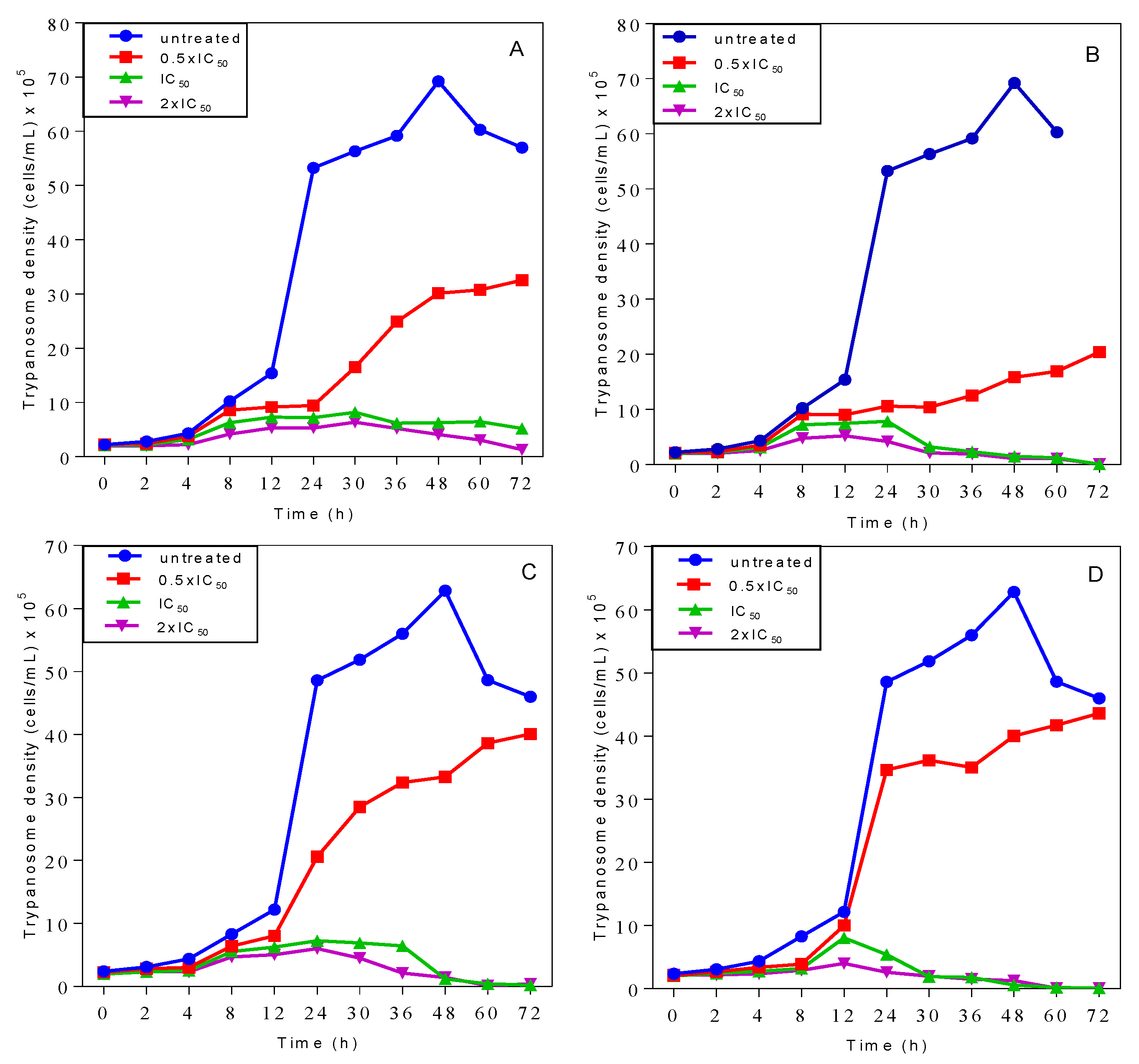
2.3. Effects on Growth Patterns of T. b. brucei Cells after Short and Long Duration of Exposure

The growth patterns of T. b. brucei cells incubated with 1 and 2 in Hirumi’s modified Iscove’s medium (HMI-9) growth media were compared with untreated cells. Both 1 and 2 caused a slow-onset growth arrest of the cells at concentrations at or above their respective IC50 values (Figure 1) with no chances of recovery from the early effects of the compounds, even after only a brief exposure of 2 h (panels C and D). However, cells incubated with either compound at half their IC50 concentrations recovered from the effects and resumed growth comparable to cells in the absence of treatment.

2.4. Propidium Iodide (PI) Assay of Cellular Integrity

The time of onset of anti-trypanosomal action of compounds 1 or 2 on T. b. brucei was determined by propidium iodide (PI) assay based on the principle that damaged cells become fluorescent as PI dye begins to enter them and binds to nucleic acids. As PI is not taken up by intact trypanosomes, this can only occur when the plasma membrane is compromised as a result of loss of cellular integrity caused directly or indirectly by 1 or 2. The effects of varying concentrations (1×, 2× and 4× the IC50 values) of 1 and 2 on the cell viability were monitored over a 6 h period (Figure 2). Both compounds showed slow but concentration-dependent trypanosomal membrane disruption effects. The time of onset of action was observed to be within 3–4 h of incubation, with peak effects achieved within 4 h.

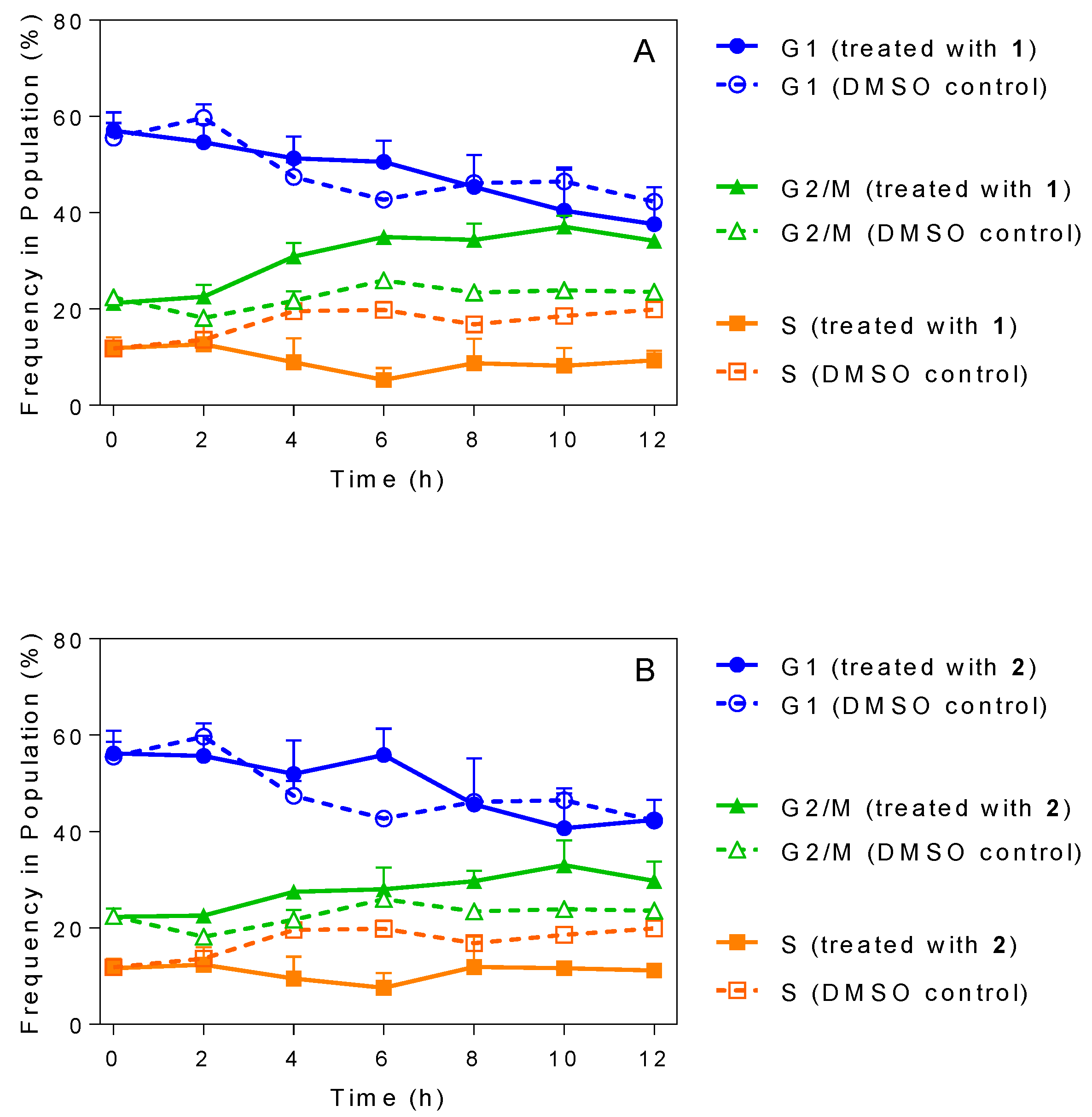
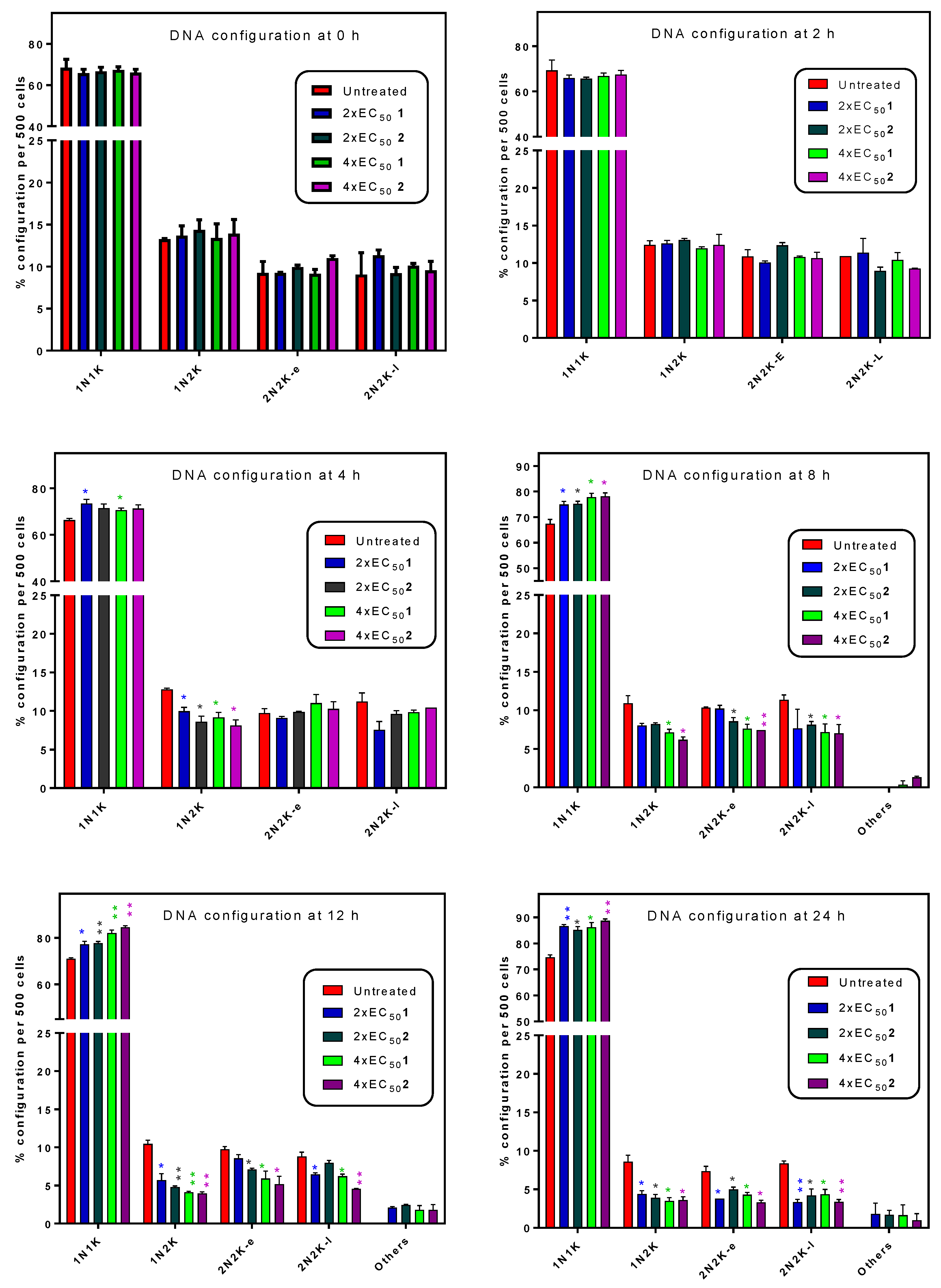
2.5. Influence of 3-Aminosteroids on DNA Content/Cell Cycle of Wild-Type T. b. brucei

Treatment with several 3-aminosteroids resulted in alterations in cellular morphology as described above. Fluorescence-activated cell sorting (FACS) flow cytometric analysis was carried out to determine the DNA content and, hence, establish the effects of 3-aminosteroids on T. b. brucei cell cycle stages (G1, S, G2/M or cytokinesis) (Figure 3, and supplementary Figure S1 for gating strategy and other details). The results showed a gradual and very similar decrease in the population of the G1 cell population and an increase in the G2/M phase for 1 and 2, both at concentrations of 4× EC50. There was also an observable effect on S cell cycle stage by either compound as the population of DNA in the phase, although appearing relatively constant, was clearly diminished relative to the control treated with the same volume of DMSO only (Figure 3). These observations seem to indicate fewer cells entering the S-phase, but S-phase cells still mostly progressing to G2/M phase, leading to the conclusion that DNA synthesis was not principally affected; 2 appeared to have a slightly more noticeable effect than 1 on the trypanosome cell cycle stages with the accumulation of about 40% DNA at the G2/M cell cycle phase after 10 h of incubation (Figure 3A) compared with about 35% observed in 1 (Figure 3B). The group treated with solvent only (DMSO) displayed the same configurations as an untreated control group assessed in parallel (not shown).
Microscopic examination of cell populations exposed for 12 h to 2× the respective EC50 concentrations of 1 or 2 prior to DAPI staining revealed swollen, misshapen cells, apparently containing replicated nuclear and kinetoplast DNA apparently stuck at G2/M phase as few cells contained more than one nucleus or kinetoplast (Figure 4), further confirming the above observed effects on DNA content analysis.
The number of nuclei (N) and kinetoplasts (K) per cell was monitored in the population by counting 500 cells and scoring them for the number of each DNA-containing organelle. Cells containing 2 nuclei and 2 kinetoplasts were further divided into early and late categories (2N2K-e, 2N2K-l) depending whether a clear ingression furrow for the longitudinal cell division could be observed. After 8 h of incubation with 1× EC50 of 1 or 2 cells started to appear that could not be classified in the regular categories and these were scored as ‘other’ (Figure 5). Observations were virtually identical for 1 and 2 throughout. Over the 24 h of the experiment, the number of 1N1K cells progressively and significantly increased relative to the control culture. Concurrently, there was a progressive decline in the number of 2N2K (early and late) cells, and the emergence of cells with an irregular DNA configuration as the cell structure deteriorated.
The morphology of the cells was studied in more detail by higher-resolution fluorescence microscopy with a DeltaVision Core microscope (Applied Precision, GE Healthcare Life Siences, Amersham, Bucks, UK). Figure 6 shows Delta Core images of cells stained with DAPI and MitoTracker after incubation for 8 h with 1× EC50 of 1 and 2, and control unexposed cells. Some of the cells incubated for 8 h with either compound appeared to have larger, elongated nuclei compared to control cells (average nuclear volume increased to 152% (p = 0.0012) and 120% (p = 0.12) of control at 8 h of culture), but otherwise the cells had not noticeably changed as a result of this mild exposure, and staining with mitotracker did not reveal any mitochondrial damage at this point. The mitochondrion also appeared to be intact, an indication that 3-aminosteroids may not directly affect mitochondrial integrity. The untreated cells retained the typical trypomastigote shape throughout the duration of the experiment. After 8 h, some more highly damaged and even lysed cells began to appear (not shown).

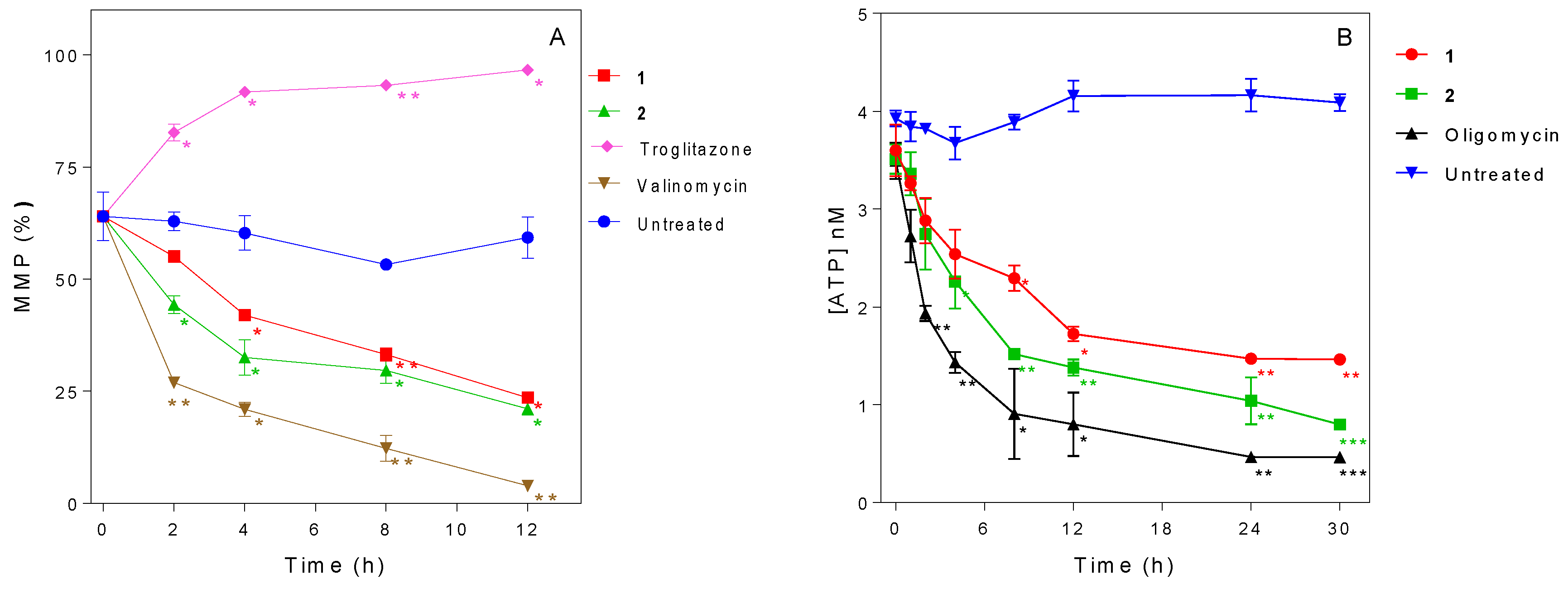
2.6. Influence of 3-Aminosteroids on Mitochondrial Membrane Potential (MMP)

We next investigated the influence of 1 and 2 on MMP. The MMP of untreated cells remained fairly unchanged while the positive controls troglitazone and valinomycin caused a strong and rapid hyperpolarization and depolarization of MMP, respectively. The 3-aminosteroids were found to cause a slower but significant depolarization of the MMP (Figure 7A). Both compounds induced a gradual MMP depolarization effect, observed as early as 2 h of incubation, which was statistically significant for 2 (p < 0.05). From 4 h the decrease was significant for both treatments, and it continued to drop to approximately 20% at 12 h. At the chosen concentration of 2× EC50, 2 had a stronger MMP depolarizing effect than 1, although this difference only showed minor significance at the 2 h point (p < 0.05), and may thus reflect a more rapid internalization of 2.

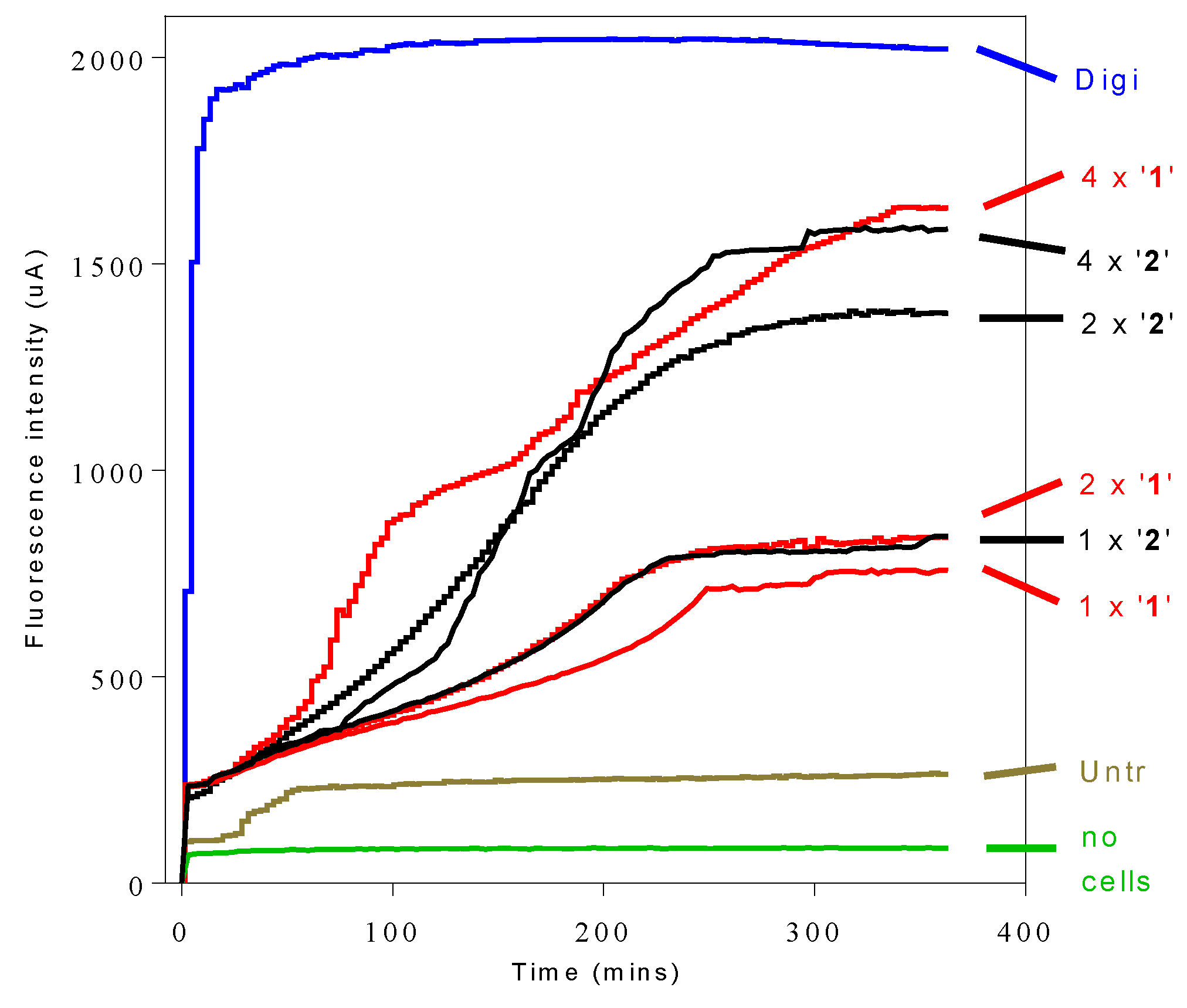
2.7. Influence of 3-Aminosteroids on Intracellular ATP Levels

Further studies to assess any cause-effect relationship between T. b. brucei intracellular ATP level and the earlier observed effects were undertaken. The results showed that 3-aminosteroids rapidly and significantly (p < 0.05) decreased the ATP level in T. b. brucei (Figure 7B). The pattern closely followed that observed for the decrease in MMP, with 2 inducing a slightly more rapid and deeper decline than 1 for the concentrations chosen, but both otherwise following very much the same trend.

3. Discussion

The 3-aminosteroids showed impressive activities against the more virulent of the two haematic parasites, Tc-IL3000, than against T. b. brucei. Both parasites, in addition to T. vivax, cause Nagana in cattle but they can also cause serious losses in pigs, camels, goats, dogs and sheep. With 2 (IC50 = 1.75 µM) and 5 (IC50 = 1.62 µM) being the most active compounds against T. b. brucei under our experimental conditions, it can be inferred that 3-aminosteroids are moderately active against T. b. brucei while much higher levels of activity had previously been determined against the human pathogen, T. b. rhodesiense, using a different protocol [14]. It is important to note that, using an assay such as the resazurin-based one, EC50 values are not constants, but very much dependent on the duration of incubation and the cell density, and should be taken as a ranking that is internally consistent but not absolute. The protocol that is used as standard in the De Koning lab is particularly rigorous and tends to give substantially higher EC50 values for some classes of compounds than similar assays that use much lower cell densities.
Our observation regarding the structure-antitrypanosomal (SAR) of the 3-aminosteroids is that methylation of the 3-amino substituent and the stereochemistry at the C-3 position affects the activity against T. b. brucei, and particularly against T. congolense. For instance, the monomethylated 3-amino derivatives were more active than the non-methylated and dimethylated derivatives, as evidenced by comparing 2, 7 and 9 with 1, 8 and 10 respectively. The α-congeners were similarly more potent than the corresponding β-congeners, comparing 3 and 5 with 1 and 4 respectively, which might in part be attributable to their apparent uptake through the TbAT1/P2 transporter, as judged by the significantly reduced sensitivity to strains lacking this carrier. There was a very clear contribution of the additional pyrroline ring, formed from the C-18-N-C-20 bridge, to the activities of 3-aminosteroids against Tc-IL3000, as observed by comparing 15 vs. 610. Interestingly, 10 (IC50 (Tc) = 0.045 µM), was ~2 times more active than the widely used veterinary drug diminazene aceturate [1] and 10 times more active than another well-known trypanocide, pentamidine, which is the standard treatment for early-stage HAT [32]. Hence, there will be a need to further explore its activity against T. congolense and carry out further SAR. The relatively higher activities and SI against Tc-IL3000 (up to 2130) of the pentacyclic compounds 610 strongly indicate a different mechanism for this aminosteroid series (whereas the very similar EC50 values for series 15 point for a similar mode of action against T. congolense and T. brucei), likely to be rooted in the biochemical and molecular differences between these species [33]. As a T. congolense-specific drug would not act against human sleeping sickness, and would have limited utility for AAT, where the infecting species is almost never known, we have here prioritised the general trypanocides 1 and 2.
The emergence of resistant parasites in both the laboratory and the field suggest that these parasites are readily capable of adapting under drug pressure [34] although it has been found impossible to induce resistance to some natural compounds, including curcumin [35]. Resistance to the current trypanocidal drugs is a major bottleneck to the control of these severe vector-borne diseases [36,37] and trypanosomes resistant to almost all the available drugs have been identified [9,38]. In order to avoid introducing new trypanocides that have resistance profiles similar to that of the standard treatments, lead compounds should be subjected to cross-resistance studies. In this study, we found almost no significant differences in the susceptibility of various drug-resistant strains and the wild-type T. b. brucei to most of the compounds, and almost all observed resistance factors were below 2.5-fold. Significant increases in IC50 values were observed following treatment with compound 3 against TbAT1-KO (5.6), AQP2/3-KO (2.7), ISMR (3.2) and B48 (5.1), 5 against B48 (2.1) and 7 against TbAT1 (2.4), ISMR (2.9) and B48 (2.4), which indicated a somewhat reduced potency against these resistant strains. However, these figures are insignificant when compared with the resistance factors of these resistant strains against the standard drugs. Furthermore, there were no similar comparable tendencies for the other aminosteroids tested, emphasising that there is no cross-resistance problem for this pharmacological class as a whole, and that the 3β-aminosteroids, in particular, are unlikely to exhibit any cross resistance with existing first line HAT and AAT drugs such as pentamidine, melarsoprol, cymelarsan, isometamidium or diminazene, whose resistance mechanisms have been well characterised [22,24,37]. The data from the cross-resistance study indicate that 3-aminosteriods, being structurally dissimilar to any current trypanocides, are most unlikely to be internalized through the same drug transporters (possible exception for TbAT1/P2 and 3α-aminosteroids), or rely on the same intracellular target, and the presented data are consistent with this notion. Aside from the conclusion about resistance-associated transporters such as TbAT1/P2 [22,37], TbAQP2/HAPT1 [37,39] and the low-affinity pentamidine transporter LAPT [23,40], the absence of resistance with R0.8 indicates that the 3-aminosteroids do not act on the trypanosome’s cAMP signalling system [25], and the similar IC50 values against ISMR show that the test compounds have probably no direct effect on the FoF1 ATPase [24].
In the presence of continuous or time-limited (2 h) exposure to the 3-aminosteroids at concentrations ≥IC50, the growth pattern of Tbb 427WT was disrupted with no possibility of recovery, although complete clearance was not observed until 2–3 days after exposure. The early growth arrest in both cases and the complete clearance observed after 72 h of incubation suggest that 3-aminosteroids are trypanocidal and, like diamidines, prone to act very slowly but irreversibly [41]. The early effects on trypanosomes being irreversible once initiated is an undoubted advantage for in vivo efficacy as a short exposure time reduces potential cytotoxicity associated with the maintenance of peak drug plasma levels over an extended period of time. Of crucial importance for the treatment of AAT, moreover, is that it is in most cases impractical to have to administer multiple doses, over an extended time, to the infected animal. To introduce such a drug would be to practically ensure non-compliance, which would lead to relapses and promote rapid-onset resistance.
PI is a red fluorophore that is not taken up by live, intact trypanosomes but does enter, and bind to trypanosome nucleic acids, when the plasma membrane is compromised as a result of loss of cellular integrity [42,43]. The effects of 3-aminosteroids differ significantly from digitonin, a known cell membrane lysis agent, which causes rapid trypanosome cell permeabilization reaching maximum fluorescence in the first few minutes [44]. The much slower, concentration-dependent membrane permeabilization effect of the 3-aminosteroids suggests that these compounds do not directly cause membrane leakage by a physicochemical effect as their main mode of action, but rather that over time an increasing number of cells becomes sufficiently permeable to PI to elicit a fluorescence response. It should be noted that, while loss of cellular integrity obviously allows binding of PI to trypanosomal nucleic acids, not all cellular entry of PI is necessarily indicative of cell death having already occurred (as opposed to a small increase in membrane permeability), and that it does not reveal whether cell death occurs by necrosis or apoptosis, as some features of necrosis and apoptosis may be shared [45].
The slow trypanocidal effects of 3-aminosteroids as observed from the growth patterns and PI-based assay leaves the likely target of the compounds open. Thus, it was explored whether these compounds target aspects of the cell cycle, bearing in mind the unusual cell cycles of kinetoplastids, in which the nucleus, kinetoplast and basal body replicate and segregate in a coordinated fashion into daughter cells during cell division [46,47,48]. The PI-associated fluorescence after binding to T. brucei DNA is proportional to the amount of DNA per cell when analysed by flow cytometry [43,49]. This revealed a possible stall during the G2/M cell cycle phase, as the cells with double the DNA content of non-dividing cells progressively increased after treatment with either 1 or 2 (Figure 4 and Figure S1), although there was no increase, but rather a gradual decrease in the number of nuclei (Figure 6 and Figure 7). These findings suggest that DNA replication was unaffected, but that instead mitosis was inhibited, leading to significantly enlarged nuclei, with increased DNA content. In bloodstream form (BSF) T. brucei, inhibition of mitosis (M) prevents cytokinesis, although not re-replication of nuclear DNA (at G2 phase) and kinetoplast DNA (at S phase), or segregation of replicated kinetoplasts (at G2 phase), resulting in cells with a single enlarged nucleus and multiple kinetoplasts [50]. However, no cells with multiple kinetoplasts but only 1 nucleus were observed, other than the regular 1N2K cells that necessarily precede mitosis into 2N2K cells, and the proportion of those cells progressively and dose-dependently declined. Thus, it appears that it is the failure to divide the mitochondrially-located kinetoplasts that halts the cell cycle at that point and leads to the observed growth arrest and ultimate cell death.
This is not entirely unexpected as the 3-aminosteroids are likely to accumulate in the trypanosomal mitochondrion, by virtue of their lipophilicity and the cationic nature of their salts, formed in aqueous media, which provides mitochondrial targeting similar to previous lipophilic cations that have shown promise against African trypanosomes [27,51,52]. Mitochondrial destruction as their mode of action could not be validated in the present studies as mitochondrial integrity remained intact, judging by fluorescence microscopy following MitoTracker staining. However, the mitochondrial accumulation has been associated with many different trypanocidal activities of various compounds disrupting various mitochondrial functions. Examples are inhibitors of TAO coupled to lipo-cations [27]; di-cationic diamidines such as pentamidine and DB75 targeting multiple mitochondrial functions including kinetoplast DNA through accumulation to high concentrations [53,54,55]; choline-based di-cations disrupting mitochondrial functions [43]; bisphosphonium compounds inhibiting the activity of the T. brucei F1-ATPase [43]; disruption of kDNA replication by ethidium [56]; selective cleavage of kinetoplast DNA minicircles by diminazene and isometamidium [57]. Many of these drugs have been documented to bind to, or otherwise affect, the kinetoplast, but in general terms the principal consequence of the mitochondrial targeting of trypanocides is that they accumulate against a concentration gradient to a very high intra-mitochondrial concentration, driven by the mitochondrial membrane potential (pentamidine accumulates to mM concentrations [58]). This makes it likely that the drug can interfere with (multiple) biochemical processes in that space.
Although no direct mitochondrial damage was established after 8 h of incubation with 1× EC50 of 1 or 2, this does not rule out either mitochondrial accumulation or a mitochondrial target for 3-aminosteroids. It has been found that kDNA replication requires mitochondrial proteins [59] that are encoded in the nuclear genome and imported into the mitochondrion. This import requires ATP and is proton-motive force-driven and hence depends on the mitochondrial membrane potential (MMP) [60]. The BSFs of T. b. brucei have a less elaborate mitochondrial metabolism than almost any other aerobic eukaryotic cell type; they lack the Krebs cycle enzymes as lack a cytochrome-dependent electron transport chain [52]. Therefore, they rely entirely on glycolysis instead of oxidative phosphorylation to generate ATP [61] and use the FoF1 ATPase to maintain mitochondrial membrane potential by pumping protons over the inner mitochondrial membrane (IMM) from the matrix to the intermembrane space [62]. We have previously reported that lipophilic bisphosphonium cations accumulate in the mitochondrion, cause a depolarisation of the IMM and reduce the cellular ATP content by inhibiting the F1 ATPase [52]. Although the lack of cross-resistance with the T. brucei ISMR strain, which derives its isometamidium resistance from a mutation in the γ-subunit of F1 [24], suggests that this ATPase is not the principal target of the aminosteroids, we found that, like the bisphosphonium salts and isometamidium, 1 and 2 rapidly depolarise the IMM, with significant decline in MMP in 4 h and 2 h, respectively. We likewise observed a rapid decline in cellular ATP levels, which could cause the IMM depolarisation, as this is directly maintained by ATP. Conversely, a functional mitochondrion is essential for ATP production in BSF trypanosomes, although the glycolysis and ATP production actually takes place in a unique trypanosomatid-specific organelle called the glycosome [63]. However, in order to maintain redox balance, the glycolysis intermediate glycerol-3-phosphate must be oxidized back to dihydroxyacetone phosphate by glycerol-3-phosphate dehydrogenase (G-3-PDH) in the mitochondrion, transferring 4 electrons to ubiquinol, which is subsequently oxidized to ubiquinone by TAO which then converts molecular oxygen to water [64,65]. This so-called glycerol-3-phosphate shuttle, and thus mitochondrial respiration, is dependent on the MMP [66], and without it BSF trypanosomes generate just one ATP per glucose, explaining the rapid and extreme decline in cellular ATP levels also observed with the Fo inhibitor oligomycin [67]. Thus, MMP and ATP content are linked, as also observed for the bisphosphonium compounds [52], but whereas those compounds caused an increase in G1-phase cells, the 3-aminosteroids caused an increase of cells arrested in G2/M phase.
Overall, we conclude that 3-aminosteroids act on the trypanosomal mitochondrion, by which they cause a strong reduction of the MMP, and ATP levels, and prevent the division of kinetoplast, resulting in irreversible G2/M phase cell cycle arrest and eventual cell death. Further studies should assess ultrastructural changes in the kinetoplast and the integrity of kDNA.

4. Materials and Methods

4.1. Parasites, Cell Lines and Cultures

The parasites used for this study include Trypanosoma congolense Tc-IL3000 (Savannah-type strain IL 3000) [27], T. brucei brucei (BSF trypomastigotes T. b. brucei strain 427 Lister WT (Tbb 427WT), MiTat 1.2/BS221) and six resistant cell lines of T. brucei brucei BSF trypomastigotes which have been described [22,23,24,25,28,68]. All the T. b. brucei strains were cultured in standard Hirumi’s modified Iscove’s medium 9 (HMI-9), supplemented with 10% heat-inactivated foetal bovine serum (FBS), 14 µL/L β-mercaptoethanol, and 3.0 g sodium hydrogen carbonate per litre of medium adjusted to pH 7.4, Tc-IL3000 strains were cultured in Dulbecco’s minimum essential medium (MEM) with β-mercaptoethanol, sodium pyruvate, sodium bicarbonate and supplemented with fresh goat serum. All the parasites were cultured in vented flasks at 37 °C in a 5% CO2 atmosphere and were passaged every 72 h.

4.2. Determination of In Vitro Anti-Trypanosomal Activity of Compounds

All tested compounds were isolated from Holarrhena africana A. DC. (a synonym of H. floribunda (G. Don) T. Durand & Schinz) (Apocynaceae) and identified as described in our previous communication [14]; 1H- and 13C-nuclear magnetic resonance (NMR) spectra to assess the identity and purity can be found in the supplementary information of [14]. The in vitro activities of 110 against BSF trypanosomes were determined using a resazurin-based assay protocol as described [24,27,42] using cell densities adjusted to 2 × 10⁵ cells/mL for T. b. brucei and 5 × 10⁵ cells/mL for Tc-IL3000. Cells were exposed to at least 11 doubling dilutions of test compound starting at 100 µL, with the last well in the row receiving medium without test compound as a drug-free control. The trypanosome culture and test compounds were incubated for a period of 48 h at 37 °C for T. b. brucei and 32 °C for Tc-IL3000 under a 5% CO2 atmosphere. In all the assays, pentamidine and diminazene aceturate were used as positive controls. Fluorescence was measured in the 96-well plates with a FLUOstar Optima (BMG Labtech, Aylesbury, Bucks, UK) at wavelengths of 544 nm for excitation and 590 nm for emission. IC50 values were calculated by non-linear regression using an equation for a sigmoidal dose-response curve with variable slope (Prism 5.0, GraphPad Software, San Diego, CA, USA).

4.3. Effect of Treatments on Growth Patterns of T. b. brucei after Short- and Long-Term Exposure

Varying concentrations of 1 and 2 were tested on trypanosomes in order to determine in vitro cell growth patterns and time-to-kill. Trypanosomes at their mid logarithmic phase of growth were taken from cultures and cell density was determined using a haemocytometer (Camlab, Over, UK). The cell density was adjusted to 2 × 10⁵ cells with fresh HMI-9 medium and the fresh cultures were incubated with varying concentrations of 1 and 2 at 37 °C under a 5% CO2 atmosphere. For the limited time of exposure assay, the cells were similarly incubated, but after 2 h of exposure to the test compounds, the cells were washed twice with fresh HMI-9 media, re-seeded at the same cell density and again incubated under the same conditions. At predetermined intervals, cell counts were taken from each culture, typically for up to 72 h, and the counts were used for plotting the growth curves.

4.4. Determination of Speed of Action by Propidium Iodide (PI) Assay

100 μL of 18 µM PI in HMI-9 was added to each well of a 96-well plate. In the first column of the wells, 200 μL solutions of test compound, also in 18 µM PI/HMI-9, was added and serially diluted across the wells. Wells receiving only media without test compound served as drug-free controls. To each well was added 100 μL of 2 × 10⁶ T. brucei 427WT cells/mL in HMI-9. Wells containing the same final concentration of 9 μM PI in HMI-9 but no cells served to record background fluorescence. The plates were incubated in a FLUOstar OPTIMA fluorimeter at 37 °C with 5% CO2 atmosphere, and the fluorescence was recorded at 544 nm excitation and 620 nm emission for 120 cycles at 180 s per cycle.

4.5. Determination of Intracellular ATP Level

Intracellular ATP was measured using a Molecular Probes ATP Determination Kit (Molecular Probes Inc., Cambridge, UK) following the manufacturer’s protocol. Trypanosome culture density was adjusted to 107 cells/mL and incubated with 1 or 2. At predetermined time intervals, 1 mL parasites culture was taken and centrifuged for 10 min at 2500× g, 4 °C. The resulting pellets were lysed by sonication and cell debris was removed using a refrigerated microcentrifuge (SciQuip, Newtown, UK) at 12,000× g for 10 min at 4 °C. The resultant supernatant was frozen in liquid nitrogen and stored at −80 °C. For the analysis, 90 μL reaction solution from the master mix was added to each designated well of a 96-well plate and the background luminescence was recorded using a FLUOstar OPTIMA fluorimeter; then 10 μL of each sample was added to the well and incubated for 15 min at 28 °C and the luminescence was recorded. A standard curve was made using serial dilution of varying concentrations of ATP (0.5–500 pM) to calculate the ATP concentrations in the samples.

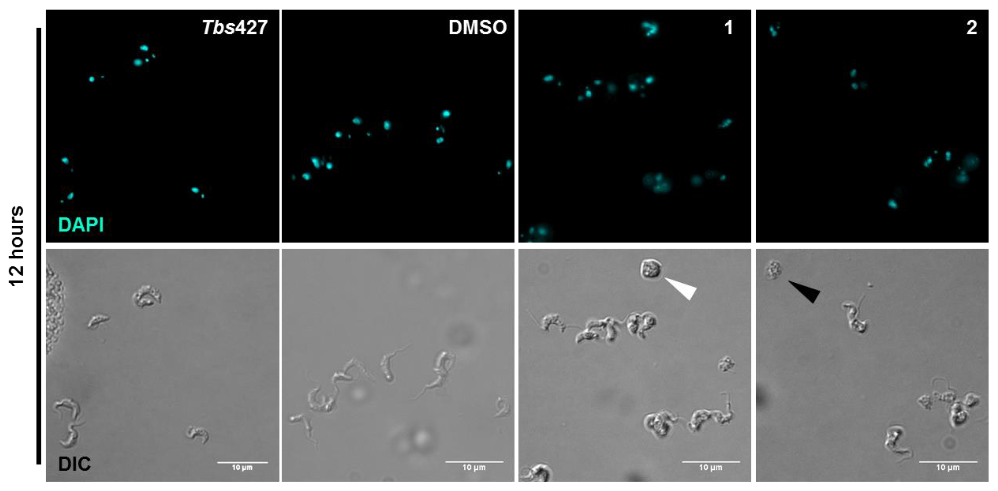
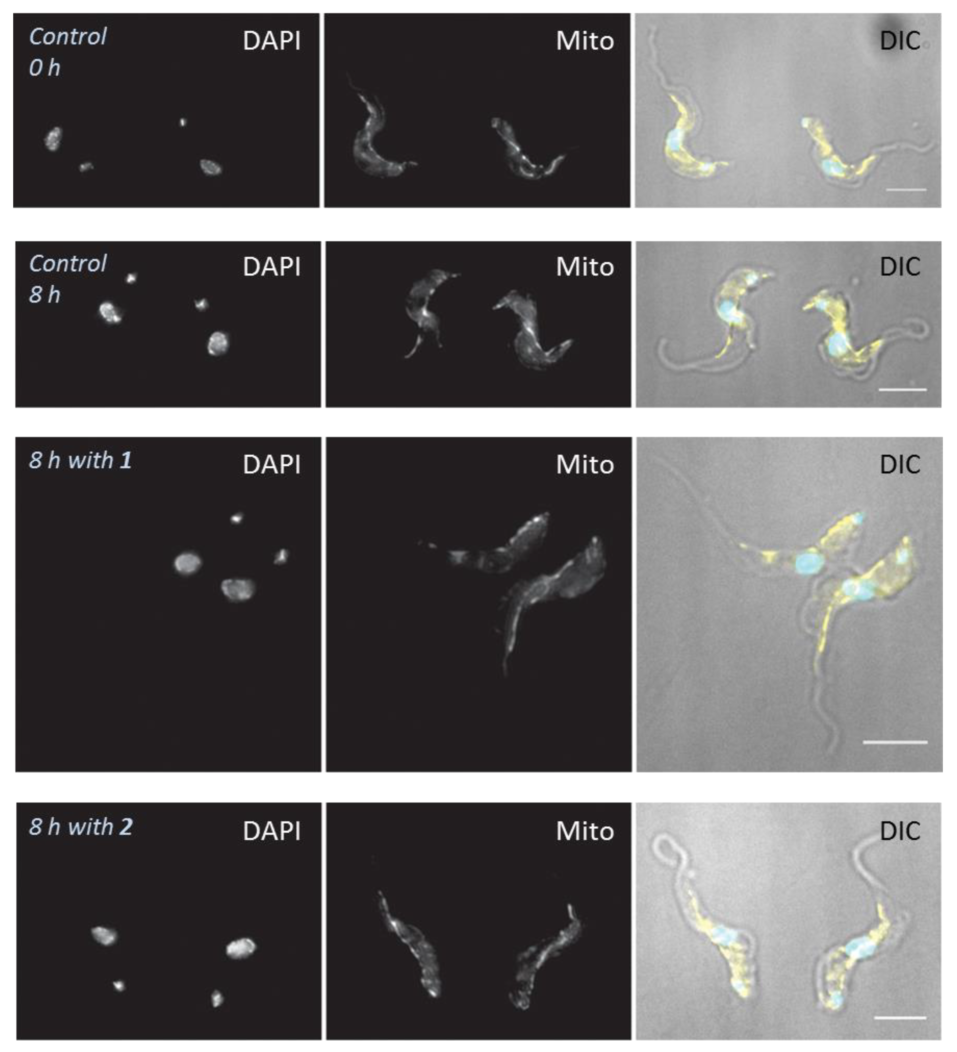
4.6. Effects of Treatments on Cell Morphology

A T. b. brucei culture was adjusted to 2 × 10⁵ cell/mL and incubated with 1 and 2 for the duration of the assay. The MitoTracker red CMXRos (New England Biolabs Ltd, Hitchin, UK) used for this assay was prepared from a stock solution of 100 μM in DMSO. From this solution, 1 μL was added to 1 mL of cell sample taken from the cell culture at each time point to make a final concentration of 100 nM of MitoTracker. The sample was then incubated at 37 °C and 5% CO2 for 5 min. The incubated sample was washed in filter-sterilized 1× phosphate buffered saline (PBS) and spun at 2600× g for 10 min (4 °C). After the final wash step, the sample was re-suspended in 1 mL of 1× PBS. A 50 μL of the re-suspended cells were spread onto a glass microscope slide (CamLab), previously coated with 50 μL of poly-L-lysine (Sigma-Aldrich, Gillingham, Dorset, UK); and were left to air dry, the cells were then fixed in 4% paraformaldehyde/1× PBS for 10 min at room temperature. The slides were covered with 1 mL of PBS and allowed to rehydrate for 10 min after which it was then allowed to evaporate but not completely. A drop of Vectashield anti-fade mounting medium with DAPI (Vector Laboratories, Burlingame, CA, USA) was added to the slides and spread by a coverslip (CamLab, Over, UK); the coverslip was then applied and the edges were sealed with nail varnish. Slides were observed for cell morphology under an Axioskop microscope (Image Solutions. Preston, UK) using softWoRx software (Suite 2.0, Applied Precision, GE Healthcare, Amersham, Bucks, UK), while DNA configuration was assessed using a Zeiss Axioplan microscope (Cambridge, UK) using Hamamatso digital camera and Openlab software. For detailed observation of single DAPI-stained cells and the measurement of nuclear volumes, Z-stacks of cells labelled for DAPI and MitoTracker were acquired using an Olympus UPLSAPO 100× oil (1.40 NA) objective on a DeltaVision Core microscope (Applied Precision, GE Healthcare Life Siences, Amersham, Bucks, UK) attached to a CoolSNAP HQ2 CCD camera (Photometrics, Tucson, AZ, USA). Deconvolution was performed using SoftWoRx Suite 2.0), and later data was analysed using FIJI [69] and/or IMARIS (Bitplane, Belfast, UK).

4.7. Determination of Mitochondrial Membrane Potential

FACS was employed in the determination of the changes in MMP due to exposure of T. b. brucei to 1 and 2 by TMRE dye incorporation as previously described [43,52]. The cell density was adjusted to 106 cells/mL and incubated with 1 or 2 at 32 °C under 5% CO2. At predetermined intervals, 1 mL of sample was washed and re-suspended in 1 mL PBS containing 200 nM of TMRE in FACS tubes, followed by incubation at 37 °C for 30 min, and analysed by a Becton Dickinson FACS Calibur (Franklin Lakes, NJ, USA) using a FL2-heigth detector and CellQuest (Becton Dickinson) and FlowJo software (V.10, Ashland, OR, USA). Valinomycin (100 nM; Sigma-Aldrich) and troglitazone (10 µM; Sigma-Aldrich) were employed as mitochondrial membrane depolarizer and hyperpolarizer control, respectively.

4.8. Determination of Cell Cycle and DNA Content Assay

FACS analysis was performed to study the effects of 1 and 2 on cell cycle of trypanosomes, essentially as described previously [49] with the following modifications. Approximately 2.5 × 10⁶ cells were harvested by centrifugation at 1500× g for 10 min at 4 °C and washed once in 1× PBS by centrifugation (3000× g for 5 min). The cells were re-suspended in 1 mL of 70% ethanol and 30% 1× PBS. The tubes with the cells were left at −20 °C to fix overnight in the dark and the samples were subsequently washed once with 1 mL 1× PBS, re-suspended in 500 µL 1× PBS containing 10 μg/mL PI and 100 μg/mL RNase A and incubated at 37 °C for 45 min before FACS analysis. The samples were analyzed by a Becton Dickinson FACSCalibur and PI incorporation was measured using the FL2-Area detector and CellQuest software. The data obtained were analyzed using FlowJo software (V.10).

Supplementary Materials

The following are available online. Figure S1: Details of the DNA content and cell cycle analysis.

Author Contributions

T.J.S. and H.P.d.K. initiated this collaborative study. T.J.S. and N.J.N. initiated the overall project on Holarrhena alkaloids. C.O.N., J.A.B. and G.U.E. conducted the experiments in the laboratories of H.P.d.K. with the compounds isolated and characterized in the laboratories of T.J.S. and wrote a draft version of the manuscript. H.P.d.K. devised and supervised the experiments, analyzed the data and refined the manuscript. L.L. performed the microscopical techniques. All authors contributed to the final manuscript which was then formatted and submitted by T.J.S.

Funding

This research was funded by a fellowship for C.O.N. from the Federal Government of Nigeria through the Tertiary Education Trust Fund (TETFund) and the University of Nigeria Nsukka to conduct his doctoral studies at WWU Münster and by a short-term scientific mission (STSM) grant from COST action CM1307 for C.O.N. to perform the studies described here at the University of Glasgow.

Acknowledgments

This work is an activity within the Research Network Natural Products against Neglected Diseases (ResNet NPND, see www.resnetnpnd.org).

Conflicts of Interest

The authors declare no conflict of interest.

References

  1. Giordani, F.; Morrison, L.J.; Rowan, T.G.; De Koning, H.P.; Barrett, M.P. The animal trypanosomiases and their chemotherapy: A review. Parasitology 2016, 143, 1862–1889. [Google Scholar] [CrossRef]
  2. PATTEC. A continental plan of action for the eradication of Tsetse and trypanosomosis. In The OAU Pathway for the PATTEC; Pan African Tsetse and Trypanosomosis Global Veterinaria, Eradication Campaign: Khartoum, Sudan, 2000. [Google Scholar]
  3. Auty, H.; Torr, S.J.; Michoel, T.; Jayaraman, S.; Morrison, L.J. Cattle trypanosomosis: The diversity of trypanosomes and implications for disease epidemiology and control. Rev. Sci. Tech. 2015, 34, 587–598. [Google Scholar] [CrossRef] [PubMed]
  4. Holt, H.R.; Selby, R.; Mumba, C.; Napier, G.B.; Guitian, J. Assessment of animal African trypanosomiasis (AAT) vulnerability in cattle-owning communities of sub-Saharan Africa. Parasites Vectors 2016, 9, 53. [Google Scholar] [CrossRef] [PubMed]
  5. CFSPH. African animal trypanosomiasis. In The Fact Sheet; The Centre for Food Security and Public Health: Victoria, BC, Canada, 2009. [Google Scholar]
  6. Shaw, A.P.M.; Cecchi, G.; Wint, G.R.W.; Mattioli, R.C.; Robinson, T.P. Mapping the economic benefits to livestock keepers from intervening against bovine trypanosomosis in Eastern Africa. Prev. Vet. Med. 2014, 113, 197–210. [Google Scholar] [CrossRef] [PubMed]
  7. Mattioli, R.C.; Feldmann, U.; Hendrickx, G.; Wint, W.; Jannin, J.; Slingenbergh, J. Tsetse and trypanosomiasis intervention policies supporting sustainable animal-agricultural development. J. Food Agric. Environ. 2004, 2, 310–314. [Google Scholar]
  8. Hursey, B.S. The programme against African trypanosomiasis: Aims, objectives and achievements. Trends Parasitol. 2001, 17, 2–3. [Google Scholar] [CrossRef]
  9. Delespaux, V.; De Koning, H.P. Drugs and drug resistance in African trypanosomiasis. Drug Resist. Updat. 2007, 10, 30–50. [Google Scholar] [CrossRef]
  10. Assefa, A.; Shibeshi, W. Drug resistance in African animal trypanosomes: A review. Afr. J. Microbiol. Res. 2018, 12, 380–386. [Google Scholar]
  11. Holmes, P. First WHO meeting of stakeholders on elimination of gambiense Human African Trypanosomiasis. PLoS Negl. Trop. Dis. 2014, 8, e3244. [Google Scholar] [CrossRef]
  12. Holmes, P. On the road to elimination of rhodesiense human African trypanosomiasis: First WHO meeting of stakeholders. PLoS Negl. Trop. Dis. 2015, 9, e0003571. [Google Scholar] [CrossRef]
  13. Morrison, L.J.; Vezza, L.; Rowan, T.; Hope, J.C. Animal African trypanosomiasis: Time to increase focus on clinically relevant parasite and host species. Trends Parasitol. 2016, 32, 599–607. [Google Scholar] [CrossRef] [PubMed]
  14. Nnadi, C.O.; Nwodo, N.J.; Kaiser, M.; Brun, R.; Schmidt, T.J. Steroid Alkaloids from Holarrhena africana with Strong Activity against Trypanosoma brucei rhodesiense. Molecules 2017, 22, 1129. [Google Scholar] [CrossRef] [PubMed]
  15. Moretti, C.; Sauvain, M.; Lavaud, C.; Massiot, G.; Bravo, J.A.; Munoz, V. A novel antiprotozoal aminosteroid from Saracha punctata. J. Nat. Prod. 1998, 61, 1390–1393. [Google Scholar] [CrossRef] [PubMed]
  16. Nnadi, C.O.; Althaus, J.B.; Nwodo, N.J.; Schmidt, T.J. A 3D-QSAR study on the antitrypanosomal and cytotoxic activities of steroid alkaloids by comparative molecular field analysis. Molecules 2018, 23, 1113. [Google Scholar] [CrossRef]
  17. Schenone, M.; Dančík, V.; Wagner, B.K.; Clemons, P.A. Target identification and mechanism of action in chemical biology and drug discovery. Nat. Chem. Biol. 2013, 9, 232. [Google Scholar] [CrossRef]
  18. McNamara, C.; Winzeler, E.A. Target identification and validation of novel antimalarials. Future Microbiol. 2011, 6, 693–704. [Google Scholar] [CrossRef] [PubMed]
  19. Xie, L.; Xie, L.; Bourne, P.E. Structure-based systems biology for analyzing off-target binding. Curr. Opin. Struct. Biol. 2011, 21, 189–199. [Google Scholar] [CrossRef] [Green Version]
  20. Apsel, B.; Blair, J.A.; Gonzalez, B.; Nazif, T.M.; Feldman, M.E.; Aizenstein, B.; Hoffman, R.; Williams, R.L.; Shokat, K.M.; Knight, Z.A. Targeted polypharmacology: Discovery of dual inhibitors of tyrosine and phosphoinositide kinases. Nat. Chem. Biol. 2008, 4, 691. [Google Scholar] [CrossRef]
  21. Das, A.; Li, H.; Liu, T.; Bellofatto, V. Biochemical characterization of Trypanosoma brucei RNA polymerase II. Mol. Biochem. Parasitol. 2006, 150, 201–210. [Google Scholar] [CrossRef]
  22. Matovu, E.; Stewart, M.L.; Geiser, F.; Brun, R.; Mäser, P.; Wallace, L.J.; Seebeck, T. Mechanisms of arsenical and diamidine uptake and resistance in Trypanosoma brucei. Eukaryot. Cell 2003, 2, 1003–1008. [Google Scholar] [CrossRef]
  23. Bridges, D.J.; Gould, M.K.; Nerima, B.; Mäser, P.; Burchmore, R.J.; De Koning, H.P. Loss of the high-affinity pentamidine transporter is responsible for high levels of cross-resistance between arsenical and diamidine drugs in African trypanosomes. Mol. Pharmacol. 2007, 71, 1098–1108. [Google Scholar] [CrossRef] [PubMed]
  24. Eze, A.A.; Gould, M.K.; Munday, J.C.; Tagoe, D.N.; Stelmanis, V.; Schnaufer, A.; De Koning, H.P. Reduced Mitochondrial Membrane Potential Is a Late Adaptation of Trypanosoma brucei brucei to Isometamidium Preceded by Mutations in the γ Subunit of the F1Fo-ATPase. PLoS Negl. Trop. Dis. 2016, 10, e0004791. [Google Scholar] [CrossRef] [PubMed]
  25. Gould, M.K.; Bachmaier, S.; Ali, J.A.; Alsford, S.; Tagoe, D.N.; Munday, J.C.; Schnaufer, D.H.; Boshart, M.; de Koning, H.P. Cyclic AMP effectors in African trypanosomes revealed by genome-scale RNA interference library screening for resistance to the phosphodiesterase inhibitor CpdA. Antimicrob. Agents Chemother. 2013, 57, 4882–4893. [Google Scholar] [CrossRef]
  26. Baker, N.; Glover, L.; Munday, J.C.; Andrés, D.A.; Barrett, M.P.; De Koning, H.P.; Horn, D. Aquaglyceroporin 2 controls susceptibility to melarsoprol and pentamidine in African trypanosomes. Proc. Natl Acad. Sci. USA 2012, 109, 10996–11001. [Google Scholar] [CrossRef] [PubMed] [Green Version]
  27. Ebiloma, G.U.; Ayuga, T.D.; Balogun, E.O.; Gil, L.A.; Donachie, A.; Kaiser, M.; Herraiz, T.; Inaoka, D.K.; Shiba, T.; Harada, S.; et al. Inhibition of trypanosome alternative oxidase without its N-terminal mitochondrial targeting signal (ΔMTS-TAO) by cationic and non-cationic 4-hydroxybenzoate and 4-alkoxybenzaldehyde derivatives active against T. brucei and T. congolense. Eur. J. Med. Chem. 2018, 150, 385–402. [Google Scholar] [CrossRef] [PubMed]
  28. Jeacock, L.; Baker, N.; Wiedemar, N.; Mäser, P.; Horn, D. Aquaglyceroporin-null trypanosomes display glycerol transport defects and respiratory-inhibitor sensitivity. PLoS Pathog. 2017, 13, e1006307. [Google Scholar] [CrossRef] [PubMed]
  29. De Koning, H.P.; Jarvis, S.M. Adenosine transporters in bloodstream forms of T. brucei: Substrate recognition motifs and affinity for trypanocidal drugs. Mol. Pharmacol. 1999, 56, 1162–1170. [Google Scholar] [CrossRef]
  30. Munday, J.C.; Tagoe, D.N.A.; Eze, A.A.; Krezdorn, J.A.; Rojas López, K.E.; Alkhaldi, A.A.M.; McDonald, F.; Still, J.; Alzahrani, K.J.; Settimo, L.; et al. Functional analysis of drug resistance-1 associated mutations in the Trypanosoma brucei Adenosine Transporter 1 (TbAT1) and the proposal of a structural model for the protein. Mol. Microbiol. 2015, 96, 887–900. [Google Scholar] [CrossRef]
  31. Collar, C.J.; Al-Salabi, M.I.; Stewart, M.L.; Barrett, M.P.; Wilson, W.D.; De Koning, H.P. Predictive computational models of substrate binding by a nucleoside transporter. J. Biol. Chem. 2009, 284, 34028–34035. [Google Scholar] [CrossRef]
  32. De Koning, H.P. The ever-increasing complexities of arsenical-diamidine cross-resistance in African trypanosomes. Trends Parasitol. 2008, 24, 345–349. [Google Scholar] [CrossRef]
  33. Utz, S.; Roditi, I.; Renggli, C.K.; Almeida, I.C.; Acosta-Serrano, A.; Bütikofer, P. Trypanosoma congolense procyclins: Unmasking cryptic major surface glycoproteins in procyclic forms. Eukaryot. Cell 2006, 5, 1430–1440. [Google Scholar] [CrossRef] [PubMed]
  34. De Koning, H.P. Drug resistance in protozoan parasites. Emerg. Top. Life Sci. 2017, 1, 627–632. [Google Scholar] [CrossRef] [Green Version]
  35. Alkhaldi, A.A.; Creek, D.J.; Ibrahim, H.; Kim, D.H.; Quashie, N.B.; Burgess, K.E.; Changtam, C.; Barrett, M.P.; Suksamrarn, A.; de Koning, H.P. Potent trypanocidal curcumin analogs bearing a monoenone linker motif act on Trypanosoma brucei by forming an adduct with trypanothione. Mol. Pharmacol. 2015, 87, 451–464. [Google Scholar] [CrossRef] [PubMed]
  36. Luscher, A.; De Koning, H.P.; Maser, P. Chemotherapeutic strategies against Trypanosoma brucei: Drug targets vs. drug targeting. Curr. Pharm. Des. 2007, 13, 555–567. [Google Scholar] [CrossRef] [PubMed]
  37. Munday, J.C.; Settimo, L.; De Koning, H.P. Transport proteins determine drug sensitivity and resistance in a protozoan parasite, Trypanosoma brucei. Front. Pharmacol. 2015, 6, 32. [Google Scholar] [CrossRef] [PubMed]
  38. Babokhov, P.; Sanyaolu, A.O.; Oyibo, W.A.; Fagbenro-Beyioku, A.F.; Iriemenam, N.C. A current analysis of chemotherapy strategies for the treatment of human African trypanosomiasis. Pathog. Glob. Health 2013, 107, 242–252. [Google Scholar] [CrossRef] [PubMed] [Green Version]
  39. Graf, F.E.; Ludin, P.; Wenzler, T.; Kaiser, M.; Brun, R.; Pyana, P.P.; Mäser, P. Aquaporin 2 mutations in Trypanosoma brucei gambiense field isolates correlate with decreased susceptibility to pentamidine and melarsoprol. PLoS Negl. Trop. Dis. 2013, 7, e2475. [Google Scholar] [CrossRef]
  40. De Koning, H.P. Uptake of pentamidine in Trypanosoma brucei brucei is mediated by three distinct transporters. Implications for crossresistance with arsenicals. Mol. Pharmacol. 2001, 59, 586–592. [Google Scholar] [CrossRef]
  41. Barrett, M.P.; Vincent, I.M.; Burchmore, R.J.; Kazibwe, A.J.; Matovu, E. Drug resistance in human African trypanosomiasis. Future Microbiol. 2011, 6, 1037–1047. [Google Scholar] [CrossRef]
  42. Gould, M.K.; Vu, X.L.; Seebeck, T.; de Koning, H.P. Propidium iodide-based methods for monitoring drug action in the kinetoplastidae: Comparison with the Alamar Blue assay. Anal. Biochem. 2008, 382, 87–93. [Google Scholar] [CrossRef]
  43. Ibrahim, H.M.; Al-Salabi, M.I.; El Sabbagh, N.; Quashie, N.B.; Alkhaldi, A.A.; Escale, R.; Smith, T.K.; Vial, H.J.; De Koning, H.P. Symmetrical choline-derived dications display strong anti-kinetoplastid activity. J. Antimicrob. Chemother. 2011, 66, 111–125. [Google Scholar] [CrossRef]
  44. Ebiloma, G.U.; Katsoulis, E.; Igoli, J.O.; Gray, A.I.; De Koning, H.P. Multi-target mode of action of an ursane-type terpenoid from Nigerian medicinal plants targeting the African trypanosomes. Sci. Rep. 2018, 8, 4613. [Google Scholar] [CrossRef]
  45. Lemasters, J.J.; Theruvath, T.P.; Zhong, Z.; Nieminen, A.L. Mitochondrial calcium and the permeability transition in cell death. Biochim. Biophys. Acta 2009, 1787, 1395–1401. [Google Scholar] [CrossRef] [PubMed] [Green Version]
  46. Matthews, K.R. The developmental cell biology of Trypanosoma brucei. J. Cell Sci. 2005, 118, 283–290. [Google Scholar] [CrossRef] [PubMed]
  47. Lacomble, S.; Vaughan, S.; Gadelha, C.; Morphew, M.K.; Shaw, M.K.; McIntosh, J.R.; Gull, K. Basal body movements orchestrate membrane organelle division and cell morphogenesis in Trypanosoma brucei. J. Cell Sci. 2010, 123, 2884–2891. [Google Scholar] [CrossRef] [PubMed]
  48. Ogbadoyi, E.O.; Robinson, D.R.; Gull, K. A high-order trans-membrane structural linkage is responsible for mitochondrial genome positioning and segregation by flagellar basal bodies in trypanosomes. Mol. Biol. Cell 2003, 14, 1769–1779. [Google Scholar] [CrossRef] [PubMed]
  49. Rodenko, B.; Wanner, M.J.; Alkhaldi, A.A.M.; Ebiloma, G.U.; Barnes, R.L.; Kaiser, M.; Brun, R.; McCulloch, R.; Koomen, G.J.; De Koning, H.P. Targeting the parasite’s DNA with methyltriazenyl purine analogs is a safe, selective and efficacious antitrypanosomal strategy. Antimicrob. Agents Chemother. 2015, 59, 6708–6716. [Google Scholar] [CrossRef]
  50. Hammarton, T.C. Cell cycle regulation in Trypanosoma brucei. Mol. Biochem. Parasitol. 2007, 153, 1–8. [Google Scholar] [CrossRef]
  51. Dardonville, C.; Alkhaldi, A.A.; De Koning, H.P. SAR studies of diphenyl cationic trypanocides: Superior activity of phosphonium over ammonium salts. ACS Med. Chem. Lett. 2014, 6, 151–155. [Google Scholar] [CrossRef]
  52. Alkhaldi, A.A.; Martinek, J.; Panicucci, B.; Dardonville, C.; Zíková, A.; de Koning, H.P. Trypanocidal action of bisphosphonium salts through a mitochondrial target in bloodstream form Trypanosoma brucei. Int. J. Parasitol. Drugs Drug Resist. 2016, 6, 23–34. [Google Scholar] [CrossRef]
  53. Soeiro, M.N.; de Castro, S.L.; de Souza, E.M.; Batista, D.G.; Silva, C.F.; Boykin, D.W. Diamidine activity against trypanosomes: The state of the art. Curr. Mol. Pharmacol. 2008, 1, 151–161. [Google Scholar] [CrossRef] [PubMed]
  54. Lanteri, C.A.; Tidwell, R.R.; Meshnick, S.R. The mitochondrion is a site of trypanocidal action of the aromatic diamidine DB75 in bloodstream forms of Trypanosoma brucei. Antimicrob. Agents Chemother. 2008, 52, 875–882. [Google Scholar] [CrossRef] [PubMed]
  55. Stewart, M.L.; Krishna, S.; Burchmore, R.J.S.; Brun, R.; De Koning, H.P.; Boykin, D.W.; Tidwell, R.R.; Hall, J.E.; Barrett, M.P. Detection of arsenical drug resistance in Trypanosoma brucei using a simple fluorescence test. Lancet 2005, 366, 486–487. [Google Scholar] [CrossRef]
  56. Roy Chowdhury, A.; Bakshi, R.; Wang, J.; Yildirir, G.; Liu, B.; Pappas-Brown, V.; Tolun, G.; Griffith, J.D.; Shapiro, T.A.; Jensen, R.E.; et al. The killing of African trypanosomes by ethidium bromide. PLoS Pathog. 2010, 6, e1001226. [Google Scholar] [CrossRef] [PubMed]
  57. Shapiro, T.A.; Englund, P.T. Selective cleavage of kinetoplast DNA minicircles promoted by antitrypanosomal drugs. Proc. Natl. Acad. Sci. USA 1990, 87, 950–954. [Google Scholar] [CrossRef] [PubMed]
  58. Damper, D.; Patton, C.L. Pentamidine transport and sensitivity in brucei-group trypanosomes. J. Protozool. 1976, 23, 349–356. [Google Scholar] [CrossRef] [PubMed]
  59. Beck, K.; Acestor, N.; Schulfer, A.; Anupama, A.; Carnes, J.; Panigrahi, A.K.; Stuart, K. Trypanosoma brucei Tb927. 2.6100 is an essential protein associated with kinetoplast DNA. Eukaryot. Cell 2013, 12, 970–978. [Google Scholar] [CrossRef]
  60. Neupert, W. Protein import into mitochondria. Ann. Rev. Biochem. 1997, 66, 863–917. [Google Scholar] [CrossRef]
  61. Panicucci, B.; Gahura, O.; Zíková, A. Trypanosoma brucei TbIF1 inhibits the essential F1-ATPase in the infectious form of the parasite. PLoS Negl. Trop. Dis. 2017, 11, e0005552. [Google Scholar] [CrossRef]
  62. Schnaufer, A.; Clark-Walker, G.D.; Steinberg, A.G.; Stuart, K. The F1-ATP synthase complex in bloodstream stage trypanosomes has an unusual and essential function. EMBO J. 2005, 24, 4029–4040. [Google Scholar] [CrossRef] [Green Version]
  63. Michels, P.A.; Bringaud, F.; Herman, M.; Hannaert, V. Metabolic functions of glycosomes in trypanosomatids. Biochim. Biophys. Acta 2006, 1763, 1463–1477. [Google Scholar] [CrossRef] [PubMed] [Green Version]
  64. Chaudhuri, M.; Ott, R.D.; Hill, G.C. Trypanosome alternative oxidase: From molecule to function. Trends Parasitol. 2006, 22, 484–491. [Google Scholar] [CrossRef] [PubMed]
  65. Ebiloma, G.U.; Balogun, E.O.; Cueto Díaz, E.; De Koning, H.P.; Dardonville, C. Alternative Oxidase inhibitors: Development and efficient mitochondrion-targeting as a strategy for new drugs against pathogenic parasites and fungi. Med. Res. Rev. 2019, in press. [Google Scholar]
  66. Miller, P.G.; Klein, R.A. Effects of oligomycin on glucose utilization and calcium transport in African trypanosomes. J. Gener. Microbiol. 1980, 116, 391–396. [Google Scholar] [CrossRef] [PubMed]
  67. Nolan, D.P.; Voorheis, H.P. The mitochondrion in bloodstream forms of Trypanosoma brucei is energized by the electrogenic pumping of protons catalysed by the F1F0-ATPase. Eur. J. Biochem. 1992, 209, 207–216. [Google Scholar] [CrossRef] [PubMed]
  68. Munday, J.C.; Eze, A.A.; Baker, N.; Glover, L.; Clucas, C.; Aguinaga Andrés, D.; Natto, M.J.; Teka, I.A.; McDonald, J.; Lee, R.S.; et al. Trypanosoma brucei Aquaglyceroporin 2 is a high affinity transporter for pentamidine and melaminophenyl arsenic drugs and is the main genetic determinant of resistance to these drugs. J. Antimicrob. Chemother. 2014, 69, 651–663. [Google Scholar] [CrossRef] [PubMed]
  69. Schindelin, J.; Arganda-Carreras, I.; Frise, E.; Kaynig, V.; Longair, M.; Pietzsch, T.; Preibisch, S.; Rueden, C.; Saalfeld, S.; Schmid, B.; et al. Fiji: An open-source platform for biological-image analysis. Nat. Methods 2012, 9, 676–682. [Google Scholar] [CrossRef] [PubMed]
Sample Availability: Samples of the compounds are not available from the authors.
Figure 1. Growth curves of wild-type T. b. brucei grown in the continuous presence of 1 (A) or 2 (B) and in Hirumi’s modified Iscove’s medium (HMI-9) standard growth media after a short incubation period (2 h) with 1 (C) or 2 (D).
Molecules 24 00268 g001
Figure 2. Speed of action profile of 3-aminosteroids. The profiles represent fluorescence intensity of nucleic acid–propidium iodide (PI) complex for T. b. brucei incubated with 1 (red traces) and 2 (black traces) at concentrations 1×, 2× and 4× IC50 values and controls: Digi, digitonin (blue trace; positive control for 100% cellular disruption); Untr, PI + untreated cells only (Tan trace); and no cell, PI background without cells (green trace, ‘no cells’).
Molecules 24 00268 g002
Figure 3. DNA content of T. b. brucei cells for (A) = cells treated with 4× EC50 1; (B) = cells treated with 4× EC50 2; error bars represent SD; data for frequency in population. Graphs depict DNA content collected by flow cytometry. The percentages of each cell cycle stage were plotted as a frequency of the total cell population (%) over 12 h.
Molecules 24 00268 g003
Figure 4. Morphological examination of DAPI-stained treated (with DMSO, 1 or 2) and untreated T. b. brucei 427WT cells; the marked cells (black and white arrow heads) were representative cells with characteristic effects on size, morphology, motility and configuration post-treatment with 1 or 2.
Molecules 24 00268 g004
Figure 5. Cell configuration in DAPI stained cell populations. Samples were taken at the indicated times from cultures incubated with or without test compounds 1 and 2, stained with the nucleic acid dye DAPI and observed by fluorescence microscopy. For each sample, 500 cells were scored in categories for the number of kinetoplasts (K) and nuclei (N), with the sub-classification early (e) indicating the absence of an ingression furrow for cell division, and late (l) indication a clear presence of an ingression furrow. Each bar represents the average and SD of two independent experiments. In each category, the treated samples were statistically compared to the treated sample by unpaired t-test. * p < 0.05; ** p < 0.01. For the category ‘others’ no statistical comparison could be performed as no ‘others’ were observed in the control culture. Observation and scoring of cells were done using a Zeiss Axioskop fluorescence microscope.
Molecules 24 00268 g005
Figure 6. High-resolution fluorescence microscopy of individual cells observed with wide-field deconvolution fluorescence microscope. In the overlay, cyan is DAPI, yellow is MitoTracker. Representative images, taken of random cells (blind study). Scale bars are 5 µm.
Molecules 24 00268 g006
Figure 7. Effects of 3-aminosteroids 1 and 2 on mitochondrial membrane potential (A) and intracellular ATP level (B) of Tbb 427WT, each at 2× EC50 concentration. * p < 0.05; ** p < 0.01; *** p < 0.001 indicate significant difference from the corresponding untreated control at the same time point (unpaired Student’s t-test (n = 3)); error bars = ± SD.
Molecules 24 00268 g007
Table 1. In vitro activities of some 3-aminosteroids against wild-type T. b. brucei (Tbb 427WT) and Tc-IL3000.
CompoundsTbb 427WTTc-IL3000
IC50, µMS. I.IC50, µMS. I.
1 Molecules 24 00268 i0014.53 ± 0.161.120.8 ± 0.840.3
2 Molecules 24 00268 i0021.75 ± 0.171.43.78 ± 1.960.7
3 Molecules 24 00268 i0033.41 ± 0.174.69.49 ±0.301.7
4 Molecules 24 00268 i0044.00 ± 0.244.32.93 ± 0.335.9
5 Molecules 24 00268 i0051.62 ± 0.0910.56.08 ± 4.972.8
6 Molecules 24 00268 i006>100<0.51.07 ± 0.3247.2
7 Molecules 24 00268 i0073.42 ± 0.218.00.22 ± 0.35123.5
8 Molecules 24 00268 i0083.51 ± 0.1017.41.65 ± 0.9237.2
9 Molecules 24 00268 i0092.20 ± 0.086.50.22 ± 0.1365.9
10 Molecules 24 00268 i0104.79 ± 0.8120.40.045 ± 0.032130
Pentamidine0.004 ± 0.001-0.448 ± 0.28-
Diminazene0.066 ± 0.002-0.093 ± 0.01-
Data are average of three independent determinations and expressed as mean IC50 ± standard deviation (SD); selectivity indices (S.I.) = cytotoxic (IC50) on L6 cell/IC50 target cell; the IC50 values for cytotoxicity against L6 rat skeletal myoblasts as well as against T. b. rhodesiense have been reported elsewhere [14].
Table 2. In vitro activities and cross resistance profile of selected 3-aminosteroids against some Tbb 427WT-derived resistant cell lines.
CompoundTbAT1-KOAQP1-3-KOAQP2/3-KOISMRB48R0.8
IC50 (µM)RFIC50 (µM)RFIC50 (µM)RFIC50 (µM)RFIC50 (µM)RFIC50 (µM)RF
13.2 ± 0.60.73.6 ± 0.20.86.6 ± 0.51.47.2 ± 0.81.64.4 ± 0.20.92.6 ± 0.30.6
21.6 ± 0.20.91.5 ± 0.20.92.7 ± 0.21.52.9 ± 0.91.72.3 ± 0.41.31.1 ± 0.10.6
319 ± 0.3 ***5.64.7 ± 0.21.49.1 ± 1.0 **2.711.0 ± 0.5 ***3.217.4 ± 0.8 ***5.15.3 ± 0.61.6
45.5 ± 0.41.42.7 ± 0.30.75.8 ± 0.41.48.0 ± 0.5 *2.07.8 ± 0.71.95.9 ± 0.61.5
51.7 ± 0.11.01.4 ± 0.10.92.6 ± 0.41.62.9 ± 0.31.83.5 ± 0.05 ***2.11.7 ± 0.61.1
6>100->100->100->100->100->100-
78.2 ± 0.2 ***2.41.7 ± 0.20.53.4 ± 0.21.010 ± 0.4 ***2.98.2 ± 0.1 ***2.46.0 ± 0.9*1.8
84.2 ± 0.11.24.6 ± 0.21.34.1 ± 0.11.22.7 ± 0.10.83.5 ± 0.41.03.4 ± 0.21.0
92.1 ± 0.10.91.8 ± 0.20.83.2 ± 0.31.43.7 ± 1.01.73.4 ± 0.21.62.6 ± 0.21.2
103.3 ± 0.20.71.4 ± 0.20.33.3 ± 0.10.76.2 ± 0.81.34.6 ± 0.10.92.6 ± 0.10.5
PMD a0.0077 ± 0.0002 **2.00.058 ± 0.003 ***150.046 ± 0.002 ***120.0065 ± 0.0010 *1.70.55 ± 0.07 **1430.0072 ± 0.0004 **1.9
DA b0.47 ± 0.02 ***7.0ND-ND-1.1 ± 0.1 ***170.74 ± 0.04 ***11ND-
a PMD, pentamidine; b DA, diminazene aceturate; RF = resistance factor = IC50 (resistant strain)/IC50 (Tbb 427WT). RF ≈ 1 or < 1 indicates no prospect for cross resistance; RF >> 1 indicates the possibility of cross resistance; RF > 1 indicates reduced potency RF < 1 indicates increased potency. p-values were calculated using the unpaired Student’s t-test (n = 3); * p < 0.05; ** p < 0.01; *** p <0.001 are significantly different from wild-type control values in Table 1. ND, not determined.

