Article

Tuning Gel State Properties of Supramolecular Gels by Functional Group Modification

by
Dipankar Ghosh
1,
Matthew T. Mulvee
2 and
Krishna K. Damodaran
1,*
1
Department of Chemistry, Science Institute, University of Iceland, Dunhagi 3, 107 Reykjavík, Iceland
2
Department of Chemistry, Durham University, South Road, Durham DH1 3LE, UK
*
Author to whom correspondence should be addressed.
Molecules 2019, 24(19), 3472; https://doi.org/10.3390/molecules24193472
Submission received: 31 August 2019 / Revised: 22 September 2019 / Accepted: 24 September 2019 / Published: 25 September 2019
(This article belongs to the Special Issue Supramolecular Gel)

Abstract

:
The factors affecting the self-assembly process in low molecular weight gelators (LMWGs) were investigated by tuning the gelation properties of a well-known gelator N-(4-pyridyl)isonicotinamide (4PINA). The N―H∙∙∙N interactions responsible for gel formation in 4PINA were disrupted by altering the functional groups of 4PINA, which was achieved by modifying pyridyl moieties of the gelator to pyridyl N-oxides. We synthesized two mono-N-oxides (INO and PNO) and a di-N-oxide (diNO) and the gelation studies revealed selective gelation of diNO in water, but the two mono-N-oxides formed crystals. The mechanical strength and thermal stabilities of the gelators were evaluated by rheology and transition temperature (Tgel) experiments, respectively, and the analysis of the gel strength indicated that diNO formed weak gels compared to 4PINA. The SEM image of diNO xerogels showed fibrous microcrystalline networks compared to the efficient fibrous morphology in 4PINA. Single-crystal X-ray analysis of diNO gelator revealed that a hydrogen-bonded dimer interacts with adjacent dimers via C―H∙∙∙O interactions. The non-gelator with similar dimers interacted via C―H∙∙∙N interaction, which indicates the importance of specific non-bonding interactions in the formation of the gel network. The solvated forms of mono-N-oxides support the fact that these compounds prefer crystalline state rather than gelation due to the increased hydrophilic interactions. The reduced gelation ability (minimum gel concentration (MGC)) and thermal strength of diNO may be attributed to the weak intermolecular C―H∙∙∙O interaction compared to the strong and unidirectional N―H∙∙∙N interactions in 4PINA.

Graphical Abstract

1. Introduction

Supramolecular gels based on low molecular weight gelators (LMWGs) [1,2,3,4,5,6,7,8,9,10] are an excellent class of soft materials with tunable gel state properties and potential applications [9,10,11,12,13,14,15,16], such as dynamic gels, cell culture, drug delivery, and media of crystal growth. LMWGs are formed by the immobilization of the solvent molecules in the three-dimensional (3-D) network of the gelator, which are stabilized by various noncovalent interactions [1,2,3,4,5,6,7,8,9], such as hydrogen bonding, van der Waals interactions, π−π stacking, etc. The gelation properties of LMWGs depend on various factors such as concentration [17,18], sonication [19,20,21], additives [22], and seeding [21,23]. The understanding and prediction of the gel structure and self-assembly process of LMWGs to control or tune the gelation properties are difficult because of the dynamic nature of noncovalent interactions [1,2,3,4,5,6,7,8,9,10]. This is due to the low molecular order of the gel state as a whole because of the variation in the length scales (ranging from nano to micro) of the gel structure. Efforts have been made to investigate the gelation mechanism and unveil the factors influencing the gel using various methods [8,19,24,25,26,27,28,29,30,31,32], such as ultraviolet–visible (UV–Vis) spectroscopy, nuclear magnetic resonance spectroscopy (NMR), Fourier transform infrared spectroscopy (FT-IR), scanning electron microscopy (SEM), atomic force microscopy (AFM), transmission electron microscopy (TEM), rheology and X-ray diffraction techniques including X-ray diffraction (XRD), small-angle X-ray scattering (SAXS), small-angle neutron scattering (SANS), etc. Although the characterization of supramolecular gels from molecular to mesoscopic scale has been challenging, X-ray diffraction techniques can be used to correlate the crystal structure of the gelator with the powder diffraction pattern of either native gel or the xerogel [4,8,33,34,35]. However, the removal of solvent to prepare a xerogel can result in artifacts due to dissolution, recrystallisation, and changes in morphology or polymorphic phase transition, but this approach still remains as one of the practical methods to gain insight into the gelator structure and aggregation behavior. Furthermore, this technique enables us to correlate the intermolecular interactions observed in the single-crystal structure with the molecular aggregation in the gel state, which may be different from its crystalline network [34]. We were interested in studying the effect of hydrogen-bonding motifs in the self-assembly process of LMWGs, to enable us to tune the gel state properties. However, the design, mechanism, and understanding of gel structure and the self-assembly process of new LMWGs are challenging because the structure and properties rely mostly on the geometry and spatial arrangement of the building blocks and also the nature of intermolecular noncovalent interactions [36,37]. Thus, modifying an existing supramolecular gel can be considered as a good strategy to analyze the self-assembly, enabling us to compare the structural and gelation properties of new LMWGs with the parent gelator [38].
Amide and urea moieties have been used extensively as supramolecular synthons [39] to generate LMWGs with tunable properties [40,41,42]. Amide-based LMWGs contain an amide N−H donor and C=O acceptor resulting in a strong N−H···O=C interaction, which self-assemble to a 3-D network via cooperative and unidirectional hydrogen bonding involving amide units [43]. These 3-D networks are capable of immobilizing solvent molecules to form organo/hydrogels [34,44,45,46,47,48,49]. Recently, we reported the structural modification of trimesic amide-based gelator N1,N3,N5-tri(pyridin-3-yl)benzene-1,3,5-tricarboxamide [50] to corresponding tris-N-oxide compound (L-3Nox) [38]. The gelation properties suggested that L-3Nox is a weaker gelator compared to the parent amide and the structure–property correlation was not performed due to the lack of a single-crystal X-ray structure. This prompted us to select low molecular weight pyridyl amide gelators, which are classified as stimuli-responsive supramolecular gels due to their tunable properties to external stimuli such as pH, coordination, salts/ions, etc. [6,51,52,53,54,55,56,57,58,59,60,61,62,63,64,65]. The advantage of using pyridyl amide is twofold: (a) the opportunity to modify each ring selectively by simple organic reactions (Scheme 1) and (b) the easiness of obtaining the crystal structure. Thus, a pyridyl amide-based hydrogelator, namely N-(4-pyridyl)isonicotinamide (4PINA) [34], with remarkably low molecular weight and minimum gel concentration was selected. The derivatives of 4PINA are ideal candidates to study the molecular aggregation enabling us to compare the role of non-bonding interactions in gel formation with 4PINA. The aggregation mode of molecules in the xerogel fibrils was found to be N―H∙∙∙N hydrogen bonding, which had a prominent effect in the self-assembly of 4PINA. The structural modification of 4PINA was achieved by oxidizing the pyridine ring to the corresponding N-oxide, which may restrict the N―H∙∙∙N hydrogen bonding-based molecular aggregation.

2. Results

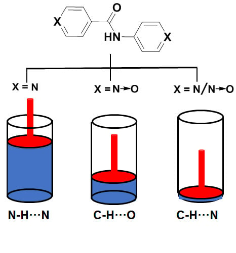
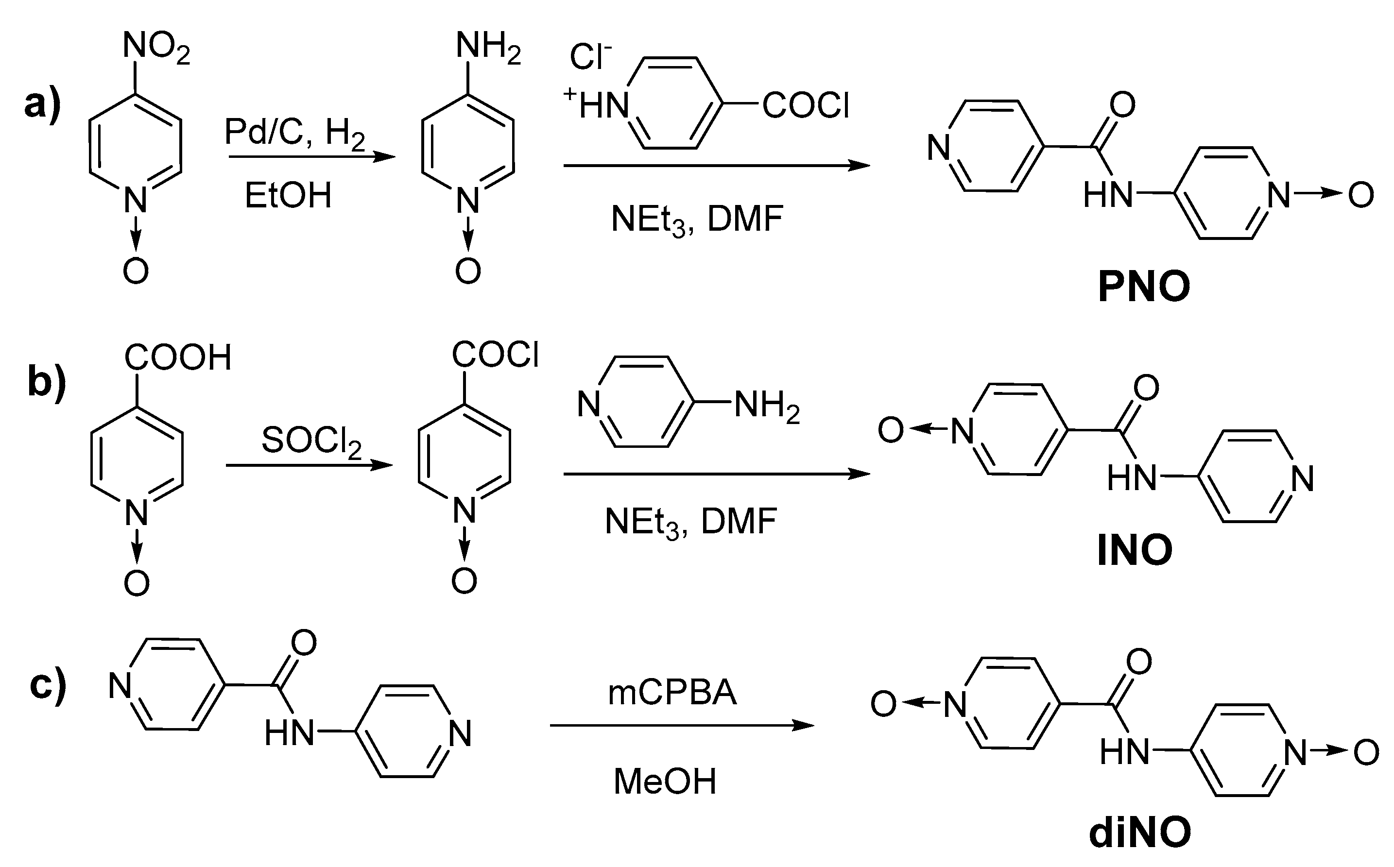
2.1. Design and Synthesis

We synthesized three N-oxides compounds by modifying the pyridyl group of N-(4-pyridyl)isonicotinamide (4PINA) [34]. This included a di-N-oxide (4-((1-oxidopyridin-4-yl)carbamoyl)pyridine-1-oxide, diNO) and two mono-N-oxides, namely 4-(isonicotinamido)pyridine-1-oxide (PNO) and 4-(pyridin-4-ylcarbamoyl)pyridine-1-oxide (INO) with the N-oxide group at the aminopyridine and isonicotinoyl end, respectively. Mono-N-oxide amides were synthesized by reacting acid chlorides with corresponding amines in anhydrous DMF in the presence of triethylamine (Scheme 1a,b). The precipitates obtained for INO and PNO were washed with aqueous sodium bicarbonate solution followed by water to ensure the removal of salts. The di-N-oxide (diNO) was synthesized by oxidizing 4PINA with m-chloroperoxybenzoic acid (Scheme 1c) and the product was washed with water and dried. All compounds were characterized by NMR, HRMS, IR, and X-ray diffraction (single-crystal and powder).

2.2. Gelation Experiments

The gelation experiments for diNO, INO, and PNO were tested in various solvents (Table S1, Supplementary Materials). In a typical experiment, a 10.0 mg portion of the compound was heated in 1.0 mL of solvent (1.0 wt %) to get a clear solution and was cooled to room temperature. The hydrogel formation of these amides tested at 1.0 wt % indicated no gelation, which prompted us to increase the concentration to 4.0 wt %. The mixture was heated (80.0–90.0 °C) and on cooling to room temperature, gelation was observed only for the di-N-oxide (diNO) after 1 h, which was confirmed via inversion test. Crystalline materials were obtained for both the mono-N-oxides (PNO and INO). The gelation experiment performed with an equimolar mixture of PNO and INO resulted in concomitant crystallization of the individual compounds, which was confirmed by single-crystal X-ray diffraction (SCXRD). The gel strength of diNO was analyzed by sol–gel transition temperature (Tgel) and minimum gel concentration (MGC) experiments. The lowest concentration at which the gel was obtained was recorded as the minimum gel concentration (MGC). The MGC experiment of the hydrogel was performed in 1.0 mL of water by weighing different amounts of diNO gelator (30.0 to 50.0 mg) in a 7.0 mL vial. The mixture was heated and then cooled to room temperature, and the vial was left undisturbed for gel formation. After 24 h, the gel formation was confirmed by the inversion test and the MGC of the gelator was found to be 4.0 wt %.

2.3. Thermal Stability
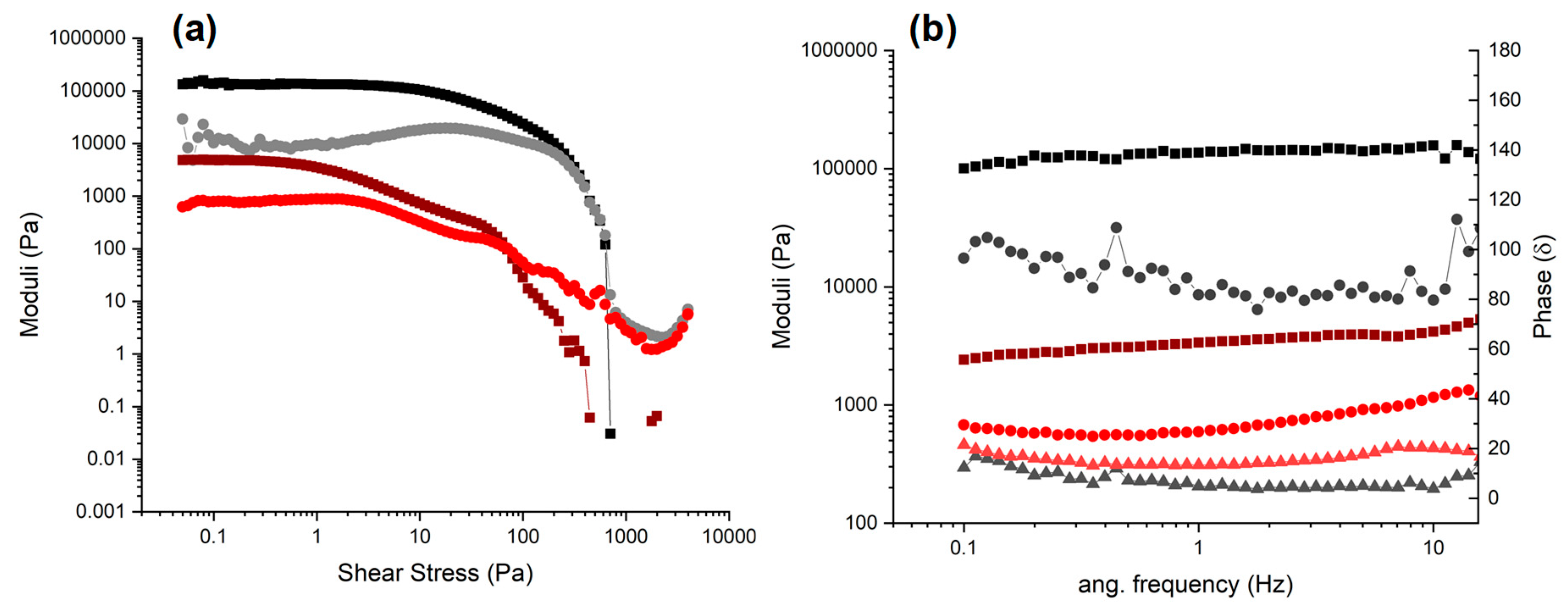
The thermal stability of the gel network of diNO was evaluated using gel-to-solution transition temperature (Tgel) experiments, which is the critical temperature at which the gel converts to a solution. The experiment was performed by placing a small spherical glass ball (92.0 mg) on top of the diNO preformed hydrogel in a standard vial. The vial was heated and the temperature at which the ball touched the bottom of the vial was recorded as Tgel. The experiments were performed at 4.0 and 6.0 wt % and the Tgel did not vary much with the concentration of diNO, which was found to be 78.0 °C and 80.0 °C at 4.0 and 6.0 wt %, respectively.

2.4. Rheology

Rheological analysis was undertaken to determine the mechanical properties of the supramolecular hydrogels (4.0 wt %) [66,67]. The oscillatory amplitude sweeps, at a constant frequency (1.0 Hz), demonstrated a linear viscoelastic region (LVR) in which the storage (elastic) G’ modulus was approximately an order of magnitude higher than the loss (viscous) G” modulus for both samples (Figure 1a) [66,68]. Thus, both samples had a linear elastic response to low stress amplitudes. This behavior is indicative of a solid-like network throughout the sample gelling the aqueous solvent. At increased shear stresses, a stress softening behavior was observed, the viscous moduli (G”) increased, and at a crossover point, there was a concomitant sharp decrease in the storage moduli (G’) indicating a transition from an elastic gel to a viscous fluid [69,70]. At low shear stresses, 4PINA had an elastic modulus of approximately 140,000 Pa and a yield stress of approximately 500 Pa. diNO was a significantly softer gel with an elastic modulus of approximately 5000 Pa and was also weaker, with a yield stress of approximately 60 Pa.
Oscillatory frequency sweeps (Figure 1b) at a fixed amplitude (0.05 Pa) demonstrated that G’, arising from the elastic network, was higher than G” across all frequencies for 4PINA, which is characteristic of the presence of a solid-like network of filaments that are temporally persistent in the viscoelastic gel. These moduli were effectively indifferent to the frequency and this was true for diNO as well at low frequencies (<10 Hz). However, with increasing frequencies G’ increased, the typical behavior of viscoelastic liquids rather than supramolecular gels [66], which further indicates that diNO formed weaker gels than 4PINA. Furthermore, the ratio of G’:G’’ is not at least an order of magnitude, the generally accepted ratio for supramolecular gels. Instead the ratio is roughly 7:1, further indicating that the modifications made to diNO severally affect its gelling ability. However, the phase angle, the lag between the applied shear stress and the measured strain, for diNO of approximately 15.0 δ is comparable to that of the original gelator, 4PINA, with a measured phase lag of approximately 7.0 δ. Moreover, the phase angle is not dependent on the frequency, a key Watson–Chambon criterion for gels. Thus, as the diNO samples do not meet the moduli requirements but demonstrated other viscoelastic behavior typical of a physical gel, it can, with caveats, be considered a very weak gel.

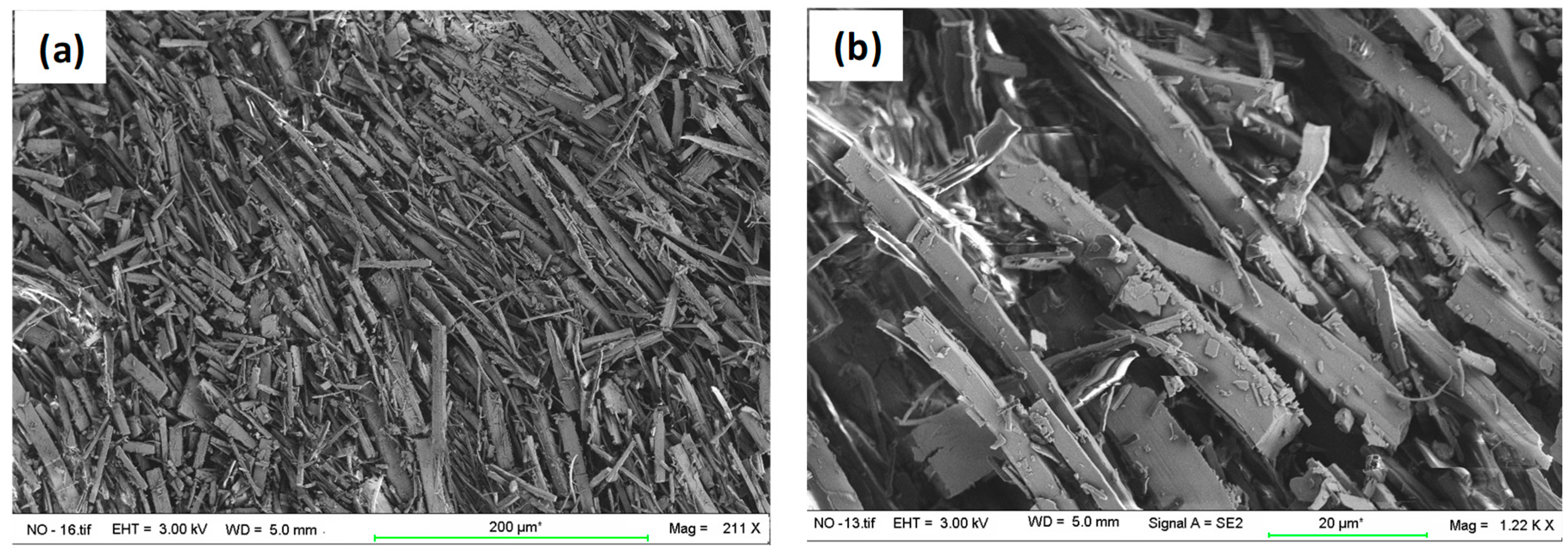
2.5. Scanning Electron Microscopy (SEM)

The morphology of gel fibers was studied by analyzing the SEM images of dried diNO gel. The gel was prepared at 4.0 wt % in water, filtered after 24 h, and dried under a fume hood. A small portion of the dried gel was placed on a carbon tab and was coated with gold for 2 min. The SEM images revealed that the xerogel displayed a rod-type architecture with needle-shaped microcrystalline materials in the network. The diameter of the small needle-shaped fibers ranged from 1.2 to 2.5 μm and the width of the larger microcrystalline rods ranged from 8.0 to 30.0 μm (Figure 2).

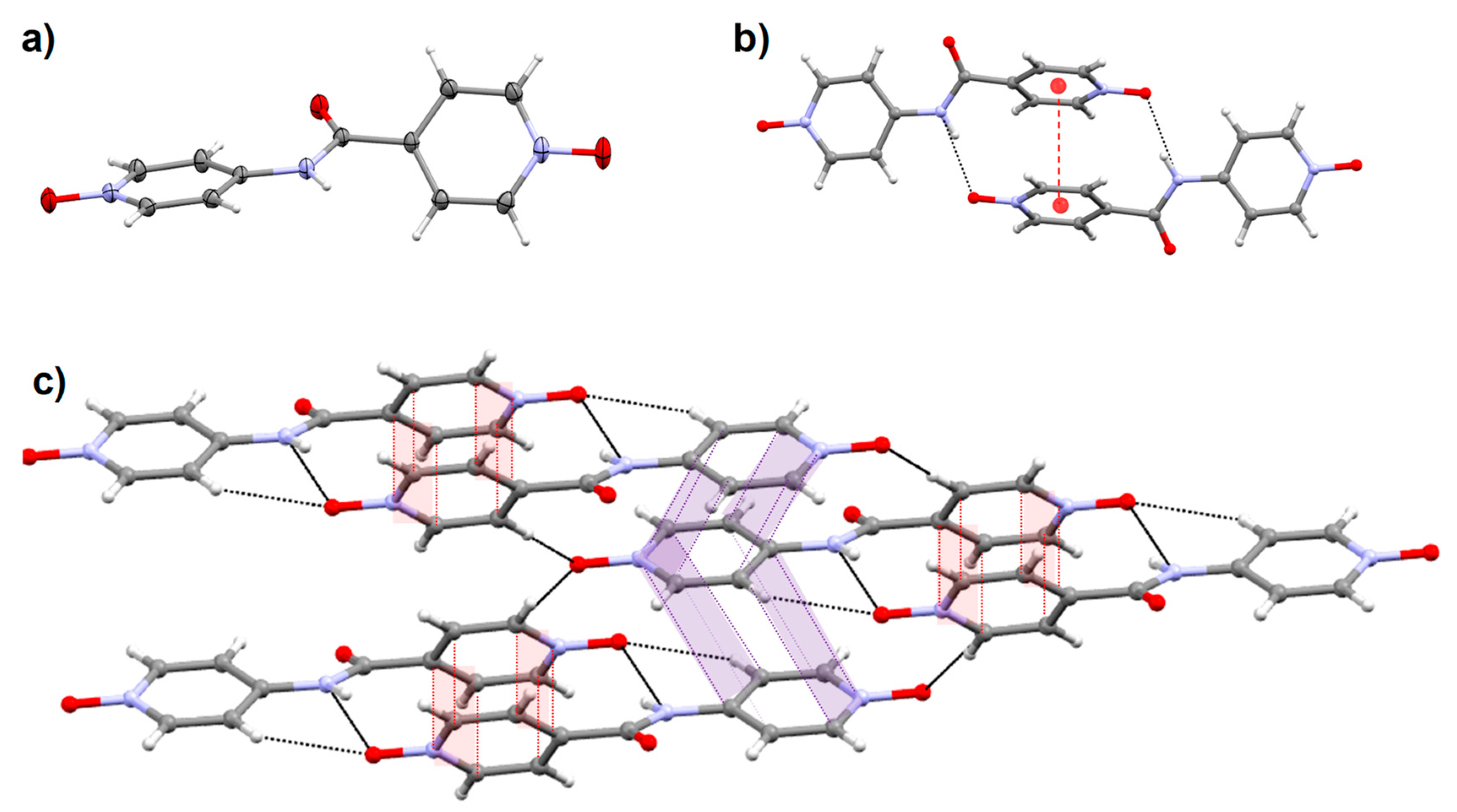
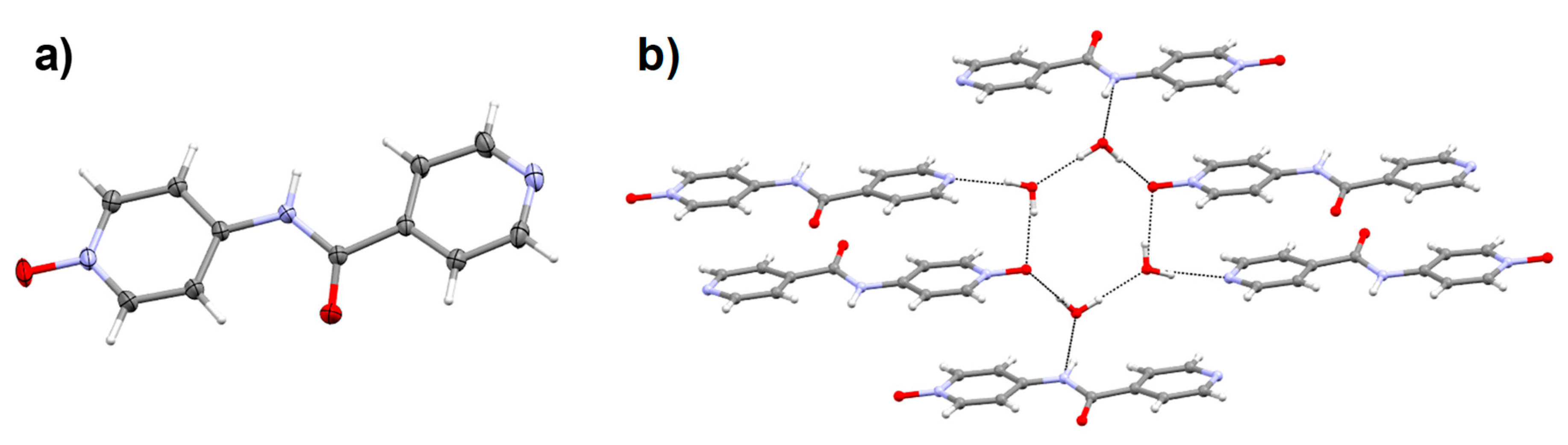
2.6. Crystal Structure

The crystallization experiments of N-oxides and an equimolar mixture of two mono-N-oxides were performed in water and various organic solvents. The compounds were proved to be sparingly soluble in most of the organic solvents, hence the experiments were carried out in a water or aqueous solution of methanol, ethanol, acetonitrile, and tetrahydrofuran. About 20.0 mg of the material were dissolved by heating in 1.0 mL of water, and the solution was cooled to room temperature. Crystallization from mixed solvents (1:1, v/v) such as methanol/water, ethanol/water, acetonitrile/water, and tetrahydrofuran/water resulted in precipitates. In some cases, needle-shaped crystals were obtained with low yield and the crystal quality was not good for single-crystal X-ray diffraction. The crystallization experiments performed in water produced X-ray quality single crystals for all N-oxides. The crystals of diNO were found to be needle-shaped, whereas block-shaped crystals were observed for both mono-N-oxides. Interestingly, crystallization of a 1:1 mixture of PNO and INO also resulted in block-shaped crystals.
The gelator diNO crystallized in a triclinic space group (Pī) with the carbonyl moiety of the amide group slightly deviated from the amide plane (15.74(8)°) and the pyridyl N-oxide rings were twisted at an angle of 58.71(3)° (Figure 3a). The N-H moiety was involved in hydrogen-bonding interaction (N―H∙∙∙O) with the oxygen atom of the adjacent molecule to form a dimer. This dimer was further stabilized by a strong π–π interaction (3.6563(9) Å) between the isonicotinoyl N-oxide ring of diNO (Figure 3b) and also N―H∙∙∙O interactions (2.8178(16) Å) between the amide N-H moiety and isonicotinoyl N-oxide moieties (Figure S9, Supplementary Materials). The aminopyridine N-oxide moieties of the diNO dimer interact with the isonicotinoyl N-oxide moieties of adjacent dimers via offset π–π interactions (3.8745(9) and 3.8219(9) Å) and also display bifurcated C―H∙∙∙O interactions (3.1789(18) and 3.1621(19) Å) with adjacent dimers (Figure 3c).
Compound INO crystalized in a triclinic space group (Pī) and the twisting of the pyridyl N-oxide ring (58.86°) was similar to diNO (Figure 4a). The hydrogen-bonding pattern was exactly the same as in diNO resulting in a dimer, which was stabilized by N―H∙∙∙O and π–π interactions. The only difference between these two structures was the connection between adjacent dimers and the dimer in INO propagates via C―H∙∙∙N interaction (3.4391(16) and 3.5489(16) Å), whereas C―H∙∙∙O and π–π interactions are observed in diNO. The solvated form of INO was also isolated (INO.2H2O) and the crystals belong to a monoclinic C2/c space group (Figure S10). The asymmetric unit contains one INO and two molecules of water, and the isonicotinoyl N-oxide and pyridyl moieties are coplanar to the amide groups compared to the de-solvated form. The hydrogen-bonding pattern of INO.2H2O was different from INO due to the presence of dimeric water clusters. The isonicotinoyl N-oxide was hydrogen-bonded to two water molecules via O―H∙∙∙O interactions (2.793(3) and 2.796(2) Å), and the pyridyl nitrogen atom interacted with a water molecule through O―H∙∙∙N interaction (2.771(2) Å). This resulted in the formation of a hydrogen-bonded macrocycle involving two INO molecules and four water molecules. The amide moieties of the macrocycle were hydrogen bonded to adjacent macrocycles via N―H∙∙∙O (2.921(2) Å) interactions to form a hydrogen-bonded 3-D network.
The structural analysis of the pyridyl N-oxide compound (PNO) revealed that the compound crystallized in a similar space group as diNO but with two solvent water molecules (PNO.2H2O, Figure 5a). The pyridyl N-oxide and isonicotinoyl moieties are almost planar to each other but slightly twisted from the amide plane (16.70 and 18.72°). The N-H moiety of the amide group, the pyridyl nitrogen atom, and the N-oxide moieties are hydrogen bonded to the water molecules. The amide acts as donor (N―H∙∙∙O = 2.8617(15) Å), whereas the N-oxide (O―H∙∙∙O = 2.7412(15) and 2.7701(16) Å) and isonicotinoyl groups (O―H∙∙∙N = 2.8598(17) Å) act as acceptor, resulting in four water molecules interacting with one PNO molecule. The hydrogen bonding between water molecules and PNO resulted in a hexagonal architecture of oxygen atoms formed by N-oxide oxygen atoms of two PNO and four water molecules, which was stabilized by various interactions with amide and pyridyl nitrogen atoms of adjacent PNO molecules (Figure 5b).
We also performed the crystallization of a 1:1 mixture of INO and PNO in water (2.0 wt %) and block-shaped crystals appeared on cooling after 1 h. The SCXRD data matched the PNO.2H2O crystals. The crystals of INO.2H2O were also obtained from the same mixture after 24 h. The experiments repeated by varying the ratios of INO and PNO (1:3 and 3:1) also resulted in the co-crystallization of the individual components.

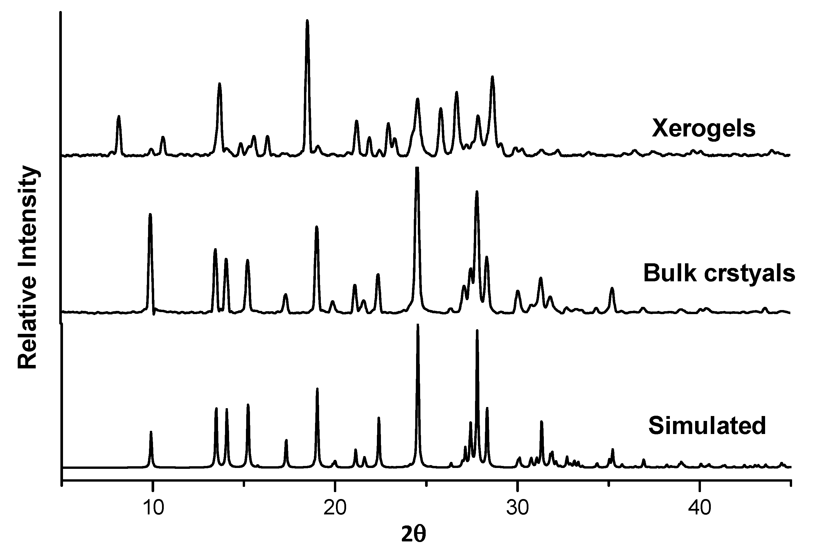
2.7. X-ray Powder Diffraction (XRPD)

The phase purity of the compounds was further analyzed by comparing the XRPD pattern of the bulk crystals with the simulated pattern obtained from the single-crystal data. XRPD was performed on the bulk solid of recrystallized diNO from water and the xerogel obtained from water at 4.0 wt %. The powder X-ray pattern of the re-crystallized diNO matches perfectly well the simulated graph obtained from the single-crystal data (Figure 6). However, the powder X-ray pattern of the xerogel did not match the calculated pattern from the crystal structure of diNO. The XRPD patterns of mono-N-oxides were performed with the recrystallized samples from water, which were filtered and dried in air. The XRPD pattern of PNO revealed that the pattern of bulk solid was virtually super-imposable on the simulated pattern of PNO (Figure S11). The XRPD pattern of the bulk crystalline material of INO was also similar to the simulated pattern of INO (Figure S12). We also performed the XRPD of the hydrated form of INO, but the pattern was different from the calculated pattern of the INO.2H2O crystal structure (Figure S13). Interestingly, the INO.2H2O bulk crystal pattern matched the simulated pattern of INO (Figure S12).

3. Discussion

The ability of pyridyl amides to form a one-dimensional (1-D) hydrogen-bonded network is well established, which makes them ideal candidates in designing LMWGs with tunable properties. The availability of single-crystal X-ray structures of many organo/hydrogelators based on pyridyl amides enabled us to identify the key factors responsible for the formation of gel networks, for example, the formation of 1-D networks. The structural analysis of pyridyl amides indicates that these compounds self-assemble through mainly two types of hydrogen bonding, namely N―H∙∙∙O synthon of the amide moiety and N―H∙∙∙N synthon involving the amide and pyridyl nitrogen. These two types of interactions are predominant in the molecular aggregation of N-(4-pyridyl)isonicotinamide (4PINA) [34] along with various other non-bonding interactions. However, the impact of these types of interactions was not studied in detail. For example, what is the role of N―H∙∙∙N synthon in gel network stabilization? What happens if these interactions are replaced by another synthon? We addressed this question by restricting the N―H∙∙∙N synthon by modifying the pyridyl nitrogen to pyridyl-N-oxide moiety. Thus, we modified the pyridyl groups of 4PINA [34] to N-oxides; two mono-N-oxides were synthesized by oxidizing the nitrogen atoms at the pyridyl amine end (PNO) and the isonicotinic acid end (INO), and oxidation of both pyridyl nitrogen atoms resulted in diNO. This restricts N―H∙∙∙N interactions between the gelators and hinders the one-dimensional growth of the gel fibril, which may facilitate the formation of either a two-dimensional (2-D) or 3-D network of gelator. Another advantage of pyridyl-N-oxides is the ease of crystallization in an aqueous medium, enabling us to compare the crystal structures of mono-N-oxides and di-N-oxide for structure–property correlation.
The gelation properties of all the N-oxides in various solvents resulted in selective gelation of diNO in the water at higher concentration (4.0 wt %). The formation of needle-shaped crystals at lower concentration revealed that crystallization and gelation properties [71] were concentration dependent. The mono-N-oxides (PNO and INO) did not form gel in any solvents but formed crystals indicating the importance of similar groups at both ends in gel formation, which corroborates well with the gelation properties of 4PINA [34]. We also prepared a mixture of PNO and INO in different ratios to test the ability of these compounds to form multi-component gels. Such gels are obtained by mixing two or more compounds (gelator/non-gelator) where individual molecules interact either constructively or destructively to form well-ordered fibers containing individual components (self-sorting), both components (specific co-assembly), or a mixture of both (random co-assembly) [72,73]. We showed that mixing enantiomeric supramolecular gels based on bis(urea) compounds tagged with a phenylalanine methyl ester leads multi-component gels with enhanced thermal and mechanical strength [74]. However, the experiments performed with the mixtures of PNO and INO resulted in concomitant crystallization of individual compounds. INO was proved to be more soluble between the two mono-N-oxides leading to slow crystallization. The crystallization of the individual compounds suggests self-sorting of the components in the mixture.
The minimum gel concentration (MGC) of diNO (4.0 wt %) indicates that the gel network formed was weaker compared to 4PINA (MGC 0.37 wt %) [34]. This may be attributed to the fact that converting the pyridyl group of 4PINA to pyridyl-N-oxide increased the hydrophilic interactions preferring crystalline form, which hindered the gel formation at low concentration [75]. The gel-to-solution transition temperature (Tgel) experiments performed at MGC of diNO to evaluate the thermal stability of the gel revealed that the gel network collapsed at 78.0 °C. This indicates that the gel network of diNO is slightly weaker compared to 4PINA and the Tgel of diNO network did not change drastically with concentration. This is in agreement with the rheological measurements that revealed that diNO gels were weaker compared to the 4PINA gelator. The rheology experiments enabled us to elucidate the information regarding factors controlling the gelation, gel strength, and the solid-like properties of pure gels, and the mechanical strength of the 4PINA and diNO gels was evaluated using rheology. The rheological measurements performed for both 4PINA and diNO gels revealed that diNO gels were significantly softer than 4PINA gels. At a low strain of 1.0%, both diNO and 4PINA samples had an effectively infinite shear stress relaxation time, typical of a temporally persistent supramolecular elastic network. On the other hand, at a higher strain of 10.0% the relaxation time for the 4PINA sample, which had sheared, now displayed a finite relaxation time and behaved like a viscous liquid (Figures S14 and S15). The morphologies of the gel fibers were analyzed using SEM images of the dried gel, and the results indicated that a microcrystalline network was observed in diNO gels. The xerogels of 4PINA at a similar concentration displayed fibrous morphology with tape-type architecture with widths ranging from 0.57 to 5.7 μm [34], which was different from diNO gels. This is quite interesting because changing the pyridyl groups to N-oxide resulted in a drastic change in the morphology of the gel fibers. The stronger gelation properties of 4PINA may be attributed to the ability of 4PINA to immobilize the solvent molecules more efficiently in the fibrous network compared to the N-oxide network.
The crystallization experiments of the N-oxides performed in aqueous solutions of polar solvents resulted in either needle-shaped crystals or precipitate, which prompted us to explore the crystallization in pure water. The crystallization of INO and PNO in water resulted in blocked-shaped crystals in 3–5 h, but needle-shaped diNO crystals were formed in 1 to 2 days. The solid-state structures of the N-oxides were analyzed by single-crystal X-ray diffraction. The structure of N-oxides with both ends modified (diNO) was compared to the parent 4PINA structure and these two structures differ mainly in the type of non-bonding interactions. The crystal structure of 4PINA showed that the pyridyl nitrogen atoms and the amide moieties were hydrogen bonded via N―H∙∙∙N interaction to form a 1-D hydrogen-bonded network. However, discrete dimers were formed in diNO by N―H∙∙∙O hydrogen bonding, which was stabilized by π–π interactions. These dimers interact with each other via bifurcated C―H∙∙∙O interactions and π–π interactions to form a 2-D supramolecular structure. The comparison of the intermolecular interactions in both the structures indicated that the N―H∙∙∙N interaction in 4PINA is replaced by bifurcated C―H∙∙∙O interactions in diNO. This explains why 4PINA is a good gelator, which indicates the importance of N―H∙∙∙N interaction in the formation of supergelators. The phase purity of the N-oxide compounds was analyzed by comparing the diffraction pattern of the crystal structures with the powder X-ray pattern of the bulk materials. The analysis of the pattern revealed that the recrystallized form of diNO matched the single-crystal structure of diNO, indicating an identical structure. However, the XRPD pattern of the xerogels of diNO did not match the pattern obtained from the crystal structure of diNO. This is presumably due to the presence of hydrogen-bonded solvent molecules in the xerogels. The XRPD data of the bulk materials of INO and PNO.2H2O matched their corresponding crystal structures. However, the XRPD pattern of INO.2H2O matched the crystal structure of the INO pattern, confirming the transformation of INO.2H2O crystals due to the loss of water molecules during the drying process.
The crystal structure analysis of INO obtained from water revealed the presence of two forms, namely INO and INO.2H2O. These two forms were crystallized from water and the rapid crystallization process produced large block-shaped crystals of INO.2H2O. The gelation experiments performed at higher concentration (>40.0 mg in 1.0 mL water) also favored the crystallization of INO.2H2O. The selective crystallization of INO was obtained by the slow crystallization in dilute aqueous solution over a period of five days. This indicates that INO.2H2O was the kinetically favored form and these crystals were slowly converted to the thermodynamically stable INO form, which was confirmed by powder X-ray analysis. The solid-state structure of these two forms was different and the molecule was planar in INO.2H2O compared to INO. This was due to the hydrogen-bonding interaction of the water molecules resulting in a hydrogen-bonded macrocycle involving two INO molecules and four water molecules. The structures of INO and diNO were compared to analyze the importance of identical groups on both ends in the gelation process. The solid-state structures of INO and diNO were similar resulting in the dimer formation, but the non-bonding interaction responsible for the propagation of the dimers was different. The dimer in diNO propagated via C―H∙∙∙O interactions (3.1789(18) and 3.1621(19) Å) and this was replaced by a C―H∙∙∙N interaction (3.4391(16) and 3.5489(16) Å) in INO. The selective gelation of diNO may be attributed to the strong C―H∙∙∙O interaction compared to the non-bonding interaction in INO. The structure analysis of PNO revealed the presence of two water molecules in the crystal structure, which helped the molecule to retain the molecular planarity. The water molecules play a crucial role in overall packing of the crystal, and a hexagonal architecture of oxygen atoms was formed by the hydrogen bonding between PNO and the water molecules. The structural analysis of 4PINA and the N-oxides confirm the importance of the non-bonding interactions and the presence of identical groups at both ends of the compounds in gel formation.

4. Materials and Methods

4.1. Chemicals and Reagents

All starting materials and solvents were purchased from Sigma-Aldrich (MEDOR ehf, Reykjavik, Iceland) and were used as supplied. Deionized water was used for all the experiments and anhydrous methanol was obtained by distilling the solvent over Mg turnings and iodine. 4-aminopyridine-1-oxide [76] and N-(4-pyridyl)isonicotinamide (4PINA) [34] were synthesized following the reported procedures. 1H NMR and IR spectra were recorded on a Bruker Advance 400 spectrometer (Rheinstetten, Germany) and a Nicolet iN10 (Thermo Fisher Scientific, Hvidovre, Denmark), respectively. Single-crystal X-ray diffraction (SCXRD) was performed on a Bruker D8 VENTURE (Karlsruhe, Germany), and X-ray powder diffraction (XRPD) was carried out using a Bruker D8 Focus instrument (Karlsruhe, Germany). The morphology of the xerogel was analyzed by scanning electron microscopy (SEM) using a Leo Supra 25 Microscope (Carl Zeiss, Oberkochen, Germany).

4.2. Synthesis

General procedure for INO and PNO: The acid chloride was prepared by stirring the carboxylic acid (10.0 mmol) with 5.0 mL of thionyl chloride overnight in a round-bottom flask at 65.0 °C. The pale-yellow solution obtained was cooled and the excess thionyl chloride was removed by distillation. The resulting white solid/crystal was transferred to a 100 mL two-neck RB flask containing 4-aminopyridine/4-aminopyridine-1-oxide (10.0 mmol). A pale yellowish solution was obtained by adding 20.0 mL anhydrous DMF to the flask, the solution was cooled to 0 °C, and triethylamine (1.5 mL, 10.8 mmol) was added dropwise to this solution. The resulting mixture was stirred overnight at room temperature and the solution was added to a beaker containing 200 mL diethyl ether resulting in a white suspension. The mixture was filtered, and the residue was stirred with 4.0% NaHCO3 for 5 h, filtered, and washed with cold water. The resulting white solid was recrystallized from water to obtain the desired product.
4-(pyridin-4-ylcarbamoyl)pyridine-1-oxide (INO): Isonicotinic acid-N-oxide (1.39 g, 10.0 mmol) and 4-aminopyridine (0.94 g, 10.0 mmol). Yield: 72.0% (1.55 g, 7.2 mmol). 1H NMR (400 MHz, DMSO-d6) δ: 10.68 (1H, s), 8.47 (2H d, J = 8.0), 8.36 (2H d, J = 8.0), 7.95 (2H d, J = 8.0), 7.73 (2H d, J = 8.0). MS (ESI) m/z for C11H9N3O2Na+: expected 238.20, found 238.20.
4-(isonicotinamido)pyridine-1-oxide (PNO): Isonicotinic acid (1.23 g, 10.0 mmol) and 4-aminopyridine-1-oxide (1.10 g, 10.0 mmol). Yield: 79.0% (1.70 g, 7.9 mmol). 1H NMR (400 MHz, DMSO-d6) δ: 10.98 (1H, s), 8.80 (2H d, J = 6.0), 8.19 (2H d, J = 7.6), 7.85 (2H d, J = 4.4, 1.4), 7.82 (2H dd, J = 5.4, 2.2). MS (ESI) m/z for C11H9N3O2Na+: expected 238.20, found 238.19.
4-((1-oxidopyridin-4-yl)carbamoyl)pyridine-1-oxide (diNO): To a solution of N-(4-pyridyl)isonicotinamide (1.35 g, 6.8 mmol) in MeOH (25 mL), m-chloroperoxybenzoic acid (3.80 g, 17.0 mmol) was added in portions for 15 min. The reaction mixture was refluxed overnight at 70.0 °C and cooled to room temperature. The solid was collected by filtration, washed with hot methanol, and then dried. Yield 0.88 g, 56.0%. 1H NMR (400 MHz, DMSO-d6) δ: 10.81 (1H, s), 8.39 (2H d, J = 7.2), 8.18 (2H d, J = 7.2), 7.95 (2H d, J = 7.2), 7.79 (2H d, J = 7.2). MS (ESI) m/z for C11H9N3O3Na+: expected 254.05, found 254.05.

4.3. Gelation Studies

Gelation experiments were initially carried out by weighing 10.0 mg of the N-oxide ligands (INO, PNO, diNO, and an equimolar mixture of INO and PNO) in a 7.0 mL vial and 1.0 mL of distilled water was added to the compounds. The mixtures were heated until a clear solution was obtained; cooling to room temperature resulted in no gelation in any case after 24 h. Since the compounds were soluble only in water, gelation experiments were carried out in aqueous solution of organic solvents. An aqueous solution (1:1, v/v) was prepared by mixing 500 µL of organic solvents (methanol, ethanol, tetrahydrofuran, acetonitrile, or nitrobenzene) with 500 µL of water. These solutions were added to 10.0 mg of the N-oxide compounds and the mixtures were heated; cooling to room temperature resulted in a white precipitate after 24 h in all cases. The higher solubility of the compounds in water compared to other solvents prompted us to check the gelation experiments at higher concentration. Crystals were obtained when the gelation experiments were performed in 1.0 mL water with 20.0 mg compounds (2.0 wt %). The gelation ability tested in mixed solvents at 2.0 wt % mostly produced precipitate, whereas some crystalline materials were observed in a few experiments (in methanol/water).
The gelation experiments at higher concentration (4.0 wt %) also resulted in crystals for INO, PNO, and their equimolar mixture but an opaque gel was formed under identical conditions (confirmed by inversion test) for diNO. The gelation experiments at higher concentration in a mixed solvent were avoided due to insolubility of these materials. The solubility of diNO was found to be approximately 60.0 mg/mL in water, and the solution formed gel at concentrations between 4.0 and 6.0 wt %. The two mono-N-oxides and their equimolar mixture at high concentration (>4.0 wt %) were dissolved in boiling water, which readily formed crystals on cooling.

4.4. Minimum Gel Concentration (MGC)

The MGC was performed by taking various amounts (30.0 to 50.0 mg) of diNO in a standard 7.0 mL vial and the vial was sealed after adding 1.0 mL of distilled water. The mixtures were heated to dissolve the compound and left undisturbed to form gel. The minimum concentration at which gel was obtained was noted as MGC. The MGC of diNO was found to be 4.0 wt % in pure water.

4.5. Tgel Experiments

The gel-to-solution transition temperature (Tgel) experiment was performed at two different concentrations (4.0 and 6.0 wt %) of diNO. The required amount (40.0 or 60.0 mg) of diNO was placed in a 7.0 mL vial and 1.0 mL of water was added. The vial was sealed, and the mixture was heated to obtain a clear solution and cooled to room temperature to form the gel. After 24 h, a small spherical glass ball (92.0 mg) was carefully placed over the gel. The vial was gradually heated in an oil bath equipped with a magnetic stirrer and a thermometer. The temperature at which the glass ball touched the bottom of the vial was recorded as Tgel. The Tgel of diNO was found to be 78.0 °C and 80.0 °C at 4.0 and 6.0 wt %, respectively.

4.6. Rheology

The rheology experiments were performed at Durham University using a TA Instruments AR 2000 (New Castle, DE, USA) fitted with a rough Peltier plate and a 25 mm rough plate geometry (gap width of 2500 µm) at 25 °C. Supramolecular gels were prepared as discussed previously at a concentration of 4.0 wt %. The top plate was lowered and the normal force was allowed to reach equilibrium. Oscillatory amplitude sweeps were performed at a constant frequency (1.0 Hz) and the oscillatory frequency sweeps were performed at a constant shear stress (0.5 Pa). These experiments were performed in triplicate and the mean moduli and standard deviation were calculated.

4.7. Scanning Electron Microscopy (SEM)

The gelator diNO (40.0 mg) was dissolved in 1.0 mL of water by heating and cooled to room temperature to form the gel. After 24 h, the yellowish opaque gel was filtered through a filter paper and the residue was air dried in a fume hood. The xerogels were gold-coated for 2 min and SEM was performed using a Leo Supra 25 Microscope.

4.8. Crystallography

X-ray quality single-crystals were isolated from water, immediately immersed in cryogenic oil, and mounted. The diffractions were collected using MoKα radiation (λ = 0.71073 Å) on a Bruker D8 VENTURE (Photon100 CMOS detector) diffractometer equipped with a Cryostream (Oxford Cryosystems, Oxford, UK) open-flow nitrogen cryostat at room temperature. The unit cell determination, data collection, data reduction, structure solution/refinement, and empirical absorption correction (SADABS) were carried out using Apex III (Bruker AXS, Madison, WI, USA). The structure was solved by a direct method and refined by the full-matrix least-squares on F2 for all data using SHELXTL [77] (Version 2017/1, University of Göttingen, Göttingen, Germany) and Olex2 [78] (Version 1.2, OlexSys Ltd., Durham, U.K.) software. All non-disordered non-hydrogen atoms were refined anisotropically, and the hydrogen atoms were placed in the calculated positions and refined using a riding model.

4.9. X-ray Powder Diffraction (XRPD)

The N-oxide compounds (20.0 mg) were dissolved in 1.0 mL of hot water and left undisturbed for crystallization. After crystallization, the mixture was filtered and the residue was dried in air. The crystals were ground to fine powder and XRPD was carried out using a Bruker D8 Focus instrument. The xerogel of diNO was prepared by heating 40.0 mg of the compound in 1.0 mL of water (4.0 wt %), and the gel formed was filtered after 24 h followed by drying the residue overnight in a fume hood.

5. Conclusions

We synthesized two mono-N-oxides (INO and PNO) and a di-N-oxide (diNO) by altering the pyridyl moieties of a known hydrogelator (4PINA) to N-oxide moieties. The gelation ability of these compounds tested in various solvent/solvent mixtures indicated that selective gelation of diNO was observed in water. The mechanical strength and the thermal stability of diNO were evaluated using rheology and gel-to-solution transition temperature (Tgel) experiments, respectively. The gelation properties of diNO were compared with the parent 4PINA gelator, which indicated that diNO is a weaker gelator. SEM images were used to visualize the changes in the morphology of the gel fibers due to the alteration in the functional groups. The effect of various non-bonding interactions in the crystalline state and dried gel was studied using X-ray diffraction. Single-crystal X-ray analysis of the di-N-oxide structure revealed the formation of a hydrogen-bonded dimer, which interacted with adjacent dimers via C―H∙∙∙O interactions to form the extended network, and similar dimers observed in non-gelator INO were interconnected through C―H∙∙∙N interaction. The strong interaction of the mono-N-oxide with water molecules indicates the increased hydrophilic nature of these compounds to form solvated crystals. The reduced gelation ability (MGC) and thermal strength of diNO may be attributed to the weak intermolecular C―H∙∙∙O interaction. The absence of strong N―H∙∙∙N interactions in the N-oxide compounds compared to 4PINA clearly indicate the role of these interactions in gel formation. The tuning of gelation ability (MGC) and thermal/mechanical strength of 4PINA by alerting the functional group proves the importance of non-bonding interactions and functional groups in designing LMWGs with tunable properties, which contributes to the ongoing efforts to identify the key structural features of gel network formation.

Supplementary Materials

The following are available online at https://www.mdpi.com/1420-3049/24/19/3472/s1, Figure S1: 1H NMR of diNO in DMSO-d6, Figure S2: 1H NMR of INO in DMSO-d6, Figure S3: 1H NMR of PNO in DMSO-d6, Figure S4: IR spectrum of diNO, Figure S5: IR spectrum of INO (anhydrous form), Figure S6: IR spectrum of INO (hydrated form), Figure S7: IR spectrum of PNO, Figure S8: IR spectrum of 1:1 mixture of INO + PNO, Figure S9: Crystal structure of diNO, Figure S10: Crystal structure of INO.2H2O, Figure S11: XRPD comparison of PNO, Figure S12: XRPD comparison of INO, Figure S13: XRPD comparison of INO.2H2O, Figure S14: Stress relaxation experiments performed at 1.0% strain, Figure S15: Stress relaxation experiments performed at 10.0% strain, Figure S16: Amplitude and frequency sweeps, Scheme S1: Chemical structure of 4PINA, Table S1: Gelation table of N-oxide compounds, Table S2: Determination of MGC of diNO, Table S3: Determination of Tgel of diNO, Table S4: Crystal data for the N-oxide compounds.

Author Contributions

Conceptualization, K.K.D.; Data curation, D.G.; Formal analysis, D.G.; Funding acquisition, K.K.D.; Investigation, D.G.; Methodology, D.G. and M.T.M.; Project administration, K.K.D.; Resources, K.K.D.; Validation, K.K.D.; Writing—original draft, D.G.; Writing—review & editing, K.K.D.

Funding

This research received no external funding.

Acknowledgments

We thank the University of Iceland Research Fund and the Science Institute for funding. D.G. thanks the University of Iceland for the Doctoral Research and Teaching Assistantship grant. We thankfully acknowledge Jonathan Steed, Durham University, Durham, UK for rheology experiments, Sigurjur Sveinn Jonsson, ISOR-Iceland for X-ray powder diffraction analysis, and Sigridur Jonsdottir, University of Iceland for NMR and mass spectroscopy. We thank Rannís Iceland for granting an infrastructure grant for a single-crystal X-ray diffractometer.

Conflicts of Interest

The authors declare no conflict of interest.

References

  1. Estroff, L.A.; Hamilton, A.D. Water Gelation by Small Organic Molecules. Chem. Rev. 2004, 104, 1201–1218. [Google Scholar] [CrossRef] [PubMed]
  2. De Loos, M.; Feringa, B.L.; van Esch, J.H. Design and Application of Self-Assembled Low Molecular Weight Hydrogels. Eur. J. Org. Chem. 2005, 2005, 3615–3631. [Google Scholar] [CrossRef]
  3. George, M.; Weiss, R.G. Molecular Organogels. Soft Matter Comprised of Low-Molecular-Mass Organic Gelators and Organic Liquids. Acc. Chem. Res. 2006, 39, 489–497. [Google Scholar] [CrossRef] [PubMed]
  4. Dastidar, P. Supramolecular gelling agents: Can they be designed? Chem. Soc. Rev. 2008, 37, 2699–2715. [Google Scholar] [CrossRef] [PubMed]
  5. Hirst, A.R.; Escuder, B.; Miravet, J.F.; Smith, D.K. High-Tech Applications of Self-Assembling Supramolecular Nanostructured Gel-Phase Materials: From Regenerative Medicine to Electronic Devices. Angew. Chem. Int. Ed. 2008, 47, 8002–8018. [Google Scholar] [CrossRef] [PubMed]
  6. Piepenbrock, M.-O.M.; Lloyd, G.O.; Clarke, N.; Steed, J.W. Metal- and Anion-Binding Supramolecular Gels. Chem. Rev. 2010, 110, 1960–2004. [Google Scholar] [CrossRef]
  7. Steed, J.W. Anion-tuned supramolecular gels: A natural evolution from urea supramolecular chemistry. Chem. Soc. Rev. 2010, 39, 3686–3699. [Google Scholar] [CrossRef]
  8. Yu, G.; Yan, X.; Han, C.; Huang, F. Characterization of supramolecular gels. Chem. Soc. Rev. 2013, 42, 6697–6722. [Google Scholar] [CrossRef]
  9. Kumar, D.K.; Steed, J.W. Supramolecular gel phase crystallization: Orthogonal self-assembly under non-equilibrium conditions. Chem. Soc. Rev. 2014, 43, 2080–2088. [Google Scholar] [CrossRef]
  10. Banerjee, S.; Das, R.K.; Maitra, U. Supramolecular gels ‘in action’. J. Mater. Chem. 2009, 19, 6649–6687. [Google Scholar] [CrossRef]
  11. Foster, J.A.; Damodaran, K.K.; Maurin, A.; Day, G.M.; Thompson, H.P.G.; Cameron, G.J.; Bernal, J.C.; Steed, J.W. Pharmaceutical polymorph control in a drug-mimetic supramolecular gel. Chem. Sci. 2017, 8, 78–84. [Google Scholar] [CrossRef] [PubMed]
  12. Terech, P.; Weiss, R.G. Low Molecular Mass Gelators of Organic Liquids and the Properties of Their Gels. Chem. Rev. 1997, 97, 3133–3160. [Google Scholar] [CrossRef] [PubMed]
  13. Du, X.; Zhou, J.; Shi, J.; Xu, B. Supramolecular Hydrogelators and Hydrogels: From Soft Matter to Molecular Biomaterials. Chem. Rev. 2015, 115, 13165–13307. [Google Scholar] [CrossRef] [PubMed]
  14. Babu, S.S.; Praveen, V.K.; Ajayaghosh, A. Functional π-Gelators and Their Applications. Chem. Rev. 2014, 114, 1973–2129. [Google Scholar] [CrossRef] [PubMed]
  15. Worthington, P.; Pochan, D.J.; Langhans, S.A. Peptide Hydrogels—Versatile Matrices for 3D Cell Culture in Cancer Medicine. Front. Oncol. 2015, 5, 92. [Google Scholar] [CrossRef] [PubMed]
  16. Truong, W.T.; Su, Y.; Meijer, J.T.; Thordarson, P.; Braet, F. Self-Assembled Gels for Biomedical Applications. Chem. Asian J. 2011, 6, 30–42. [Google Scholar] [CrossRef] [PubMed]
  17. Li, J.-L.; Wang, R.-Y.; Liu, X.-Y.; Pan, H.-H. Nanoengineering of a Biocompatible Organogel by Thermal Processing. J. Phys. Chem. B 2009, 113, 5011–5015. [Google Scholar] [CrossRef] [PubMed]
  18. Li, J.-L.; Liu, X.-Y.; Wang, R.-Y.; Xiong, J.-Y. Architecture of a Biocompatible Supramolecular Material by Supersaturation-Driven Fabrication of its Fiber Network. J. Phys. Chem. B 2005, 109, 24231–24235. [Google Scholar] [CrossRef] [PubMed]
  19. Afrasiabi, R.; Kraatz, H.-B. Sonication-Induced Coiled Fibrous Architectures of Boc-L-Phe-L-Lys(Z)-OMe. Chem. Eur. J. 2013, 19, 1769–1777. [Google Scholar] [CrossRef] [PubMed]
  20. Dhibar, S.; Dey, A.; Majumdar, S.; Ghosh, D.; Mandal, A.; Ray, P.P.; Dey, B. A supramolecular Cd(ii)-metallogel: An efficient semiconductive electronic device. Dalton Trans. 2018, 47, 17412–17420. [Google Scholar] [CrossRef] [PubMed]
  21. Wang, R.-Y.; Liu, X.-Y.; Li, J.-L. Engineering Molecular Self-Assembled Fibrillar Networks by Ultrasound. Cryst. Growth Des. 2009, 9, 3286–3291. [Google Scholar] [CrossRef]
  22. Alexander, S.L.M.; Korley, L.T.J. Nucleation effects of high molecular weight polymer additives on low molecular weight gels. Polym. J. 2018, 50, 775–786. [Google Scholar] [CrossRef]
  23. Wang, R.-Y.; Liu, X.-Y.; Narayanan, J.; Xiong, J.-Y.; Li, J.-L. Architecture of Fiber Network:  From Understanding to Engineering of Molecular Gels. J. Phys. Chem. B 2006, 110, 25797–25802. [Google Scholar] [CrossRef] [PubMed]
  24. Draper, E.R.; Adams, D.J. How should multicomponent supramolecular gels be characterised? Chem. Soc. Rev. 2018, 47, 3395–3405. [Google Scholar] [CrossRef] [PubMed] [Green Version]
  25. Cao, X.; Meng, L.; Li, Z.; Mao, Y.; Lan, H.; Chen, L.; Fan, Y.; Yi, T. Large Red-Shifted Fluorescent Emission via Intermolecular π–π Stacking in 4-Ethynyl-1,8-naphthalimide-Based Supramolecular Assemblies. Langmuir 2014, 30, 11753–11760. [Google Scholar] [CrossRef] [PubMed]
  26. Wang, C.; Zhang, D.; Zhu, D. A Chiral Low-Molecular-Weight Gelator Based on Binaphthalene with Two Urea Moieties:  Modulation of the CD Spectrum after Gel Formation. Langmuir 2007, 23, 1478–1482. [Google Scholar] [CrossRef]
  27. Wang, C.; Zhang, D.; Xiang, J.; Zhu, D. New Organogels Based on an Anthracene Derivative with One Urea Group and Its Photodimer:  Fluorescence Enhancement after Gelation. Langmuir 2007, 23, 9195–9200. [Google Scholar] [CrossRef]
  28. Wang, G.; Cheuk, S.; Yang, H.; Goyal, N.; Reddy, P.V.N.; Hopkinson, B. Synthesis and Characterization of Monosaccharide-Derived Carbamates as Low-Molecular-Weight Gelators. Langmuir 2009, 25, 8696–8705. [Google Scholar] [CrossRef]
  29. Gao, W.; Liang, Y.; Peng, X.; Hu, Y.; Zhang, L.; Wu, H.; He, B. In situ injection of phenylboronic acid based low molecular weight gels for efficient chemotherapy. Biomaterials 2016, 105, 1–11. [Google Scholar] [CrossRef] [Green Version]
  30. Das, U.K.; Banerjee, S.; Dastidar, P. Remarkable Shape-Sustaining, Load-Bearing, and Self-Healing Properties Displayed by a Supramolecular Gel Derived from a Bis-pyridyl-bis-amide of L-Phenyl Alanine. Chem. Asian J. 2014, 9, 2475–2482. [Google Scholar] [CrossRef]
  31. Tang, Y.-T.; Dou, X.-Q.; Ji, Z.-A.; Li, P.; Zhu, S.-M.; Gu, J.-J.; Feng, C.-L.; Zhang, D. C2-symmetric cyclohexane-based hydrogels: A rational designed LMWG and its application in dye scavenging. J. Mol. Liq. 2013, 177, 167–171. [Google Scholar] [CrossRef]
  32. Li, P.; Dou, X.-Q.; Tang, Y.-T.; Zhu, S.; Gu, J.; Feng, C.-L.; Zhang, D. Gelator-polysaccharide hybrid hydrogel for selective and controllable dye release. J. Colloid Interface Sci. 2012, 387, 115–122. [Google Scholar] [CrossRef] [PubMed]
  33. Ghosh, D.; Lebedytė, I.; Yufit, D.S.; Damodaran, K.K.; Steed, J.W. Selective gelation of N-(4-pyridyl)nicotinamide by copper(ii) salts. CrystEngComm 2015, 17, 8130–8138. [Google Scholar] [CrossRef]
  34. Kumar, D.K.; Jose, D.A.; Dastidar, P.; Das, A. Nonpolymeric Hydrogelator Derived from N-(4-Pyridyl)isonicotinamide. Langmuir 2004, 20, 10413–10418. [Google Scholar] [CrossRef] [PubMed]
  35. Sahoo, P.; Kumar, D.K.; Raghavan, S.R.; Dastidar, P. Supramolecular Synthons in Designing Low Molecular Mass Gelling Agents: L-Amino Acid Methyl Ester Cinnamate Salts and their Anti-Solvent-Induced Instant Gelation. Chem. Asian J. 2011, 6, 1038–1047. [Google Scholar] [CrossRef] [PubMed]
  36. Bhattacharjee, S.; Bhattacharya, S. Role of synergistic [small pi]-[small pi] stacking and X-H[three dots, centered]Cl (X = C, N, O) H-bonding interactions in gelation and gel phase crystallization. Chem. Commun. 2015, 51, 7019–7022. [Google Scholar] [CrossRef] [PubMed]
  37. Jones, C.D.; Steed, J.W. Gels with sense: Supramolecular materials that respond to heat, light and sound. Chem. Soc. Rev. 2016, 45, 6546–6596. [Google Scholar] [CrossRef]
  38. Ghosh, D.; Ferfolja, K.; Drabavičius, Ž.; Steed, J.W.; Damodaran, K.K. Crystal habit modification of Cu(ii) isonicotinate–N-oxide complexes using gel phase crystallisation. New J. Chem. 2018, 42, 19963–19970. [Google Scholar] [CrossRef]
  39. Desiraju, G.R. Supramolecular Synthons in Crystal Engineering—A New Organic Synthesis. Angew. Chem. Int. Ed. 1995, 34, 2311–2327. [Google Scholar] [CrossRef]
  40. Weiss, R.G.; Terech, P. Molecular Gels: Materials with Self-Assembled Fibrillar Networks; Springer: Heidelberg, Germany, 2006; p. 978. [Google Scholar]
  41. Fages, F.; Voegtle, F.; Zinic, M. Systematic design of amide- and urea-type gelators with tailored properties. Top. Curr. Chem. 2005, 256, 77–131. [Google Scholar]
  42. Isare, B.; Pensec, S.; Raynal, M.; Bouteiller, L. Bisurea-based supramolecular polymers: From structure to properties1. C. R. Chim. 2016, 19, 148–156. [Google Scholar] [CrossRef]
  43. Gale, P.A.; Busschaert, N.; Haynes, C.J.E.; Karagiannidis, L.E.; Kirby, I.L. Anion receptor chemistry: Highlights from 2011 and 2012. Chem. Soc. Rev. 2014, 43, 205–241. [Google Scholar] [CrossRef] [PubMed]
  44. Dzolic, Z.; Cametti, M.; Dalla Cort, A.; Mandolini, L.; Zinic, M. Fluoride-responsive organogelator based on oxalamide-derived anthraquinone. Chem. Commun. 2007, 34, 3535–3537. [Google Scholar] [CrossRef] [PubMed]
  45. Kotova, O.; Daly, R.; dos Santos, C.M.G.; Boese, M.; Kruger, P.E.; Boland, J.J.; Gunnlaugsson, T. Europium-Directed Self-Assembly of a Luminescent Supramolecular Gel from a Tripodal Terpyridine-Based Ligand. Angew. Chem. Int. Ed. 2012, 51, 7208–7212. [Google Scholar] [CrossRef] [PubMed]
  46. Feng, L.; Cavicchi, K.A. Investigation of the relationships between the thermodynamic phase behavior and gelation behavior of a series of tripodal trisamide compounds. Soft Matter 2012, 8, 6483–6492. [Google Scholar] [CrossRef] [Green Version]
  47. Mukhopadhyay, S.; Ira; Krishnamoorthy, G.; Maitra, U. Dynamics of Bound Dyes in a Nonpolymeric Aqueous Gel Derived from a Tripodal Bile Salt. J. Phys. Chem. B 2003, 107, 2189–2192. [Google Scholar] [CrossRef]
  48. Li, Z.; Cao, J.; Li, H.; Liu, H.; Han, F.; Liu, Z.; Tong, C.; Li, S. Self-assembled drug delivery system based on low-molecular-weight bis-amide organogelator: Synthesis, properties and in vivo evaluation. Drug Deliv. 2016, 23, 3168–3178. [Google Scholar] [CrossRef] [PubMed]
  49. Bradberry, S.J.; Dee, G.; Kotova, O.; McCoy, C.P.; Gunnlaugsson, T. Luminescent lanthanide (Eu(iii)) cross-linked supramolecular metallo co-polymeric hydrogels: The effect of ligand symmetry. Chem. Commun. 2019, 55, 1754–1757. [Google Scholar] [CrossRef] [PubMed]
  50. Kumar, D.K.; Jose, D.A.; Dastidar, P.; Das, A. Nonpolymeric Hydrogelators Derived from Trimesic Amides. Chem. Mater. 2004, 16, 2332–2335. [Google Scholar] [CrossRef]
  51. Meazza, L.; Foster, J.A.; Fucke, K.; Metrangolo, P.; Resnati, G.; Steed, J.W. Halogen-bonding-triggered supramolecular gel formation. Nat. Chem. 2013, 5, 42–47. [Google Scholar] [CrossRef]
  52. Pal, A.; Ghosh, Y.K.; Bhattacharya, S. Molecular mechanism of physical gelation of hydrocarbons by fatty acid amides of natural amino acids. Tetrahedron 2007, 63, 7334–7348. [Google Scholar] [CrossRef]
  53. Kawamoto, K.; Grindy, S.C.; Liu, J.; Holten-Andersen, N.; Johnson, J.A. Dual Role for 1,2,4,5-Tetrazines in Polymer Networks: Combining Diels–Alder Reactions and Metal Coordination to Generate Functional Supramolecular Gels. ACS Macro Lett. 2015, 4, 458–461. [Google Scholar] [CrossRef]
  54. Das, U.K.; Banerjee, S.; Dastidar, P. Primary Ammonium Monocarboxylate Synthon in Designing Supramolecular Gels: A New Series of Chiral Low-Molecular-Weight Gelators Derived from Simple Organic Salts that are Capable of Generating and Stabilizing Gold Nanoparticles. Chem. Asian J. 2013, 8, 3022–3031. [Google Scholar] [CrossRef] [PubMed]
  55. Jin, Q.; Zhang, L.; Liu, M. Solvent-Polarity-Tuned Morphology and Inversion of Supramolecular Chirality in a Self-Assembled Pyridylpyrazole-Linked Glutamide Derivative: Nanofibers, Nanotwists, Nanotubes, and Microtubes. Chem. Eur. J. 2013, 19, 9234–9241. [Google Scholar] [CrossRef] [PubMed]
  56. Karak, S.; Kumar, S.; Bera, S.; Diaz, D.D.; Banerjee, S.; Vanka, K.; Banerjee, R. Interplaying anions in a supramolecular metallohydrogel to form metal organic frameworks. Chem. Commun. 2017, 53, 3705–3708. [Google Scholar] [CrossRef] [PubMed] [Green Version]
  57. Kartha, K.K.; Praveen, V.K.; Babu, S.S.; Cherumukkil, S.; Ajayaghosh, A. Pyridyl-Amides as a Multimode Self-Assembly Driver for the Design of a Stimuli-Responsive π-Gelator. Chem. Asian J. 2015, 10, 2250–2256. [Google Scholar] [CrossRef] [PubMed]
  58. Liu, Q.; Wang, Y.; Li, W.; Wu, L. Structural Characterization and Chemical Response of a Ag-Coordinated Supramolecular Gel. Langmuir 2007, 23, 8217–8223. [Google Scholar] [CrossRef] [PubMed]
  59. Lee, H.H.; Jung, S.H.; Park, S.; Park, K.-M.; Jung, J.H. A metal-organic framework gel with Cd2+ derived from only coordination bonds without intermolecular interactions and its catalytic ability. New J. Chem. 2013, 37, 2330–2335. [Google Scholar] [CrossRef]
  60. Aoyama, R.; Sako, H.; Amakatsu, M.; Yamanaka, M. Palladium ion-induced supramolecular gel formation of tris-urea molecules. Polym. J. 2015, 47, 136–140. [Google Scholar] [CrossRef]
  61. Ghosh, K.; Panja, S.; Bhattacharya, S. Naphthalene linked pyridyl urea as a supramolecular gelator: A new insight into naked eye detection of I- in the gel state with semiconducting behaviour. RSC Adv. 2015, 5, 72772–72779. [Google Scholar] [CrossRef]
  62. Kumar, D.P. Synthesis of gold nanoparticles and nanoclusters in a supramolecular gel and their applications in catalytic reduction of p-nitrophenol to p-aminophenol and Hg(ii) sensing. RSC Adv. 2014, 4, 45449–45457. [Google Scholar] [CrossRef]
  63. Byrne, P.; Lloyd, G.O.; Applegarth, L.; Anderson, K.M.; Clarke, N.; Steed, J.W. Metal-induced gelation in dipyridyl ureas. New J. Chem. 2010, 34, 2261–2274. [Google Scholar] [CrossRef]
  64. Pandurangan, K.; Kitchen, J.A.; Blasco, S.; Paradisi, F.; Gunnlaugsson, T. Supramolecular pyridyl urea gels as soft matter with antibacterial properties against MRSA and/or E. coli. Chem. Commun. 2014, 50, 10819–10822. [Google Scholar] [CrossRef] [PubMed]
  65. Stanley, C.E.; Clarke, N.; Anderson, K.M.; Elder, J.A.; Lenthall, J.T.; Steed, J.W. Anion binding inhibition of the formation of a helical organogel. Chem. Commun. 2006, 30, 3199–3201. [Google Scholar] [CrossRef] [PubMed]
  66. Goodwin, J.W.; Hughes, R.W. Rheology for Chemists: An Introduction, 2nd ed.; Royal Society of Chemistry: Cambridge, UK, 2008; p. 264. [Google Scholar]
  67. Guenet, J.-M. Rheological Aspects. In Organogels: Thermodynamics, Structure, Solvent Role, and Properties; Guenet, J.-M., Ed.; Springer International Publishing: Cham, Switzerland, 2016; pp. 83–94. [Google Scholar]
  68. Sathaye, S.; Mbi, A.; Sonmez, C.; Chen, Y.; Blair, D.L.; Schneider, J.P.; Pochan, D.J. Rheology of peptide- and protein-based physical hydrogels: Are everyday measurements just scratching the surface? Wiley Interdiscip. Rev. Nanomed. Nanobiotechnol. 2015, 7, 34–68. [Google Scholar] [CrossRef] [PubMed]
  69. Zuidema, J.M.; Rivet, C.J.; Gilbert, R.J.; Morrison, F.A. A protocol for rheological characterization of hydrogels for tissue engineering strategies. J. Biomed. Mater. Res. B 2014, 102, 1063–1073. [Google Scholar] [CrossRef]
  70. Gaharwar, A.K.; Avery, R.K.; Assmann, A.; Paul, A.; McKinley, G.H.; Khademhosseini, A.; Olsen, B.D. Shear-Thinning Nanocomposite Hydrogels for the Treatment of Hemorrhage. ACS Nano 2014, 8, 9833–9842. [Google Scholar] [CrossRef] [Green Version]
  71. Dasgupta, D.; Thierry, A.; Rochas, C.; Ajayaghosh, A.; Guenet, J.M. Key role of solvent type in organogelation. Soft Matter 2012, 8, 8714–8721. [Google Scholar] [CrossRef]
  72. Colquhoun, C.; Draper, E.R.; Eden, E.G.B.; Cattoz, B.N.; Morris, K.L.; Chen, L.; McDonald, T.O.; Terry, A.E.; Griffiths, P.C.; Serpell, L.C.; et al. The effect of self-sorting and co-assembly on the mechanical properties of low molecular weight hydrogels. Nanoscale 2014, 6, 13719–13725. [Google Scholar] [CrossRef] [Green Version]
  73. Raeburn, J.; Adams, D.J. Multicomponent low molecular weight gelators. Chem. Commun. 2015, 51, 5170–5180. [Google Scholar] [CrossRef]
  74. Tómasson, D.A.; Ghosh, D.; Kržišnik, Z.; Fasolin, L.H.; Vicente, A.A.; Martin, A.D.; Thordarson, P.; Damodaran, K.K. Enhanced Mechanical and Thermal Strength in Mixed-Enantiomers-Based Supramolecular Gel. Langmuir 2018, 34, 12957–12967. [Google Scholar] [CrossRef] [PubMed] [Green Version]
  75. Sauvée, C.; Ström, A.; Haukka, M.; Sundén, H. A Multi-Component Reaction towards the Development of Highly Modular Hydrogelators. Chem. Eur. J. 2018, 24, 8071–8075. [Google Scholar] [CrossRef]
  76. Gardner, J.N.; Katritzky, A.R. 875. N-oxides and related compounds. Part V. The tautomerism of 2- and 4-amino- and -hydroxy-pyridine 1-oxide. J. Chem. Soc. 1957, 4375–4385. [Google Scholar] [CrossRef]
  77. Sheldrick, G.M. Crystal structure refinement with SHELXL. Acta Crystallogr. Sect. C Struct. Chem. 2015, 71 Pt 1, 3–8. [Google Scholar] [CrossRef] [PubMed]
  78. Dolomanov, O.V.; Bourhis, L.J.; Gildea, R.J.; Howard, J.A.K.; Puschmann, H. OLEX2: A complete structure solution, refinement and analysis program. J. Appl. Crystallogr. 2009, 42, 339–341. [Google Scholar] [CrossRef]
Sample Availability: Samples of the compounds diNO, INO, and PNO are available from the authors.
Scheme 1. Synthesis of the N-oxide amides: (a) 4-(isonicotinamido)pyridine-1-oxide (PNO), (b) 4-(pyridin-4-ylcarbamoyl)pyridine-1-oxide (INO), and (c) 4-((1-oxidopyridin-4-yl)carbamoyl)pyridine-1-oxide (diNO).
Molecules 24 03472 sch001
Figure 1. (a) Oscillatory amplitude sweeps and (b) oscillatory frequency sweeps, for N-(4-pyridyl)isonicotinamide (4PINA) (black) and diNO (red). For both, ▪ refers to G’ and ● refers to G”, the storage modulus and viscous modulus, at 4.0 wt %, respectively. ▲ refers to the phase lag (δ).
Molecules 24 03472 g001
Figure 2. SEM images of diNO show the microcrystalline fibrous network: (a) overview; (b) magnified images.
Molecules 24 03472 g002
Figure 3. (a) Crystal structure of diNO, (b) showing hydrogen-bonded dimer stabilized with π–π interaction and (c) interaction of the individual dimer with adjacent dimer via various non-bonding interactions (dotted lines); black lines indicate hydrogen bonding, red line for π–π interactions of the individual dimer, and blue line for π–π interactions with adjacent dimers.
Molecules 24 03472 g003
Figure 4. (a) Crystal structure of INO and (b) interaction of individual dimers via various non-bonding interactions; black dotted lines indicate hydrogen bonding and red dotted lines represent the π–π interactions.
Molecules 24 03472 g004
Figure 5. Single-crystal structure of PNO: (a) Asymmetric unit (water molecules not shown); (b) two-dimensional (2-D) sheet constructed by H-bonding between the ligand and solvent (water) molecules.
Molecules 24 03472 g005
Figure 6. X-ray powder diffraction (XRPD) comparison of diNO: simulated pattern from single-crystal X-ray diffraction (SCXRD) data, bulk crystals, and xerogel obtained from water.
Molecules 24 03472 g006

