Article

Synthesis and Antiproliferative Activity of Novel A-Ring Cleaved Glycyrrhetinic Acid Derivatives

by
Daniela P.S. Alho
1,2,
Jorge A.R. Salvador
1,2,*,
Marta Cascante
3,4 and
Silvia Marin
3,4,*
1
Laboratory of Pharmaceutical Chemistry, Faculty of Pharmacy, University of Coimbra, 3000-548 Coimbra, Portugal
2
Centre for Neuroscience and Cell Biology, 3000-504 Coimbra, Portugal
3
Department of Biochemistry and Molecular Biomedicine, Faculty of Biology, University of Barcelona, Diagonal 643, 08028 Barcelona, Spain
4
Centro de Investigación Biomédica en Red de Enfermedades Hepáticas y Digestivas (CIBEREHD), Instituto de Salud Carlos III (ISCIII), 28029 Madrid, Spain
*
Authors to whom correspondence should be addressed.
Molecules 2019, 24(16), 2938; https://doi.org/10.3390/molecules24162938
Submission received: 27 July 2019 / Revised: 9 August 2019 / Accepted: 12 August 2019 / Published: 14 August 2019
(This article belongs to the Section Medicinal Chemistry)

Abstract

:
A series of new glycyrrhetinic acid derivatives was synthesized via the opening of its ring A along with the coupling of an amino acid. The antiproliferative activity of the derivatives was evaluated against a panel of nine human cancer cell lines. Compound 17 was the most active compound, with an IC50 of 6.1 µM on Jurkat cells, which is 17-fold more potent than that of glycyrrhetinic acid, and was up to 10 times more selective toward that cancer cell line. Further biological investigation in Jurkat cells showed that the antiproliferative activity of compound 17 was due to cell cycle arrest at the S phase and induction of apoptosis.

Graphical Abstract

1. Introduction

Cancer is a leading cause of death worldwide. Its global incidence continues to rise due to the aging and growth of the world population and the increasing adoption of lifestyle choices associated with cancer in developed countries [1]. Plants have been a major source of highly effective conventional drugs for cancer treatment and nowadays they play an important role as a source of leads for the development of potential new agents [2,3]. The plant-derived triterpenoids are proving to be interesting leading compounds as reported in a large number of scientific papers emerging in this field [4,5,6,7,8,9,10,11,12,13,14,15].
Glycyrrhetinic acid (GA) 1 is the hydrolyzed metabolite of glycyrrhizin, a major pentacyclic triterpenoid saponin obtained from the roots of licorice (Glycyrrhiza species) in high yields up to 24% [16,17]. This compound has been shown to inhibit tumor initiation [18,19,20,21,22] and proliferation in several cancer cell lines; its antiproliferative activity is mediated by cell cycle arrest [23,24] and induction of apoptosis [21,25,26,27,28]. The antitumor effects of GA 1 were also observed in animal models [20,29,30]. Nevertheless, it lacks potency and selectivity as an antitumor agent. Many derivatizations have been performed in order to enhance the potency of GA 1 [31,32,33,34,35,36]. However, the cleavage of its ring A is still poorly explored [37]. On the other hand, is well known that the conjugation of an amino acid moiety to pentacyclic triterpenoids improves their cytotoxicity and their selectivity towards tumor cells [38,39,40]. These findings prompted us to synthesize new GA 1 derivatives via the opening of its ring A along with the coupling with an amino acid. The novel semisynthetic derivatives were tested for their antiproliferative activity against a panel of nine human cancer cell lines. Further biological assays were conducted for the most potent compound 17 in the cancer cell line that yielded the best results (Jurkat cells), to investigate its preliminary mechanism of action. The study of selectivity was performed on human fibroblasts (BJ).

2. Results and Discussion
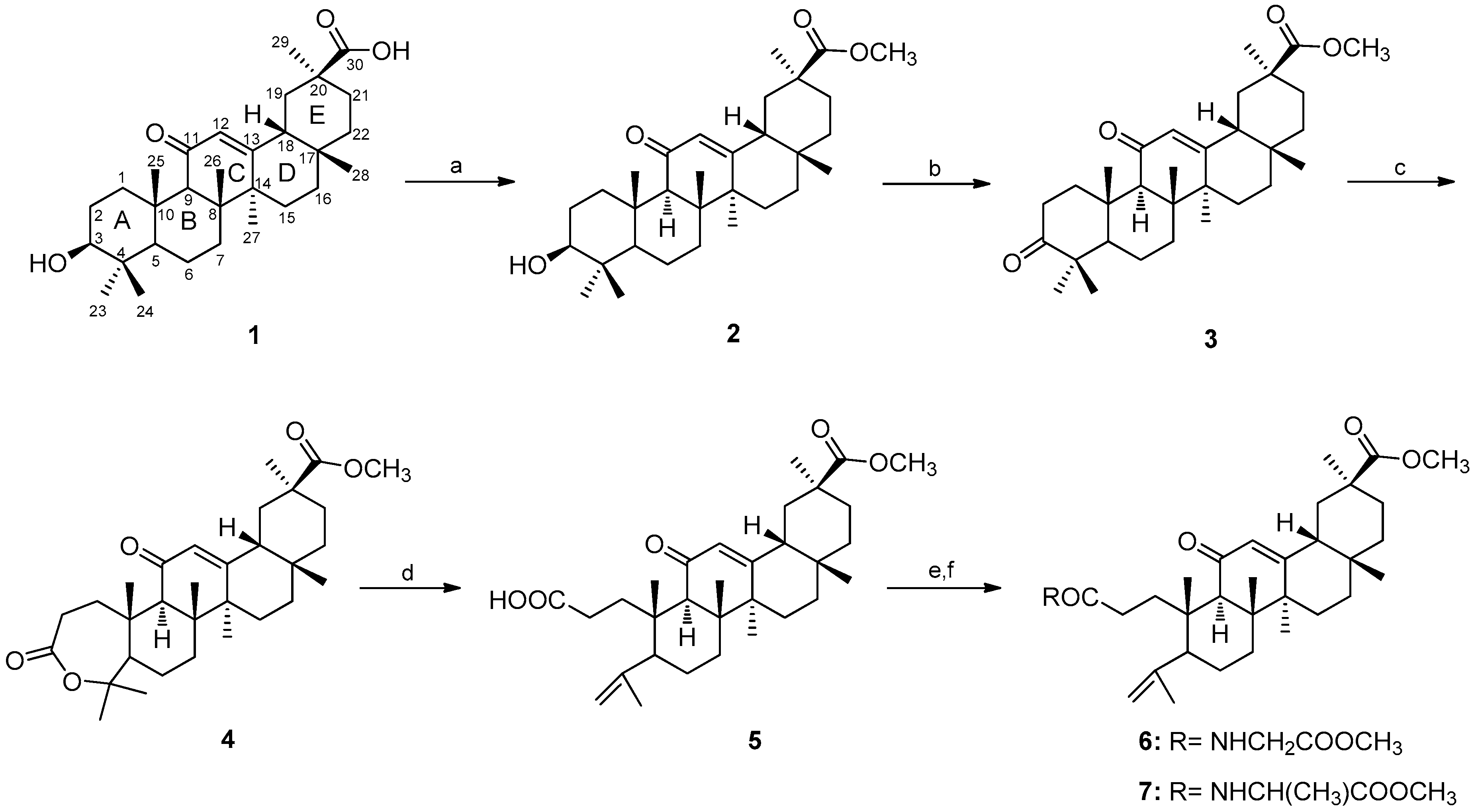
2.1. Chemistry

The synthesis of the glycyrrhetinic acid 1 derivatives is outlined in Scheme 1, Scheme 2 and Scheme 3. Full structural elucidation of the new glycyrrhetinic acid derivatives was achieved using nuclear magnetic resonance (NMR), mass spectrometry (MS) and elemental analysis. The analytical data obtained for the known compounds 15 and 810 were in agreement with those reported in the literature [39,41,42,43].
The synthesis of compounds 27 is summarized in Scheme 1. Methyl ester 2 was obtained from the reaction of compound 1, the starting material, with methyl iodide in the presence of potassium carbonate [39]. The 3β-hydroxyl group of compound 2 was then oxidized using the Jones reagent [41] to give the 3-keto derivative 3. The reaction of this derivative with m-chloroperbenzoic acid (m-CPBA) provided lactone 4. The lactone ring of 4 was opened by treatment with p-toluenesulfonic acid (p-TSA) in dichloromethane [42]. Reaction of compound 5 with bis(2-methoxyethyl)aminosulfur trifluoride (Deoxo-Fluor®) [44] provided the acyl fluoride intermediate which was reacted either with glycine methyl ester hydrochloride or with L-alanine methyl ester hydrochloride to afford compounds 6 and 7, in yields of 69% and 61%, respectively. We found that the acyl fluoride, in this position of the structure, decomposes on standing. For that reason, the crude compound was employed without further purification, and immediately, in the subsequent reactions. The preparation of compounds 6 and 7 was confirmed by the presence of the proton signals of the amino acid side chains. On the 1H NMR spectrum of compound 6, the δ signals of the glycine methyl ester side chain were observed around 6.1 ppm (NH), 4.0 ppm (NCH2) and 3.7 ppm (CH3). Compound 7, with an alanine methyl ester side chain, had δ signals around 6.1 ppm (NH), 4.6 ppm (NCH) and 3.7 ppm (CH3).
Compounds 8–17 were synthesized as depicted in Scheme 2. Compound 1 was oxidized using the Jones reagent [41] to afford compound 8, which was reacted with m-CPBA to give the derivative 9. The lactone ring of 9 was cleaved by treatment with p-TSA in methanol and dichloromethane [42] to provide compound 10. The derivative 11 and the three pairs of compounds synthesized in the following steps were prepared to explore the influence of the keto group in position C-11 on the antiproliferative activity. The removal of the keto group was performed by a Clemmensen reduction [45] with zinc dust and concentrated HCl in dioxane at room temperature to afford 11 (75%). The reduction was confirmed on the 13C NMR spectrum, by the absence of the δ signal around 200 ppm, which corresponds to the carbonyl group in ring C. Acyl fluorides 12 and 13 were obtained from the reaction of compounds 10 and 11 with Deoxo-Fluor, in yields of 75% and 61%, respectively. The synthesis of acyl fluorides was detected on the 13C NMR spectra. The carbon C30 appeared as a doublet with a δ signal around 166 ppm and a coupling constant of 375 Hz, in both compounds 12 and 13. These derivatives were reacted either with glycine methyl ester hydrochloride or with L-alanine methyl ester hydrochloride to afford compounds 1417, in yields ranging from 43% to 83%. The glycine methyl ester side chain of compounds 14 and 15 was detected on the 1H NMR spectra. Its proton signals were observed around 6.2 ppm (NH), 4.0 ppm (NCH2) and 3.8 ppm (CH3). Compounds 16 and 17, with an alanine methyl ester side chain, had δ signals around 6.2 ppm (NH), 4.6 ppm (NCH) and 3.8 ppm (CH3).
Deprotection of the carboxyl group of the amino acid chain was performed in compounds 6, 7, 14 and 16, by alkaline hydrolysis (Scheme 3). This reaction also caused deprotection of the other carboxyl group on compounds 14 and 16. Compounds 1821 were obtained in yields ranging from 94% to 98%. Deprotection of the carboxyl group of the amino acid chains was confirmed by the absence of δ signals around 3.7–3.8 ppm, on the 1H NMR spectra of compounds 1821. The loss the other methyl group of compounds 14 and 16 was detected by the absence of the δ signal around 3.6 ppm, on the 1H NMR spectra of compounds 20 and 21.

2.2. Biology

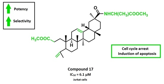
2.2.1. Antiproliferative Activity

Several cancer cell lines were used to evaluate the potential cytotoxicity of the synthesized compounds against human cancers. This evaluation was based on the determination of the concentration that inhibits cell proliferation at 50% (IC50), using 3-(4,5-dimethylthiazol-2-yl)-3,5-diphenyltetrazolium bromide (MTT) or 2,3-bis(2-methoxy-4-nitro- 5-sulphophenyl)-2H-tetrazolium-5-carboxanilide (XTT) assays, after 72 h of treatment with the compounds.
Compounds 1, 57 and 1021 were screened for their antiproliferative activity on A549 (lung adenocarcinoma) and HT-29 (colon adenocarcinoma) cell lines (Table 1). Compounds 24, 8 and 9 were not evaluated because they have already been tested with no improvements in potency and/or selectivity [37,39,46,47]. Intermediates 5 and 10, afforded by the cleavage of the ring A, were more potent than the parental compound GA 1. Removal of the keto group from ring C, that provided compound 11, resulted in an increment of cytotoxicity. Acyl fluorides 12 and 13 were less potent compared to their substrates. Analysis of the IC50 values of the derivatives 6, 7 and 1417 showed that the conjugation of an amino acid methyl ester provided more potent compounds. Deprotection of the carboxyl groups resulted in a loss of cytotoxicity. Compounds 1821, afforded by the alkaline hydrolysis, were further tested in Jurkat (acute T-cell leukemia) and MOLT-4 (acute lymphoblastic leukemia) cell lines (Table 2). The results of these assays confirmed that the deprotection provided less active compounds. Derivatives 6, 7 and 1417 and the parental compound GA 1 were also screened for their antiproliferative activity in seven additional human cancer cell lines: Jurkat, MOLT-4, MIAPaca 2 (pancreas adenocarcinoma), MCF7 (breast adenocarcinoma), HeLa (cervix adenocarcinoma), A375 (melanoma) and HepG2 (hepatocellular carcinoma). Comparing IC50 values of compounds 6 and 7 with those obtained for compounds 14 and 16, no significant differences were found regarding the position in which the amino acid methyl ester was introduced. Derivatives 15 and 17 were respectively more potent than compounds 14 and 16 in all tested cell lines, which confirmed that the removal of the keto group from ring C enhanced the cytotoxicity. Compounds 7, 16 and 17, which have an alanine methyl ester chain, were more active than compounds 6, 14 and 15, with a glycine methyl ester chain, respectively. These results suggest that the type of amino acid moiety introduced influences the antiproliferative activity. Within the newly synthesized derivatives, compound 17, with a reduced ring C and with an alanine methyl ester chain, was the most potent derivative. This compound was 5 to 17-fold more active than GA 1, depending on the cancer cell line.
The selectivity towards cancer cells was studied for GA 1 and compound 17 by incubating them with a human nontumoral cell line (BJ) (Table 2). GA 1 and compound 17 showed IC50 values that were 1.6 and more than 16.4 times lower on Jurkat cells than on the nontumoral BJ cells, respectively. Therefore, the novel derivative 17 was up to 10 times more selective towards malignant cells than its parental compound 1. This compound also showed a significant improvement in selectivity compared to the chemotherapy agent cisplatin. Considering also the Jurkat cell line, cisplatin presented an IC50 value that was 5.3 times lower than on BJ cells (Table 2); therefore, compound 17 was up to 3 times more selective than cisplatin towards Jurkat cells.

2.2.2. Analysis of Cell Cycle Distribution and Apoptosis

The Jurkat cell line which was the most susceptible to these derivatives was selected to investigate the mechanism of action of compound 17. To evaluate the effects on the cell cycle distribution, Jurkat cells were treated with compound 17, at a concentration corresponding to its IC50 value at 72 h of treatment, for 24, 48 and 72 h and then analyzed by flow cytometry. The calculation of the fraction of cells in G0/G1, S and G2/M phases was performed using the fraction of live cells. Treatment for 24 h induced significant increase in the population at S phase with respect to untreated cells (Figure 1); after 48 h this effect has decreased and after 72 h it was no longer observed. DNA fragmentation was detected after 72 h of incubation based on the appearance of a sub-G0 peak. This sequence of events suggests that the cell cycle arrest at S phase may have led cells to undergo apoptosis.
Apoptosis assays were then performed to better elucidate the mechanism of cell death involved in the cytotoxic effect of compound 17. The Annexin V-FITC/PI flow cytometric assay employs the property of fluorescein isothiocyanate (FITC) conjugated to Annexin V (Annexin V-FITC) to bind to phosphatidylserine (PS) and the property of propidium iodide (PI) to enter cells with damaged cell membranes and to bind to DNA. Early apoptosis is characterized by the loss of membrane asymmetry, with translocation of PS from the inner to the outer membrane, prior to the loss of membrane integrity. Therefore, this assay allows the discrimination of live cells (Annexin-V/ PI) from early apoptotic (Annexin-V+/PI), late apoptotic (Annexin-V+/PI+) or necrotic cells (Annexin-V+/PI+). The experiments were conducted on Jurkat cells treated with compound 17 at a concentration corresponding to its IC50 value at 72 h of treatment (6.1 μM) for 24 and 48 h, and at concentrations of 6.1 μM and 12.2 μM for 72 h. Exposure to this compound for 24 and 48 h did not change significantly the apoptotic (Figure 2A) and necrotic (data not shown) populations. Treatment for 72 h with compound 17 at concentrations of 6.1 μM and 12.2 μM increased the early apoptotic population by 19% and 30%, respectively. No significant changes were observed in the late apoptotic population. These results were in good agreement with those obtained in the cell cycle experiments.
The induction of apoptosis was further confirmed by the observation of its characteristic morphological changes. Hoechst 33342 staining showed volume reduction, chromatin condensation and apoptotic bodies in Jurkat cells treated in the same conditions for 72 h (Figure 2B). In contrast, untreated cells presented a normal morphological profile.
Taken together, the results described above suggest that compound 17 inhibits cell growth through cell cycle arrest at the S phase and induction of apoptosis. Its mechanism of action needs to be studied further.

3. Materials and Methods

3.1. Chemistry

Glycyrrhetinic acid and all reagents were purchased from Sigma-Aldrich Co. (Saint Louis, MO, USA) The solvents used in the reactions were obtained from Merck Co. (Kenilworth, NJ, USA) and were purified and dried according to the literature procedures. The solvents used in workups were purchased from VWR Portugal. Thin layer chromatography (TLC) analysis was performed in Kieselgel 60HF254/Kieselgel 60G. Purification of compounds by flash column chromatography (FCC) was carried out using Kiesegel 60 (230–400 mesh, Merck). Melting points were determined using a BUCHI melting point B-540 apparatus and were uncorrected. 1H and 13C NMR spectra were recorded on a Bruker Avance-400 Digital NMR spectrometer, in CDCl3, with Me4Si as the internal standard (see Supplementary Materials). Chemical shifts values (δ) are given in parts per million (ppm) and coupling constants (J) are presented in hertz (Hz). Mass spectra were obtained using a Quadrupole/Ion Trap Mass Spectrometer (QIT-MS) (LCQ Advantage MAX, THERMO FINNINGAN). Elemental analysis was performed in an Analyzer Elemental Carlo Erba 1108 by chromatographic combustion.
Methyl 3β-hydroxy-11-oxo-olean-12-en-30-oate(2): Compound 2 was prepared according to the literature [39], from 1 to give a colorless solid (90%). m.p.: 254–256 °C.
Methyl 3,11-dioxo-olean-12-en-30-oate(3): Preparation of 3 was performed according to a previously described method [41], from 2 providing a white solid (94%). m.p.: 248–250 °C.
Methyl 3,11-dioxo-4-oxa-A-homo-olean-12-en-30-oate(4): Compound 4 was prepared according to the literature [42], from 3 to give a white solid (77%). m.p.: 168–170 °C.
3,4-seco-30-methyloxicarbonyl-11-oxo-olean-4(23),12-dien-3-oic acid(5): Preparation of 5 was performed according to a previously described method [42], from 4 providing a white solid (72%). m.p.: 90–92 °C.
Methyl 3,4-seco-3-N-methylglycinamido-11-oxo-olean-4(23),12-dien-30-oate(6): To a solution of compound 5 (300 mg, 0.60 mmol) in dichloromethane (8 mL), Deoxo-Fluor (50% in THF, 0.52 mL, 1.20 mmol) was added and the reaction mixture was stirred, at room temperature, for 2.5 h, after which additional Deoxo-Fluor (50% in THF, 0.26 mL, 0.60 mmol) was added. After 2 h, the reaction was completed. The reaction mixture was quenched by addition of water (2 mL). The organic layer was diluted with chloroform (40 mL), washed with water (2 × 30 mL), dried over Na2SO4, filtered and evaporated to the dryness (280 mg, 93%). The residue was dissolved in dichloromethane (6 mL) and glycine methyl ester hydrochloride (105 mg, 0.84 mmol) and triethylamine (0.15 mL, 1.12 mmol) were added. After 1 h under magnetic stirring at room temperature, the reaction was completed. The reaction mixture was evaporated to dryness and ethyl acetate (40 mL) and water (30 mL) were added to the residue. The aqueous phase was further extracted with ethyl acetate (2 × 40 mL). The combined organic phase was washed with 5% aqueous HCl (2 × 30 mL), 10% aqueo+us NaHCO3 (2 × 30 mL), water (30 mL) and brine (30 mL), dried over Na2SO4, filtered and the solvent was removed under reduced pressure to afford a white solid. The solid was purified by flash column chromatography (FCC) with petroleum ether/ethyl acetate (2:1) to afford compound 6 as a white solid (69%). m.p.: 199–201 °C. 1H NMR (400 MHz, CDCl3): δ 6.14 (1H, m, NH), 5.69 (1H, s, H-12), 4.90 (1H, br s, H-23), 4.73 (1H, br s, H-23), 3.93-4.05 (2H, m, NCH2), 3.74 (3H, s, COOCH3), 3.69 (3H, s, COOCH3), 1.76 (3H, s), 1.38 (3H, s), 1.16 (3H, s), 1.15 (6H, s), 0.81 (3H, s). 13C NMR (100 MHz, CDCl3): δ 200.3 (C11), 177.1, 173.7, 170.6, 170.3, 146.7, 128.6 (C12), 114.5 (C23), 53.3, 52.4, 51.9, 51.3, 48.5, 45.2, 44.2, 43.8, 41.4, 41.3, 39.1, 37.9, 35.7, 32.0, 31.8, 31.5, 31.3, 28.7, 28.4, 26.7, 26.6, 24.0, 23.6, 23.5, 19.7, 18.8. ESI-MS m/z: 570.34 ([M + H] +, 100%). Found C 70.78, H 9.39, N 2.41, calcd for C34H51NO6·0.25H2O: C 71.11, H 9.04, N 2.44%.
Methyl 3,4-seco-3-N-methyalaninamido-11-oxo-olean-4(23),12-dien-30-oate(7): The method followed followed that described for compound 6. The resulting solid (274 mg, 0.55 mL; 91%) from the first step was dissolved in dichloromethane (6 mL) and alanine methyl ester hydrochloride (116 mg, 0.83 mmol) and triethylamine (0.15 mL, 1.10 mmol) were added. After 1 h, the reaction was completed. The workup was performed as described for compound 6. The resulting solid was subjected to FCC with petroleum ether/ethyl acetate (2:1) to afford compound 7 as a white solid (61%). m.p.: 193–195 °C. 1H NMR (400 MHz, CDCl3): δ 6.09 (1H, m, NH), 5.70 (1H, s, H-12), 4.91 (1H, br s, H-23), 4.74 (1H, br s, H-23), 4.55 (1H, m, -NCH(CH3)-), 3.73 (3H, s, COOCH3), 3.69 (3H, s, COOCH3), 1.77 (3H, s), 1.38 (3H, s), 1.37 (3H, m, -NCH(CH3)-), 1.17 (3H, s), 1.15 (3H, s), 1.15 (3H,s), 0.82 (3H, s). 13C NMR (100 MHz, CDCl3): δ 200.3 (C11), 177.1, 173.8, 173.0, 170.2, 146.7, 128.6 (C12), 114.5 (C23), 53.3, 52.5, 51.9, 51.1, 48.5, 48.1, 45.2, 44.2, 43.9, 41.4, 39.1, 37.9, 35.8, 32.0, 31.9, 31.5, 31.3, 28.8, 28.4, 26.7, 26.6, 24.0, 23.7, 23.5, 19.7, 18.8, 18.5. ESI-MS m/z: 584.29 ([M + H] +, 100%). Found C 71.40, H 9.31, N 2.32, calcd for C35H53NO6·0.25H2O: C 71.46, H 9.17, N 2.38 %.
3,11-Dioxo-olean-12-en-30-oic acid(8): Compound 8 was prepared according to the literature [41], from 1 to give a colorless solid (92%). m.p.: 308–310 °C.
3,11-Dioxo-4-oxa-A-homo-olean-12-en-30-oic acid(9): Compound 9 was prepared from 8, using the same method as for the preparation of 4, with the obtention of a white solid. (75%). m.p.: 268–270 °C.
3,4-seco-3-methyloxicarbonyl-11-oxo-olean-4(23),12-dien-30-oic acid(10): Preparation of 10 was performed according to a previously described method [42], from 9 providing a colorless solid (62%). m.p.: 130–132 °C.
3,4-seco-3-methyloxicarbonyl-olean-4(23),12-dien-30-oic acid(11): Preparation of 11 was done according to a previously described method [45]. Compound 10 (900 mg, 1.80 mmol) was dissolved in dioxane (25 mL) and Zn powder (941 mg, 14.40 mmol) was added. Concentrated HCl (37%, 3.6 mL, 43.20 mmol) was added dropwise for 30 min with stirring. After 4.5 h under magnetic stirring at room temperature, the reaction was completed. The reaction mixture was filtered and the solvent was removed under pressure. Diethyl ether (75 mL) and water (60 mL) were added to the residue. The aqueous phase was further extracted with diethyl ether (2 × 70 mL). The combined organic extract was then washed with 5% aqueous HCl (2 × 50 mL), 10% aqueous NaHCO3 (2 × 50 mL), water (50 mL) and brine (50 mL), dried over Na2SO4, filtered and evaporated to the dryness. The resulting solid was purified by (FCC) with petroleum ether/ ethyl acetate (1:1) to afford compound 11 as a white solid (75%). m.p.: 138–140 °C. 1H NMR (400 MHz, CDCl3): δ 5.32 (1H, m, H-12), 4.87 (1H, br, s, H-23), 4.67 (1H, br, H-23), 3.65 (3H, s, COOCH3), 1.75 (3H, s), 1.21 (3H, s), 1.16 (3H, s), 1.02 (3H, s), 0.94 (3H, s), 0.82 (3H, s). 13C NMR (100 MHz, CDCl3): δ 183.1 (C30), 174.8 (C3), 147.6, 144.3, 122.8 (C12), 113.7 (C23), 51.8, 50.6, 48.2, 44.2, 42.7, 42.2, 39.7, 39.3, 38.4, 38.0, 34.1, 32.2, 31.5, 31.2, 28.8, 28.6, 28.3, 27.1, 26.2, 26.0, 24.7, 23.8, 23.6, 19.7, 17.0. ESI-MS m/z: 118.11 (19%), 274.50 (32), 318.46 (75), 346.48 (11), 362.45 (15), 485.38 ([M + H] +, 100%).
Methyl 3,4-seco-30-fluorcarbonyl-11-oxo-olean-4(23),12-dien-3-oate(12): To a solution of compound 10 (500 mg, 1.00 mmol) in dichloromethane (10 mL), Deoxo-Fluor (50% in THF, 0.87 mL, 2.00 mmol) was added. After 1.5 h under magnetic stirring at room temperature, the reaction was completed. The reaction mixture was quenched by addition of water (3 mL). The organic layer was diluted with chloroform (50 mL), washed with water (2 × 40 mL), dried over Na2SO4, filtered and evaporated to the dryness. The resulting solid was subjected to (FCC) [petroleum ether/ethyl acetate from (6:1) to (4:1)] to afford compound 12 as a white solid (75%). m.p.: 183–185 °C. 1H NMR (400 MHz, CDCl3): δ 5.71 (1H, s, H-12), 4.90 (1H, br s, H-23), 4.69 (1H, br s, H-23), 3.62 (3H, s, COOCH3), 1.75 (3H, s), 1.38 (3H, s), 1.30 (3H, s), 1.17 (3H, s), 1.16 (3H, s), 0.86 (3H, s). 13C NMR (100 MHz, CDCl3): δ 199.5 (C11), 174.5 (C3), 168.1, 166.3 (J = 374.6 Hz, C30), 146.7, 129.1 (C12), 114.4 (C23), 53.0, 51.7, 51.0, 48.1, 45.3, 44.4 (J = 42.0 Hz), 43.8, 40.8, 38.9, 37.6, 34.6, 32.1, 31.6, 30.9, 29.4, 28.5, 27.0, 26.6, 26.5, 23.9, 23.6, 23.5, 19.7, 18.8. ESI-MS m/z: 501.16 ([M + H] +, 100%).
Methyl 3,4-seco-30-fluorcarbonyl-olean-4(23),12-dien-3-oate(13): The method followed that described for compound 12 but using compound 11 (600 mg, 1.24 mmol) and Deoxo-Fluor (50% in THF, 1.08 mL, 2.48 mmol) in dichloromethane (12 mL) for 2 h. The resulting solid was purified by FCC [petroleum ether /ethyl acetate from (10:1) to (4:1)] to afford compound 13 as a white solid (61%). m.p.: 174–176 °C. 1H NMR (400 MHz, CDCl3): δ 5.32 (1H, m, H-12), 4.87 (1H, br s, H-23), 4.67 (1H, br s, H-23), 3.65 (3H, s, COOCH3), 1.75 (3H, s), 1.27 (3H, s), 1.15 (3H, s), 1.02 (3H, s), 0.94 (3H, s), 0.83 (3H, s). 13C NMR (100 MHz, CDCl3): δ 174.7 (C3), 167.0 (J = 375.0 Hz, C30), 147.5, 143.6, 123.4 (C12), 113.7 (C23), 51.7, 50.6, 48.3, 44.6 (J = 41.2 Hz), 42.4, 42.2, 39.7, 39.3, 38.1, 38.0, 34.1, 32.2, 31.5, 31.1, 28.6, 28.1, 27.2, 27.0, 26.1, 26.0, 24.6, 23.8, 23.6, 19.7, 17.0. ESI-MS m/z: 439.51 (38%), 440.52 (14), 485.44 (27), 487.36 ([M + H] +, 100%).
Methyl 3,4-seco-30-N-methylglycinamido-11-oxo-olean-4(23),12-dien-3-oate(14): To a solution of compound 12 (300 mg, 0.60 mmol) and glycine methyl ester hydrochloride (226 mg, 1.80 mmol) in dichloromethane (8 mL), triethylamine (0.33 mL, 2.40 mmol) was added. After 13 h under magnetic stirring at room temperature, the reaction was completed. The reaction mixture was evaporated to dryness and ethyl acetate (50 mL) and water (40 mL) were added to the residue. The aqueous phase was further extracted with ethyl acetate (2 × 50 mL). The resulting organic phase was washed with 5% aqueous HCl (2 × 30 mL), 10% aqueous NaHCO3 (2 × 30 mL), water (30 mL) and brine (30 mL), dried over Na2SO4, filtered and the solvent was removed under reduced pressure to afford a solid. The solid was subjected to (FCC) [petroleum ether /ethyl acetate from (2:1) to (1:1)] to afford compound 14 as a white solid (77%). m.p.:126–128 °C. 1H NMR (400 MHz, CDCl3): δ 6.17 (1H, m, NH), 5.74 (1H, s, H-12), 4.88 (1H, br s, H-23), 4.68 (1H, br s, H-23), 3.95-4.14 (2H, m, NCH2), 3.76 (3H, s, COOCH3), 3.61 (3H, s, COOCH3), 1.75 (3H, s), 1.39 (3H, s), 1.16 (6H, s), 1.15 (3H, s), 0.83 (3H, s). 13C NMR (100 MHz, CDCl3): δ 199.7 (C11), 176.3, 174.5, 170.7, 169.5, 146.7, 128.6 (C12), 114.3 (C23), 52.9, 52.5, 51.7, 51.0, 47.9, 45.2, 43.8 (2), 42.1, 41.3, 38.9, 37.4, 34.6, 32.0, 31.6 (2), 29.5, 29.4, 28.6, 26.7, 26.5, 23.9, 23.6, 23.4, 19.6, 18.8. ESI-MS m/z: 570.32 ([M + H] +, 100%). Found C 70.83, H 9.27, N 2.49, calcd for C34H51NO6·0.25H2O: C 71.11, H 9.04, N 2.44%.
Methyl 3,4-seco-30-N-methylglycinamido-olean-4(23),12-dien-3-oate(15): The method followed that described for compound 14 but using compound 13 (290 mg, 0.60 mmol), glycine methyl ester hydrochloride (226 mg, 1.80 mmol) and triethylamine (0.33 mL, 2.40 mmol) in dichloromethane (8 mL). The workup was performed after 16 h. The resulting solid was purified by FCC with petroleum ether/ ethyl acetate (3:2) to afford compound 15 as a white solid (43%). m.p.: 190–192 °C. 1H NMR (400 MHz, CDCl3): δ 6.18 (1H, m, NH), 5.34 (1H, m, H-12), 4.87 (1H, br s, H-23), 4.67 (1H, br s, H-23), 3.97-4.17 (2H, m, NCH2), 3.77 (3H, s, COOCH3), 3.65 (3H, s, COOCH3), 1.75 (3H, s), 1.17 (3H, s), 1.13 (3H, s), 1.01 (3H, s), 0.93 (3H, s), 0.79 (3H, s). 13C NMR (100 MHz, CDCl3): δ 177.0, 174.7, 171.0, 147.6, 144.4, 122.8 (C12), 113.7 (C23), 52.5, 51.7, 50.6, 48.1, 44.1, 43.6, 42.2, 41.4, 39.7, 39.3, 38.0 (2),34.2, 32.2, 31.7, 31.5, 29.8, 28.6, 28.3, 27.0, 26.2, 25.9, 24.6, 23.9, 23.6, 19.7, 17.0. ESI-MS m/z: 274.37 (25%), 318.32 (27), 556.24 ([M + H] +, 100%). Found C 73.18, H 9.83, N 2.55, calcd for C34H53NO5: C 73.47, H 9.61, N 2.52 %.
Methyl 3,4-seco-30-N-methylalaninamido-11-oxo-olean-4(23),12-dien-3-oate(16): To a solution of compound 12 (300 mg, 0.60 mmol) and alanine methyl ester hydrochloride (251 mg, 1.80 mmol) in dichloromethane (8 mL), triethylamine (0.33 mL, 2.40 mmol) was added and the reaction mixture was stirred, at room temperature, for 8 h, after which additional dichloromethane (1 mL) and triethylamine (0.17 mL, 1.20 mmol) were added. After 16 h, the reaction was completed. The workup was performed as described for compound 14. The solid was subjected to FCC with petroleum ether/ethyl acetate (1:2) to afford compound 16 as a white solid (83%). m.p.: 130–132 °C. 1H NMR (400 MHz, CDCl3): δ 6.15 (1H, m, NH), 5.77 (1H, s, H-12), 4.89 (1H, br s, H-23), 4.69 (1H, br s, H-23), 4.62 (1H, m, -NCH(CH3)-), 3.76 (3H, s, COOCH3), 3.61(3H, s, COOCH3), 1.75 (3H, s), 1.39 (6H, m), 1.17 (3H, s), 1.16 (3H, s), 1.14 (3H, s), 0.82 (3H, s). 13C NMR (100 MHz, CDCl3): δ 199.7 (C11), 175.4, 174.5, 173.7, 169.5, 146.7, 128.6 (C12), 114.3 (C23), 53.0, 52.7, 51.7, 51.0, 47.9 (2), 45.2, 43.8, 43.7, 42.1, 38.9, 37.5, 34.6, 32.0, 31.6, 31.5, 29.5, 29.4, 28.6, 26.7, 26.5, 24.0, 23.6, 23.4, 19.7, 18.8, 18.6. ESI-MS m/z: 570.36 (17%), 584.36 ([M + H] +, 100%). Found C 71.09, H 9.51, N 2.35, calcd for C35H53NO6·0.25H2O: C 71.46, H 9.17, N 2.38%.
Methyl 3,4-seco-30-N-methylalaninamido-olean-4(23),12-dien-3-oate(17): To a solution of compound 13 (290 mg, 0.60 mmol) and alanine methyl ester hydrochloride (251 mg, 1.80 mmol) in dichloromethane (8 mL), triethylamine (0.33 mL, 2.40 mmol) was added and the reaction mixture was stirred, at room temperature, for 10 h, after which additional dichloromethane (2 mL) and triethylamine (0.17 mL, 1.20 mmol) and alanine methyl ester hydrochloride (84 mg, 0.60 mmol) were added. After 24 h, the reaction was completed. The work-up was performed as described for compound 15. The solid was purified by FCC with petroleum ether/ ethyl acetate (3:2) to afford compound 17 as a white solid (48%). m.p.: 100–102 °C. 1H NMR (400 MHz, CDCl3): δ 6.22 (1H, m, NH), 5.37 (1H, m, H-12), 4.87 (1H, br s, H-23), 4.67 (1H, br s, H-23), 4.63 (1H, m, -NCH(CH3)-), 3.76 (3H, s, COOCH3), 3.65 (3H, s, COOCH3), 1.75 (3H, s), 1.40 (3H, m, -NCH(CH3)-), 1.17 (3H, s), 1.11 (3H, s), 1.01 (3H, s), 0.94 (3H, s), 0.79 (3H, s). 13C NMR (100 MHz, CDCl3): δ 176.2, 174.7, 173.9, 147.6, 144.5, 122.7 (C12), 113.7 (C23), 52.6, 51.7, 50.6, 48.0, 47.9, 44.0, 43.6, 42.2, 39.7, 39.3, 38.1, 38.0, 34.2, 32.2, 31.6, 31.5, 29.8, 28.6, 28.3, 27.0, 26.2, 25.9, 24.6, 23.9, 23.6, 19.7, 18.7, 17.0. ESI-MS m/z: 570.33 ([M + H] +, 100%). Found C 73.04, H 9.94, N 2.45, calcd for C35H55NO5·0.25H2O: C 73.20, H 9.74, N 2.44%.
Methyl 3,4-seco-3-N-glycinamido-11-oxo-olean-4(23),12-dien-30-oate(18): To a solution of compound 6 (120 mg, 0.21 mmol) in methanol (1 mL) and tetrahydrofuran (THF) (1.5 mL), KOH 4N (0.53 ml, 2.10 mmol) was added. After 10 min under magnetic stirring at room temperature, the reaction was completed. The pH of the reaction mixture was neutralized with 10% aqueous HCl. Dichloromethane (30 mL) and water (20 mL) were added to the mixture. The aqueous phase was further extracted with dichloromethane (2 × 30 mL). The combined organic phase was washed with water (2 × 30 mL) and brine (30 mL), dried over Na2SO4, filtered and the solvent was removed under reduced pressure to afford compound 18 as a white solid (98%). m.p.: 223–225 °C. 1H NMR (400 MHz, CDCl3): δ 6.76 (1H, m, NH), 5.70 (1H, s, H-12), 4.93 (1H, br s, H-23), 4.78 (1H, br s, H-23), 3.88-4.25 (2H, m, NCH2), 3.70 (3H, s, COOCH3), 1.80 (3H, s), 1.36 (3H, s), 1.17 (3H, s), 1.15 (3H, s), 1.14 (3H, s), 0.81 (3H, s). 13C NMR (100 MHz, CDCl3): δ 202.2 (C11), 177.0, 174.5, 173.3, 172.3, 146.5, 127.7 (C12), 114.8 (C23), 53.3, 52.0, 50.8, 48.6, 45.4, 44.2, 44.1, 41.8, 41.4, 39.2, 37.8, 36.1, 32.0, 31.9, 31.5, 31.3, 28.8, 28.3, 26.7, 26.5, 23.9, 23.8, 23.4, 19.7, 18.7. ESI-MS m/z: 556.26 ([M + H] +, 100%).
Methyl 3,4-seco-3-N-alaninamido-11-oxo-olean-4(23),12-dien-30-oate(19): Compound 19 was prepared using the same method as for the preparation of 18, but using compound 7 (116 mg, 0.20 mmol), methanol (1 mL), THF (1.5 mL) and KOH 4N (0.50 mL, 2.00 mmol), at room temperature for 10 min, to afford a white solid (94%). m.p.: 216–218 °C. 1H NMR (400 MHz, CDCl3): δ 6.89 (1H, m, NH), 5.69 (1H, s, H-12), 4.94 (1H, br s, H-23), 4.78 (1H, br s, H-23), 4.61 (1H, m, -NH(CH3)-), 3.69 (3H, s, COOCH3), 1.80 (3H, s), 1.42 (3H, m, -NH(CH3)-), 1.35 (3H, s), 1.16 (3H, s), 1.14 (6H, s), 0.81 (3H, s). 13C NMR (100 MHz, CDCl3): δ 202.0 (C11), 176.8, 175.3, 174.0, 173.1, 146.3, 127.6 (C12), 114.6 (C23), 53.2, 51.9, 50.6, 48.5, 48.0, 45.3, 44.0 (2), 41.2, 39.0, 37.7, 36.1, 31.9, 31.8, 31.3, 31.1, 28.6, 28.1, 26.5, 26.4, 23.8 (2), 23.2, 19.5, 18.7, 18.6. ESI-MS m/z: 570.26 ([M + H] +, 100%).
3,4-seco-30-N-glycinamido-11-oxo-olean-4(23),12-dien-3-oic acid(20): The method followed that of compound 18, using compound 14 (145 mg, 0.26 mmol), methanol (1 mL), THF (1.5 mL) and KOH 4N (0.65 mL, 2.60 mmol), at room temperature for 10 min, to afford compound 20 as a white solid (97%). m.p.: 213–215 °C. 1H NMR (400 MHz, CDCl3): δ 7.58 (1H, m, NH), 5.77 (1H, s, H-12), 4.88 (1H, br s, H-23), 4.62 (1H, br s, H-23), 3.50-4.48 (2H, m, NCH2), 1.72 (3H, s), 1.42 (3H, s), 1.21 (3H, s), 1.16 (3H, s), 1.09 (3H, s), 0.83 (3H, s). 13C NMR (100 MHz, CDCl3): δ 202.1 (C11), 181.0, 177.3, 173.1, 172.7, 146.5, 128.0 (C12), 114.5 (C23), 52.5, 50.6, 47.5, 45.7, 44.2, 44.0, 42.1, 41.1, 39.5, 37.5, 34.5, 31.8, 31.6 (2), 30.2, 29.1, 28.7, 26.8 (2), 23.7, 23.4, 22.9, 19.8, 18.8. ESI-MS m/z: 542.26 ([M + H] +, 100%).
3,4-seco-30-N-alaninamido-11-oxo-olean-4(23),12-dien-3-oic acid(21): Compound 21 was prepared using the same method as for the preparation of 18, but using compound 16 (170 mg, 0.29 mmol), methanol (1 mL), THF (1.5 mL) and KOH 4N (0.73 mL, 2.90 mmol), at room temperature for 10 min, to afford a white solid (98%). m.p.: 224–226 °C. 1H NMR (400 MHz, CDCl3): δ 7.37 (1H, m, NH), 5.74 (1H, s, H-12), 4.88 (1H, br s, H-23), 4.78 (1H, m, -NH(CH3)-), 4.68 (1H, br s, H-23), 1.74 (3H, s), 1.15-1.35 (15H, m), 0.82 (3H, s). 13C NMR (100 MHz, CDCl3): δ 201.1 (C11), 179.8, 176.9, 176.3, 171.8, 146.6, 128.1 (C12), 114.5 (C23), 52.7, 50.7, 47.8, 47.7, 45.7, 44.0, 43.8, 41.9, 39.2, 37.6, 34.1, 31.9, 31.6 (2), 29.6, 29.1, 28.7, 26.8, 26.7, 23.8, 23.5, 23.2, 19.8, 18.8, 17.6. ESI-MS m/z: 272.01 (10%), 527.35 (12), 556.25 ([M + H] +, 100%).

3.2. Biology

A549, HT-29, Jurkat, MOLT-4, MIA Paca 2, MCF7, HeLa, A375, HepG2, and BJ cells were obtained from the American Type Culture Collection (ATCC, Rockville, MD, USA). Dulbecco´s Modified Eagle Medium (DMEM), Roswell Park Memorial Institute (RPMI)-1640 medium, Phosphate Buffered Saline (PBS), glucose 45%, human insulin 10 mg/mL, dimethyl sulfoxide (DMSO), 3-(4,5-dimethylthiazol-2-yl)-2,5-diphenyl tetrazolium bromide (MTT) powder, Trypan Blue (TB) 0.4%, propidium iodide (PI) and Hoescht 33342 were purchased from Sigma Aldrich Co. (St Louis, MO, USA). Minimum Essential Medium (MEM), penicillin/streptomycin (P/S) and l-glutamine were purchased from Gibco-BRL (Eggenstein, Germany). Sodium pyruvate, trypsin/EDTA (0.05%/0.02%) and MEM-Eagle Non-Essential Aminoacids 100× were obtained from Biological Industries (Kibbutz Beit Haemek, Israel). Fetal Bovine Serum (FBS) was obtained from PAA Laboratories (Pasching, Austria), the Cell Proliferation Kit II (XTT kit) was purchased from Roche (Roche Molecular Biochemicals, Indianapolis, IN, USA) and annexin V-FITC was obtained from Bender MedSystems (Vienna, Austria).
Stock solutions of 20 mM in DMSO of the synthesized compounds were prepared and stored at −20 °C. Working solutions were prepared in culture medium and appropriate amounts of DMSO were included in controls; all solutions had a final concentration of 0.5% DMSO.

3.2.1. Cell Culture

A549, HT-29, MIA Paca 2, HeLa and A375 cells were cultured in DMEM supplemented with 10% heat-inactivated FBS and 1% P/S. HepG2 and BJ cells were grown in DMEM supplemented with 10% heat-inactivated FBS, 1% P/S and 1mM sodium pyruvate. Jurkat and MOLT-4 cells were cultured in RPMI-1640 medium supplemented with 10% heat-inactivated FBS, 1% P/S and 2 mM l-glutamine. MCF7 cells were maintained in MEM supplemented with 10% heat-inactivated FBS, 0.1% P/S, 1mM sodium pyruvate, 2 mM l-glutamine, 1× MEM-Eagle Non-Essential Aminoacids, 0.01 mg/mL insulin human and 10 mM glucose.
All cell cultures were performed at 37 °C in an atmosphere of 5% CO2.

3.2.2. Antiproliferative Activity Assays

The antiproliferative activity of the synthesized compounds on A549, HT-29, MIA Paca 2, MCF7, HeLa, A375, HepG2 and BJ adherent cells was determined by the MTT assay. Exponentially growing cells were plated in 96-well plates at a density of 1–8 × 10³ cells/ well. After 24 h, cells were attached to the plate, and the growth medium was replaced with fresh medium containing either the compounds dissolved in DMSO at different concentrations or only DMSO, in triplicate, and the cells were continued to culture for 72 h. After incubation with the compounds, the medium was removed and 100 µL of MTT solution (0.5 mg/mL) were added to each well and the plates were incubated for 1 h. MTT was removed and 100 µL of DMSO was added to dissolve the formazan crystals. The absorbance was immediately read at 550 nm on an ELISA read plater (Tecan Sunrise MR20-301, TECAN, Austria). For Jurkat and MOLT-4 non-adherent cells, the antiproliferative activity was determined by XTT assay. These cell lines were plated with 5.5 × 10³ and 1 × 10⁴ cells/well, respectively, in 96-well plates in 100 µL medium. The seeding was executed simultaneously with the addition of the different concentrations of compounds or vehicle, in triplicate, and cells were allowed to incubate for 72 h. After that incubation period, 100 μL of the XTT labelling mixture were added to each well and the plates were incubated again for 4 h. Then, the absorbance was read at 450 nm on the ELISA plate reader.
Concentrations that inhibit cell proliferation by 50% (IC50) represent an average of a minimum of three independent experiments and were expressed as means ± standard deviation (SD).

3.2.3. Cell Cycle Analysis

Cell cycle was assessed by flow cytometry using a fluorescence-activated cell sorter (FACS). Jurkat cells were plated in six-well plates at a density of 1.6 × 10⁵ cells/well, simultaneously with the addition of compound 17, at a concentration corresponding to its IC50 value at 72 h of treatment, or with only the vehicle, in a total volume of 2 mL of medium. The cells were allowed to incubate for 24, 48 and 72 h. After incubation, cells were collected and centrifuged. The supernatant was removed and the pellet was resuspended in 1 mL of TBS containing 1 mg/mL PI, 10 mg/mL RNase free of DNase and 0.1% Igepal CA-630, for 1 h, at 4 °C. FACS analysis was performed at 488 nm in an Epics XL flow cytometer (Coulter Corporation, Hialeah, FL, USA). Data were collected and analyzed using the Multicycle software (Phoenix Flow Systems, San Diego, CA, USA). Three independent experiments were performed, with two replicates per experiment.

3.2.4. Annexin V-FITC/PI Flow Cytometry Assay

Apoptosis was assessed by flow cytometry using a FACS. Jurkat cells were plated in six-well plates at a density of 1.6 × 10⁵ cells/well, simultaneously with the addition of compound 17, at specified concentrations, or with only the vehicle, in a total volume of 2 mL of medium. The cells were allowed to incubate for 24, 48 and 72 h. After incubation, cells were collected and centrifuged. The supernatant was removed and the pellet was resuspended in 95 µL of binding buffer (10 mM HEPES/NaOH, pH 7.4, 140 mM NaCl, 2.5 mM CaCl2). Annexin V-FITC conjugate (3 µL) was added and cells were incubated for 30 min, at room temperature, in darkness. After incubation, 0.8 mL of binding buffer were added. Just before the FACS analysis, cells were stained with 20 µL of 1 mg/mL PI solution. Three independent experiments were performed, with two replicates per experiment.

3.2.5. Hoechst 33342 Staining

The morphological changes were observed by fluorescence microscopy using Hoechst staining. Jurkat cells were plated in six-well plates at a density of 1.6 × 10⁵ cells/well, simultaneously with the addition of compound 17, at specified concentrations, or with only the vehicle, in a total volume of 2 mL of medium. The cells were incubated for 72 h. After incubation, cells were collected by centrifugation, washed twice with PBS and stained with 500 µL of Hoechst 33342 solution (2 µg/ml in PBS), for 15 min, at room temperature, in darkness. Finally, cells were washed and resuspended in 10 µL PBS. The samples were mounted on a slide and observed with a fluorescence microscope (DMRB, Leica Microssystems, Wetzlar, Germany) with a 4′,6-diamidine-2′-phenylindole dihydrochloride (DAPI) filter. Three independent experiments were conducted.

4. Conclusions

In summary, we synthesized a series of new GA derivatives via the opening of its ring A along with the coupling of an amino acid. Antiproliferative activity assays in a panel of nine human cancer cell lines showed that the most potent compound 17 was 5 to 17-fold more active than GA 1. The study of selectivity revealed that this new derivative was up to 10 times more selective towards malignant cells than its parental compound. Preliminary mechanism investigation indicated that compound 17 may act through arresting cell cycle progression at the S phase and inducing apoptosis. The enhanced potency and the high selectivity of this new GA derivative warrant further biological evaluation.

Supplementary Materials

1H-NMR and 13C-NMR spectra of selected compounds are available online.

Author Contributions

Conceptualization, J.A.R.S. and S.M.; Synthesis and Structural Characterization, D.P.S.A. and J.A.R.S.; Biological Experiments, D.P.S.A. and S.M.; Writing—Original Draft Preparation, D.P.S.A.; Writing—Review and Editing, J.A.R.S., M.C., and S.M.; Supervision, J.A.R.S. and S.M.; Project Administration, J.A.R.S. and M.C.; Funding Acquisition, J.A.R.S. and M.C.

Funding

Jorge A. R. Salvador thanks PT2020 (Programa Operacional do Centro 2020), project nº 3269, drugs2CAD, and the financial support by FEDER (European Regional Development Fund) through the COMPETE 2020 Programme (Operational Programme for Competitiveness and Internationalisation). Jorge A. R. Salvador also wishes to thank Universidade de Coimbra for financial support. Daniela P. S. Alho thanks FEDER (Programa Operacional Factores de Competitividade—COMPETE 2020) and Fundação para a Ciência e Tecnologia (FCT) through Projecto Estratégico: UID/NEU/04539/2013 and the financial support for the PhD grant SFRH/BD/66020/2009. Marta Cascante and Silvia Marin thank MINECO-European Commission FEDER—Una manera de hacer Europa (SAF2017-89673-R) and Agència de Gestió d’Ajuts Universitaris i de Recerca (AGAUR)—Generalitat de Catalunya (2017SGR1033). Marta Cascante also acknowledges the support received through the prize “ICREA Academia” for excellence in research, funded by ICREA Foundation—Generalitat de Catalunya. The authors acknowledge UC-NMR facility, which is supported by FEDER and FCT funds through the grants REEQ/481/QUI/2006, RECI/QEQ-FI/0168/2012, and CENTRO-07-CT62-FEDER-002012, and Rede Nacional de Ressonância Magnética Nuclear (RNRMN), for NMR data.

Acknowledgments

The authors are grateful to Laboratory of Mass Spectrometry (LEM) of the Node UC integrated in the National Mass Spectrometry Network (RNEM) of Portugal, for the MS analyses, and Centro de Apoio Científico e Tecnolóxico á Investigación (CACTI), Universidade de Vigo, for elemental analysis. The authors acknowledge Centres Científics I Tecnològics de la UB (CCiTUB) for flow cytometry analysis support.

Conflicts of Interest

The authors declare no conflicts of interest.

References

  1. Bray, F.; Ferlay, J.; Soerjomataram, I.; Siegel, R.L.; Torre, L.A.; Jemal, A. Global cancer statistics 2018: Globocan estimates of incidence and mortality worldwide for 36 cancers in 185 countries. CA-Cancer J. Clin. 2018, 68, 394–424. [Google Scholar] [CrossRef] [PubMed]
  2. Cragg, G.M.; Newman, D.J. Plants as a source of anti-cancer agents. J. Ethnopharmacol. 2005, 100, 72–79. [Google Scholar] [CrossRef] [PubMed] [Green Version]
  3. Newman, D.J.; Cragg, G.M. Natural products as sources of new drugs from 1981 to 2014. J. Nat. Prod. 2016, 79, 629–661. [Google Scholar] [CrossRef] [PubMed]
  4. Salvador, J.A.R.; Moreira, V.M.; Goncalves, B.M.F.; Leal, A.S.; Jing, Y.K. Ursane-type pentacyclic triterpenoids as useful platforms to discover anticancer drugs. Nat. Prod. Rep. 2012, 29, 1463–1479. [Google Scholar] [CrossRef] [PubMed]
  5. Salvador, J.A.R.; Leal, A.S.; Alho, D.P.S.; Goncalves, B.M.F.; Valdeira, A.S.; Mendes, V.I.S.; Jing, Y.K. Highlights of pentacyclic triterpenoids in the cancer settings. In Studies in Natural Products Chemistry; AttaUrRahman, F.R.S., Ed.; Elsevier Science Bv: Amsterdam, The Netherlands, 2014; Volume 41, pp. 33–73. [Google Scholar]
  6. Chudzik, M.; Korzonek-Szlacheta, I.; Krol, W. Triterpenes as potentially cytotoxic compounds. Molecules 2015, 20, 1610–1625. [Google Scholar] [CrossRef] [PubMed]
  7. Salvador, J.A.R.; Leal, A.S.; Valdeira, A.S.; Goncalves, B.M.F.; Alho, D.P.S.; Figueiredo, S.A.C.; Silvestre, S.M.; Mendes, V.I.S. Oleanane-, ursane-, and quinone methide friedelane-type triterpenoid derivatives: Recent advances in cancer treatment. Eur. J. Med. Chem. 2017, 142, 95–130. [Google Scholar] [CrossRef] [PubMed]
  8. Heller, L.; Schwarz, S.; Per, V.; Kowitsch, A.; Siewert, B.; Csuk, R. Incorporation of a michael acceptor enhances the antitumor activity of triterpenoic acids. Eur. J. Med. Chem. 2015, 101, 391–399. [Google Scholar] [CrossRef] [PubMed]
  9. Wiemann, J.; Heller, L.; Csuk, R. Targeting cancer cells with oleanolic and ursolic acid derived hydroxamates. Bio Org. Med. Chem. Lett. 2016, 26, 907–909. [Google Scholar] [CrossRef]
  10. Goncalves, B.M.F.; Salvador, J.A.R.; Marin, S.; Cascante, M. Synthesis and anticancer activity of novel fluorinated asiatic acid derivatives. Eur. J. Med. Chem. 2016, 114, 101–117. [Google Scholar] [CrossRef]
  11. Sommerwerk, S.; Heller, L.; Kuhfs, J.; Csuk, R. Urea derivates of ursolic, oleanolic and maslinic acid induce apoptosis and are selective cytotoxic for several human tumor cell lines. Eur. J. Med. Chem. 2016, 119, 1–16. [Google Scholar] [CrossRef]
  12. Sommerwerk, S.; Heller, L.; Kerzig, C.; Kramell, A.E.; Csuk, R. Rhodamine b conjugates of triterpenoic acids are cytotoxic mitocans even at nanomolar concentrations. Eur. J. Med. Chem. 2017, 127, 1–9. [Google Scholar] [CrossRef] [PubMed]
  13. Spivak, A.; Khalitova, R.; Nedopekina, D.; Dzhemileva, L.; Yunusbaeva, M.; Odinokov, V.; D’Yakonov, V.; Dzhemilev, U. Synthesis and evaluation of anticancer activities of novel c-28 guanidine-functionalized triterpene acid derivatives. Molecules 2018, 23, 22. [Google Scholar] [CrossRef] [PubMed]
  14. Valdeira, A.S.C.; Ritt, D.A.; Morrison, D.K.; McMahon, J.B.; Gustafson, K.R.; Salvador, J.A.R. Synthesis and biological evaluation of new madecassic acid derivatives targeting erk cascade signaling. Front. Chem. 2018, 6, 20. [Google Scholar] [CrossRef] [PubMed]
  15. Sheng, L.X.; Huang, J.Y.; Liu, C.M.; Zhang, J.Z.; Cheng, K.G. Synthesis of oleanolic acid/ursolic acid/glycyrrhetinic acid-hydrogen sulfide donor hybrids and their antitumor activity. Med. Chem. Res. 2019, 28, 1212–1222. [Google Scholar] [CrossRef]
  16. Asl, M.N.; Hosseinzadeh, H. Review of pharmacological effects of glycyrrhiza sp and its bioactive compounds. Phytother. Res. 2008, 22, 709–724. [Google Scholar] [CrossRef] [PubMed]
  17. Baltina, L.A. Chemical modification of glycyrrhizic acid as a route to new bioactive compounds for medicine. Curr. Med. Chem. 2003, 10, 155–171. [Google Scholar] [CrossRef] [PubMed]
  18. Agarwal, M.K.; Iqbal, M.; Athar, M. Inhibitory effect of 18 beta-glycyrrhetinic acid on 12-o-tetradecanoyl phorbol-13-acetate-induced cutaneous oxidative stress and tumor promotion in mice. Redox Rep. 2005, 10, 151–157. [Google Scholar] [CrossRef]
  19. Kowsalya, R.; Vishwanathan, P.; Manoharan, S. Chemopreventive potential of 18beta-glycyrrhetinic acid: An active constituent of liquorice, in 7,12-dimethylbenz(a)anthracene induced hamster buccal pouch carcinogenesis. Pak. J. Biol. Sci. 2011, 14, 619–626. [Google Scholar] [CrossRef]
  20. Hasan, S.K.; Khan, R.; Ali, N.; Khan, A.Q.; Rehman, M.U.; Tahir, M.; Lateef, A.; Nafees, S.; Mehdi, S.J.; Rashid, S.; et al. 18-glycyrrhetinic acid alleviates 2-acetylaminofluorene-induced hepatotoxicity in wistar rats: Role in hyperproliferation, inflammation and oxidative stress. Hum. Exp. Toxicol. 2015, 34, 628–641. [Google Scholar] [CrossRef]
  21. Hasan, S.K.; Siddiqi, A.; Nafees, S.; Ali, N.; Rashid, S.; Ali, R.; Shahid, A.; Sultana, S. Chemopreventive effect of 18 beta-glycyrrhetinic acid via modulation of inflammatory markers and induction of apoptosis in human hepatoma cell line (hepg2). Mol. Cell. Biochem. 2016, 416, 169–177. [Google Scholar] [CrossRef]
  22. Cao, D.H.; Jiang, J.; Zhao, D.; Wu, M.H.; Zhang, H.J.; Zhou, T.Y.; Tsukamoto, T.; Oshima, M.; Wang, Q.; Cao, X.Y. The protective effects of 18 beta-glycyrrhetinic acid against inflammation microenvironment in gastric tumorigenesis targeting pge2-ep2 receptor-mediated arachidonic acid pathway. Eur. J. Inflamm. 2018, 16, 7. [Google Scholar] [CrossRef]
  23. Satomi, Y.; Nishino, H.; Shibata, S. Glycyrrhetinic acid and related compounds induce g1 arrest and apoptosis in human hepatocellular carcinoma hepg2. Anticancer Res. 2005, 25, 4043–4047. [Google Scholar]
  24. Zhu, J.; Chen, M.J.; Chen, N.; Ma, A.Z.; Zhu, C.Y.; Zhao, R.L.; Jiang, M.; Zhou, J.; Ye, L.H.; Fu, H.A.; et al. Glycyrrhetinic acid induces g1-phase cell cycle arrest in human non-small cell lung cancer cells through endoplasmic reticulum stress pathway. Int. J. Oncol. 2015, 46, 981–988. [Google Scholar] [CrossRef]
  25. Lee, C.S.; Kim, Y.J.; Lee, M.S.; Han, E.S.; Lee, S.J. 18 beta-glycyrrhetinic acid induces apoptotic cell death in siha cells and exhibits a synergistic effect against antibiotic anti-cancer drug toxicity. Life Sci. 2008, 83, 481–489. [Google Scholar] [CrossRef]
  26. Sharma, G.; Kar, S.; Palit, S.; Das, P.K. 18 beta-glycyrrhetinic acid (concur) induces apoptosis through modulation of akt/foxo3a/bim pathway in human breast cancer mcf-7 cells. J. Cell. Physiol. 2012, 227, 1923–1931. [Google Scholar] [CrossRef]
  27. Wang, S.S.; Shen, Y.; Qiu, R.F.; Chen, Z.L.; Chen, Z.H.; Chen, W.B. 18 beta-glycyrrhetinic acid exhibits potent antitumor effects against colorectal cancer via inhibition of cell proliferation and migration. Int. J. Oncol. 2017, 51, 615–624. [Google Scholar] [CrossRef]
  28. Cai, Y.; Zhao, B.X.; Liang, Q.Y.; Zhang, Y.Q.; Cai, J.Y.; Li, G.F. The selective effect of glycyrrhizin and glycyrrhetinic acid on topoisomerase ii alpha and apoptosis in combination with etoposide on triple negative breast cancer mda-mb-231 cells. Eur. J. Pharmacol. 2017, 809, 87–97. [Google Scholar] [CrossRef]
  29. Shanmugam, M.K.; Nguyen, A.H.; Kumar, A.P.; Tan, B.K.H.; Sethi, G. Targeted inhibition of tumor proliferation, survival, and metastasis by pentacyclic triterpenoids: Potential role in prevention and therapy of cancer. Cancer Lett. 2012, 320, 158–170. [Google Scholar] [CrossRef] [Green Version]
  30. Kuang, P.H.; Zhao, W.X.; Su, W.X.; Zhang, Z.Q.; Zhang, L.; Liu, J.M.; Ren, G.L.; Yin, Z.Y.; Wang, X.M. 18 beta-glycyrrhetinic acid inhibits hepatocellular carcinoma development by reversing hepatic stellate cell-mediated immunosuppression in mice. Int. J. Cancer 2013, 132, 1831–1841. [Google Scholar] [CrossRef]
  31. Lai, Y.S.; Shen, L.H.; Zhang, Z.Z.; Liu, W.Q.; Zhang, Y.H.; Ji, H.; Tian, J. Synthesis and biological evaluation of furoxan-based nitric oxide-releasing derivatives of glycyrrhetinic acid as anti-hepatocellular carcinoma agents. Bio Org. Med. Chem. Lett. 2010, 20, 6416–6420. [Google Scholar] [CrossRef]
  32. Salomatina, O.V.; Markov, A.V.; Logashenko, E.B.; Korchagina, D.V.; Zenkova, M.A.; Salakhutdinov, N.F.; Vlassov, V.V.; Tolstikov, G.A. Synthesis of novel 2-cyano substituted glycyrrhetinic acid derivatives as inhibitors of cancer cells growth and no production in lps-activated j-774 cells. Bio Org. Med. Chem. 2014, 22, 585–593. [Google Scholar] [CrossRef]
  33. Li, Y.; Feng, L.; Song, Z.F.; Li, H.B.; Huai, Q.Y. Synthesis and anticancer activities of glycyrrhetinic acid derivatives. Molecules 2016, 21, 20. [Google Scholar] [CrossRef]
  34. Guo, W.B.; Yan, M.M.; Xu, B.; Chu, F.H.; Wang, W.; Zhang, C.Z.; Jia, X.H.; Han, Y.T.; Xiang, H.J.; Zhang, Y.Z.; et al. Design, synthesis, and biological evaluation of the novel glycyrrhetinic acid-cinnamoyl hybrids as anti-tumor agents. Chem. Cent. J. 2016, 10, 11. [Google Scholar] [CrossRef]
  35. Xu, B.; Wu, G.R.; Zhang, X.Y.; Yan, M.M.; Zhao, R.; Xue, N.N.; Fang, K.; Wang, H.; Chen, M.; Guo, W.B.; et al. An overview of structurally modified glycyrrhetinic acid derivatives as antitumor agents. Molecules 2017, 22, 24. [Google Scholar] [CrossRef]
  36. Wang, R.; Li, Y.; Huai, X.D.; Zheng, Q.X.; Wang, W.; Li, H.J.; Huai, Q.Y. Design and preparation of derivatives of oleanolic and glycyrrhetinic acids with cytotoxic properties. Drug Des. Dev. Ther. 2018, 12, 1321–1336. [Google Scholar] [CrossRef]
  37. Lin, K.W.; Huang, A.M.; Hour, T.C.; Yang, S.C.; Pu, Y.S.; Lin, C.N. Beta 18b-glycyrrhetinic acid derivatives induced mitochondrial-mediated apoptosis through reactive oxygen species-mediated p53 activation in ntub1 cells. Bio Org. Med. Chem. 2011, 19, 4274–4285. [Google Scholar] [CrossRef]
  38. Drag-Zalesinska, M.; Kulbacka, J.; Saczko, J.; Wysocka, T.; Zabel, M.; Surowiak, P.; Drag, M. Esters of betulin and betulinic acid with amino acids have improved water solubility and are selectively cytotoxic toward cancer cells. Bio Org. Med. Chem. Lett. 2009, 19, 4814–4817. [Google Scholar] [CrossRef]
  39. Schwarz, S.; Csuk, R. Synthesis and antitumour activity of glycyrrhetinic acid derivatives. Bio Org. Med. Chem. 2010, 18, 7458–7474. [Google Scholar] [CrossRef]
  40. Csuk, R.; Schwarz, S.; Siewert, B.; Kluge, R.; Strohl, D. Synthesis and cytotoxic activity of methyl glycyrrhetinate esterified with amino acids. Z. Fur Nat. Sect. B J. Chem. Sci. 2012, 67, 731–746. [Google Scholar] [CrossRef]
  41. Rao, G.; Kondaiah, P.; Singh, S.K.; Ravanan, P.; Sporn, M.B. Chemical modifications of natural triterpenes-glycyrrhetinic and boswellic acids: Evaluation of their biological activity. Tetrahedron 2008, 64, 11541–11548. [Google Scholar] [CrossRef]
  42. Maitraie, D.; Hung, C.F.; Tu, H.Y.; Liou, Y.T.; Wei, B.L.; Yang, S.C.; Wang, J.P.; Lin, C.N. Synthesis, anti-inflammatory, and antioxidant activities of 18 beta-glycyrrhetinic acid derivatives as chemical mediators and xanthine oxidase inhibitors. Bio Org. Med. Chem. 2009, 17, 2785–2792. [Google Scholar] [CrossRef]
  43. Sakano, K.; Ohshima, M. Microbial conversion of glycyrrhetinic acids .2. Microbial conversion of 18beta-glycyrrhetinic acid and 22-alpha-hydroxy-18beta-glycyrrhetinic acid by chainia-antibiotica. Agric. Biol. Chem. 1986, 50, 1239–1245. [Google Scholar] [CrossRef]
  44. Lal, G.S.; Pez, G.P.; Pesaresi, R.J.; Prozonic, F.M.; Cheng, H.S. Bis(2-methoxyethyl)aminosulfur trifluoride: A new broad-spectrum deoxofluorinating agent with enhanced thermal stability. J. Org. Chem 1999, 64, 7048–7054. [Google Scholar] [CrossRef]
  45. High yield 11-de:Oxo:Glycyrrhetic Acid Prepn–by Reducing Glycyrrhetic Acid in Solvent Using Zinc and Hydrochloric Acid. JP59070638-A.; JP90024264-B, JP59070638-A. 21 April 1984.
  46. Csuk, R.; Schwarz, S.; Siewert, B.; Kluge, R.; Strohl, D. Synthesis and antitumor activity of ring a modified glycyrrhetinic acid derivatives. Eur. J. Med. Chem. 2011, 46, 5356–5369. [Google Scholar] [CrossRef]
  47. Liu, D.; Song, D.D.; Guo, G.; Wang, R.; Lv, J.L.; Jing, Y.K.; Zhao, L.X. The synthesis of 18 beta-glycyrrhetinic acid derivatives which have increased antiproliferative and apoptotic effects in leukemia cells. Bio Org. Med. Chem 2007, 15, 5432–5439. [Google Scholar] [CrossRef]
  48. Porchia, M.; Dolmella, A.; Gandin, V.; Marzano, C.; Pellei, M.; Peruzzo, V.; Refosco, F.; Santini, C.; Tisato, F. Neutral and charged phosphine/scorpionate copper(i) complexes: Effects of ligand assembly on their antiproliferative activity. Eur. J. Med. Chem. 2013, 59, 218–226. [Google Scholar] [CrossRef]
  49. Chu, F.H.; Xu, X.; Li, G.L.; Gu, S.; Xu, K.; Gong, Y.; Xu, B.; Wang, M.N.; Zhang, H.Z.; Zhang, Y.Z.; et al. Amino acid derivatives of ligustrazine-oleanolic acid as new cytotoxic agents. Molecules 2014, 19, 18215–18231. [Google Scholar] [CrossRef]
  50. Antunovic, M.; Kriznik, B.; Ulukaya, E.; Yilmaz, V.T.; Mihalic, K.C.; Madunic, J.; Marijanovic, I. Cytotoxic activity of novel palladium-based compounds on leukemia cell lines. Anti-Cancer Drugs 2015, 26, 180–186. [Google Scholar] [CrossRef]
  51. Rajic, Z.; Zorc, B.; Raic-Malic, S.; Ester, K.; Kralj, M.; Pavelic, K.; Balzarini, J.; De Clercq, E.; Mintas, M. Hydantoin derivatives of l- and d-amino acids: Synthesis and evaluation of their antiviral and antitumoral activity. Molecules 2006, 11, 837–848. [Google Scholar] [CrossRef]
  52. Goncalves, B.M.F.; Salvador, J.A.R.; Marin, S.; Cascante, M. Synthesis and biological evaluation of novel asiatic acid derivatives with anticancer activity. RSC Adv. 2016, 6, 3967–3985. [Google Scholar] [CrossRef]
Sample Availability: Samples of all compounds are available from the authors.
Scheme 1. Reagents and conditions: (a) CH3I, K2CO3, DMF, r.t.; (b) Jones reagent, acetone, 0 °C; (c) m-CPBA, CH2Cl2, r.t.; (d) p-TSA, CH2Cl2, r.t.; (e) Deoxo-Fluor®, CH2Cl2, r.t.; (f) glycine methyl ester hydrochloride or L-alanine methyl ester hydrochloride, Et3N, CH2Cl2, r.t.
Molecules 24 02938 sch001
Scheme 2. Reagents and conditions: (a) Jones reagent, acetone, 0 °C; (b) m-CPBA,CH2Cl2, r.t.; (c) MeOH, p-TSA, CH2Cl2, r.t.; (d) zinc dust, conc. HCl, dioxane, r.t.; (e) Deoxo-Fluor®, CH2Cl2, r.t.; (f) glycine methyl ester hydrochloride or L-alanine methyl ester hydrochloride, Et3N, CH2Cl2, r.t.
Molecules 24 02938 sch002
Scheme 3. Reagents and conditions: (a) KOH 4N, THF/MeOH, r.t.
Molecules 24 02938 sch003
Figure 1. Effect of compound 17 on cell cycle distribution. Cell cycle analysis of Jurkat cells untreated (Control) or treated with 6.1 μM compound 17 for 24 h (A), 48 h (B), and 72 h (C). After treatment, cells were stained with PI and DNA content analyzed by flow cytometry. A representative histogram is shown for each incubation time and condition. Results are presented as means ± SD of three independent experiments.
Molecules 24 02938 g001
Figure 2. Induction of apoptosis by compound 17. (A) Flow cytometry quantification of apoptosis in Jurkat cells untreated (Control) or treated with compound 17 at specified concentrations for 24, 48 and 72 h. After treatment, cells were stained with annexin V-FITC/PI and analyzed by flow cytometry. The percentage of early (dark gray bar) and late (light gray bar) apoptotic cells in each condition is represented as a bar diagram, calculated from dot plots. Results are presented as means ± SD of three independent experiments. (B) Upper panel: Representative dot plots of annexin V-FITC/PI assays of Jurkat cells untreated (Control) or treated with compound 17 at specified concentrations for 72 h; the right quadrants of each diagram (annexin+/PI and annexin+/PI+) represent apoptotic cells. Lower panel: Representative fluorescence microscopic images of Jurkat cells untreated (Control) or treated with compound 17 at specified concentrations for 72 h; Jurkat cells were stained with Hoechst 33342 before analysis by fluorescence microscopy.
Molecules 24 02938 g002
Table 1. Antiproliferative activities of GA 1, its derivatives 57 and 1021, and cisplatin against A549 and HT-29 cell lines.
CompoundCell line (IC50, µM) 1
A549HT-29
1110.5 ± 3.9115.7 ± 1.6
559.4 ± 2.166.6 ± 3.2
631.6 ± 1.537.4 ± 1.0
726.2 ± 2.424.4 ± 1.7
1052.2 ± 3.061.7 ± 1.4
1133.7 ± 2.043.0 ± 1.5
12> 100> 100
13> 100> 100
1433.7 ± 1.835.0 ± 1.7
1526.4 ± 2.224.7 ± 0.9
1624.4 ± 1.423.8 ± 0.3
1714.8 ± 0.913.0 ± 0.5
18> 100> 100
19> 100> 100
20> 100> 100
21> 100> 100
Cisplatin12.6 ± 0.8 [48]6.1 [49]
1 The cell lines were treated with different concentrations of each compound for 72 h. IC50 values were determined by MTT assay and are expressed as means ± SD (standard deviation) of three independent experiments.
Table 2. Antiproliferative activities of GA 1, its derivatives 6, 7 and 14–21, and cisplatin against several cancer cell lines and the human nontumoral BJ cell line.
CompoundCell line (IC50, µM) 1
JurkatMOLT-4MIAPaca2MCF7HeLaA375HepG2BJ
1105.6 ± 5.095.5 ± 3.9101.6 ± 1.697.8 ± 3.9107.2 ± 2.5112.2 ± 2.6125.1 ± 9.1165.0 ± 7.1
611.9 ± 0.218.9 ± 1.628.2 ± 0.532.9 ± 1.634.5 ± 2.530.0 ± 1.530.6 ± 0.5N.D.
711.7 ± 0.618.5 ± 0.924.9 ± 1.224.9 ± 0.925.7 ± 0.624.5 ± 1.024.8 ± 0.4N.D.
1413.3 ± 1.123.5 ± 0.832.5 ± 3.228.8 ± 0.734.2 ± 2.430.0 ± 2.234.7 ± 1.1N.D.
1512.5 ± 0.518.9 ± 1.620.2 ± 1.224.8 ± 1.322.2 ± 0.318.8 ± 1.125.4 ± 1.3N.D.
169.6 ± 0.419.1 ± 1.322.6 ± 0.623.8 ± 1.619.1 ± 0.517.0 ± 1.125.7 ± 0.8N.D.
176.1 ± 0.215.3 ± 0.711.8 ± 1.121.6 ± 0.613.0 ± 0.511.3 ± 0.416.0 ± 0.3> 100
1846.4 ± 3.751.9 ± 2.5N.D.N.D.N.D.N.D.N.D.N.D.
1940.8 ± 2.749.0 ± 1.6N.D.N.D.N.D.N.D.N.D.N.D.
20> 100> 100N.D.N.D.N.D.N.D.N.D.N.D.
21> 100> 100N.D.N.D.N.D.N.D.N.D.N.D.
Cisplatin1.9 [50]1.4 [50] 5.0 ± 1.0 [51]19.1 ± 4.5 [52]2.3 ± 0.3 [52]3.1 ± 1.0 [48]2.9 [49]10.1 ± 2.0 [52]
1 The cell lines were treated with different concentrations of each compound for 72 h. IC50 values were determined by XTT assay in Jurkat and MOLT-4 cells and by MTT assay in all the other cell lines. Results are expressed as means ± SD of three independent experiments. N.D.: not determined.

