Article

Stable Deuterium Labeling of Histidine-Rich Lysine-Based Dendrimers

by
Nadezhda N. Sheveleva
1,
Denis A. Markelov
1,*,
Mikhail A. Vovk
1,
Irina I. Tarasenko
2,
Mariya E. Mikhailova
1,
Maxim Yu Ilyash
3,
Igor M. Neelov
3 and
Erkki Lahderanta
4
1
Saint Petersburg State University, 7/9 Universitetskaya nab, 199034 Saint Petersburg, Russia
2
Institute of Macromolecular Compounds, Russian Academy of Sciences, Bolshoi Prospect 31, V.O., 199004 Saint Petersburg, Russia
3
Saint Petersburg National Research University of Information Technologies, Mechanics and Optics (ITMO University), Kronverkskiy pr. 49, 197101 Saint Petersburg, Russia
4
Department of Physics, LUT University, Box 20, 53851 Lappeenranta, Finland
*
Author to whom correspondence should be addressed.
Molecules 2019, 24(13), 2481; https://doi.org/10.3390/molecules24132481
Submission received: 15 June 2019 / Revised: 2 July 2019 / Accepted: 4 July 2019 / Published: 6 July 2019
(This article belongs to the Special Issue Dendrimers as Nanocarriers for Drug Delivery)

Abstract

:
Peptide dendrimers, due to their biocompatibility and low toxicity, are highly promising candidates as nanocarriers for drugs and genes. The development of this kind of delivery system requires reliable monitoring of their metabolic and biological pathways. In this respect, hydrogen isotope labeling has tremendous importance, being a safe tool for detection of the labeled nanocarriers. In this work, we have synthesized new histidine-rich lysine-based dendrimers (Lys-2His dendrimer) with two linear histidine (His) residues in every inner segment. The presence of His residues has enabled us to perform controlled deuteration of Lys-2His dendrimers. The high deuteration degree (around 70%) does not practically change after redissolving the samples in H2O and heating them at 40 °C, which indicates the isotopic label stability.

1. Introduction

Dendrimers are hyperbranched, monodisperse macromolecules with a well-defined structure, multivalency, and nanoscale sizes. Dendrimers have great potential in biomedical applications due to their unique properties [1,2,3]. It has been shown that some types of dendrimers exhibit antibacterial, antiviral, and antitumor activity [4,5,6], but more often they are considered as nanocarriers for drugs and genes [2,7,8]. There is a huge variety of dendrimer structures that differ in core type, branching units, and surface functional groups. Many of the dendrimers have been investigated extensively and are already used in drug and gene delivery [1,9,10,11]. Poly-l-lysine (PLL) dendrimers have attracted considerable attention by many researchers who attempt to synthesize a safe and reliable dendrimer-based delivery system [12,13]. PLL dendrimers differ from synthetic analogs in their synthesis that is conducted using natural compounds (amino acid residues). Good biocompatibility and low toxicity are among the main favorable characteristics of these dendrimers [2,9,14]. NMR studies of PLL dendrimers were performed by us earlier [15,16,17,18]. In order to improve physicochemical and especially biological properties, the dendrimers are modified using various strategies, for example, surface functionalization, PEGylation, and acetylation [19,20]. One highly promising strategy for modifying bioactive molecules is the introduction the stable isotopes into their internal structure. The isotopes serve as a label for a compound that can be detected using NMR and mass spectrometry [21,22]. Among the most commonly used stable isotopes, deuterium finds important applications in the pharmaceutical industry for drug discovery and development [23,24].
Incorporation of His residues containing imidazole groups gives several advantages to a PLL dendrimer as a system for drug and gene delivery. The imidazole ring group has a pKa of ~6.0 [25]. Since the imidazole group can be neutral or cationic at different pH values, histidine-rich molecules show the proton sponge effect that helps to overcome physiological barriers inside the cell [10,13,26,27,28]. Protonation of these groups leads to disruption of the endosomal membrane and early release of entrapped molecules [29]. Therefore, the high proton buffering capacity of histidine-modified dendrimers results in increasing their transfection efficiency which improves intracellular DNA transport [27].
One unique property of histidine residue is the possibility of its selective deuteration. C2 protons of the histidine imidazole ring are substituted by deuterium in heavy water (D2O) [30,31,32]. After removing D2O and redissolving peptide samples in water, labile deuterons in amine, imine, and amide NH groups were replaced by protons rapidly, while deuteration at the C2 atom of the imidazole ring was preserved [33]. In comparison with NH or NH2 groups, the isotope exchange reaction at the C2 atom of an imidazole ring occurs more slowly and has a half-life on the order of days [31,34]. Mobility of histidine residues and their accessibility to the aqueous solvent influence the rate of isotope exchange. The latter was higher in proteins where His residues were situated on the surface of the molecule [30,34,35]. Deuteration of l-histidine was carried out to synthesize deuterium-labeled peptides and proteins [32]. Moreover, peptides and proteins were deuterated to study their conformation, dynamics, and biological and metabolic pathways [31,33,36,37]. Proteins are completely denatured when exposed to high temperatures. Therefore, the range of temperatures used for their deuteration is severely limited. Generally, the hydrogen–deuterium exchange reaction in peptides and proteins containing histidine residues was conducted by incubating samples at 37–40 °C over 2–7 days [30,35,37,38].
The hydrogen–deuterium exchange reaction can be an invaluable tool for the preparation of deuterium-labeled peptide dendrimers. It should be noted that the synthesis of peptide dendrimers is a complex and multistage process. Intermediate products are purified after completion of each step of the synthesis. Significant reduction in deuterated histidine residues can occur under rather harsh synthesis conditions. At the same time, the overall yield of dendrimer after synthesis is about 30% [5]. Therefore, the use of pre-deuterated histidine residues can be resource-consuming.
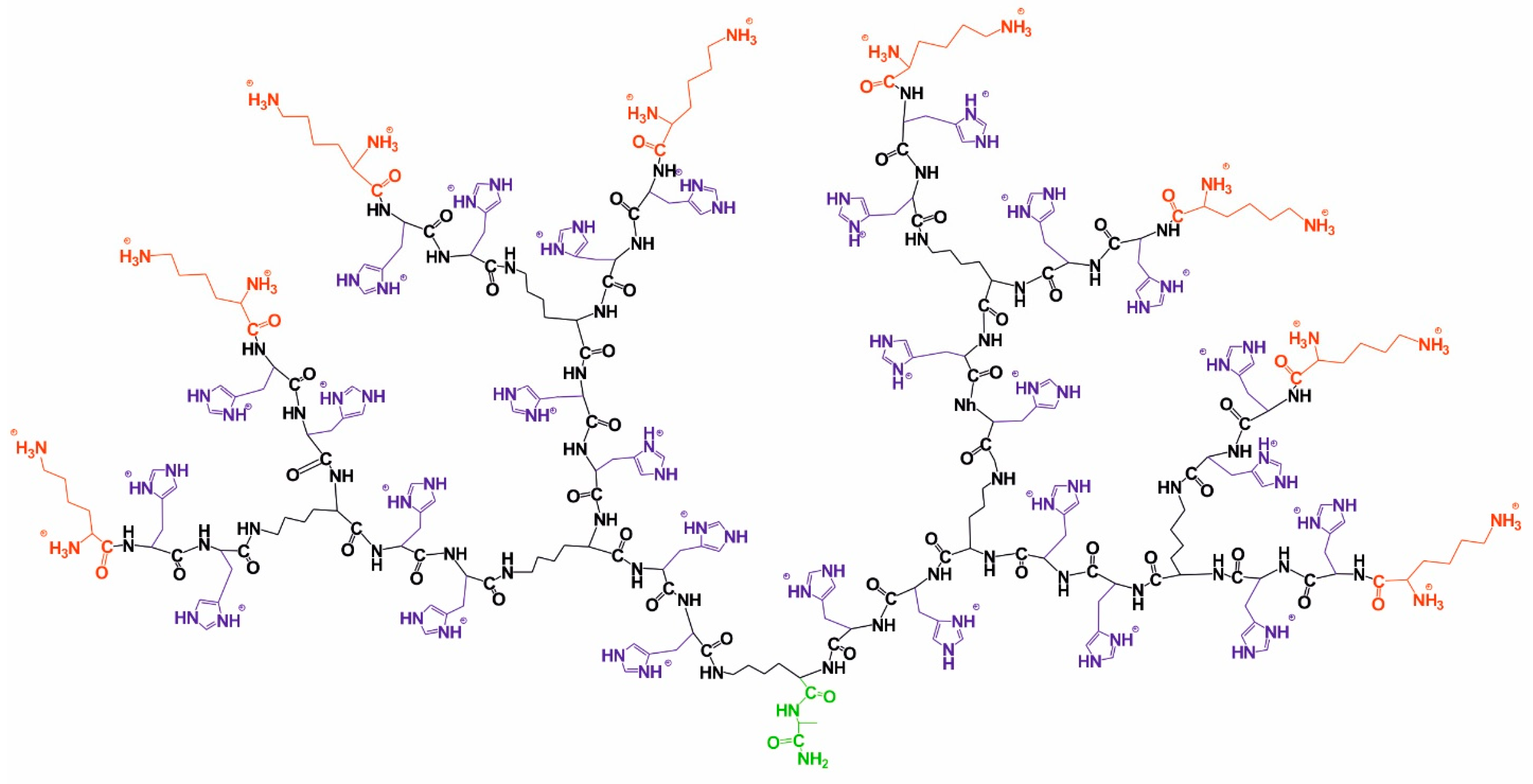
Here, we have synthesized and deuterated a second generation lysine-based dendrimer by inserting two histidine amino acid residues into every inner segment (Figure 1). By analogy with our previous works [39,40], we have modified a dendrimer interior. The insertion of arginine or histidine amino acid residues into inner segments of lysine dendrimer could enhance its potential for drug and gene delivery (see, for instance, a recent review [19]). It is believed that the mechanism of this improved delivery is different. In particular, arginine residues allow more tight DNA packing. At the same time, histidine residues sensitize the delivery vehicles for acidic pH and manipulate the pH-based lysosomal escape. This work is devoted to the possibility of stable selective deuteration of Lys-2His dendrimers. The hydrogen–deuterium exchange at the C2 atoms of the histidine imidazole groups was used for controlled isotope labeling. We prepared deuterium-labeled histidine-containing lysine-based dendrimers using heavy water and by heating the samples. We showed that the labeling is stable at physiological conditions. These findings open new perspectives of biomedical applications of His-modified peptide dendrimers, especially in drug and gene delivery.
The paper is organized as follows. In Section 2, we describe the synthesis of a lysine-based dendrimer with double histidine residues and the preparation of samples for the experiments. Section 3 is devoted to the confirmation of the Lys-2His dendrimer structure by various NMR methods. In Section 4, we present the description of the deuteration process and the analysis of the obtained results. In the last section, we shortly summarize the results and conclusions.

2. Synthesis and Sample Preparation

Histidine-modified lysine-based dendrimer of the second generation (Lys-2His dendrimer) is shown in Figure 1. The core of Lys-2His dendrimer consists of alanine amino acid residue. The inner segments contain ε- or α-part of Lys and two His residues. The side segment consists of β-part of His residue containing imidazole group. The terminal segments contain Lys residues.
Lys-2His dendrimer was synthesized by the solid peptide phase reaction (SPPS) that has been performed manually in polypropylene syringes with a porous membrane on polymer support using the BOC-strategy with DIC/HOBt as a condensing mixture. Trifluoroacetic acid was used for deblocking at the acylation stage. An alanine residue was introduced at the C-terminus of the dendrimer. Nε, Nα-di-(tert-butylhydroxycarbonyl)lysine was introduced into the branching points and, subsequently, double the amounts of amino acid derivatives were added. 4-N, N-dimethylaminopyridine (DMAP) was added to the reaction mixture as a catalyst for complete conversion on the last stage of the dendrimer growth. At the final stage of the synthesis, the target dendrimer molecule was cleaved from the polymeric carrier with complete deprotection of the TFMSA/TFA system in the presence of scavengers. Purification of the crude dendrimer was performed by gel-filtration on a Sephadex G-50 column (Pharmacia Fine Chemicals, Inc., Uppsala, Sweden). The corresponding fraction was dialyzed. The purification degree (95%) of the product was analyzed by RP-HPLC. A detailed description of the synthesis is presented in the Supplementary Materials (SM).
For the deuteration procedure, Lys-2His dendrimer was dissolved in D2O at a concentration of 1.55 g/dl with or without 0.157 M NaCl (saline solution).

3. H and 13C Spectral Characterization

NMR measurements were carried out on a Bruker Avance III 500 (Bruker BioSpin AG, Fällanden, Switzerland). One-dimensional 1H (500 MHz) and 13C (126 MHz) NMR spectra were recorded. We also performed two-dimensional 1H-1H COSY, 1H-13C HSQC, and HMBC NMR experiments for structural characterization of Lys-2His dendrimer using standard pulse sequences.
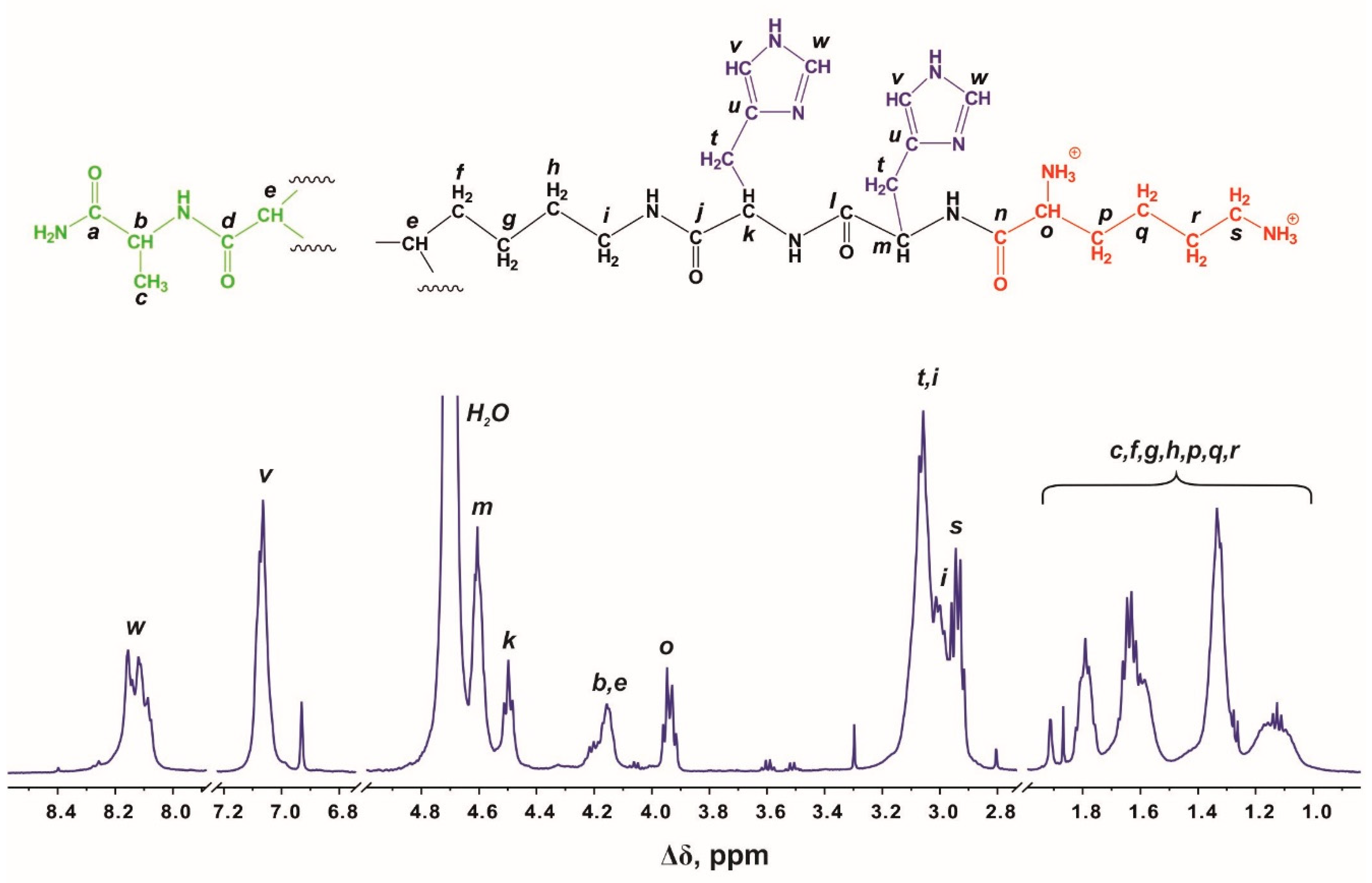
There are four regions in the 1H NMR spectrum (Figure 2) where peaks are observed: the first region at 8.35–6.90 ppm refers to the protons of imidazole rings in His residues; the second region from 4.55 to 3.85 ppm refers to protons in CH groups; the third region from 3.25 to 2.82 ppm is attributed to protons in CH2 groups that are adjacent to nitrogen atoms; and the fourth region at 1.95–1.00 ppm refers to methyl and methylene groups. The ratios of the integrated areas of each type of protons match the expected values (Table 1).
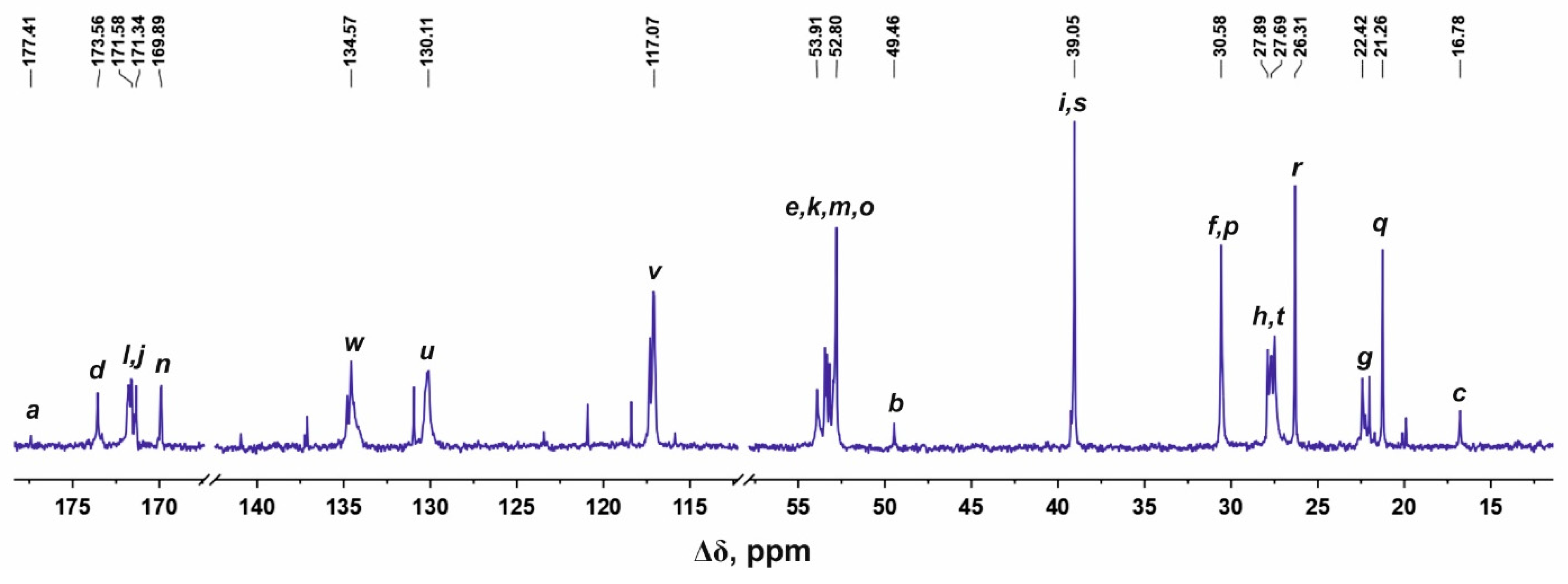
Figure 3 shows the 13C NMR spectrum of the Lys-2His dendrimer. The signals in the range from 177.50 to 169.60 ppm refer to carbon atoms located in carboxyl groups. The peaks at 134.57, 130.11, and 117.07 ppm correspond to carbons in imidazole rings of His. The signals from carbons of CH groups are located in the region 54.20–49.00 ppm. The peak 30.95 ppm is attributed to the CH2 groups adjacent to nitrogen atoms in the inner and terminal Lys segments. The CH2 groups bonded to imidazole rings of His residues have the signal at about 27.50 ppm. In the region of 31.00–16.75 ppm there are peaks from carbons in the CH2 groups of the aliphatic part of the dendrimer.
We measured two-dimensional 1H-1H COSY, 1H-13C HSQC, and HMBC spectra for accurate correlation of peaks in the 1H and 13C NMR spectra. The detailed analysis of 1H-13C two-dimensional spectra and chemical shift assignments are provided in the SM. In conclusion of this section, we found that the claimed structure of Lis-2His dendrimer was totally confirmed by NMR spectroscopy (Figure 1).

4. Deuteration (Hydrogen–Deuterium Exchange)

Here, we present the results of deuteration of the histidine-modified lysine-based dendrimer. Samples of Lys-2His dendrimer were dissolved in heavy water and heated from 25 to 70 °C in increments of 5 °C. Samples were held at each temperature for 2 h. The deuterium incorporation was detected and confirmed by the proton NMR spectra which were recorded after each step.
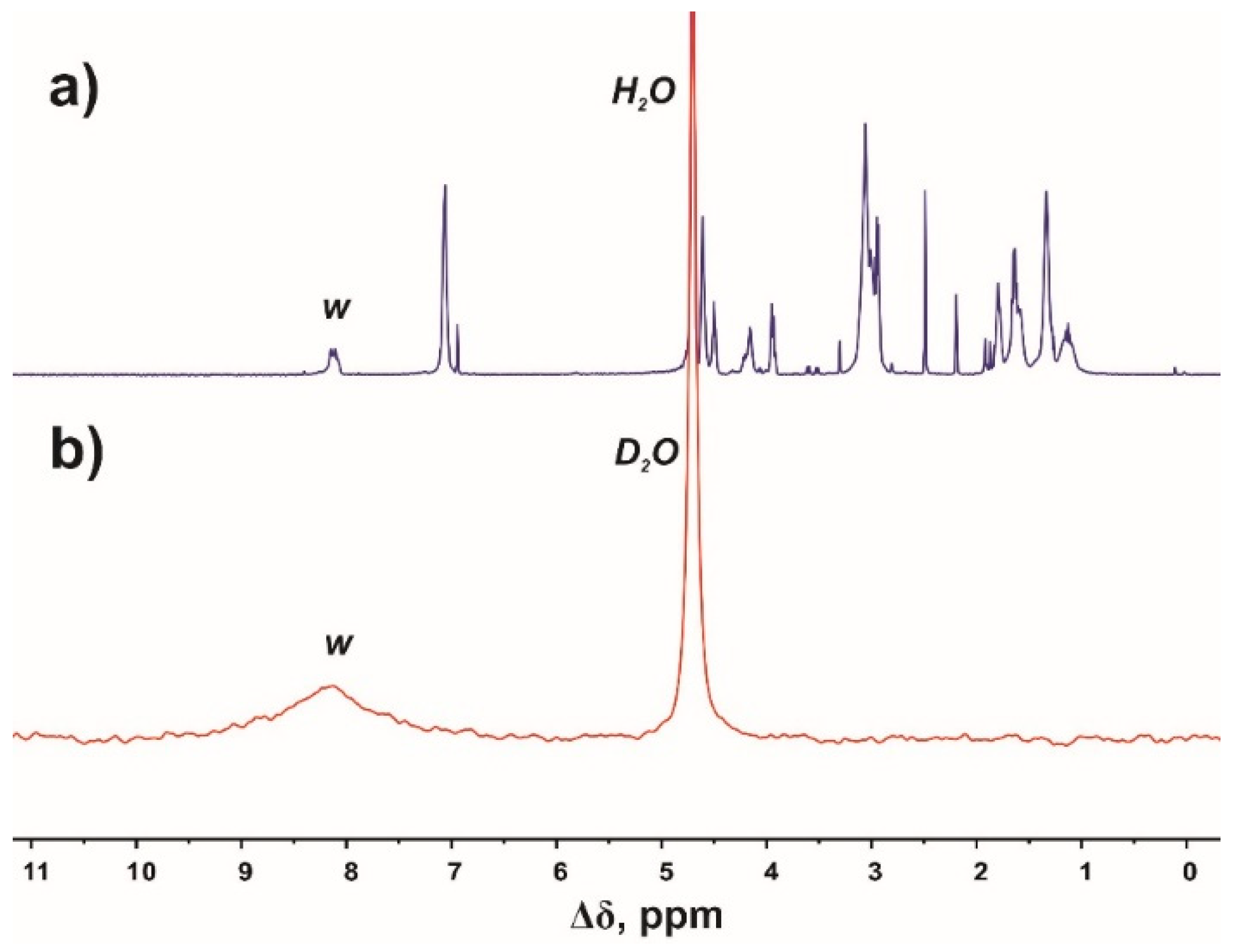
In Figure 4, we compare the 1H (Figure 4a) and 2H (Figure 4b) NMR spectra of Lys-2His dendrimer in D2O after heating. We are interested in the peak (w) with the chemical shift at 8.12 ppm that corresponds to the signal from protons at the C2 carbons of imidazole rings. As shown in Figure 4b, the signal from deuterons appears at 8.12 ppm. The presence of this peak in the 2H NMR spectrum indicates that substitution of hydrogens by deuterons at the C2 carbons of imidazole rings has occurred.
The temperature dependence of the concentration of protons at the C2 carbons of imidazole rings in His residues is presented in Figure 5. As seen in Figure 5, the proton concentration decreases up to 30% after increasing temperature from 25 to 70 °C. Therefore, the deuteration degree at the C2 position in the imidazole rings is around 70%. This result indicates that His residues in the inner segments are available to the water solvent. This fact is in agreement with the data of the atomistic modeling for the dendrimer that we carried out earlier [41]. We found that in the absence of NaCl in the solvent, the deuteration process has practically the same dependence, but the maximal value of the deuteration degree is smaller (~60%) than in the saline solution. We think that this result is caused by the presence of the salt ions in the solvent. Particularly, chlorine ions were used in histidine deuteration experiments [32].
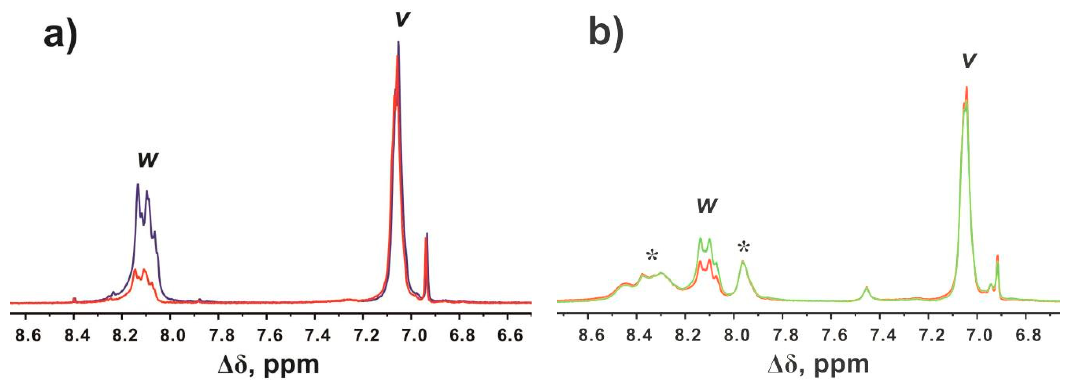
It is important to determine that deuterium labels will be stable during biological and medical studies. We have to make sure that the deuterated C2 carbons of the imidazole rings in histidine residues do not undergo the back-exchange reaction under physiological conditions. For this purpose, we removed D2O from the solution and redissolved Lys-2His dendrimer in H2O. The concentration of NaCl in the solution was kept the same. Heating at 40 °C over four hours did not change the integral value of the peak (w) at 8.12 ppm. It means that the deuteration degree remains constant under physiological conditions. The replacement of deuterons with protons was observed when the dendrimer solution was heated at 70 °C over four hours. Figure 6 illustrates the evolution of the peak (w) at 8.12 ppm during hydrogen–deuterium exchange (a) and deuterium-hydrogen exchange (b) reactions. As can be seen in Figure 6b, recovery of the integral value of the peak (w) has partially occurred. It should be noted that the additional peaks at 7.95 and 8.30 ppm (Figure 6b) are assigned to protons in the NH groups and appeared as a result of the reverse exchange in these groups in H2O.

5. Conclusions

We synthesized a new histidine-modified lysine-based dendrimer using the solid peptide phase reaction. Lys-2His dendrimer was dialyzed, and its purification degree was analyzed by RP-HPLC. The claimed structure was confirmed and characterized by one- and two-dimensional 1H and 13C NMR spectroscopy. In particular, 1H-1H COSY, 1H-13C HSQC, and HMBC spectra were recorded and analyzed. It was established that isotope exchange occurs in Lys-2His dendrimer at the C2 carbons of the imidazole rings of histidine residues during heating. The hydrogen–deuterium exchange has been more intensive in the temperature range 55−70 °C in heavy water. The high level of deuteration (around 70%) was achieved. Deuterons were partially replaced by hydrogens at the C2 carbons of the imidazole ring of histidine residues after heating at 70 °C. However, the carbon at the C2 position in the imidazole rings remained deuterated in aqueous solution under physiological conditions, particularly up to 40 °C.
Here, we have demonstrated that it is possible to prepare a deuterium-labeled histidine-containing lysine-based dendrimer using a rapid and simple method without specific requirements. The chemical structure of Lys-2His dendrimer remains stable at relatively high temperatures during the isotope exchange reaction. We believe that the possibility of direct deuteration will expand the range of potential biological and medical applications of His-modified peptide dendrimers.

Supplementary Materials

The following are available online. Figure S1: The principal scheme of the synthesis of Lys-2His dendrimer from the core to the first generation, Figure S2: 1H-13C HSQC spectrum of Lys-2His dendrimer in D2O at 25 °C. The letter symbols correspond to the designations of the groups in Figure 2, Figure S3: 1H-13C HMBC spectrum of Lys-2His dendrimer in D2O at 25 °C at the range from 5.0 ppm to 1.0 ppm. The letter symbols correspond to the designations of the groups in Figure 1, Figure S4: 1H-13C HMBC spectrum of Lys-2His dendrimer in D2O at 25 °C at the range from 8.5 ppm to 6.5 ppm. The letter symbols correspond to the designations of the groups in Figure 1.

Author Contributions

Conceptualization, D.A.M., and I.M.N.; Methodology, N.N.S., M.A.V., I.I.T., and M.E.M.; Validation, N.N.S., M.A.V., and D.A.M.; Formal Analysis, M.A.V. and M.Y.I.; Investigation, N.N.S., M.A.V., and M.Y.I.; Resources, I.I.T. and M.E.M.; Writing—Original Draft Preparation, N.N.S.; Writing—Review & Editing, N.N.S., D.A.M., M.A.V., I.I.T., M.E.M., M.Y.I., I.M.N., and E.L.; Visualization, N.N.S.; Supervision, D.A.M., I.M.N., and E.L.; Project Administration, D.A.M.; Funding Acquisition, D.A.M. and E.L.

Funding

This research was funded by the Russian Science Foundation [grant number 19-13-00087] and the Researcher Mobility Grant of the Academy of Finland [grant number 318608].

Acknowledgments

The NMR measurements were carried out in the Center for Magnetic Resonance of Research Park of St. Petersburg State University. This work is supported by the Russian Science Foundation (grant No. 19-13-00087). M.Y.I. was supported by Government of Russian Federation (subsidy 08-08). E.L. and N.N.S. are thankful for the Researcher Mobility Grant of the Academy of Finland (No. 318608).

Conflicts of Interest

The authors declare no conflicts of interest.

References

  1. Hsu, H.J.; Bugno, J.; Lee, S.R.; Hong, S. Dendrimer-based nanocarriers: A versatile platform for drug delivery. Wiley Interdiscip. Rev. Nanomed. Nanobiotechnol. 2017, 9, e1409. [Google Scholar] [CrossRef] [PubMed]
  2. Palmerston Mendes, L.; Pan, J.; Torchilin, V. Dendrimers as nanocarriers for nucleic acid and drug delivery in cancer therapy. Molecules 2017, 22, 1401. [Google Scholar] [CrossRef] [PubMed]
  3. Lallana, E.; Sousa-Herves, A.; Fernandez-Trillo, F.; Riguera, R.; Fernandez-Megia, E. Click chemistry for drug delivery nanosystems. Pharm. Res. 2012, 29, 1–34. [Google Scholar] [CrossRef] [PubMed]
  4. Svenson, S.; Tomalia, D.A. Dendrimers in biomedical applications-reflections on the field. Adv. Drug Deliv. Rev. 2005, 57, 2106–2129. [Google Scholar] [CrossRef]
  5. Mintzer, M.A.; Grinstaff, M.W. Biomedical applications of dendrimers: A tutorial. Chem. Soc. Rev. 2011, 40, 173–190. [Google Scholar] [CrossRef]
  6. Felczak, A.; Wrońska, N.; Janaszewska, A.; Klajnert, B.; Bryszewska, M.; Appelhans, D.; Voit, B.; Różalska, S.; Lisowska, K. Antimicrobial activity of poly (propylene imine) dendrimers. New J. Chem. 2012, 36, 2215–2222. [Google Scholar] [CrossRef]
  7. Abbasi, E.; Aval, S.F.; Akbarzadeh, A.; Milani, M.; Nasrabadi, H.T.; Joo, S.W.; Hanifehpour, Y.; Nejati-Koshki, K.; Pashaei-Asl, R. Dendrimers: Synthesis, applications, and properties. Nanoscale Res. Lett. 2014, 9, 247. [Google Scholar] [CrossRef]
  8. Szulc, A.; Pulaski, L.; Appelhans, D.; Voit, B.; Klajnert-Maculewicz, B. Sugar-modified poly (propylene imine) dendrimers as drug delivery agents for cytarabine to overcome drug resistance. Int. J. Pharm. 2016, 513, 572–583. [Google Scholar] [CrossRef]
  9. Kuanga, T.; Fub, D.; Changb, L.; Yangb, Z.; Chenc, Z.; Jine, L.; Chenb, F.; Peng, X. Recent progress in dendrimer-based gene delivery systems. Curr. Org. Chem. 2016, 20, 1820–1826. [Google Scholar] [CrossRef]
  10. Milanizadeh, S.; Parhiz, H.; Abnous, K.; Tabatabai, S.M.; Ramezani, M.; Amel Farzad, S.; Hashemi, M. Gene delivery efficiency and cytotoxicity of heterocyclic amine-modified PAMAM and PPI dendrimers. Mater. Sci. Eng. C 2016, 61, 791–800. [Google Scholar]
  11. Gorzkiewicz, M.; Appelhans, D.; Boye, S.; Lederer, A.; Voit, B.; Klajnert-Maculewicz, B. Effect of the structure of therapeutic adenosine analogues on stability and surface electrostatic potential of their complexes with poly (propyleneimine) dendrimers. Macromol. Rapid Commun. 2019, 1900181. [Google Scholar] [CrossRef] [PubMed]
  12. Ko, K.S.; Yu, G.S.; Bae, Y.M.; Kim, J.Y.; Choi, J.S.; Han, J. Amino acid-modified bioreducible poly(amidoamine) dendrimers: Synthesis, characterization and in vitro evaluation. Macromol. Res. 2012, 20, 1156–1162. [Google Scholar]
  13. Mutalik, S.; Nayak, U.Y.; Kalra, R.; Kumar, A.; Kulkarni, R.V.; Parekh, H.S. Sonophoresis-mediated permeation and retention of peptide dendrimers across human epidermis. Ski. Res. Technol. 2012, 18, 101–107. [Google Scholar] [CrossRef] [PubMed]
  14. Neelov, I.M.; Janaszewska, A.; Klajnert, B.; Bryszewska, M.; Makova, N.Z.; Hicks, D.; Pearson, H.A.; Vlasov, G.P.; Ilyash, M.Y.; Vasilev, D.S.; et al. Molecular properties of lysine dendrimers and their Interactions with Aβ-peptides and neuronal cells. Curr. Med. Chem. 2013, 20, 134–143. [Google Scholar] [CrossRef] [PubMed]
  15. Neelov, I.; Falkovich, S.; Markelov, D.; Paci, E.; Darinskii, A.; Tenhu, H. Molecular dynamics of lysine dendrimers. computer simulation and NMR. In Dendrimers in Biomedical Applications; The Royal Society of Chemistry: London, UK, 2013; pp. 99–114. ISBN 978-1-84973-611-4. [Google Scholar]
  16. Neelov, I.M.; Markelov, D.A.; Falkovich, S.G.; Ilyash, M.Y.; Okrugin, B.M.; Darinskii, A.A. Mathematical simulation of lysine dendrimers: temperature dependences. Polym. Sci. Ser. C 2013, 55, 154–161. [Google Scholar] [CrossRef]
  17. Markelov, D.A.; Falkovich, S.G.; Neelov, I.M.; Ilyash, M.Y.; Matveev, V.V.; Lähderanta, E.; Ingman, P.; Darinskii, A.A. Molecular dynamics simulation of spin–lattice NMR relaxation in poly-L-lysine dendrimers: Manifestation of the semiflexibility effect. Phys. Chem. Chem. Phys. 2015, 17, 3214–3226. [Google Scholar] [CrossRef] [PubMed]
  18. Markelov, D.A.; Dolgushev, M.; Lähderanta, E. Chapter One-NMR Relaxation in Dendrimers. In Annual Reports on NMR Spectroscopy; Webb, G.A., Ed.; Academic Press: Cambridge, MA, USA, 2017; Volume 91, pp. 1–66. ISBN 0066-4103. [Google Scholar]
  19. Yang, J.; Zhang, Q.; Chang, H.; Cheng, Y. Surface-engineered dendrimers in gene delivery. Chem. Rev. 2015, 115, 5274–5300. [Google Scholar] [CrossRef] [PubMed]
  20. Ghaffari, M.; Dehghan, G.; Abedi-Gaballu, F.; Kashanian, S.; Baradaran, B.; Dolatabadi, J.E.N.; Losic, D. Surface functionalized dendrimers as controlled-release delivery nanosystems for tumor targeting. Eur. J. Pharm. Sci. 2018, 122, 311–330. [Google Scholar] [CrossRef]
  21. LeMaster, D.M. Deuterium labelling in NMR structural analysis of larger proteins. Q. Rev. Biophys. 1990, 23, 133–174. [Google Scholar] [CrossRef]
  22. Sattler, M.; Fesik, S.W. Use of deuterium labeling in NMR: Overcoming a sizeable problem. Structure 1996, 4, 1245–1249. [Google Scholar] [CrossRef]
  23. Atzrodt, J.; Derdau, V.; Kerr, W.J.; Reid, M. Deuterium-and tritium-labelled compounds: applications in the life sciences. Angew. Chemie Int. Ed. 2018, 57, 1758–1784. [Google Scholar] [CrossRef] [PubMed]
  24. Liu, C.; Chen, Z.; Su, C.; Zhao, X.; Gao, Q.; Ning, G.-H.; Zhu, H.; Tang, W.; Leng, K.; Fu, W.; et al. Controllable deuteration of halogenated compounds by photocatalytic D2O splitting. Nat. Commun. 2018, 9, 80. [Google Scholar] [CrossRef] [PubMed]
  25. Khandogin, J.; Brooks, C.L. Toward the accurate first-principles prediction of ionization equilibria in proteins. Biochemistry 2006, 45, 9363–9373. [Google Scholar] [CrossRef] [PubMed]
  26. Okuda, T.; Sugiyama, A.; Niidome, T.; Aoyagi, H. Characters of dendritic poly(L-lysine) analogues with the terminal lysines replaced with arginines and histidines as gene carriers in vitro. Biomaterials 2004, 25, 537–544. [Google Scholar] [CrossRef]
  27. Yu, G.S.; Bae, Y.M.; Choi, H.; Kong, B.; Choi, I.S.; Choi, J.S. Synthesis of PAMAM dendrimer derivatives with enhanced buffering capacity and remarkable gene transfection efficiency. Bioconjugate Chem. 2011, 22, 1046–1055. [Google Scholar] [CrossRef] [PubMed]
  28. Shao, N.; Wang, F.; Chen, Y.; Wang, H.; Cheng, Y.; Wang, Y. Synergistic effect of amino acids modified on dendrimer surface in gene delivery. Biomaterials 2014, 35, 9187–9198. [Google Scholar]
  29. Varkouhi, A.K.; Scholte, M.; Storm, G.; Haisma, H.J. Endosomal escape pathways for delivery of biologicals. J. Control. Release 2011, 151, 220–228. [Google Scholar] [CrossRef] [PubMed]
  30. Bradbury, J.H.; Chapman, B.E.; Crompton, M.W.; Norton, R.S.; Teh, J.S. Hydrogen–deuterium exchange of the C-2 protons of histidine and histidine peptides and proteins. J. Chem. Soc. Perkin Trans. 2 1980, 693–700. [Google Scholar] [CrossRef]
  31. Cebo, M.; Kielmas, M.; Adamczyk, J.; Cebrat, M.; Szewczuk, Z.; Stefanowicz, P. Hydrogen-deuterium exchange in imidazole as a tool for studying histidine phosphorylation. Anal. Bioanal. Chem. 2014, 406, 8013–8020. [Google Scholar] [CrossRef]
  32. Matthews, H.R.; Matthews, K.S.; Opella, S.J. Selectively deuterated amino acid analogues synthesis, Incorporation into proteins and NMR properties. BBA-Gen. Subj. 1977, 497, 1–13. [Google Scholar] [CrossRef]
  33. Miyagi, M.; Nakazawa, T. Determination of pKa values of individual histidine residues in proteins using mass spectrometry. Anal. Chem. 2008, 80, 6481–6487. [Google Scholar] [CrossRef] [PubMed]
  34. Cobb, N.J.; Apostol, M.I.; Chen, S.; Smirnovas, V.; Surewicz, W.K. Conformational stability of mammalian prion protein amyloid fibrils is dictated by a packing polymorphism within the core region. J. Biol. Chem. 2014, 289, 2643–2650. [Google Scholar] [CrossRef] [PubMed]
  35. Takesada, H.; Nakanishi, M.; Tsuboi, M.; Ajisaka, K. Hydrogen-deuterium exchange in the histidine residues of bovine α-lactalbumin. J. Biochem. 1976, 80, 969–974. [Google Scholar] [CrossRef] [PubMed]
  36. Cesarotti, E.; Rimoldi, I.; Zerla, D.; Aldini, G. Histidine and deuterium labelled histidine by asymmetric catalytic reduction with gaseous H2 or D2; the role of strong non-coordinating acids. Tetrahedron Asymmetry 2008, 19, 273–278. [Google Scholar] [CrossRef]
  37. Hayashi, N.; Kuyama, H.; Nakajima, C.; Kawahara, K.; Miyagi, M.; Nishimura, O.; Matsuo, H.; Nakazawa, T. Imidazole C-2 hydrogen/deuterium exchange reaction at histidine for probing protein structure and function with matrix-assisted laser desorption ionization mass spectrometry. Biochemistry 2014, 53, 1818–1826. [Google Scholar] [CrossRef] [PubMed]
  38. Miyagi, M.; Wan, Q.; Ahmad, M.F.; Gokulrangan, G.; Tomechko, S.E.; Bennett, B.; Dealwis, C. Histidine hydrogen-deuterium exchange mass spectrometry for probing the microenvironment of histidine residues in dihydrofolate reductase. PLoS ONE 2011, 6, e17055. [Google Scholar] [CrossRef] [PubMed]
  39. Sheveleva, N.N.; Markelov, D.A.; Vovk, M.A.; Mikhailova, M.E.; Tarasenko, I.I.; Neelov, I.M.; Lähderanta, E. NMR studies of excluded volume interactions in peptide dendrimers. Sci. Rep. 2018, 8, 1–7. [Google Scholar] [CrossRef] [Green Version]
  40. Sheveleva, N.N.; Markelov, D.A.; Vovk, M.A.; Mikhailova, M.E.; Tarasenko, I.I.; Tolstoy, P.M.; Neelov, I.M.; Lahderanta, E. Lysine-based dendrimer with double arginine residues. RSC Adv. 2019, 9, 18018–18026. [Google Scholar] [CrossRef] [Green Version]
  41. Falkovich, S.; Markelov, D.; Neelov, I.; Darinskii, A. Are structural properties of dendrimers sensitive to the symmetry of branching? Computer simulation of lysine dendrimers. J. Chem. Phys. 2013, 139, 064903. [Google Scholar] [CrossRef]
Sample Availability: Samples of the compounds Lys-2His dendrimers are available from the authors.
Figure 1. Structural formula of Lis-2His dendrimer. Green color marks the core, black corresponds to the main chain, violet marks the side segments, and red corresponds to the terminal segments.
Molecules 24 02481 g001
Figure 2. 1H NMR spectrum of Lis-2His dendrimer in D2O at 25 °C.
Molecules 24 02481 g002
Figure 3. 13C NMR spectrum of Lis-2His dendrimer in D2O at 25 °C. The letter symbols correspond to the designations of the groups in Figure 2.
Molecules 24 02481 g003
Figure 4. (a) 1H and (b) 2H NMR spectra of Lys-2His dendrimer in D2O after heating. 2H NMR spectrum was recorded using a pulse sequence that suppresses the solvent signal. The letter symbols correspond to the designations of the groups in Figure 2.
Molecules 24 02481 g004
Figure 5. The temperature dependence of the concentration of protons at the C2 carbons of histidine imidazole rings (8.12 ppm) during heating.
Molecules 24 02481 g005
Figure 6. Evolution of the peak at 8.12 ppm from protons at the C2 carbons of the imidazole rings of histidine residues in Lys-2His dendrimer: (a) 1H NMR spectra of Lys-2His dendrimer in D2O before (blue) and after (red) heating; (b) 1H NMR spectra of Lys-2His dendrimer in H2O before (red) and after (green) heating. The peaks from protons in the NH groups are indicated by *.
Molecules 24 02481 g006
Table 1. Chemical shift assignments and integral values.
PeakType of GroupChemical Shift, ppmIntegral ValueNumber of Protons in Groups
wCH-(N)
(in imidazole ring)
8.122628
vCH-(N)
(in imidazole ring)
7.062628
m,k,b,e,oCH-(N)4.64–3.884444
i,t,sCH2-(N) 3.22–2.838686
c,f,g,h,p,q,rCH2, CH3a1.93–1.0010290 + 3 a
a In the core.