
Six New Methyl Apiofuranosides from the Bark of Phellodendron chinense Schneid and Their Inhibitory Effects on Nitric Oxide Production

 and
and
Abstract
1. Introduction
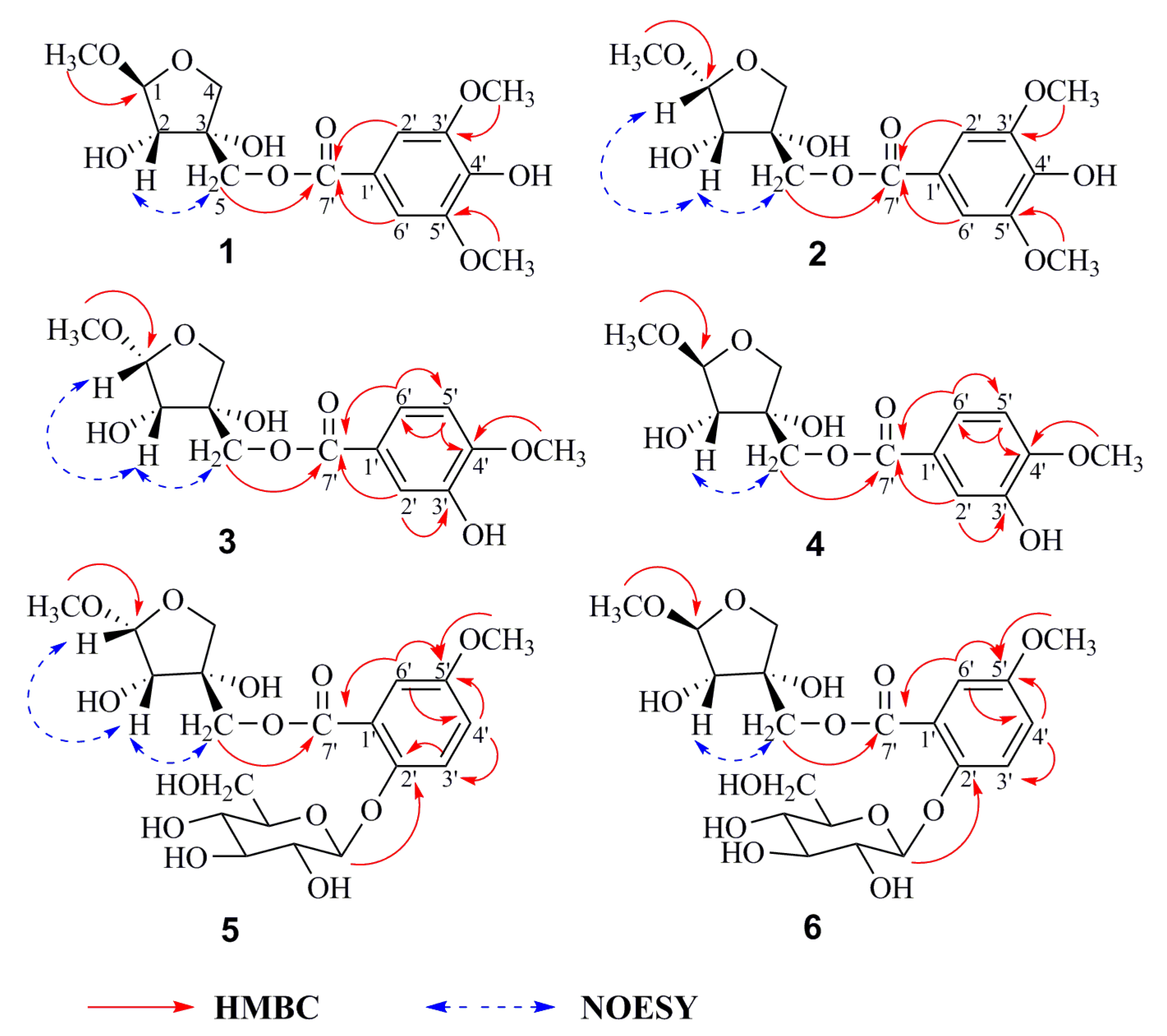
2. Results
3. Materials and Methods
3.1. General Experimental Procedure
3.2. Plant Material
3.3. Extraction and Isolation
3.4. Sugar Identification
3.5. Cell Culture
3.6. Cell Viability Assay
3.7. Measurement of NO Release
Supplementary Materials
Author Contributions
Funding
Conflicts of Interest
Abbreviations
| DMEM | dulbecco’s modified eagle medium |
| DMSO | Dimethyl Sulfoxide |
| HMBC | Heteronuclear Multiple Bonding Correlation |
| HSQC | Heteronuclear Single Quantum Correlation |
| MTT | 3-(4,5-dimethyl-2-thiazolyl)-2,5-diphenyl-2-H-tetrazolium bromide |
| NOE | Nuclear Overhauser Enhancement |
| NOESY | Nuclear Overhauser Enhancement Spectroscopy |
| RAW | leukemia cellsin mouse macrophage |
References
- Shiao, P.G. Photocatalogue of Chinese Traditional Medicine; Taiwan Business Publication Company: Taipei, Taiwan, 1990; Volume 10, p. 86. [Google Scholar]
- Hsu, K.J. Chinese Traditional Medicine; Chinese Pharmaceutical Science and Technology Publication Company: Beijing, China, 1996; p. 802. [Google Scholar]
- Chinese Pharmacopoeia Commission. Pharmacopoeia of the People’s Republic of China; Chemical Industry Press: Beijing, China, 2015; pp. 305–306. [Google Scholar]
- Chen, M.L.; Xian, Y.F.; Ip, S.P.; Tsai, S.H.; Yang, J.Y. Chemical and Biological Differentiation of Cortex Phellodendri Chinensis and Cortex Phellodendri Amurensis. Planta Med. 2010, 76, 1530–1535. [Google Scholar] [CrossRef] [PubMed]
- Wu, T.S.; Hsu, M.Y.; Kuo, P.C.; Sreenivasulu, B.; Damu, A.G.; Su, C.R. Constituents from the leaves of Phellodendron amurense var. wilsonii and their bioactivity. J. Nat. Prod. 2003, 66, 1207–1211. [Google Scholar] [CrossRef] [PubMed]
- Yan, C.; Zhang, Y.D.; Wang, X.H.; Geng, S.D.; Wang, T.Y.; Sun, M. Tirucallane-type triterpenoids from the fruits of Phellodendron chinense Schneid and their cytotoxic activities. Fitoterapia 2016, 113, 132–138. [Google Scholar] [CrossRef] [PubMed]
- Cuéllar, M.J.; Giner, R.M.; Recio, M.C.; Máñez, S.; RíOs, J.L. Topical anti-inflammatory activity of some Asian medicinal plants used in dermatological disorders. Fitoterapia 2001, 72, 221–229. [Google Scholar] [CrossRef]
- Park, S.D.; Lai, Y.S.; Kim, C.H. Immunopontentiating and antitumor activities of the purified polysaccharides from Phellodendron chinense SCHNEID. Life Sci. 2004, 75, 2621–2632. [Google Scholar] [CrossRef]
- Ishii, T.; Yanagisawa, M. Synthesis, separation and NMR spectral analysis of methyl apiofuranosides. Carbohydr. Res. 1998, 313, 189–192. [Google Scholar] [CrossRef]
- Ishiia, T.; Ono, H. NMR ctroscopic analysis of the borate diol esters of methyl apiofuranosides. Carbohydr. Res. 1999, 321, 257–260. [Google Scholar] [CrossRef]
- Park, S.; Goo, Y.M.; Na, D.S. Isolation and structure elucidation of a catechin glycoside with phospholipase A2 inhibiting activity from Ulmi cortex. Bull. Korean Chem. Soc. 1996, 17, 101–103. [Google Scholar]
- Angyal, S.J.; Bodkin, C.L.; Mills, J.A.; Pojer, P.M. ChemInform Abstract: Complexes of carbohydrates with metal cations. IX Synthesis of the methyld D-tagatosides, D-psicosides, D-apiosides, and D-erythrosides. Cheminform 1977, 8, 1259–1268. [Google Scholar] [CrossRef]
- Tronchet, J.M.J.; Tronchet, J. 9-(3-C-hydroxymethyl-α-L-threofuranosyl) adenine (9-(β-Dapio-L-furanosyl) adenine). Carbohydr. Res. 1974, 34, 263–270. [Google Scholar] [CrossRef]
- Zhang, N.; Huang, W.X.; Xia, G.Y.; Oppong, M.B.; Ding, L.Q.; Li, P.; Qiu, F. Methods for determination of absolute configuration of monosaccharides. Chin. Herb. Med. 2018, 10, 14–22. [Google Scholar] [CrossRef]
- Mathias, L.; Vieira, I.J.C.; Filho, R.B.; Filho, R.E. A New Isoflavone Glycoside from Dalbergia nigra. J. Nat. Prod. 1998, 61, 1158–1161. [Google Scholar] [CrossRef]
- Zhou, K.L.; Zhao, F.; Liu, Z.H.; Zhuang, Y.L.; Chen, L.X.; Qiu, F. Triterpenoids and Flavonoids from Celery (Apium graveolens). J. Nat. Prod. 2009, 72, 1563–1567. [Google Scholar] [CrossRef]
- Tanaka, T.; Nakashima, T.; Ueda, T. Facile Discrimination of Aldose Enantiomers by Reversed-Phase HPLC. Chem. Pharm. Bull. 2007, 55, 899–901. [Google Scholar] [CrossRef]
- Murata, T.; Endo, Y.; Miyase, T.; Yoshizaki, F. Iridoid glycoside constituents of Stachys lanata. J. Nat. Prod. 2008, 71, 1768–1770. [Google Scholar] [CrossRef]
- Johnsson, P.; Peerlkamp, N. Polymeric fractions containing phenol glucosides in flaxseed. Food Chem. 2002, 6, 207–212. [Google Scholar] [CrossRef]
- Rowe, J.W.; Bower, C.L.; Wagner, E.R. Extractives of jack pine bark: Occurrence of cis- and trans-pinosylvin dimethyl ether and ferulic acid esters. Phytochemistry 1969, 8, 235–241. [Google Scholar] [CrossRef]
- Andrade, P.B.; Leitão, R.; Seabra, R.M. 3,4-Dimethoxycinnamic acid levels as a tool for differentiation of Coffea canephora var. robusta and Coffea arabica. Food Chem. 1998, 61, 511–514. [Google Scholar] [CrossRef]
- Gopalakrishnan, S.; Subbarao, G.V.; Nakahara, K. Nitrification inhibitors from the root tissues of Brachiaria humidicola, a tropical grass. J. Agric. Food. Chem. 2007, 55, 1385–1388. [Google Scholar] [CrossRef]
- Vongvanich, N.; Kittakoop, P.; Charoenchai, P. Antiplasmodial, Antimycobacterial, and Cytotoxic Principles from Camchaya calcarea. Planta Med. 2006, 72, 1427–1430. [Google Scholar] [CrossRef]
- Xiao, H.; Parkin, K.L. Isolation and identification of potential cancer chemopreventive agents from methanolic extracts of green onion (Allium cepa). Phytochemistry 2007, 68, 1059–1067. [Google Scholar] [CrossRef]
- Nakamura, S.; Fujimoto, K.; Matsumoto, T. Acylated sucroses and acylated quinic acids analogs from the flower buds of Prunus mume and their inhibitory effect on melanogenesis. Phytochemistry 2013, 92, 128–136. [Google Scholar] [CrossRef]
- Youn, U.J.; Lee, J.H.; Lee, Y.J. Regulation of the 5-HT3A Receptor-Mediated Current by Alkyl 4-Hydroxybenzoates Isolated from the Seeds of Nelumbo nucifera. Clin. Biochem. 2010, 7, 2296–2302. [Google Scholar]
- Chai, W.M.; Shi, Y.; Feng, H.L. NMR, HPLC-ESI-MS, and MALDI-TOF MS Analysis of Condensed Tannins from Delonix regia (Bojer ex Hook.) Raf. and Their Bioactivities. J. Agric. Food. Chem. 2012, 60, 5013–5022. [Google Scholar] [CrossRef] [PubMed]
- Majumder, P.L.; Banerjee, S.; Sen, S. Three stilbenoids from the orchid Agrostophyllum callosum. Phytochemistry 1996, 42, 847–852. [Google Scholar] [CrossRef]
- David, A.G.; Timothy, R.B. Molecular biology of nitric oxide synthases. Cancer Metast. Rev. 1998, 17, 7–23. [Google Scholar]
- Beckman, J.S.; Koppenol, W.H. Nitric oxide, superoxide, and peroxynitrite: the good, the bad, and ugly. Am. J. Physiol. 1996, 1424–1437. [Google Scholar] [CrossRef]
- Bungorn, S.; Jintana, J.; Nawarat, W.; Doosadee, H. Anti-inflammatory effect of Streblus asperleaf extract in rats and its modulationon inflammation-associated genes expression in RAW 264.7 macrophage cells. J. Ethnopharmacol. 2009, 124, 566–570. [Google Scholar]
- Guzik, T.J.; Korbut, R.; Adamek, G.T. Nitric oxide and superoxide in inflammation and immune regulation. J. Physiol. Pharmacol. 2003, 54, 469. [Google Scholar]
- Naohiro, O.; Tomofumi, S.; Yuji, N.; Noriyasu, H.; Fumiyuki, K. Quantitative analysis of the anti-inflammatory activity of orengedokuto II: berberine is responsible for the inhibition of NO production. J. Nat. Med. 2018, 72, 706–714. [Google Scholar]
- Sun, L.D.; Wang, F.; Dai, F.; Wang, Y.H.; Lin, D.; Zhou, B. Development and mechanism investigation of a new piperlongumine derivative as a potent anti-inflammatory agent. Biochem. Pharmacol. 2015, 95, 156–169. [Google Scholar] [CrossRef] [PubMed]
- Xue, G.M.; Li, X.Q.; Chen, C. Highly Oxidized Guaianolide Sesquiterpenoids with Potential Antiinflammatory Activity from Chrysanthemum indicum. J. Nat. Prod. 2018, 81, 378–386. [Google Scholar] [CrossRef]
- Zhao, F.; Wang, L.; Liu, K. In vitro anti-inflammatory effects of arctigenin, a lignan from Arctium lappa L. through inhibition on iNOS pathway. J. Ethnopharmacology. 2009, 122, 457–462. [Google Scholar] [CrossRef] [PubMed]
- Choi, Y.Y.; Kim, M.H.; Han, J.M.; Hong, J.K.; Lee, T.H.; Kim, S.H. The anti-inflammatory potential of Cortex Phellodendron in vivo and in vitro: down-regulation of NO and iNOS through suppression of NF-κB and MAPK activation. Int. J. Immunopharmacol. 2014, 19, 214–220. [Google Scholar] [CrossRef] [PubMed]
Sample Availability: Samples of the compounds are not available from the authors. |


| 1 a | 2 a | 3 a | 4 a | |||||
|---|---|---|---|---|---|---|---|---|
| Position | δC | δH (J in Hz) | δC | δH (J in Hz) | δC | δH (J in Hz) | δC | δH (J in Hz) |
| Api-1 | 111.6 | 4.86 (1H, d, J = 2.5 Hz) | 104.7 | 4.90 (1H, d, J = 4.6 Hz) | 104.7 | 4.90 (1H, d, J = 4.6 Hz) | 111.6 | 4.86 (1H, d, J = 2.6 Hz) |
| 2 | 78.9 | 3.97 (1H, d, J = 2.5 Hz) | 74.5 | 3.99 (1H, d, J = 4.6 Hz) | 74.4 | 3.99 (1H, d, J = 4.6 Hz) | 78.8 | 3.96 (1H, d, J = 2.6 Hz) |
| 3 | 79.1 | 76.6 | 76.7 | 79.1 | ||||
| 4 | 75.0 | 4.04 (1H, d, J = 9.8 Hz) | 75.4 | 4.07 (1H, d, J = 9.9 Hz) | 75.4 | 4.07 (1H, d, J = 9.8 Hz) | 75.0 | 4.03 (1H, d, J = 9.8 Hz) |
| 3.89 (1H, d, J = 9.8 Hz) | 3.91 (1H, d, J = 9.9 Hz) | 3.91 (1H, d, J = 9.8 Hz) | 3.89 (1H, d, J = 9.8 Hz) | |||||
| 5 | 67.7 | 4.32 (2H, m) | 68.7 | 4.29 (2H, m) | 68.4 | 4.28 (2H, m) | 67.5 | 4.31 (2H, m) |
| 1′ | 121.2 | 121.2 | 125.4 | 125.4 | ||||
| 2′ | 108.5 | 7.35 (1H, s) | 108.5 | 7.35 (1H, s) | 116.1 | 7.59 (1H, d, J = 1.9 Hz) | 116.1 | 7.59 (1H, d, J = 1.9 Hz) |
| 3′ | 149.1 | 149.1 | 148.9 | 148.9 | ||||
| 4′ | 142.3 | 142.3 | 153.2 | 153.2 | ||||
| 5′ | 149.1 | 149.1 | 113.9 | 6.84 (1H, d, J = 8.8 Hz) | 113.8 | 6.85 (1H, d, J = 8.0 Hz) | ||
| 6′ | 108.5 | 7.35 (1H, s) | 108.5 | 7.35 (1H, s) | 122.4 | 7.57 (1H, dd, J = 8.8, 1.9 Hz) | 122.4 | 7.58 (1H, dd, J = 8.0, 1.9 Hz) |
| 7′ | 167.9 | 167.9 | 168.0 | 168.0 | ||||
| 3′-OCH3 | 57.0 | 3.89 (3H, s) | 57.0 | 3.88 (3H, s) | ||||
| 4′-OCH3 | 56.6 | 3.90 (3H, s) | 56.6 | 3.90 (3H, s) | ||||
| 5′-OCH3 | 57.0 | 3.89 (3H, s) | 57.0 | 3.88 (3H, s) | ||||
| 1-OCH3 | 56.0 | 3.37 (3H, s) | 56.0 | 3.43 (3H, s) | 55.7 | 3.43 (3H, s) | 55.7 | 3.38 (3H, s) |
| 5 a | 6 a | |||
|---|---|---|---|---|
| Position | δC | δH (J in Hz) | δC | δH (J in Hz) |
| Api-1 | 104.7 | 4.90 (1H, d, J = 4.5 Hz) | 111.6 | 4.86 (1H, d, J = 2.6 Hz) |
| 2 | 74.5 | 4.00 (1H, d, J = 4.5 Hz) | 78.8 | 3.96 (1H, d, J = 2.6 Hz) |
| 3 | 76.6 | 79.1 | ||
| 4 | 75.4 | 4.07 (1H, d, J = 10.0 Hz) | 75.0 | 4.03 (1H, d, J = 9.8 Hz) |
| 3.92 (1H, d, J = 10.0 Hz) | 3.90 (1H, d, J = 9.8 Hz) | |||
| 5 | 68.7 | 4.31 (2H, m) | 67.8 | 4.33 (2H, m) |
| 1′ | 125.3 | 125.3 | ||
| 2′ | 152.5 | 152.5 | ||
| 3′ | 116.6 | 7.22 (1H, d, J = 8.5 Hz) | 116.6 | 7.22 (1H, d, J = 8.5 Hz) |
| 4′ | 124.9 | 7.68 (1H, dd, J = 8.5, 2.0 Hz) | 124.9 | 7.67 (1H, dd, J = 8.5, 2.0 Hz) |
| 5′ | 150.6 | 150.6 | ||
| 6′ | 114.5 | 7.64 (1H, d, J = 2.0 Hz) | 114.5 | 7.64 (1H, d, J = 2.0 Hz) |
| 7′ | 167.6 | 167.5 | ||
| 5′-OCH3 | 56.9 | 3.90 (3H, s) | 56.9 | 3.91 (3H, s) |
| 1-OCH3 | 55.7 | 3.43 (3H, s) | 56.0 | 3.38 (3H, s) |
| Glc-1′′ | 102.1 | 5.03 (1H, d, J = 7.6 Hz) | 102.1 | 5.03 (1H, d, J = 7.6 Hz) |
| 2′′ | 74.9 | 3.53 (1H, m) | 75.0 | 3.53 (1H, m) |
| 3′′ | 78.6 | 3.47 (1H, m) | 78.5 | 3.47 (1H, m) |
| 4′′ | 71.4 | 3.40 (1H, m) | 71.4 | 3.42 (1H, m) |
| 5′′ | 78.0 | 3.49 (1H, m) | 78.0 | 3.49 (1H, m) |
| 6′′ | 62.6 | 3.70 (1H, dd, J = 12.0, 5.6 Hz) | 62.6 | 3.69 (1H, dd, J = 12.0, 5.6 Hz) |
| 3.87 (1H, m) | 3.88 (1H, m) | |||
© 2019 by the authors. Licensee MDPI, Basel, Switzerland. This article is an open access article distributed under the terms and conditions of the Creative Commons Attribution (CC BY) license (http://creativecommons.org/licenses/by/4.0/).
Share and Cite
Wang, P.-F.; Li, Y.-P.; Ding, L.-Q.; Cao, S.-J.; Wang, L.-N.; Qiu, F. Six New Methyl Apiofuranosides from the Bark of Phellodendron chinense Schneid and Their Inhibitory Effects on Nitric Oxide Production. Molecules 2019, 24, 1851. https://doi.org/10.3390/molecules24101851
Wang P-F, Li Y-P, Ding L-Q, Cao S-J, Wang L-N, Qiu F. Six New Methyl Apiofuranosides from the Bark of Phellodendron chinense Schneid and Their Inhibitory Effects on Nitric Oxide Production. Molecules. 2019; 24(10):1851. https://doi.org/10.3390/molecules24101851
Chicago/Turabian StyleWang, Peng-Fei, Yan-Ping Li, Li-Qin Ding, Shi-Jie Cao, Li-Ning Wang, and Feng Qiu. 2019. "Six New Methyl Apiofuranosides from the Bark of Phellodendron chinense Schneid and Their Inhibitory Effects on Nitric Oxide Production" Molecules 24, no. 10: 1851. https://doi.org/10.3390/molecules24101851
APA StyleWang, P.-F., Li, Y.-P., Ding, L.-Q., Cao, S.-J., Wang, L.-N., & Qiu, F. (2019). Six New Methyl Apiofuranosides from the Bark of Phellodendron chinense Schneid and Their Inhibitory Effects on Nitric Oxide Production. Molecules, 24(10), 1851. https://doi.org/10.3390/molecules24101851