Novel Potent Hypoglycemic Compounds from Euonymus laxiflorus Champ. and Their Effect on Reducing Plasma Glucose in an ICR Mouse Model

 ,
,  ,
,  ,
,  ,
, 

Abstract
:
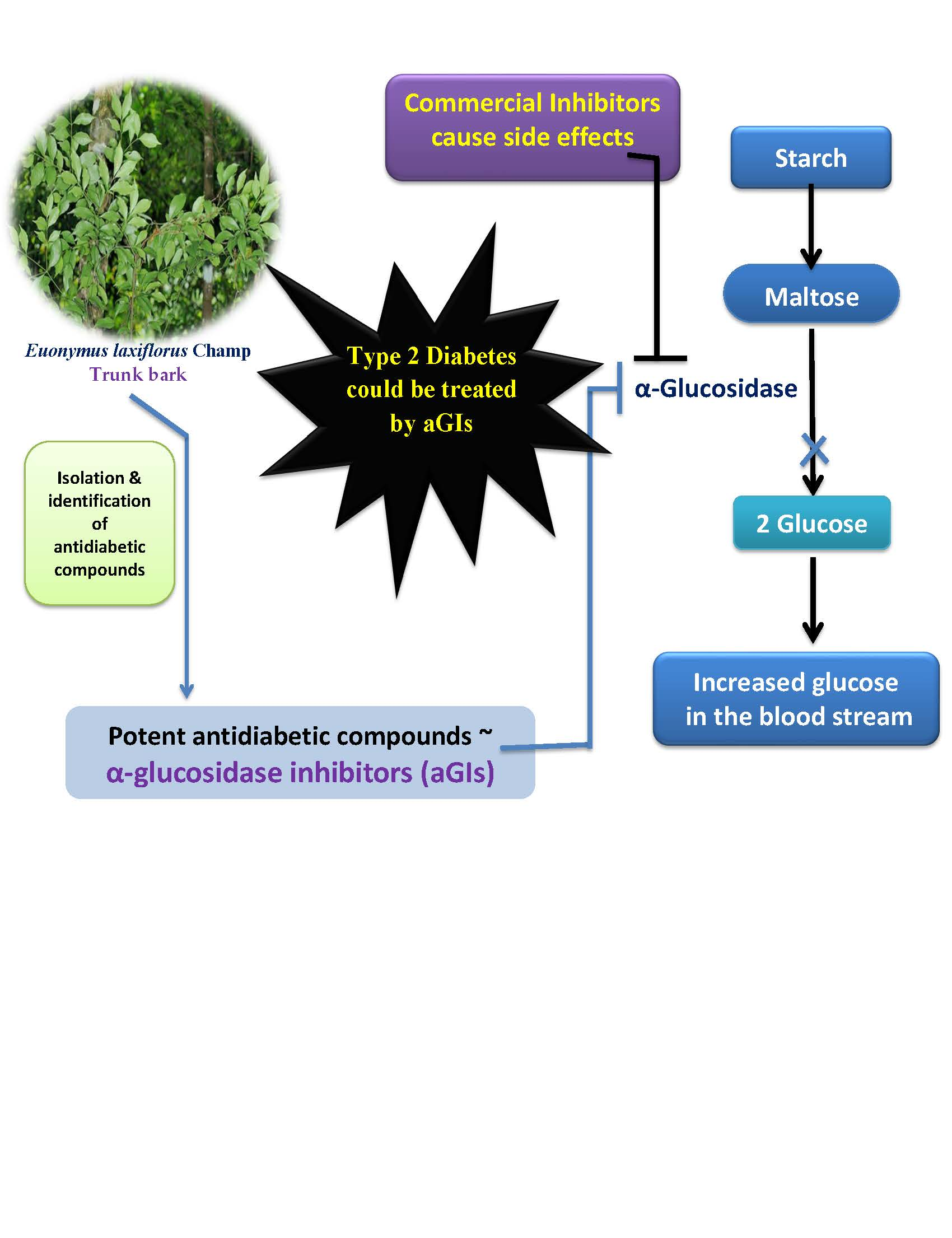
1. Introduction
2. Results and Discussion
2.1. Reclamation of Euonymus laxiflorus Champ. Extracts as a Potent Source of Natural aGIs
2.2. Purification of Active aGIs from MeOH Extract of ELCTB
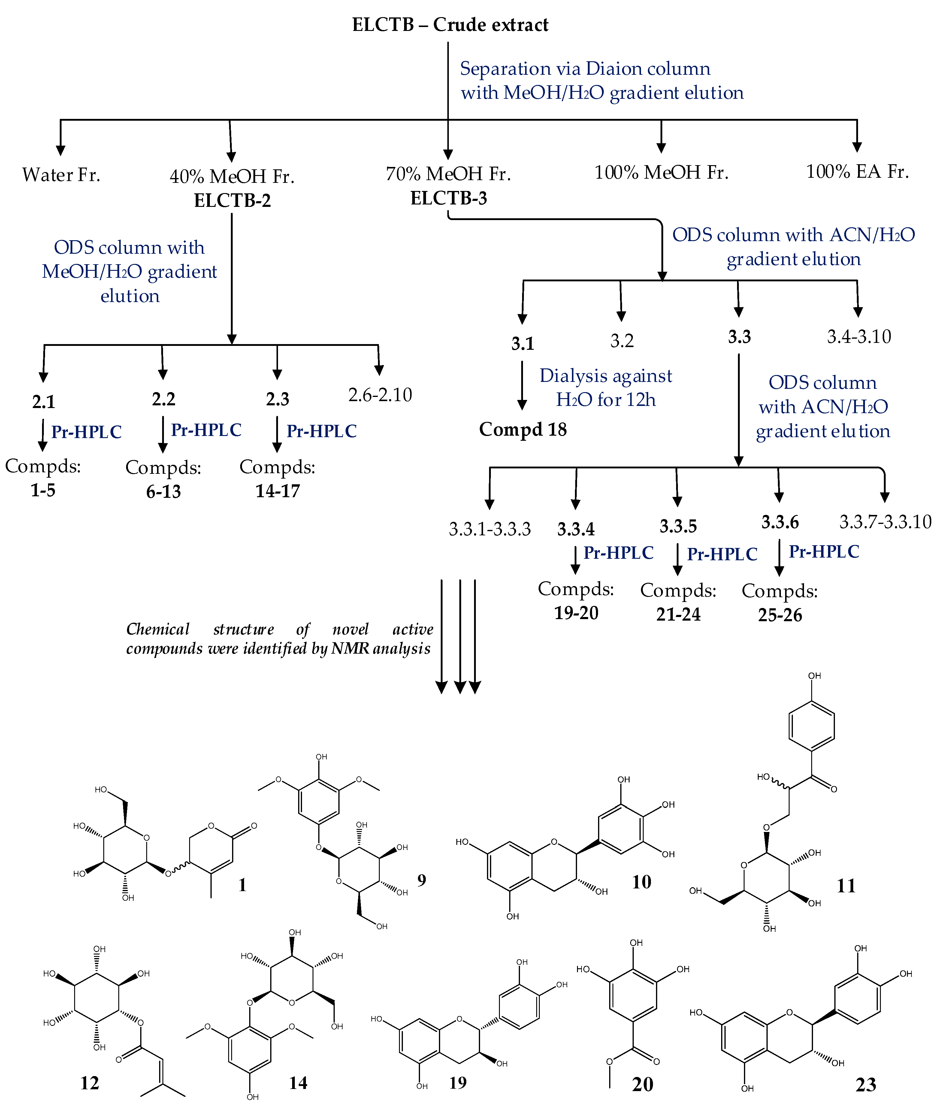
2.2.1. Separation, Subfractionation and Isolation of Compounds from ELCTB Extract
2.2.2. Evaluation and Identification of Active aGIs
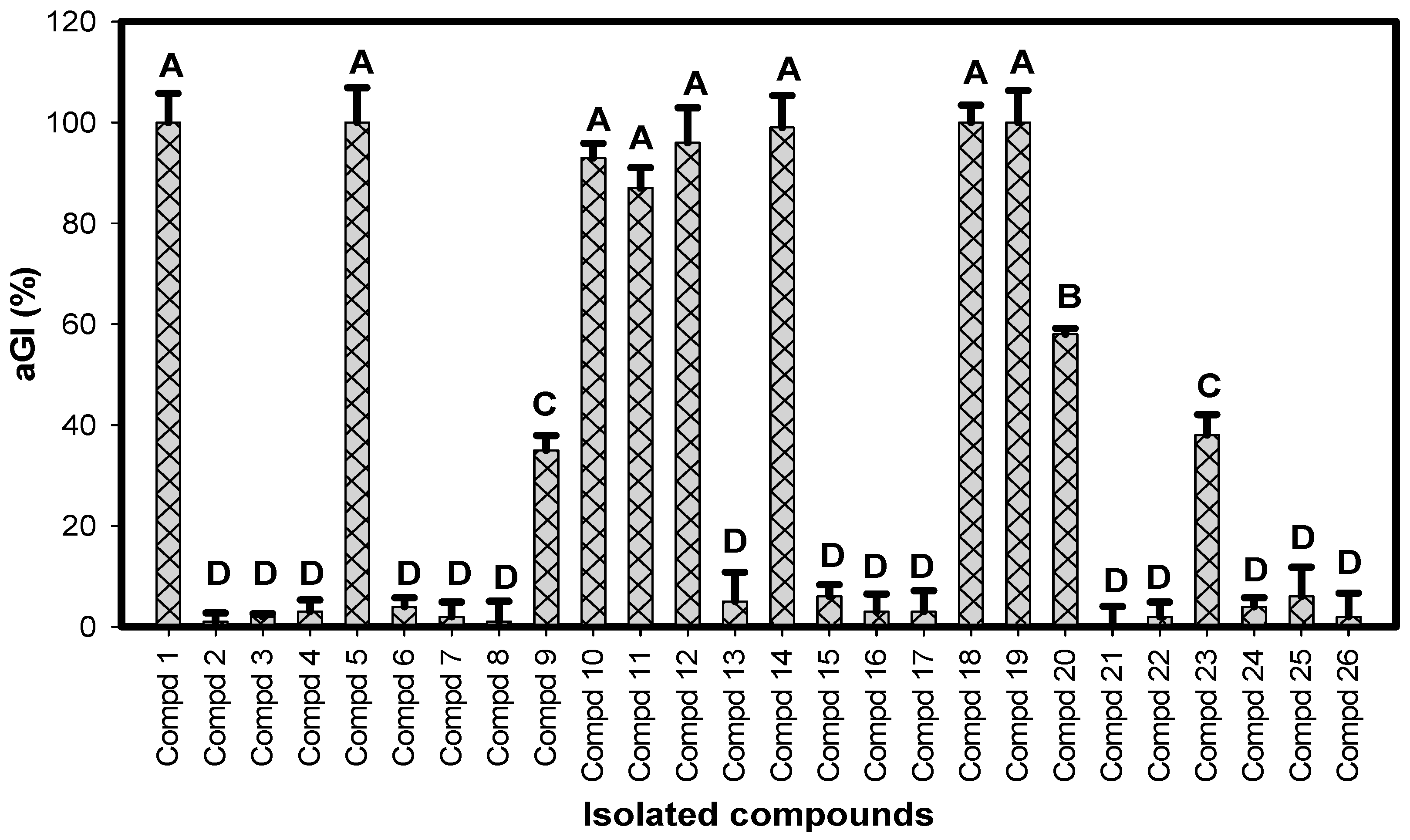
2.3. Comparision of α-Glucosidase Inhibitory Activity of Identified Compounds
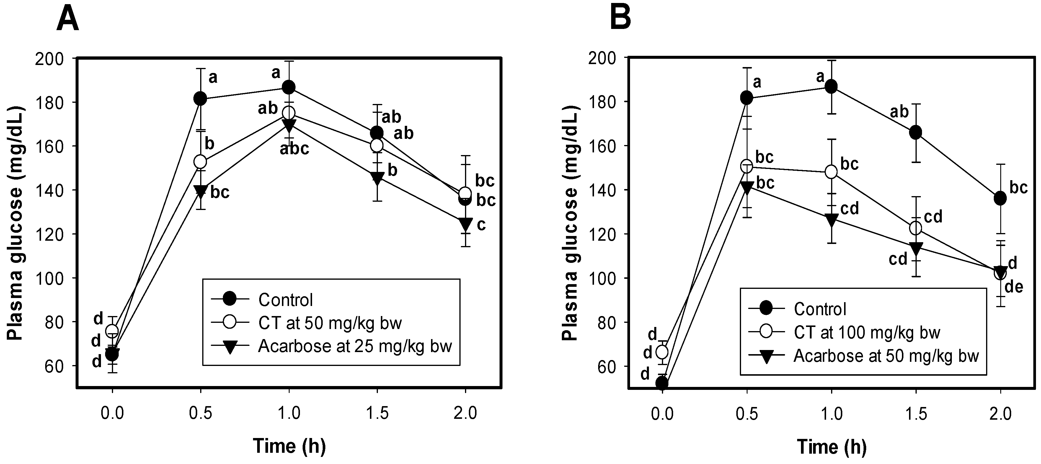
2.4. The Effect of Condensed Tannin (CT) on Reducing Plasma Glucose in a Mouse Model
3. Materials and Methods
3.1. Materials
3.2. Biological Activity Assays
3.2.1. α-Glucosidase Inhibitory Activity Determination
3.2.2. Experimental Animal Protocol
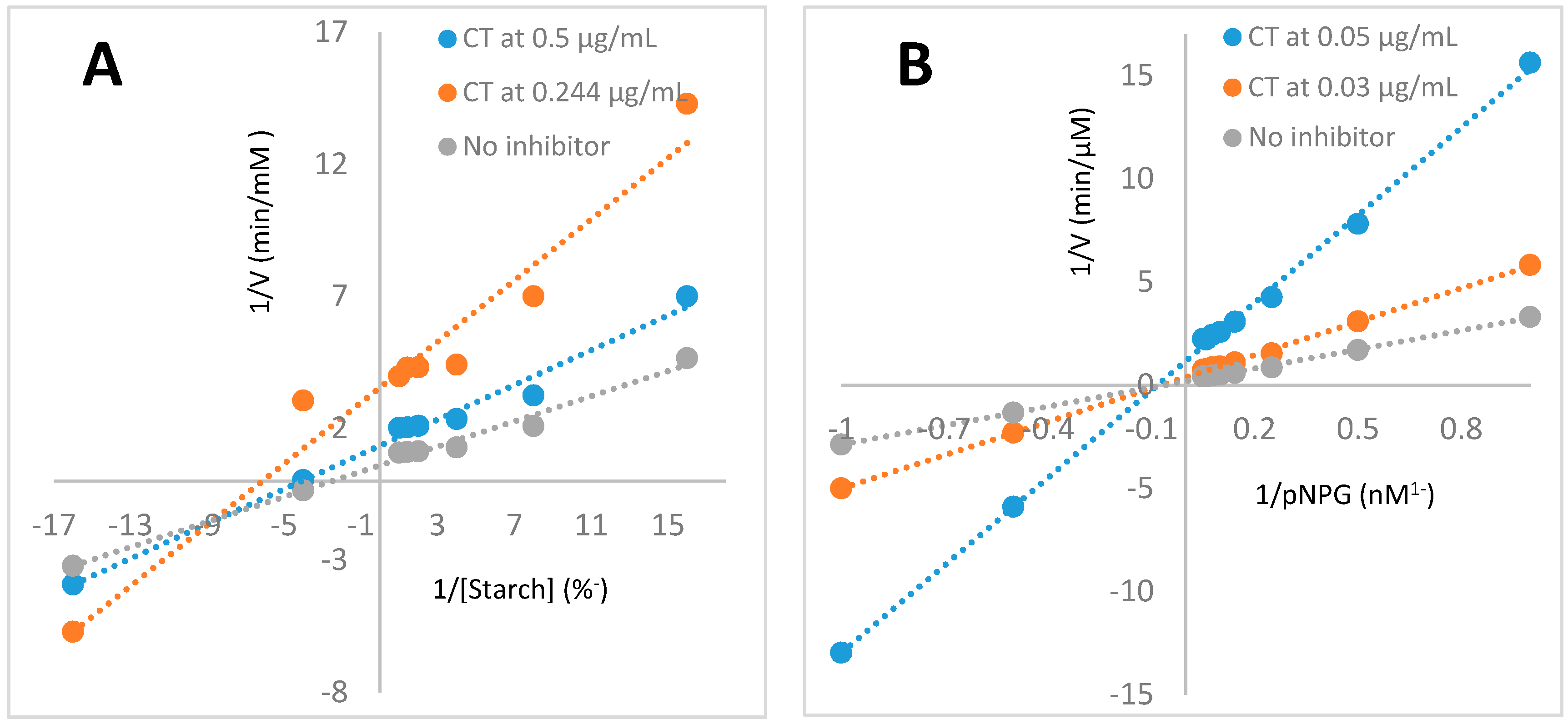
3.2.3. Determination of Enzymatic Inhibition Modes of Isolated Condensed Tannin
3.3. Purification and Identification Procedures of the Active aGIs
3.4. Characteristics and NMR Data of Identified Compounds
3.5. Statistical Analysis
4. Conclusions
Supplementary Materials
Authors Contributions
Funding
Acknowledgments
Conflicts of Interest
References
- Nguyen, V.B.; Nguyen, T.H.; Doan, C.T.; Tran, T.N.; Nguyen, A.D.; Kuo, Y.H.; Wang, S.L. Production and bioactivity-guided isolation of antioxidants with α-glucosidase inhibitory and anti-NO properties from marine chitinous materials. Molecules 2018, 23, 1124. [Google Scholar] [CrossRef] [PubMed]
- Nguyen, V.B.; Nguyen, A.D.; Wang, S.L. Utilization of fishery processing by-product squid pens for α-glucosidase inhibitors production by Paenibacillus sp. Mar. Drugs 2017, 15, 274. [Google Scholar] [CrossRef] [PubMed]
- DeMelo, E.B.; Gomes, A.; Carvalha, I. α-and β-Glucosidase inhibitors: Chemical structure and biological activity. J. Tetrahedr. 2006, 62, 10277–10302. [Google Scholar]
- Nguyen, V.B.; Nguyen, Q.V.; Nguyen, A.D.; Wang, S.L. Screening and evaluation of α-glucosidase inhibitors from indigenous medicinal plants in Dak Lak Province, Vietnam. Res. Chem. Intermed. 2017, 43, 3599–3612. [Google Scholar] [CrossRef]
- Nguyen, Q.V.; Nguyen, V.B.; Eun, J.B.; Wang, S.L.; Nguyen, D.H.; Tran, T.N.; Nguyen, A.D. Anti-oxidant and antidiabetic effect of some medicinal plants belong to Terminalia species collected in Dak Lak Province, Vietnam. Res. Chem. Intermed. 2016, 42, 5859–5871. [Google Scholar] [CrossRef]
- Nguyen, Q.V.; Wang, S.L.; Nguyen, A.D. In vitro α-glucosidase and α-amylase inhibition, and in vivo anti-hyperglycemic effects of Psidium littorale Raddi leaf extract. Res. Chem. Intermed. 2018, 44, 1745–1753. [Google Scholar] [CrossRef]
- Nguyen, V.B.; Wang, S.-L.; Nhan, N.T.; Nguyen, T.H.; Nguyen, N.P.D.; Nghi, D.H.; Cuong, N.M. New records of potent in-vitro antidiabetic properties of Dalbergia tonkinensis heartwood and the bioactivity-guided isolation of active compounds. Molecules 2018, 23, 1589. [Google Scholar] [CrossRef] [PubMed]
- Nguyen, V.B.; Wang, S.L. Reclamation of marine chitinous materials for the production of α-glucosidase inhibitors via microbial conversion. Mar. Drugs 2017, 15, 350. [Google Scholar] [CrossRef] [PubMed]
- Wang, S.L.; Su, Y.C.; Nguyen, V.B.; Nguyen, A.D. Reclamation of shrimp heads for the production of α-glucosidase inhibitors by Staphylococcus sp. TKU043. Res. Chem. Intermed. 2018, 44, 4929–4937. [Google Scholar] [CrossRef]
- Hsu, C.H.; Nguyen, V.B.; Nguyen, A.D.; Wang, S.L. Conversion of shrimp heads to α-glucosidase inhibitors via co-culture of Bacillus mycoides TKU040 and Rhizobium sp. TKU041. Res. Chem. Intermed. 2017, 44, 4597–4607. [Google Scholar] [CrossRef]
- Nguyen, V.B.; Nguyen, A.D.; Kuo, Y.H.; Wang, S.L. Biosynthesis of α-glucosidase inhibitors by a newly isolated bacterium, Paenibacillus sp. TKU042 and its effect on reducing plasma glucose in mouse model. Int. J. Mol. Sci. 2017, 18, 700. [Google Scholar] [CrossRef] [PubMed]
- Nguyen, V.B.; Wang, S.L. New novel α-glucosidase inhibitors produced by microbial conversion. Process Biochem. 2018, 65, 228–232. [Google Scholar] [CrossRef]
- Wang, G.; Peng, Z.; Wang, J.; Li, X.; Li, J. Synthesis, in vitro evaluation and molecular docking studies of novel triazine-triazole derivatives as potential α-glucosidase inhibitors. Eur. J. Med. Chem. 2017, 125, 423–429. [Google Scholar] [CrossRef] [PubMed]
- Ghani, U. Re-exploring promising a-glucosidase inhibitors for potential development into oral anti-diabetic drugs: Finding needle in the haystack. Eur. J. Med. Chem. 2015, 103, 133–162. [Google Scholar] [CrossRef] [PubMed]
- Flora of China. Available online: http://www.efloras.org/florataxon.aspx?flora_id=2&taxon_id=200012808 (accessed on 10 May 2018).
- Nguyen, V.B.; Nguyen, Q.V.; Nguyen, A.D.; Wang, S.L. Porcine pancreatic α-amylase inhibitors from Euonymus laxiflorus Champ. Res. Chem. Intermed. 2017, 43, 259–269. [Google Scholar] [CrossRef]
- Nguyen, Q.V.; Nguyen, N.H.; Wang, S.L.; Nguyen, V.B.; Nguyen, A.D. Free radical scavenging and antidiabetic activities of Euonymus laxiflorus champ extract. Res. Chem. Intermed. 2017, 43, 5615–5624. [Google Scholar] [CrossRef]
- Nguyen, V.B.; Wang, S.L.; Nguyen, A.D.; Vo, T.P.K.; Zhang, L.J.; Nguyen, Q.V.; Kuo, Y.H. Isolation and identification of novel α-amylase inhibitors from Euonymus laxiflorus Champ. Res. Chem. Intermed. 2018, 44, 1411–1424. [Google Scholar] [CrossRef]
- Liu, L.M.; Cheng, S.F.; Shieh, P.C.; Lee, J.C.; Chen, J.J.; Ho, C.T.; Kuo, S.C.; Kuo, D.H.; Huang, L.J.; Way, T.D. The methanol extract of Euonymus laxiflorus, Rubia lanceolata and Gardenia jasminoides inhibits xanthine oxidase and reduce serum uric acid level in rats. Food Chem. Toxicol. 2014, 70, 179–184. [Google Scholar] [CrossRef] [PubMed]
- Shao, J.H.; Chen, J.; Zhao, C.C.; Shen, J.; Liu, W.Y.; Gu, W.Y.; Li, K.H. Insecticidal and α-glucosidase inhibitory activities of chemical constituents from Viburnum fordiae Hance. Nat. Prod. Res. 2018, 27, 1–6. [Google Scholar] [CrossRef] [PubMed]
- Nomizu, K.; Hashida, K.; Makino, R.; Ohara, S. Antioxidants from steamed used tea leaves and their reaction behavior. Biosci. Biotechnol. Biochem. 2008, 72, 1682–1689. [Google Scholar] [CrossRef] [PubMed]
- Shrestha, S.; Lee, D.Y.; Park, J.H.; Cho, J.G.; Lee, D.S.; Li, B.; Kim, Y.C.; Kim, G.; Bang, M.H.; Baek, N.I. Phenolic components from Rhus parviflora fruits and their inhibitory effects on lipopolysaccharide-induced nitric oxide production in RAW 264.7 macrophages. Nat. Prod. Res. 2013, 27, 2244–2247. [Google Scholar] [CrossRef] [PubMed]
- Li, X.; Liu, Z.; Zhang, X.F.; Wang, L.J.; Zheng, Y.N.; Yuan, C.C.; Sun, G.Z. Isolation and characterization of phenolic compounds from the leaves of Salix matsudana. Molecules 2008, 13, 1530–1537. [Google Scholar] [CrossRef] [PubMed]
- Guo, P.; Anderson, J.D.; Bozell, J.J.; Zivanovic, S. The effect of solvent composition on grafting gallic acid onto chitosan via carbodiimid. Carbohydr. Polym. 2016, 140, 171–180. [Google Scholar] [CrossRef] [PubMed]
- Schofield, P.; Mbugua, D.M.; Pell, A.N. Analysis of condensed tannins: A review. Anim. Feed. Sci. Technol. 2001, 91, 21–40. [Google Scholar] [CrossRef]
- Li, C.; Leverence, R.; Trombley, J.D.; Xu, S.; Yang, J.; Tian, Y.; Reed, J.D.; Hagerman, A.E. High molecular weight persimmon (Diospyros kaki) proanthocyanidin: A highly galloylated, a-linked tannin with an unusual flavonol terminal unit, myricetin. J. Agric. Food Chem. 2010, 58, 9033–9042. [Google Scholar] [CrossRef] [PubMed]
- Chai, W.M.; Shi, Y.; Feng, H.L.; Qiu, L.; Zhou, H.C.; Deng, Z.W.; Yan, C.L.; Chen, Q.X. NMR, HPLC-ESI-MS, and MALDI-TOF MS analysis of condensed tannins from Delonix regia (Bojer ex Hook.) Raf. and their bioactivities. J. Agric. Food Chem. 2012, 60, 5013–5022. [Google Scholar] [CrossRef] [PubMed]
- Sadik, G.; Islam, R.; Rahman, M.M.; Khondkar, P.; Rashid, M.A.; Sarker, S.D. Antimicrobial and cytotoxic constituents of Loranthus globosus. Fitoterapia 2003, 74, 308–311. [Google Scholar] [CrossRef]
- Kwon, D.H.; Choi, W.J.; Lee, C.H.; Kim, J.H.; Kim, M.B. Flavonoid Compound Having an Antiviral Activity. U.S. Patent 7,998,937, 16 August 2011. [Google Scholar]
- Lianda, R.L.P.; Santana, L.D.; Echevarria, A.; Castro, R.N. Polyphenolic compounds in the fruits of Egyptian medicinal plants (Terminalia bellerica, Terminalia chebula and Terminalia horrida): Characterization, quantitation and determination of antioxidant capacities. J. Braz. Chem. Soc. 2012, 23, 618–627. [Google Scholar]
- Jing, H.; Xing, Y.F.; Huang, B.; Zhang, Y.Z.; Zeng, C.M. Tea catechins induce the conversion of preformed lysozyme amyloid fibrils to amorphous aggregates. J. Agric. Food Chem. 2009, 57, 11391–11396. [Google Scholar]
- Ngoumfo, R.M.; Ngounou, G.E.; Tchamadeu, C.V.; Qadir, M.I.; Mbazoa, C.D.; Begum, A.; Ngninzeko, F.N.; Lontsi, D.; Choudhary, M.I. Inhibitory effect of macabarterin, a polyoxygenated ellagitannin from Macaranga barteri, on human neutrophil respiratory burst activity. J. Nat. Prod. 2008, 71, 1906–1910. [Google Scholar] [CrossRef] [PubMed]
- Rawat, P.; Khan, M.F.; Kumar, M.; Tamarkar, A.K.; Srivastava, A.K.; Arya, K.R.; Maurya, R. Constituents from fruits of Cupressus sempervirens. Fitoterapia 2010, 81, 162–166. [Google Scholar] [CrossRef] [PubMed]
- Zhao, J.; Zhou, X.W.; Chen, X.B.; Wang, Q.X. α-Glucosidase inhibitory constituents from Toona sinensis. Chem. Nat. Compd. 2009, 45, 244–246. [Google Scholar] [CrossRef]
- Sanae, F.; Miyaichi, Y.; Kizu, H.; Hayashi, H. Effects of catechins on vascular tone in rat thoracic aorta with endothelium. Life Sci. 2002, 71, 2553–2562. [Google Scholar] [CrossRef]
- Yusoff, N.A.; Ahmad, M.; al Hindi, B.; Widyawati, T.; Yam, M.F.; Mahmud, R.; Razak, K.N.A.; Asmawi, M.Z. Aqueous extract of Nypa fruticans wurmb. Vinegar alleviates postprandial hyperglycemia in normoglycemic rats. Nutrients 2015, 7, 7012–7026. [Google Scholar] [CrossRef] [PubMed]
- Bryans, J.A.; Judd, P.A.; Ellis, P.R. The effect of consuming instant black tea on postprandial plasma glucose and insulin concentrations in healthy humans. J. Am. Coll. Nutr. 2007, 26, 471–478. [Google Scholar] [CrossRef] [PubMed]
- Igarashi, M.; Satoh, T.; Yamashita, H.; Wantanabe, K. Black tea inhibits small intestinal α-glucosidase activity in db/db mouse. Jpn. J. Complem. Altern. Med. 2014, 11, 25–33. [Google Scholar] [CrossRef]
- da Silva, S.M.; Koehnlein, E.A.; Bracht, A.; Castoldi, R.; de Morais, G.R.; Baesso, M.L.; Peralta, R.A.; de Souza, C.G.M.; de Sa-Nakanishi, A.B.; Peralta, R.M. Inhibition of salivary and pancreatic α-amylases by a pinhão coat (Araucaria angustifolia) extract rich in condensed tannin. Food Res. Int. 2014, 56, 1–8. [Google Scholar] [CrossRef]
- Ogawa, S.; Yazaki, Y. Tannins from acacia mearnsii de wild. bark: Tannin determination and biological activities. Molecules 2018, 23, 837. [Google Scholar] [CrossRef] [PubMed]
- Kumari, M.; Jain, S. Tannins: An antinutrient with positive effect to manage diabetes. Res. J. Recent Sci. 2012, 1, 70–73. [Google Scholar]
- Palmer, T. Understanding Enzymes, 3rd ed.; Ellis Horwood Limited: Chichester, UK, 1991; p. 155. [Google Scholar]
Sample Availability: Samples of the compounds including Walterolactone A/B β-d-pyranoglucoside, and condensed tannin-ELCTB-3.1 (compound 18) isolated from ELCTB extract are available from the authors. |


| Scientific Name of Medicinal Plants | Part Used | IC50 (µg/mL) | Ref. |
|---|---|---|---|
| Euonymus laxiflorus Champ. | Trunk bark | 360 ± 28.9 d,e | Nguyen et al., 2017 [4] |
| Euonymus laxiflorus Champ. | Leaves | 670 ± 40.4 d | Nguyen et al., 2017 [4] |
| Cinnamomum cassia J. S. Presl. | Trunk bark | 1080 ± 103.9 c | Nguyen et al., 2017 [4] |
| Terminalia bellirica | Leaves | 660 ± 63.5 d | Nguyen et al., 2017 [4] |
| Terminalia bellirica | Trunk bark | 410 ± 30.4 d,e | Nguyen et al., 2016 [5] |
| Terminalia corticosa | Trunk bark | 1420 ± 20.2 b,c | Nguyen et al., 2016 [5] |
| Psidium littorale Raddi | Leaves | 250 ± 10.4 e | Nguyen et al., 2018 [6] |
| Dalbergia tonkinensis | Heartwood | 1720 ± 116 b | Nguyen et al., 2018 [7] |
| Dalbergia tonkinensis | Trunk bark | 2910 ± 289 a | Nguyen et al., 2018 [7] |
| Dalbergia tonkinensis | Leaves | 2780 ± 173 a | Nguyen et al., 2018 [7] |
| Fractions/Subfractions | % Gradient of Solvent in Water | α-Glucosidase Inhibitory Activity | |
|---|---|---|---|
| IC50 (μg/mL) | Inhibition (%) * | ||
| Separation of ELCTB via a Diaion column with MeOH in H2O | |||
| ELCTB | 7.16 ± 0.21 | 98 ± 2.5 | |
| ELCTB-1 | 0 | 21.75 ± 1.32 | 67 ± 3.3 |
| ELCTB-2 | 40 | 2.80 ± 0.03 | 99 ± 3.1 |
| ELCTB-3 | 70 | 3.50 ± 0.12 | 98 ± 1.4 |
| ELCTB-4 | 100 | 18.50 ± 1.39 | 97 ± 2.3 |
| ELCTB-5 | 100% EA | - | - |
| Separation of ELCTB-2 via an ODS column with MeOH in H2O | |||
| ELCTB-2.1 | 0 | 6.20 ± 0.43 | 97 ± 3.5 |
| ELCTB-2.2 | 5 | 85.30 ± 4.03 | 97 ± 2.9 |
| ELCTB-2.3 | 10 | 26.03 ± 1.83 | 98 ± 1.8 |
| ELCTB-2.4 | 15 | 14.96 ± 0.78 | 98 ± 3.0 |
| ELCTB-2.5 | 20 | 6.56 ± 0.45 | 97 ± 2.0 |
| ELCTB-2.6 | 25 | 6.92 ± 0.73 | 99 ± 1.3 |
| ELCTB-2.7 | 30 | 72.95 ± 3.93 | 98 ± 1.0 |
| ELCTB-2.8 | 35 | 6.42 ± 0.41 | 98 ± 2.8 |
| ELCTB-2.9 | 40 | 10.75 ± 0.63 | 95 ± 3.7 |
| ELCTB-2.10 | 100 | - | - |
| Separation of ELCTB-3 via an ODS column with ACN in H2O | |||
| ELCTB-3.1 | 10 | 1.12 ± 0.03 | 100 ± 2.3 |
| ELCTB-3.2 | 15 | 6.21 ± 0.05 | 97 ± 2.0 |
| ELCTB-3.3 | 20 | 2.48 ± 0.07 | 97 ± 2.4 |
| ELCTB-3.4 | 25 | 1.64 ± 0.04 | 99 ± 3.6 |
| ELCTB-3.5 | 30 | 1.95 ± 0.08 | 97 ± 2.7 |
| ELCTB-3.6 | 35 | 1.89 ± 0.0 | 100 ± 2.0 |
| ELCTB-3.7 | 40 | 4.87 ± 0.12 | 99 ± 3.0 |
| ELCTB-3.8 | 45 | 4.34 ± 0.21 | 99 ± 3.4 |
| ELCTB-3.9 | 50 | 7.506 ± 0.32 | 96 ± 3.8 |
| ELCTB-3.10 | 100 | - | - |
| Acarbose | 1239 ± 78 | 64 ± 2.4 | |
| No. | Compound | IC50 (μg/mL) | Maximum Inhibition (%) |
|---|---|---|---|
| 1 | Walterolactone A/B β-d-pyranoglucoside | 0.907 ± 0.102 e | 100 ± 5.8 a |
| 5 | Condensed tannin-ELCTB-2.1.2 | 0.083 ± 0.004 e | 100 ± 6.9 a |
| 9 | 1-β-d-Glucopyranosyloxy-3,5-dimethoxy-4-hydroxybenzene | UD | 35 ± 2.9 c |
| 10 | (−)-Gallocatechin | 11.9 ± 1.674 d | 93 ± 2.9 a |
| 11 | Schweinfurthinol 9-O-β-d-pyranoglucoside | 31.6 ± 0.924 b | 87 ± 4.0 a |
| 12 | 1-O-(3-Methyl)-butenoyl-myo-inositol | 27.1 ± 1.212 c | 96 ± 6.9 a |
| 14 | Leonuriside | 0.926 ± 0.043 e | 99 ± 6.4 a |
| 18 | Condensed tannin-ELCTB-3.1. | 0.076 ± 0.008 e | 100 ± 6.2 a |
| 19 | (+)-Catechin | 0.113 ± 0.008 e | 100 ± 5.9 a |
| 20 | Methyl galloate | 110 ± 1.732 a | 58 ± 1.2 b |
| 23 | (−)-Catechin | UD | 38 ± 4.0 c |
| Acarbose (positive control) | 1345 ± 89 | 65 ± 2.7 | |
| Coefficient of variation (%) | 6.510292 | 8.746013 |
© 2018 by the authors. Licensee MDPI, Basel, Switzerland. This article is an open access article distributed under the terms and conditions of the Creative Commons Attribution (CC BY) license (http://creativecommons.org/licenses/by/4.0/).
Share and Cite
Nguyen, V.B.; Wang, S.-L.; Nguyen, T.H.; Nguyen, M.T.; Doan, C.T.; Tran, T.N.; Lin, Z.-H.; Nguyen, Q.V.; Kuo, Y.-H.; Nguyen, A.D. Novel Potent Hypoglycemic Compounds from Euonymus laxiflorus Champ. and Their Effect on Reducing Plasma Glucose in an ICR Mouse Model. Molecules 2018, 23, 1928. https://doi.org/10.3390/molecules23081928
Nguyen VB, Wang S-L, Nguyen TH, Nguyen MT, Doan CT, Tran TN, Lin Z-H, Nguyen QV, Kuo Y-H, Nguyen AD. Novel Potent Hypoglycemic Compounds from Euonymus laxiflorus Champ. and Their Effect on Reducing Plasma Glucose in an ICR Mouse Model. Molecules. 2018; 23(8):1928. https://doi.org/10.3390/molecules23081928
Chicago/Turabian StyleNguyen, Van Bon, San-Lang Wang, Thi Hanh Nguyen, Minh Trung Nguyen, Chien Thang Doan, Thi Ngoc Tran, Zhi-Hu Lin, Quang Vinh Nguyen, Yao-Haur Kuo, and Anh Dzung Nguyen. 2018. "Novel Potent Hypoglycemic Compounds from Euonymus laxiflorus Champ. and Their Effect on Reducing Plasma Glucose in an ICR Mouse Model" Molecules 23, no. 8: 1928. https://doi.org/10.3390/molecules23081928
APA StyleNguyen, V. B., Wang, S.-L., Nguyen, T. H., Nguyen, M. T., Doan, C. T., Tran, T. N., Lin, Z.-H., Nguyen, Q. V., Kuo, Y.-H., & Nguyen, A. D. (2018). Novel Potent Hypoglycemic Compounds from Euonymus laxiflorus Champ. and Their Effect on Reducing Plasma Glucose in an ICR Mouse Model. Molecules, 23(8), 1928. https://doi.org/10.3390/molecules23081928