
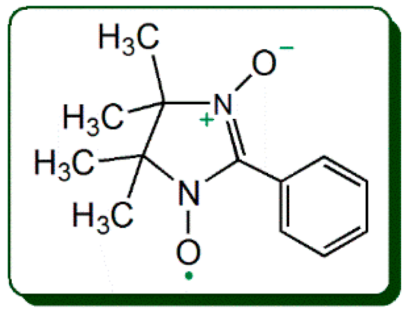
2-Phenyl-4,4,5,5-tetramethylimidazoline-1-oxyl 3-oxide Radical (PTIO•) Trapping Activity and Mechanisms of 16 Phenolic Xanthones


Abstract
:1. Introduction
2. Results and Discussion
2.1. Antioxidant Activity and Mechanism in PTIO•-Trapping Assay
2.2. Antioxidant Role of Phenolic -OH: Evidence From PTIO•-Trapping Assay
2.3. Antioxidant Role of Other Substituents: Evidence from the PTIO•-Trapping Assay
3. Materials and Methods
3.1. Chemicals and Animals
3.2. PTIO•-Trapping Colorimetric Assay
3.3. UPLC−ESI−Q−TOF−MS/MS Analysis of Xanthone Reaction Product with PTIO•
3.4. Statistical Analysis
4. Conclusions
Supplementary Materials
Author Contributions
Funding
Acknowledgments
Conflicts of Interest
Abbreviations
| ABTS•+ | 2,2′-azino-bis(3-ethylbenzothiazoline-6-sulphonic acid) |
| DPPH• | 1,1-diphenyl-2-picryl-hydrazl radical |
| ET | electron transfer |
| ET-PT | electron transfer−proton transfer |
| HAT | hydrogen atom transfer |
| PCET | proton coupled electron transfer |
| PTIO• | 2-phenyl-4,4,5,5-tetramethylimidazoline-1-oxyl 3-oxide radical |
| RAF | radical adduct formation |
| ROS | reactive oxygen species |
| SD | standard deviation |
| SPLET | sequential proton-loss electron-transfer |
| Trolox | (±)-6-hydroxy-2,5,7,8-tetramethlychromane-2-carboxylic acid |
| UPLC−ESI−Q−TOF−MS/MS | ultra-performance liquid chromatography coupled with electrospray ionization quadrupole time-of-flight tandem mass spectrometry |
References
- Han, Q.B.; Yang, L.; Wang, Y.L.; Qiao, C.F.; Song, J.Z.; Sun, H.D.; Xu, H.X. A pair of novel cytotoxic polyprenylated xanthone epimers from gamboges. Chem. Biodivers. 2006, 3, 101–105. [Google Scholar] [CrossRef] [PubMed]
- Li, H.L.; Li, X.M.; Liu, H.; Meng, L.H.; Wang, B.G. Two New Diphenylketones and a New Xanthone from Talaromyces islandicus EN-501, an Endophytic Fungus Derived from the Marine Red Alga Laurencia okamurai. Mar. Drugs 2016, 14, 223. [Google Scholar] [CrossRef] [PubMed]
- Yang, R.; Li, P.; Li, N.; Zhang, Q.; Bai, X.; Wang, L.; Xiao, Y.; Sun, L.; Yang, Q.; Yan, J. Xanthones from the Pericarp of Garcinia mangostana. Molecules 2017, 22, 683. [Google Scholar] [CrossRef] [PubMed]
- Jung, H.A.; Su, B.N.; Keller, W.J.; Mehta, R.G.; Kinghorn, A.D. Antioxidant xanthones from the pericarp of Garcinia mangostana (Mangosteen). J. Agric. Food Chem. 2006, 54, 2077–2082. [Google Scholar] [CrossRef] [PubMed]
- Ramirez, J.E.; Zambrano, R.; Sepúlveda, B.; Simirgiotis, M.J. Antioxidant Properties and Hyphenated HPLC-PDA-MS Profiling of Chilean Pica Mango Fruits (Mangifera indica L. Cv. piqueño). Molecules 2014, 19, 438–458. [Google Scholar] [CrossRef] [PubMed]
- Schieber, A.; Berardini, N.; Carle, R. Identification of flavonol and xanthone glycosides from mango (Mangifera indica L. Cv. Tommy Atkins) peels by high-performance liquid chromatography-electrospray ionization mass spectrometry. J. Agric. Food Chem. 2003, 51, 5006–5011. [Google Scholar] [CrossRef] [PubMed]
- Panda, S.S.; Chand, M.; Sakhuja, R.; Jain, S.C. Xanthones as Potential Antioxidants. Cur. Med. Chem. 2013, 20, 4481–4507. [Google Scholar] [CrossRef]
- Priya, V.; Jainu, M.; Mohan, S. Biochemical Evidence for the Antitumor Potential of Garcinia mangostana Linn. On Diethylnitrosamine-Induced Hepatic Carcinoma. Pharmacogn. Mag. 2018, 14, 186–190. [Google Scholar] [PubMed]
- Kryston, T.B.; Georgiev, A.B.; Pissis, P.; Georgakilas, A.G. Role of oxidative stress and DNA damage in human carcinogenesis. Mutat. Res. 2011, 711, 193–201. [Google Scholar] [CrossRef] [PubMed]
- Li, X. 2-Phenyl-4,4,5,5-tetramethylimidazoline-1-oxyl 3-Oxide (PTIO•) Radical Scavenging: A New and Simple Antioxidant Assay In Vitro. J. Agric. Food Chem. 2017, 65, 6288–6297. [Google Scholar] [CrossRef] [PubMed]
- Amorati, R.; Valgimigli, L. Methods to Measure the Antioxidant Activity of Phytochemicals and Plant Extracts. J. Agric. Food Chem. 2018, 66, 3324–3329. [Google Scholar] [CrossRef] [PubMed]
- Litwinienko, G.; Ingold, K.U. Solvent Effects on the Rates and Mechanisms of Reaction of Phenols with Free Radicals. Acc. Chem. Res. 2007, 40, 222–230. [Google Scholar] [CrossRef] [PubMed]
- Qin, H.L.; Yu, D.Q. 1H-NMR Spectroscopic Databook of Natural Products, 1st ed.; Chemical Industry Press: Beijing, China, 2011; pp. 881–2235. [Google Scholar]
- Yang, J.S. 13C-NMR Spectroscopic Databook of Natural Products; Chemical Industry Press: Beijing, China, 2011; pp. 1630–2285. [Google Scholar]
- Gómez-Zaleta, B.; Ramírez-Silva, M.T.; Gutiérrez, A.; González-Vergara, E.; Güizado-Rodríguez, M.; Rojas-Hernández, A. UV/vis, 1H, and 13C NMR spectroscopic studies to determine mangiferin pKa values. Spectrochim. Acta A Mol. Biomol. Spectrosc. 2006, 64, 1002–1009. [Google Scholar] [CrossRef] [PubMed]
- Bondet, V.; Brand-Williams, W.; Berset, C. Kinetics and mechanisms of antioxidant activity using the DPPH free radical method. LWT-Food Sci. Technol. 1997, 30, 609–615. [Google Scholar] [CrossRef]
- Krishnamachari, V.; Levine, L.H.; Zhou, C.; Pare, P.W. In vitro flavon-3-ol oxidation mediated by a B ring hydroxylation pattern. Chem. Res. Toxicol. 2004, 17, 795–804. [Google Scholar] [CrossRef] [PubMed]
- Fourre, I.; Di Meo, F.; Podloucka, P.; Otyepka, M.; Trouillas, P. Dimerization of quercetin, Diels-Alder vs. radical-coupling approach: A joint thermodynamics, kinetics, and topological study. J. Mol. Model. 2016, 22, 190. [Google Scholar] [CrossRef] [PubMed]
- Krishnamachari, V.; Levine, L.H.; Pare, P.W. Flavonoid oxidation by the radical generator AIBN: A unified mechanism for quercetin radical scavenging. J. Agric. Food Chem. 2002, 50, 4357–4363. [Google Scholar] [CrossRef] [PubMed]
- Burton, G.W.; Doba, T.; Gabe, E.; Ingold, K.U. Autoxidation of biological molecules. 4. Maximizing the antioxidant activity of phenols. J. Am. Chem. Soc. 1985, 107, 7053–7065. [Google Scholar] [CrossRef]
- Lucarini, M.; Pedulli, G.F. Free radical intermediates in the inhibition of the autoxidation reaction. Chem. Soc. Rev. 2010, 39, 2106–2119. [Google Scholar] [CrossRef] [PubMed]
- Heim, K.E.; Tagliaferro, A.R.; Bobilya, D.J. Flavonoid antioxidants: Chemistry, metabolism and structure-activity relationships. J. Nutr. Biochem. 2002, 13, 572–584. [Google Scholar] [CrossRef]
- Li, X.; Li, K.; Xie, H.; Xie, Y.; Li, Y.; Zhao, X.; Jiang, X.; Chen, D. Antioxidant and Cytoprotective Effects of the Di-O-Caffeoylquinic Acid Family: The Mechanism, Structure-Activity Relationship, and Conformational Effect. Molecules 2018, 23, 222. [Google Scholar] [CrossRef] [PubMed]
- De La Cruz, J.P.; Ruiz-Moreno, M.I.; Guerrero, A.; Lopez-Villodres, J.A.; Reyes, J.J.; Espartero, J.L.; Labajos, M.T.; Gonzalez-Correa, J.A. Role of the catechol group in the antioxidant and neuroprotective effects of virgin olive oil components in rat brain. J. Nutr. Biochem. 2015, 26, 549–555. [Google Scholar] [CrossRef] [PubMed]
- Li, X.; Wu, X.; Huang, L. Correlation between antioxidant activities and phenolic contents of radix angelicae sinensis (danggui). Molecules 2009, 14, 5349–5361. [Google Scholar] [CrossRef] [PubMed]
- Liu, J.; Li, X.; Lin, J.; Li, Y.; Wang, T.; Jiang, Q.; Chen, D. Sarcandra glabra (Caoshanhu) protects mesenchymal stem cells from oxidative stress: A bioevaluation and mechanistic chemistry. BMC Complement. Altern. Med. 2016, 16, 423. [Google Scholar] [CrossRef] [PubMed]
- Li, X.; Xie, Y.; Xie, H.; Yang, J.; Chen, D. π-π Conjugation Enhances Oligostilbene’s Antioxidant Capacity: Evidence from α-Viniferin and Caraphenol A. Molecules 2018, 23, 694. [Google Scholar] [CrossRef] [PubMed]
- Ali, H.M.; Ali, I.H. Energetic and electronic computation of the two-hydrogen atom donation process in catecholic and non-catecholic anthocyanidins. Food Chem. 2018, 243, 145–150. [Google Scholar] [CrossRef] [PubMed]
- Yamaguchi, L.F.; Lago, J.H.; Tanizaki, T.M.; Mascio, P.D.; Kato, M.J. Antioxidant activity of prenylated hydroquinone and benzoic acid derivatives from Piper crassinervium Kunth. Phytochemistry 2006, 67, 1838–1843. [Google Scholar] [CrossRef] [PubMed]
- Dong, L.M.; Jia, X.C.; Luo, Q.W.; Zhang, Q.; Luo, B.; Liu, W.B.; Zhang, X.; Xu, Q.L.; Tan, J.W. Phenolics from Mikania micrantha and Their Antioxidant Activity. Molecules 2017, 22, 1140. [Google Scholar] [CrossRef] [PubMed]
- Woodman, O.; Meeker, W.; Boujaoude, M. Vasorelaxant and antioxidant activity of flavonols and flavones: Structure-activity relationships. J. Cardiovasc. Pharm. 2005, 46, 302–309. [Google Scholar] [CrossRef]
- Valgimigli, L.; Amorati, R.; Fumo, M.G.; Dilabio, G.A.; Pedulli, G.F.; Ingold, K.U.; Pratt, D.A. The unusual reaction of semiquinone radicals with molecular oxygen. J. Org. Chem. 2008, 73, 1830–1841. [Google Scholar] [CrossRef] [PubMed]
- Li, X.; Wang, L.; Han, W.; Mai, W.; Han, L.; Chen, D. Amentoflavone protects against hydroxyl radical-induced DNA damage via antioxidant mechanism. Turk. J. Biochem. 2014, 39, 30–36. [Google Scholar] [CrossRef]
- Wang, T.T.; Li, X.C.; Li, Y.R.; Chen, D.F. Mechanistic chemistry of extraordinary capacity of salvianolic acid B on oxidatively damaged mesenchymal stem cells. J. Chin. Chem. Soc. 2016, 63, 924–929. [Google Scholar] [CrossRef]
- Apak, R.; Ozyurek, M.; Guclu, K.; Capanoglu, E. Antioxidant Activity/Capacity Measurement. 1. Classification, Physicochemical Principles, Mechanisms, and Electron Transfer (ET)-Based Assays. J. Agric. Food Chem. 2016, 64, 997–1027. [Google Scholar] [CrossRef] [PubMed]
- Li, T.; Zhang, H. Identification and comparative determination of rhodionin in traditional tibetan medicinal plants of fourteen Rhodiola species by high-performance liquid chromatography-photodiode array detection and electrospray ionization-mass spectrometry. Chem. Pharm. Bull. (Tokyo) 2008, 56, 807–814. [Google Scholar] [CrossRef] [PubMed]
- Guerreiro, E.; Kavka, J.; Giordano, O.S. 5,8-Dihydroxy-3,6,7-trimethoxyflavone from Gnaphalium gaudichaudianum. Phytochemistry 1982, 21, 2601–2603. [Google Scholar] [CrossRef]
- Briante, R.; Ferdinando Febbraio, A.; Nucci, R. Antioxidant Properties of Low Molecular Weight Phenols Present in the Mediterranean Diet. J. Agric. Food Chem. 2003, 51, 6975–6981. [Google Scholar] [CrossRef] [PubMed]
- Markovic, S.; Tosovic, J. Comparative study of the antioxidative activities of caffeoylquinic and caffeic acids. Food Chem. 2016, 210, 585–592. [Google Scholar] [CrossRef] [PubMed]
- Zhang, H.; Wu, W.; Mo, Y. Study of proton-coupled electron transfer (PCET) with four explicit diabatic states at the ab initio level. Comput. Theor. Chem. 2017, 1116, 50–58. [Google Scholar] [CrossRef]
- Amorati, R.; Baschieri, A.; Morroni, G.; Gambino, R.; Valgimigli, L. Peroxyl Radical Reactions in Water Solution: A Gym for Proton-Coupled Electron-Transfer Theories. Chem. Eur. J. 2016, 22, 7924–7934. [Google Scholar] [CrossRef] [PubMed]
- Chen, D.; Li, X.; Xu, Z.; Liu, X.; Du, S.; Li, H.; Zhou, J.; Zeng, H.; Hua, Z. Hexadecanoic Acid from Buzhong Yiqi Decoction Induced Proliferation of Bone Marrow Mesenchymal Stem Cells. J. Med. Food 2010, 13, 967–975. [Google Scholar] [CrossRef] [PubMed]
- Li, X.; Hu, Q.; Jiang, S.; Li, F.; Lin, J.; Han, L.; Hong, Y.; Lu, W.; Gao, Y.; Chen, D. Flos Chrysanthemi Indici protects against hydroxyl-induced damages to DNA and MSCs via antioxidant mechanism. J. Saudi. Chem. Soc. 2015, 19, 454–460. [Google Scholar] [CrossRef]
- Lin, J.; Li, X.; Chen, L.; Lu, W.; Chen, X.; Han, L.; Chen, D. Protective effect against hydroxyl radical-induced DNA damage and antioxidant mechanism of [6]-gingerol: A. Chemical Study. Bull. Korean Chem. Soc. 2014, 35, 1633–1638. [Google Scholar] [CrossRef]
- Li, X.; Han, L.; Li, Y.; Zhang, J.; Chen, J.; Lu, W.; Zhao, X.; Lai, Y.; Chen, D.; Wei, G. Protective Effect of Sinapine against Hydroxyl Radical-Induced Damage to Mesenchymal Stem Cells and Possible Mechanisms. Chem. Pharm. Bull. (Tokyo) 2016, 64, 319–325. [Google Scholar] [CrossRef] [PubMed]
- Li, X.; Liu, J.; Zhao, Z.; Wang, T.; Lin, J.; Chen, D. Effects of Natural Chalcone-Tannin Hybrids Protecting Mesenchymal Stem Cells against ROS-mediated Oxidative Damage and Indexes for Antioxidant Mechanisms. Chem. Lett. 2016, 45, 743–745. [Google Scholar] [CrossRef]
- Marin, M.; Manez, S. Recent trends in the pharmacological activity of isoprenyl phenolics. Curr. Med. Chem. 2013, 20, 272–279. [Google Scholar] [CrossRef] [PubMed]
- Xie, H.; Li, X.; Ren, Z.; Qiu, W.; Chen, J.; Jiang, Q.; Chen, B.; Chen, D. Antioxidant and cytoprotective effects of Tibetan tea and its phenolic components. Molecules 2018, 23, 179. [Google Scholar] [CrossRef] [PubMed]
- Li, X.; Xie, Y.; Li, K.; Wu, A.; Xie, H.; Guo, Q.; Xue, P.; Maleshibek, Y.; Zhao, W.; Guo, J.; et al. Antioxidation and Cytoprotection of Acteoside and Its Derivatives: Comparison and Mechanistic Chemistry. Molecules 2018, 23, 498. [Google Scholar] [CrossRef] [PubMed]
- Jiang, Q.; Li, X.C.; Tian, Y.G.; Lin, Q.Q.; Xie, H.; Lu, W.B.; Chi, Y.G.; Chen, D.F. Lyophilized Aqueous Extracts of Mori Fructus and Mori Ramulus Protect Mesenchymal Stem Cells from •OH-Treated Damage: Bioassay and Antioxidant Mechanism. BMC Complement. Altern. Med. 2017, 17, 242. [Google Scholar] [CrossRef] [PubMed]
Sample Availability: Sample of the mangiferin is available from the authors. |


| No | Xanthone | Main Results of UPLC-ESI-Q-TOF-MS/MS Analysis of Reaction Products | IC50 in Colorimetric Analysis/μM | Activity | ||||
|---|---|---|---|---|---|---|---|---|
| Retention Time/Min | Primary MS Spectra | MS/MS Spectra | Product | pH 4.5 | pH 7.4 | |||
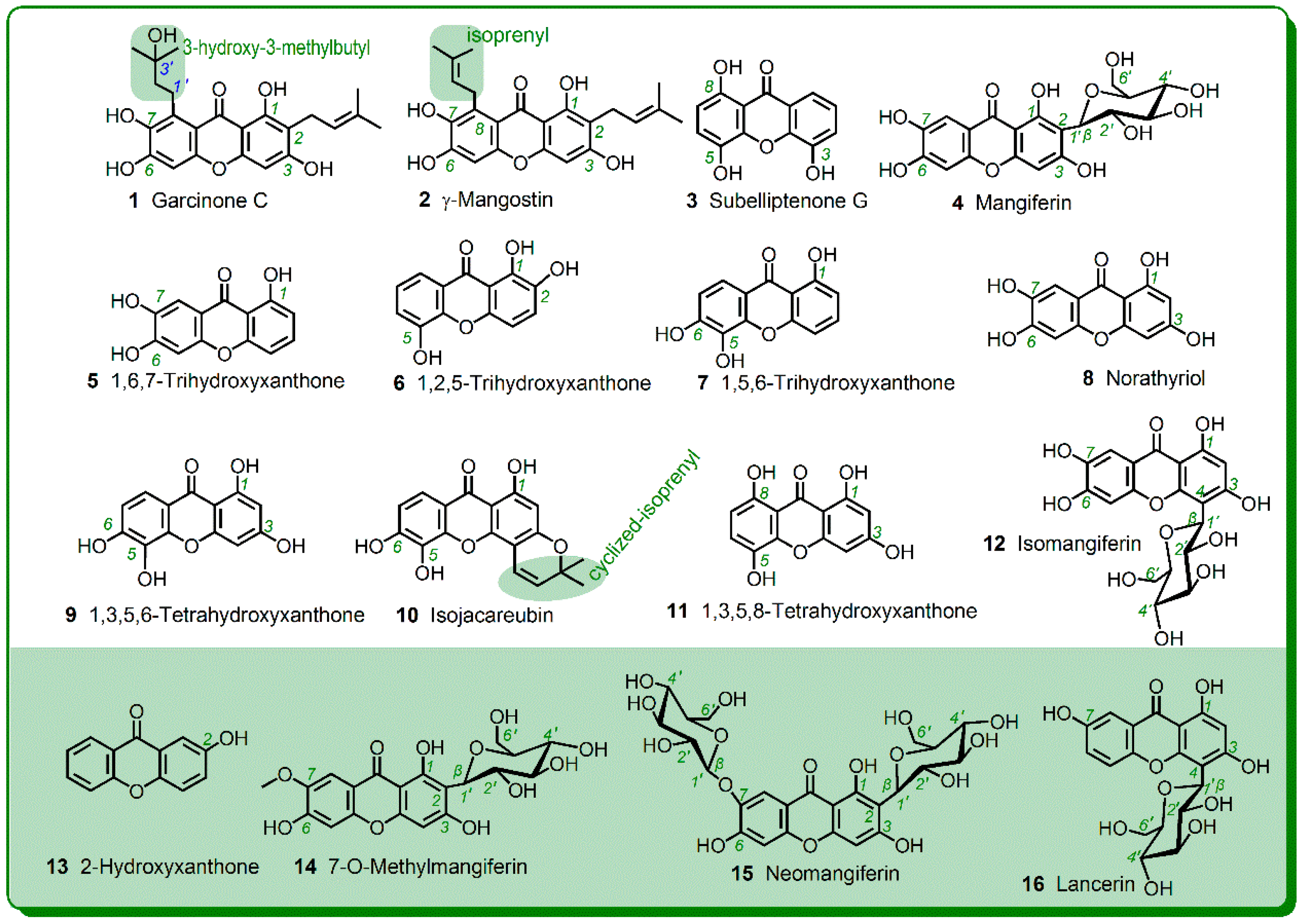
| 1 | Garcinone C | 5.961 | 825, 826, 827 | 353, 411, 825 | dimer | 36.0 ± 3.1 | 40.8 ± 2.0 | Strong |
| 2 | γ-Mangostin | 9.847 | 789, 790, 791 | 375, 393, 394, 395, 677 | dimer | 45.5 ± 2.4 | 60.4 ± 1.8 | |
| 3 | Subelliptenone G | 1.739 | 487, 488 | No data | dimer | 63.4 ± 13.3 | 110.9 ± 3.8 | |
| 4 | Mangiferin | 1.105 | 841, 842, 843, 844 | 329, 419, 601, 631, 661, 721,751, 823 | dimer | 64.1 ± 8.5 | 38.0 ± 2.7 | |
| 5 | 1,6,7-Trihydroxyxanthone | 1.813 | 485, 846, 487 | 243, 349 | dimer | 83.0 ± 2.2 | 90.3 ± 3.0 | |
| 6 | 1,2,5-Trihydroxyxanthone | 1.648 | 487, 488 | No data | dimer | 89.1 ± 7.4 | 112.2 ± 0.2 | |
| 7 | 1,5,6-Trihydroxyxanthone | 2.044 | 485, 486, 487, 488 | 243, 485 | dimer | 101.3 ± 16.6 | 116.3 ± 2.2 | |
| 8 | Norathyriol | 1.341 | 517, 518, 519, 520 | 229, 257, 258, 259, 365, 499, 517 | dimer | 103.0 ± 3.7 | 54.1 ± 0.9 | |
| 9 | 1,3,5,6-Tetrahydroxyxanthone | 1.349 | 517, 518, 519, 520 | 229, 257, 258, 259, 365, 499, 517 | dimer | 108.1 ± 19.4 | 102.7 ± 4.7 | |
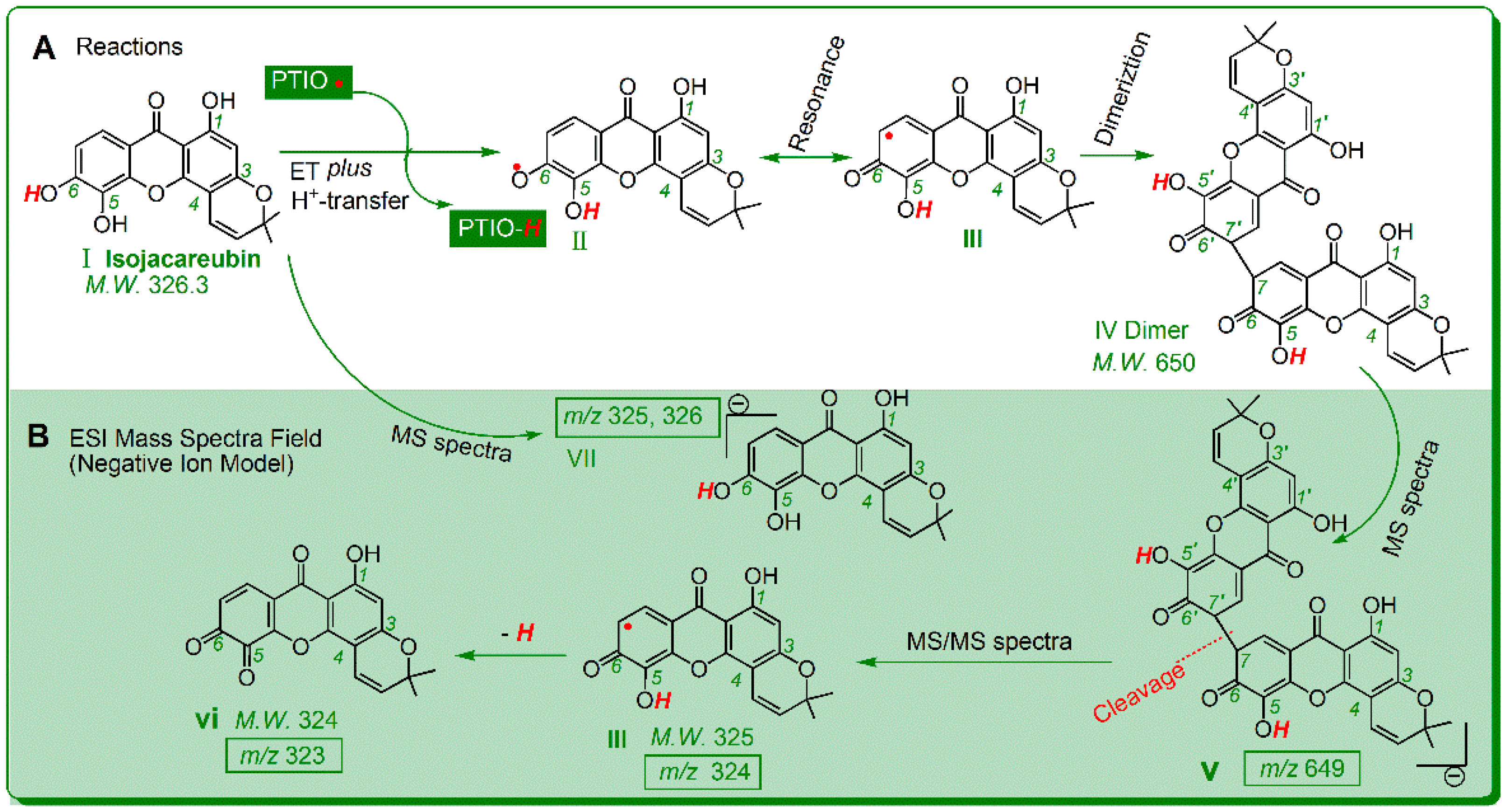
| 10 | Isojacareubin | 8.483 | 649, 650, 651, 652 | 323-325, 649 | dimer | 108.7 ± 0.1 | 136.7 ± 7.3 | |
| 11 | 1,3,5,8-Tetrahydroxyxanthone | 1.726 | 519, 520, 521 | 215, 259, 260 | dimer | 116.7 ± 12.6 | 133.1 ± 29.4 | |
| 12 | Isomangiferin | 1.105 | 841, 842, 843, 844 | 329, 419, 601, 631, 661, 721,751, 823 | dimer | 121.3 ± 8.3 | 104.1 ± 12.3 | |
| 13 | 2-Hydroxyxanthone | 1.885 | 423, 424 | No data | dimer | 284.2 ± 48.8 | 142.5 ± 13.1 | Weak |
| 14 | 7-O-Methylmangiferin | 1.204 | 871, 872 | No data | dimer | 387.2 ± 37.5 | 234.9 ± 1.7 | |
| 15 | Neomangiferin | No product | No product | No product | No product | 545.2 ± 15.2 | 213.1 ± 23.8 | |
| 16 | Lancerin | 1.165–1.191 | 811, 812 | No date | dimer | 681.2 ± 7.9 | 1106.3 ± 202.6 | |
© 2018 by the authors. Licensee MDPI, Basel, Switzerland. This article is an open access article distributed under the terms and conditions of the Creative Commons Attribution (CC BY) license (http://creativecommons.org/licenses/by/4.0/).
Share and Cite
Li, X.; Chen, B.; Zhao, X.; Chen, D. 2-Phenyl-4,4,5,5-tetramethylimidazoline-1-oxyl 3-oxide Radical (PTIO•) Trapping Activity and Mechanisms of 16 Phenolic Xanthones. Molecules 2018, 23, 1692. https://doi.org/10.3390/molecules23071692
Li X, Chen B, Zhao X, Chen D. 2-Phenyl-4,4,5,5-tetramethylimidazoline-1-oxyl 3-oxide Radical (PTIO•) Trapping Activity and Mechanisms of 16 Phenolic Xanthones. Molecules. 2018; 23(7):1692. https://doi.org/10.3390/molecules23071692
Chicago/Turabian StyleLi, Xican, Ban Chen, Xiaojun Zhao, and Dongfeng Chen. 2018. "2-Phenyl-4,4,5,5-tetramethylimidazoline-1-oxyl 3-oxide Radical (PTIO•) Trapping Activity and Mechanisms of 16 Phenolic Xanthones" Molecules 23, no. 7: 1692. https://doi.org/10.3390/molecules23071692
APA StyleLi, X., Chen, B., Zhao, X., & Chen, D. (2018). 2-Phenyl-4,4,5,5-tetramethylimidazoline-1-oxyl 3-oxide Radical (PTIO•) Trapping Activity and Mechanisms of 16 Phenolic Xanthones. Molecules, 23(7), 1692. https://doi.org/10.3390/molecules23071692