Article

Anthocidins A–D, New 5-Hydroxyanthranilic Acid Related Metabolites from the Sea Urchin-Associated Actinobacterium, Streptomyces sp. HDa1

1
Key Laboratory of Biology and Genetic Resources of Tropical Crops, Ministry of Agriculture, Institute of Tropical Bioscience and Biotechnology, Chinese Academy of Tropical Agricultural Sciences, Haikou 571101, China
2
Hainan Academy of Ocean and Fisheries Sciences, Haikou 570203, China
3
Hainan Testing Center for the Quality and Safety of Aquatic Products, Haikou 570206, China
*
Author to whom correspondence should be addressed.
These authors contributed equally to this work.
Molecules 2018, 23(5), 1032; https://doi.org/10.3390/molecules23051032
Submission received: 4 April 2018 / Revised: 24 April 2018 / Accepted: 25 April 2018 / Published: 27 April 2018

Abstract

:
Four new 5-hydroxyanthranilic acid related compounds, named anthocidins A–D (14), two known analogues n-lauryl 5-hydroxyanthranilate (5) and isolauryl 5-hydroxyanthranilate (6), together with benzamide (7), 3-hydroxy-4-methoxycinnamamide (8), and (3S-cis)-hexahydro-3-[(3,4-dihydroxyphenyl)methyl]pyrrolo[1,2-a]pyrazine-1,4-dione (9), were isolated from the fermentation broth of the marine-derived actinomycete, Streptomyces sp. HDa1, which was isolated from the gut of a sea urchin, Anthocidaris crassispina, collected from Hainan Island, China. The structures of these secondary metabolites were elucidated on the basis of their 1D and 2D-NMR and mass spectroscopic data, and anthocidin A was confirmed by single-crystal X-ray diffraction with Cu Kα radiation. Anthocidins A–D (14) feature an acetyl group substitution at the amino group and varying alkyl side chains at the carboxyl group of 5-hydroxyanthranilic acid, and compound 5 was isolated as a natural product for the first time. The cytotoxic and antibacterial activity of compounds 19 were evaluated.

1. Introduction

Natural products from actinomycetes have played a key role in drug discovery for the treatment of human diseases as exemplified by the immunosuppressant rapamycin; the antifungal agent nystatin; and the antibiotics tetracyclines, erythromycin, and vancomycin [1]. Despite the previous success of pharmaceutical compounds from actinomycetes, the constant need for the discovery of bioactive natural products has prompted the microbial natural product chemists to apply diverse strategies to identify novel secondary metabolites. One strategy is the exploration of the actinomycetes that inhabit un- or under-explored environments such as marine ecosystems. Marine-sourced actinomycetes have been proven to be rich sources of structurally diverse and biological active natural products [2,3]. A number of bioactive secondary metabolites featuring interesting structural properties have been discovered recently in marine-derived actinomycetes, such as tetrocarcin Q [4], fluostatins M–Q [5], succinilenes A–D [6], strepchazolins A and B [7], and a new napyradiomycin analogue [8], showing various potential antibacterial, antifungal, and antitumor activities. The under-explored marine animal-symbiont associations could provide a tremendous opportunity for the natural products discovery [9,10,11]. In our continuing efforts to search for novel bioactive natural products from marine microbes [12,13,14], recently we isolated an actinomycete strain Streptomyces sp. HDa1 from the gut of a sea urchin, Anthocidaris crassispina, collected from Hainan Island, China. Subsequent chemical study on the large-scale fermentation broth of this strain led to the discovery of four new 5-hydroxyanthranilic acid related compounds, anthocidins A–D (14), two known analogues n-lauryl 5-hydroxyanthranilate (5) and isolauryl 5-hydroxyanthranilate (6) [15], and benzamide (7) [16], 3-hydroxy-4-methoxycinnamamide (8) [17], and (3S-cis)-hexahydro-3-[(3,4-dihydroxyphenyl)methyl]pyrrolo[1,2-a]pyrazine-1,4-dione (9) [18] (Figure 1). Unlike the known analogues 5 and 6, anthocidins A–D feature varying alkyl side chains and possess an N-acetyl group at the C-2 position. In this paper, we describe the isolation and structure elucidation of these four new 5-hydroxyanthranilic acid derivatives as well as their cytotoxic and antibacterial activity.

2. Results

Anthocidin A (1) was obtained as light yellow needles. Its molecular formula was determined as C21H33NO4 on the basis of the high resolution electrospray ionization mass spectroscopy (HRESIMS) data at m/z 386.2309 [M + Na]+ (calcd for C21H33NO4Na, 386.2308) together with its NMR data (Table 1). In the 1H-NMR spectrum, the splitting patterns for three coupled aromatic protons (δH 8.53, d, J = 9.1 Hz, H-3; 7.15, dd, J = 9.1, 3.0 Hz, H-4; 7.55, d, J = 3.0 Hz, H-6) indicated the presence of a 1,2,4-trisubstituted benzene ring. The 1H-NMR data of 1 also showed three methyl groups (δH 2.25, 3H, s, H3-9; 0.87, 6H, d, J = 6.6 Hz, H3-11′ and H3-12′) and one exchangeable proton (δH 10.93, s, 2-NH). The 13C and DEPT135-NMR spectra revealed the presence of two carbonyl carbons (δC 169.4 and 168.1), six aromatic carbons (δC 116.8–151.7) (including three methine and three quaternary carbons), one sp3 methine (δC 27.9), nine sp3 methylene (δC 26.0–65.7) and three methyl carbons (δC 22.7–25.2). The HSQC spectrum of 1 allowed all protons to be assigned to their respective carbons and the structure of anthocidin A (1) was elucidated by the interpretation of its HMBC and 1H-1H COSY correlations (Figure 2). In the HMBC spectrum, the correlations from the aromatic signal H-3 to C-1 (δC 116.8) and C-5 (δC 151.7), from the aromatic signal H-6 to C-2 (δC 134.1), C-4 (δC 121.8) and the carboxyl carbon C-7 (δC 168.1) and from 2-NH to C-1 and C-3 (δC 122.1) indicated the presence of a 1,2,4-trisubstituted benzene, which could be also supported by the 1H-1H COSY correlation from H-3 to H-4. In addition, the HMBC correlations from 2-NH and H3-9 to the amide carbon C-8 (δC 169.4) suggested an N-acetyl group at the C-2 position. The last partial structure was identified as an isolauryl alcohol moiety which was deduced by the interpretation of the clear 1H-1H COSY correlations from two terminal methyl groups (H-11′ and H-12′) to a methine proton H-10′ (δH 1.53, m), from H-9′ (δH 1.16, m) to H-10′ and H-8′ (δH 1.20–1.38, m), from H-2′ (δH 1.77, m) to H-1′ (δH 4.31, t, J = 6.7 Hz) and H-3′ (δH 1.45, m), and the overlapped COSY correlations among the methylene protons. The connection of the isolauryl alcohol moiety to the C-7 on the 1,2,4-trisubstituted benzene ring through an oxygen bridge was secured by the HMBC correlations from the oxygenated methylene protons H-1′ to C-7. In addition, analysis of the HRESIMS and NMR data revealed one hydroxyl group could be located at C-5 on the 1,2,4-trisubstituted benzene ring. Finally, the structure of anthocidin A (1) was elucidated as shown, which was also confirmed by single-crystal X-ray crystallographic analysis in a Cu Kα radiation in low temperature (Figure 3).
Anthocidin B (2) was obtained as light yellow needles with the molecular formula of C21H33NO4 as deduced from the positive HRESIMS data at m/z 386.2310 [M + Na]+ (calcd for C21H33NO4Na, 386.2308)), 1H and 13C-NMR data. The 1H and 13C-NMR data of 2 was almost identical to those of 1 (Table 1). Extensive comparative analysis of the MS and 1H, 13C, DEPT135, and HSQC NMR data of 2 with those of 1 disclosed the presence of one additional methylene group and the absence of one terminal methyl group in the lipophilic chain of 2. Therefore, the structure of 2 was determined as an analogue of 1 with a lauryl alcohol chain by complete analysis of the HSQC, 1H-1H COSY, and HMBC spectra.
Anthocidin C (3) was isolated as a light yellow solid and had the molecular formula C22H35NO4 by analysis of HRESIMS data at m/z 400.2466 [M + Na]+ (calcd for C22H35NO4Na, 400.2464) and NMR data (Table 1). Its mass data was found to be 14 Da higher than that of 1. The 1H and 13C-NMR data of 3 was almost identical to those of 1 except for one more methylene group in the lipid chain of 3. Thus, the structure of 3 was deduced to be an analogue of 1 with an isotridecyl alcohol chain by extensive NMR analysis.
Anthocidin E (4), isolated as a light yellow amorphous powder, gave a molecular formula of C20H31NO4 based on the HRESIMS data at m/z 372.2154 [M + Na]+ (calcd for C20H31NO4Na, 372.2151), and 1H and 13C-NMR data (Table 2). Comparison of the NMR data of 4 with those of 2 revealed that they are almost identical except for absence of one methylene group in 4. On the basis of the 2D NMR data including the HSQC, 1H–1H COSY, and HMBC data of 4, its structure was unambiguously assigned as shown, possessing an undecyl alcohol side chain.
Compounds 5 and 6 were characterized as n-lauryl 5-hydroxyanthranilate, a chemically-synthesized 5-hydroxyanthranilate ester, and isolauryl 5-hydroxyanthranilate, respectively, by comparison of their NMR data with those reported data [15]. The literature also reported that these compounds possessed potent in vitro 5-lipoxygenase inhibitory activity. Herein, compound 5 was isolated and reported as a natural product for the first time.
Compounds 19 were screened for their in vitro antibacterial activities against a variety of bacteria including Staphylococcus aureus, Streptococcus pyogenes, Bacillus subtilis, Vibrio harveyi, and Vibrio alginolyticus at a concentration of 10 mg/mL. As a result, only compound 8 showed weak activity against the Gram-positive bacterium Bacillus subtilis with inhibition zone of 3 mm, while compound 9 displayed weak activity against the Gram-negative bacterium Vibrio harveyi with inhibition zone of 1.5 mm. Due to their weak activity at a high concentration, we did not determine the minimum inhibitory concentration. Also, their in vitro cytotoxic activities against the human melanoma cell line A375 and human ovarian carcinoma cell line CaoV3 were tested. However, none of these natural products exhibited potent cytotoxicity against these human cancer cell lines at a concentration of 10 µM.

3. Materials and Methods

3.1. General Experimental Procedures

NMR data were recorded in CDCl3 using a Bruker DRX-600 spectrometer (600 MHz for 1H-NMR and 150 MHz for 13C-NMR) with TMS (tetramethylsilane) as the internal standard (δ in ppm, J in Hz) (Bruker Corporation, Karlsruhe, Germany). High resolution electrospray ionization mass spectra were obtained on an Agilent 6210 TOF LC-MS spectrometer (Agilent Technologies Inc., Palo Alto, CA, USA). Silica gel (200–300 mesh, Qingdao Marine Chemical Factory, Qingdao, China) and Sephadex LH-20 (GE Healthcare Bio-Sciences AB, Uppsala, Sweden) were used for column chromatography. Semipreparative reverse-phase (RP) HPLC was conducted on an Hitachi HPLC system (Hitachi High-Technologies Corporation, Tokyo, Japan) consisting of a Hitachi L-7110 pump (Hitachi) and a Hitachi L-7420 UV–vis detector equipped with a Hypersil RP-C18 column (5 µm, 250 × 10.0 mm, Thermo Fisher Scientific, Waltham, MA, USA).

3.2. Strain Isolation and Cultivation

The strain HDa1 was isolated by one of the authors (R.W.) from the gut of a sea urchin, Anthocidaris crassispina, collected from Hainan Island, China, using the ISP4 agar medium (consisting of 10.0 g/L soluble starch, 1.0 g/L K2PO4, 1.0 g/L MgSO4·7H2O, 1.0 g/L NaCl, 2.0 g/L (NH4)2SO4, 2.0 g/L CaCO3, 0.001 g/L FeSO4·7H2O, 0.001 g/L MnCl2·7H2O, 20 g/L agar, and deionized water, pH 7.2) supplemented with a final concentration of 50 µg/mL potassium dichromate. It has been identified as Streptomyces sp. by 16S rRNA sequence analysis (GenBank accession number is MG745333). A voucher specimen (HNHDa1) has been deposited in the Institute of Tropical Bioscience and Biotechnology, Chinese Academy of Tropical Agricultural Sciences. The strain was cultivated on ISP4 agar plates at 28 °C. After five days, the spores were inoculated into 10 500 mL-Erlenmeyer flasks, each containing 100 mL of sterile seed medium (Tryptone soy broth, 30 g/L) and cultivated for 3 days at 28 °C with 160 rpm/min. Then, 10 mL of the seed cultures were inoculated into 100 × 1000 mL-Erlenmeyer flasks with 200 mL of a fermentation medium consisting of 4 g/L yeast extract, 10 g/L malt extract, and 4 g/L glucose (pH 7.2) and fermented on a rotary shaker with 140 rpm/min at 28 °C for 10 days.

3.3. Extraction and Purification

The entire filtrate of the fermentation broth (about 20 L) was harvested and 4% XAD-16N resin was added. Then the mixture was oscillated on a rotary shaker with 160 rpm/min for 4 h. After that, the resin was collected and extracted four times with methanol at room temperature. Subsequently, the methanol extract was evaporated to dryness under reduced pressure to yield a crude extract (8.0 g), which was then fractionated by silica gel column chromatography (CC) using a gradient elution of petroleum ether/EtOAc (v/v, 100:0, 50:1, 20:1, 10:1, 5:1, 2:1, 1:1, and 0:100) to give eight fractions (Fr.1–Fr.8). Fr.5 (petroleum ether/EtOAc, v/v, 5:1) was subsequently subjected to ODS CC with a gradient of MeOH/H2O (v/v, 30:70, 40:60, 50:50, 60:40, 70:30, and 100:0) to give six subfractions (Fr.5.1–Fr.5.6). Fr.5.3 (MeOH/H2O, 50:50) was further purified by semipreparative RP-HPLC to yield compounds 1 (6.5 mg), 2 (5.1 mg), 3 (3.0 mg), and 7 (2.3 mg). Fr.6 (petroleum ether/EtOAc, v/v, 2:1) was subjected to Sephadex LH-20 CC using MeOH as eluents to give eight subfractions, which were further purified by semipreparative RP-HPLC to yield compounds 4 (2.0 mg) and 8 (4.1 mg). Fr.7 (petroleum ether/EtOAc, v/v, 1:1) was subjected to ODS CC with a gradient of MeOH/H2O (v/v, 30:70, 40:60, 50:50, 60:40, 70:30, and 100:0) to give six subfractions. Fr.5.4 (MeOH/H2O, 60:40) was further purified by Sephadex LH-20 CC eluted by MeOH and finally by semipreparative RP-HPLC to generate compounds 5 (6.0 mg), 6 (8.2 mg), and 9 (3.2 mg).

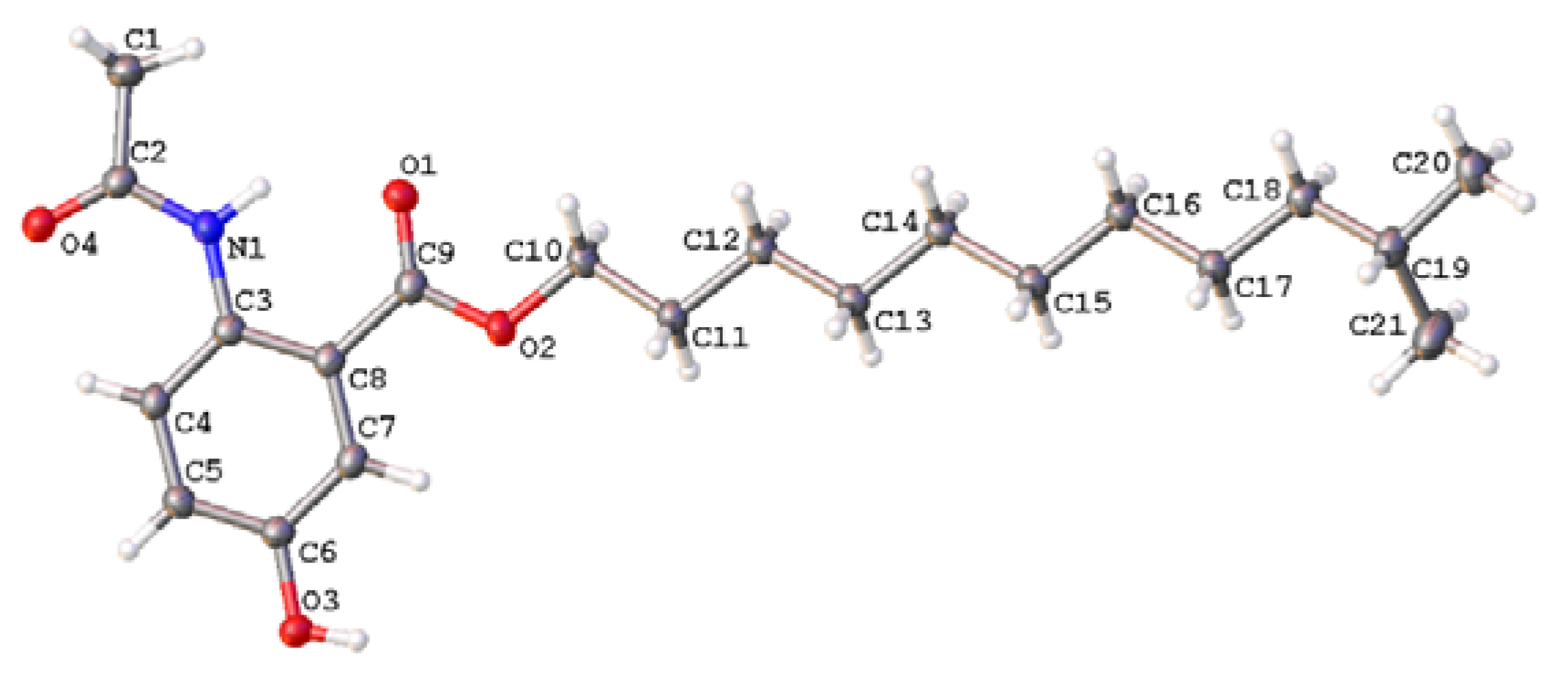
3.4. X-ray Single-Crystal Data of 1

The crystals of 1 were obtained by crystallization from a solution of MeOH/CH2Cl2 (v/v, 1:1). The single crystal X-ray diffraction data was obtained on a Bruker APEX-II diffractometer with Cu Kα radiation (λ = 1.54178 Å) at 130 K. The structure was solved using the program SHELXS-97 and refined by full-matrix least-squares on F2. Crystal data of compound 1 have been deposited with the Cambridge Crystallographic Data Center (deposition no. CCDC 1814418), which can be obtained free of charge via www.ccdc.cam.ac.uk/data_request/cif.
Crystal data for 1: molecular formula C21H33NO4, Mr = 363.48, monoclinic crystals, a = 5.3049 (7) Å, b = 15.916 (2) Å, c = 24.348 (3) Å, α = 90.00°, β = 97.794(11)°, γ = 90.00°, Z = 4, µ = 0.642 mm−1, F (000) = 792, and T = 130 K; Crystal dimensions: 0.12 × 0.08 × 0.06 mm3, Volume = 2055.6 (5) Å3, 9576 reflections measured, 3635 independent reflections (Rint = 0.0522), the final R indices [I > 2σ(I)] R1 = 0.0519, wR2 = 0.1348, R indices (all data) R1 = 0.0701, wR2 = 0.1482. The goodness of fit on F2 was 1.041.

3.5. Biological Assays

Cytotoxic activities of compounds 19 against the human melanoma cell line A375 and human ovarian carcinoma cell line CaoV3 were evaluated with the MTT assay [19]. The antibacterial activity of these compounds were also tested against Staphylococcus aureus, Streptococcus pyogenes, Bacillus subtilis, Vibrio harveyi, and Vibrio alginolyticus, in accordance with the previously reported method [20]. In the assays, the antibacterial activities were tested using the agar diffusion method with 7 mm paper discs containing 100 µg of compounds and rifampicin as the positive control. All tested compounds were dissolved in dimethyl sulfoxide (DMSO).

4. Conclusions

Four new 5-hydroxyanthranilic acid derivatives, anthocidins A–D (14), and n-lauryl 5-hydroxyanthranilate (5), isolauryl 5-hydroxyanthranilate (6), benzamide (7), 3-hydroxy-4-methoxycinnamamide (8), and (3S-cis)-hexahydro-3-[(3,4-dihydroxyphenyl)methyl]- pyrrolo[1,2-a]pyrazine-1,4-dione (9), were isolated from a sea urchin (Anthocidaris crassispina)-associated actinomycete, Streptomyces sp. HDa1. Their structures were determined by 1D and 2D-NMR and mass spectroscopic data, and anthocidin A was confirmed by single-crystal X-ray diffraction with Cu Kα radiation. Anthocidins A–D feature an acetamide group substitution at the amino group and varying ester chains at the carboxyl group of 5-hydroxyanthranilic acid, and compound 5 was isolated as a natural product for the first time. In the biological assays, compound 8 showed weak activity against the Gram-positive bacterium Bacillus subtilis, while compound 9 displayed weak activity against the Gram-negative bacterium Vibrio harveyi. However, none of these natural products exhibited potent cytotoxicity against these human cancer cell lines at a concentration of 10 µM. The result showed that the marine animal-symbiont actinomycetes could provide a good opportunity for discovering diverse new or bioactive natural products.

Supplementary Materials

The 1D and 2D-NMR spectra for compounds 14 are available online.

Author Contributions

Zhi-Kai Guo conceived and designed the experiments; Zhi-Kai Guo and Rong Wang cultured, isolated, and identified the compounds and performed the biological tests; Zhi-Kai Guo, Rong Wang, Shi-Quan Chen, Fu-Xiao Chen, Tian-Mi Liu, and Ming-Qiu Yang analyzed the data; Zhi-Kai Guo and Rong Wang wrote the paper.

Acknowledgments

This work was financially supported by the Hainan Provincial Natural Science Foundation of China (318QN300) and National Natural Science Foundation of China (41406083, 41776093, 41506096).

Conflicts of Interest

The authors declare no conflict of interest.

References

  1. Baltz, R.H. Renaissance in antibacterial discovery from actinomycetes. Curr. Opin. Pharmacol. 2008, 8, 557–563. [Google Scholar] [CrossRef] [PubMed]
  2. Blunt, J.W.; Copp, B.R.; Keyzers, R.A.; Munro, M.H.G.; Prinsep, M.R. Marine natural products. Nat. Prod. Rep. 2017, 34, 235–294. [Google Scholar] [CrossRef] [PubMed]
  3. Abdelmohsen, U.R.; Yang, C.; Horn, H.; Hajjar, D.; Ravasi, T.; Hentschel, U. Actinomycetes from red sea sponges: sources for chemical and phylogenetic diversity. Mar. Drugs 2014, 12, 2771–2789. [Google Scholar] [CrossRef] [PubMed] [Green Version]
  4. Gong, T.; Zhen, X.; Li, X.L.; Chen, J.J.; Chen, T.J.; Yang, J.L.; Zhu, P. Tetrocarcin Q, a new spirotetronate with a unique glycosyl group from a marine-derived actinomycete Micromonospora carbonacea LS276. Mar. Drugs 2018, 16, 74. [Google Scholar] [CrossRef] [PubMed]
  5. Jin, J.; Yang, X.Y.; Liu, T.; Xiao, H.; Wang, G.Y.; Zhou, M.J; Liu, F.W.; Zhang, Y.T.; Liu, D.; Chen, M.H.; et al. Fluostatins M–Q featuring a 6-5–6-6 ring skeleton and high oxidized A-rings from marine Streptomyces sp. PKU-MA00045. Mar. Drugs 2018, 16, 87. [Google Scholar] [CrossRef] [PubMed]
  6. Bae, M.; Park, S.H.; Kwon, Y.; Lee, S.K.; Shin, J.; Nam, J.W.; Oh, D.C. QM-HiFSA-aided structure determination of succinilenes A–D, new triene polyols from a marine-derived Streptomyces sp. Mar. Drugs 2017, 15, 38. [Google Scholar] [CrossRef] [PubMed]
  7. Yang, C.L.; Wang, Y.S.; Liu, C.L.; Zeng, Y.J.; Cheng, P.; Jiao, R.H.; Bao, S.X.; Huang, H.Q.; Tan, R.X.; Ge, H.M. Strepchazolins A and B: Two new alkaloids from a marine Streptomyces chartreusis NA02069. Mar. Drugs 2017, 15, 244. [Google Scholar] [CrossRef] [PubMed]
  8. Lacret, R.; Pérez-Victoria, I.; Oves-Costales, D.; De la Cruz, M.; Domingo, E.; Martín, J.; Díaz, C.; Vicente, F.; Genilloud, O.; Reyes, F. MDN-0170, a new napyradiomycin from Streptomyces sp. Strain CA-271078. Mar. Drugs 2016, 14, 188. [Google Scholar] [CrossRef] [PubMed]
  9. Abdelmohsen, U.R.; Bayer, K.; Hentschel, U. Diversity, abundance and natural products of marine sponge-associated actinomycetes. Nat. Prod. Rep. 2014, 31, 381–399. [Google Scholar] [CrossRef] [PubMed]
  10. Eltamany, E.E.; Abdelmohsen, U.R.; Ibrahim, A.K.; Hassanean, H.A.; Hentschel, U.; Ahmed, S.A. New antibacterial xanthone from the marine sponge-derived Micrococcus sp. EG45. Bioorg. Med. Chem. Lett. 2014, 24, 4939–4942. [Google Scholar] [CrossRef] [PubMed]
  11. Abdelmohsen, U.R.; Cheng, C.; Viegelmann, C.; Zhang, T.; Grkovic, T.; Ahmed, S.; Quinn, R.J.; Hentschel, U.; Edrada-Ebel, R. Dereplication strategies for targeted isolation of new antitrypanosomal actinosporins A and B from a marine sponge associated-Actinokineospora. sp. EG49. Mar. Drugs 2014, 12, 1220–1244. [Google Scholar] [CrossRef] [PubMed] [Green Version]
  12. Guo, Z.K.; Zhou, Y.Q.; Han, H.; Wang, W.; Xiang, L.; Deng, X.Z.; Ge, H.M.; Jiao, R.H. New antibacterial phenone derivatives asperphenone A–C from mangrove-derived fungus Aspergillus sp. YHZ-1. Mar. Drugs 2018, 16, 45. [Google Scholar] [CrossRef] [PubMed]
  13. Guo, Z.K.; Gai, C.J.; Cai, C.H.; Chen, L.L.; Liu, S.B.; Zeng, Y.B.; Yuan, J.Z.; Mei, W.L.; Dai, H.F. Metabolites with insecticidal activity from Aspergillus fumigatus JRJ111048 isolated from mangrove plant Acrostichum. specioum endemic to Hainan island. Mar. Drugs 2017, 15, 381. [Google Scholar] [CrossRef] [PubMed]
  14. Wang, R.; Liu, T.M.; Shen, M.H.; Yang, M.Q.; Feng, Q.Y.; Tang, X.M.; Li, X.M. Spiculisporic acids B–D, three new γ-butenolide derivatives from a sea urchin-derived fungus Aspergillus sp. HDf2. Molecules 2012, 17, 13175–13182. [Google Scholar] [CrossRef] [PubMed]
  15. Ohkuma, H.; Tomita, K.; Hoshino, Y.; Suzuki, K.; Hasegawa, M.; Sawada, Y.; Konishi, M.; Hook, D.J.; Oki, T. 5-Hydroxyanthranilic acid derivatives as potent 5-lipoxygenase inhibitors. J. Antibiot. 1993, 46, 705–711. [Google Scholar] [CrossRef] [PubMed]
  16. De Kowalewski, D.G.; Kowalewski, V.J.; Botek, E.; Contreras, R.H.; Facelli, J.C. Experimental and theoretical study of the ethoxy group conformational effect on 13C chemical shifts in ortho-substituted phenetols. Magn. Reson. Chem. 1997, 35, 351–356. [Google Scholar] [CrossRef]
  17. Linder, J.; Moody, C.J. The total synthesis of siphonazole, a structurally unusual bis-oxazole natural product. Chem. Commun. 2007, 15, 1508–1509. [Google Scholar] [CrossRef] [PubMed]
  18. Bobylev, M.M.; Bobyleva, L.I.; Strobel, G.A. Synthesis and bioactivity of analogs of maculosin, a host-specific phytotoxin produced by Alternaria alternata on spotted knapweed (Centaurea maculosa). J. Agric. Food Chem. 1996, 44, 3960–3964. [Google Scholar] [CrossRef]
  19. Mosmann, T. Rapid colorimetric assay for cellular growth and survival: Application to proliferation and cytotoxicity assays. J. Immunol. Methods 1983, 65, 55–63. [Google Scholar] [CrossRef]
  20. Wiegand, I.; Hilpert, K.; Hancock, R.E.W. Agar and broth dilution methods to determine the minimal inhibitory concentration (MIC) of antimicrobial substances. Nat. Protoc. 2008, 3, 163–175. [Google Scholar] [CrossRef] [PubMed]
Sample Availability: Samples of the compounds 19 are available from the authors.
Figure 1. The structures of compounds 19.
Molecules 23 01032 g001
Figure 2. Key 1H–1H COSY (bold lines) and HMBC (arrows) correlations of 1.
Molecules 23 01032 g002
Figure 3. X-ray single-crystal structure of 1.
Molecules 23 01032 g003
Table 1. 1H and 13C-NMR data for anthocidins A–C (13) in CDCl3. a.
PositionAnthocidin A (1)Anthocidin B (2)Anthocidin C (3)
δCδH (J in Hz)δCδH (J in Hz)δCδH (J in Hz)
1116.8, C 116.8, C 116.7, C
2134.1, C 134.3, C 135.1, C
2-NH 10.93, s 10.88, s 10.84, s
3122.1, CH8.53, d (9.1)122.1, CH8.50, d (9.0)122.2, CH8.53, d (8.9)
4121.8, CH7.15, dd (9.1, 3.0)121.8, CH7.10, dd (9.0, 2.4)121.9, CH7.07, dd (8.9, 3.0)
5151.7, C 151.7, C 150.9, C
6116.9, CH7.55, d (3.0)116.9, CH7.52, d (2.4)116.8, CH7.50, d (3.0)
7168.1, C 168.1, C 168.1, C
8169.4, C 169.3, C 169.1, C
925.2, CH32.25, s25.3, CH32.22, s26.2, CH32.21, s
1′65.7, CH24.31, t (6.7)65.7, CH24.28, t (6.6)65.8, CH24.30, t (6.7)
2′28.6, CH21.77, m28.6, CH21.75, m28.7, CH21.76, m
3′26.0, CH21.45, m26.0, CH21.42, m26.2, CH21.43, m
4′27.4, CH21.20–1.38, m29.3 c, CH21.24–1.36, m22.0–39.0 d, CH21.20–1.40, m
5′29.3 b, CH21.20–1.38, m29.4 c, CH21.24–1.36, m22.0–39.0 d, CH21.20–1.40, m
6′29.5 b, CH21.20–1.38, m29.5 c, CH21.24–1.36, m22.0–39.0 d, CH21.20–1.40, m
7′29.7 b, CH21.20–1.38, m29.6 c, CH21.24–1.36, m22.0–39.0 d, CH21.20–1.40, m
8′29.9 b, CH21.20–1.38, m29.6 c, CH21.24–1.36, m22.0–39.0 d, CH21.20–1.40, m
9′39.1, CH21.16, m29.7 c, CH21.24–1.36, m22.0–39.0 d, CH21.20–1.40, m
10′27.9, CH1.53, m39.1, CH21.24–1.36, m22.0–39.0 d, CH21.20–1.40, m
11′22.7, CH30.87, d (6.6)22.8, CH21.24–1.36, m28.1, CH1.51, m
12′22.7, CH30.87, d (6.6)14.1, CH30.87, t (6.8)19.4, CH30.85, d (6.4)
13′ 11.5, CH30.83, d (6.4)
a 1H and 13C-NMR data were obtained at 600 and 150 MHz, respectively. δ in ppm. b–d interchangeable.
Table 2. 1H and 13C-NMR data for anthocidin D (4) in CDCl3. a
PositionAnthocidin D (4)
δCδH (J in Hz)PositionδCδH (J in Hz)
1116.4, C 1′65.7, CH24.30, t (6.7)
2135.2, C 2′28.6, CH21.77, m
2-NH 10.81, s3′26.0, CH21.43, m
3122.1, CH8.56, d (9.0)4′29.5 b, CH21.20–1.40, m
4121.7, CH7.04, dd (9.0, 3.0)5′29.6 b, CH21.20–1.40, m
5150.4, C 6′29.6 b, CH21.20–1.40, m
5-OH 7′29.3 b, CH21.20–1.40, m
6116.5, CH7.48, d (3.0)8′29.2 b, CH21.20–1.40, m
7167.9, C 9′31.9, CH21.20–1.40, m
8168.8, C 10′22.7, CH21.20–1.40, m
925.3, CH32.21, s11′14.1, CH30.88, t (7.0)
a 1H and 13C-NMR data were obtained at 600 and 150 MHz, respectively. δ in ppm. b interchangeable.

