Article

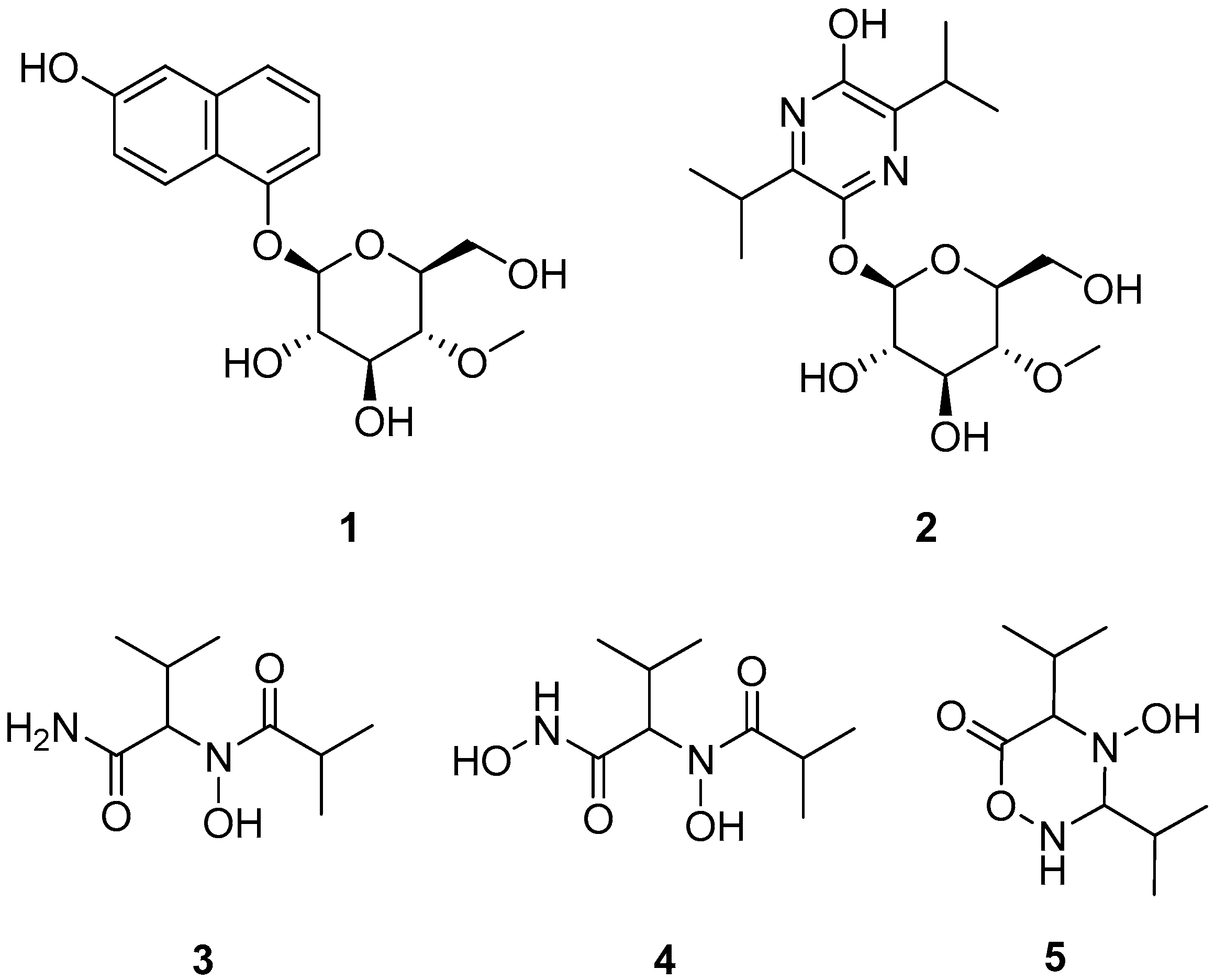
Five Unprecedented Secondary Metabolites from the Spider Parasitic Fungus Akanthomyces novoguineensis

by
Soleiman E. Helaly
1,2,†,
Wilawan Kuephadungphan
1,3,†,
Souwalak Phongpaichit
3,4,
Janet Jennifer Luangsa-ard
5,
Vatcharin Rukachaisirikul
6,7 and
Marc Stadler
1,*
1
Department of Microbial Drugs, Helmholtz Centre for Infection Research, 38124 Braunschweig, Germany
2
Department of Chemistry, Faculty of Science, Aswan University, Aswan 81528, Egypt
3
Department of Microbiology, Faculty of Science, Prince of Songkla University, Songkhla 90112, Thailand
4
Natural Products Research Center of Excellence and Department of Microbiology, Prince of Songkla University, Songkhla 90112, Thailand
5
National Centre for Genetic Engineering and Biotechnology (BIOTEC), Pathumthani 12120, Thailand
6
Department of Chemistry, Faculty of Science, Prince of Songkla University, Songkhla 90112, Thailand
7
Center of Excellence for Innovation in Chemistry, Prince of Songkla University, Songkhla 90112, Thailand
*
Author to whom correspondence should be addressed.
These authors contributed equally to this work.
Molecules 2017, 22(6), 991; https://doi.org/10.3390/molecules22060991
Submission received: 4 May 2017 / Revised: 31 May 2017 / Accepted: 1 June 2017 / Published: 14 June 2017

Abstract

:
Five new compounds including the glycosylated β-naphthol (1, akanthol), a glycosylated pyrazine (2, akanthozine), and three amide derivatives including a hydroxamic acid derivative (35) were isolated from the spider-associated fungus Akanthomyces novoguineensis (Cordycipitaceae, Ascomycota). Their structures were elucidated by using high resolution mass spectrometry (HRMS) and NMR spectroscopy. In this study, the antimicrobial, cytotoxic, anti-biofilm, and nematicidal activities of the new compounds were evaluated. The distribution pattern of secondary metabolites in the species was also revealed in which more isolates of A. novoguineensis were encountered and their secondary metabolite profiles were examined using analytical HPLC with diode array and mass spectrometric detection (HPLC-DAD/MS). Remarkably, all isolated compounds are specifically produced by A. novoguineensis.

Graphical Abstract

1. Introduction

For over decades, fungi that are closely associated with invertebrates are accepted as a rich source for a wide range of bioactive secondary metabolites [1,2]. The largest numbers of invertebrate-pathogenic fungi belong to the order Hypocreales in which many species have been investigated for a long time and have proven to be effective biocontrol agents as well as prolific producers of biologically active secondary metabolites [2,3]. So far, certain ecological groups of hypocrealean fungi, and in particular the parasites of spiders have not yet received much attention, despite the fact that their close phylogenetic relatives have been proven to be an attractive source of natural products. In a concurrent study, four unprecedented glycosylated α-pyrone derivatives, akanthopyrones A–D, were obtained from a culture of A. novoguineensis [4]. These findings lead us to further investigate this species and its allies. As a part of our ongoing research on biologically active fungal metabolites [5], the constituents of several A. novoguineensis isolates were compared. Scale-up fermentation and subsequent extensive chromatography of the organic crude extracts led to the isolation of five new compounds. Herein, we report on the isolation, structure elucidation, biological activities of the new compounds (15), and the species-specific pattern of secondary metabolite production in A. novoguineensis.

2. Results and Discussion

Compounds 13 were isolated from the ethyl acetate extract of A. novoguineensis BCC47894 as described in the experimental section, while Compounds 4 and 5 were obtained from the ethyl acetate extract of A. novoguineensis BCC47881 (Figure 1).
Compound 1 was obtained as a pale brown gum. HRMS afforded the molecular ion cluster at m/z 359.1107 [M + Na]+, which furnished the molecular formula of C17H28O7 (calcd. for [M + Na]+ 359.1101). The 1H- and 13C-NMR data revealed the presence of six sp2 olefinic carbons, five sp3 methine carbons, one of which is anomeric carbon (δH 5.07, δC 102.5), hence suggesting the presence of sugar moiety in 1, one methylene carbon, one methoxy carbon, and four sp2 quaternary carbons. The sugar moiety was assigned as 4-O-methyl-glucopyranose based on COSY and HMBC spectra. In addition to the sugar signals, only signals in the aromatic region were observed. Thus, the aglycone was determined by COSY correlation between 3-H/4-H and between 6-H/7-H/8-H together with a series of HMBC correlations from 8-H to C-1/C-4a/C-6, from 6-H to C-4/C-4a/C-8, from 7-H to C-5/C-8a, and from 4-H to C-2/C-8a/C-5 allowed the construction of a 2-naphthol substructure (Figure 2). Finally, the glycosidic linkage at C-5 was evident by HMBC correlation from the anomeric methine (H-1′) to C-5.
The relative configuration of the sugar moiety in 1 was determined by NOESY data and coupling constant analysis. Starting from a characteristic H-1′ signal of a β-glycoside at δH 5.07 with J1′,2′ = 8.2 Hz a chain of vicinal trans couplings of J2′,3′ = 8.6 Hz, J3′,4′ = 8.6 Hz, and J4′,5′ = 9.5 Hz supported by NOEs between H-1′ and H-3′/H-5′ determined the configuration of the sugar moiety as 4-O-methyl-β-glucopyranose. Its β-glycoside connection was evident by a 1JC,H coupling of 162 Hz for the anomeric methine. The d-configuration of the sugar was established by comparing the specific rotation of the aqueous layer of its acid hydrolysate [ α ] D 25 +36, c 0.05, MeOH) with that of 4-O-methyl-d-glucopyranose [ α ] D 25 +80, c 1.3, MeOH) [6]. Consequently, Compound 1 was determined as the new natural product, β-naphthol bearing 4-O-methyl-β-d-glucopyranose, for which we have proposed the trivial name akanthol.
Akanthozine (2) possesses the molecular formula of C17H28N2O7 as determined from the molecular ion peak at m/z 373.1971 [M + H]+ (calcd. 373.1969). The sugar moiety signals were preserved in the NMR data of 2. In addition, the remaining 10 carbon signals were determined as follow: four douplets assigned to two sets of germinal methyls, two methines, and four sp2 quaternary carbons. COSY spectra showed correlations between the germinal methyls H3-8/H3-9 and the methine H-7 and between the germinal methyls H3-11/H3-12 and the methine H-10. Furthermore, HMBC correlations from the germinal methyls H3-8/H3-9 to C-3, from H3-11/H-12 to C-6, and from H-7 to C-2, together with the two nitrogen atoms, implied by the molecular formula, determined the agylcone as 3,6-diisopropylpyrazine-2-ol (Figure 2). Finally, HMBC correlation from the anomeric proton H-1′ to C-5 established the glycosidic bond in 2. Similarly to 1, NOESY data and coupling constant values for 2 determined the sugar unit as 4-O-methyl-β-d-glucopyranose. The co-occurrence and the positive specific rotation value of 2 [ α ] D 25 +20°, c 0.17, MeOH) suggested that the sugar in 2 possesses the same absolute d-configuration as 1. Consequently, Compound 2 was determined as a new natural product, 3,6-diisopropylpyrazine-2-ol bearing 4-O-methyl-β-d-glucopyranose and was named akanthozine.
Compound 3 has the molecular formula of C9H18N2O3 as determined from the molecular in peak at m/z 203.1389 [M + H]+ (calcd. 203.1390) with two unsaturation degrees. The relatively small molecular weight suggested the lack of the sugar moiety in 3. 13C- and HSQC NMR data revealed the presence of four methyls, three methines, and two carbonyls, which is required for the unsaturation degrees, so Compound 3 must be acyclic. COSY and HMBC data established two substructures 3-methylbutanamide and N-hydroxyisobutyramide as shown in Figure 2. An HMBC correlation from the methine 2-H (δH 4.60, δC 65.8) to the carbonyl carbon 1′-C established the linkage of the N-hydroxyisobutyramide at C-2 of the 3-methylbutanamide by a C–N bond. Furthermore, 1H-15N HSQC and 1H-15N HMBC experiments were performed in order to confirm the structure of 3, which showed correlations from 2-H/3-H to the tertiary amide nitrogen (δN 177.2) and from 2-H to the primary amide nitrogen (δN 106.4). Additionally, HR-ESI-MS spectra of 3 showed two characteristic fragments at m/z 186.1133 (C9H16NO3; assigned to the loss of NH2 group) and at m/z 158.1178 (C8H16NO2, attributed to the cleavage of amide group (CONH2). This further confirmed the structure of 3 (Figure 3). Consequently, Compound 3 was determined as new butanamide derivative, namely 2-(N-hydroxyisobutyramide)-3-methylbutanamide. To the best of our knowledge, this is the first report on this compound.
Compound 4 was obtained as a white amorphous powder; it showed the molecular formula of C9H18N2O4 calculated from the ion peak at m/z 219.1343 [M + H]+ (calcd. 219.1339). At first glance, Compound 4 differs from 3 by the presence of an additional oxygen atom. The 1H- and 13C-NMR data of 4 revealed a high similarity to those of 3; nevertheless, slight shifts were observed. Particularly, in the 13C-NMR spectrum, the carbonyl carbon C-1 was shifted from δC 175.2 in 3 to δC 168.9 in 4 suggested that the additional oxygen could be attached to the amide group (C-1). The 2D NMR data for 4 showed no further remarkable differences to those of 3. Nevertheless, the HRMS spectrum of 4 showed a different fragmentation pattern to that of 3. A fraction ion peak at m/z 186.1129 (C9H16NO3) was assigned to the cleavage of hydroxyamide (NHOH) and a fraction peak at m/z 158.1180 (C8H16NO2) was assigned to the cleavage of hydroxamic group (CONHOH) (Figure 3). This was supported by the singlet band in the IR spectrum at 3466 cm−1 (secondary amine). Thus, the structure for 4 was determined as a new hydroxamic acid derivative, namely N-hydroxy-2-(N-hydroxyisobutyramido)-3-methylbutanamide. The newly released ACD labs’ Structure elucidator was used to confirm the suggested structure of 4; the structure showed the lowest ΔδCExperimental − δCalculated) dN13C = 1.34 and thus was assigned as the best structure among the 54 generated structures by the software [7].
Compound 5 shared the same molecular formula with Compound 3 (C9H18N2O3) with two unsaturation degrees. The NMR data of 5 resemble those of 3 and 4. Nevertheless, instead of two carbonyl carbons in 3 and 4, the 13C-NMR showed resonances for one carbonyl carbon and signals for a methine carbon (δH 4.39, δC 85.4). Compound 5 must be monocyclic due to the lack of a second double bond, which is required for the unsaturation degrees implied by the molecular formula. Furthermore, the significant up field shift for the 13C resonance of the methine 1′-C (δC 85.4) suggested that 1′-C was connected to two nitrogen atoms. COSY and HMBC data allowed the construction of the structure as a monocyclic compound (4-hydroxy-3,5-diisopropyl-1,2,4-oxadiazinan-6-one) as shown in Figure 2. HRMS spectrum of 5 lacks the fragment peaks at m/z 158.1180 and 186.1129, which were observed for 3 and 4, and showed instead a peak at m/z 170.1180 (C9H16NO2), which was assigned to the loss of NH2OH (Figure 3) further confirmed the structure of 5. Based on the comparison of chemical structures, we postulate that 5 is a cyclization product of 4, so 4 could be an intermediate in the biosynthesis of 5.
In order to evaluate the biological activity of Compounds 15, they were tested for their antimicrobial, cytotoxic, anti-biofilm, and nematicidal activities, but none of them displayed any activity, even at the highest tested concentration of 300, 37, 33.33, and 100 μg/mL for those assays, respectively. Notably, Compound 4 did not possess any significant cytotoxicity against the tested human cervical carcinoma cell lines, although it has the same functional group as hydroxamic acid derivatives, which have been proposed by Pal and Saha [8] as powerful anticancer molecules. Moreover, it has also been found that pyrazine and a 2-naphthol bearing glucoside have lost the activity even though pyrazine and its analogues have been reported for decades to possess broad spectrum biological activities [9,10,11,12]. Even 2-naphthol has been proved to be an efficient moiety for the design of antimicrobial agents [13,14].
Due to further fieldwork, six more isolates of A. novoguineensis were encountered. Their HPLC profiles were generated and then compared with each other in order to study the distribution pattern of secondary metabolites in the species. Our concurrently reported akanthopyrones A and B were detected in all isolates in which half of them could produce akanthopyrone A as major metabolite (Figure 4). Most of the crude extracts contained Compounds 2 and 3 in low amounts except the strains BCC47878 and BCC47876, which showed great ability to produce those compounds as major components. Compounds 4 and 5 could not be found in any other isolates except BCC47895. Although the HPLC-UV profiles revealed that this isolate tended to produce trace amounts of compound 5, the mass spectra did not exactly match. So far, there have been many reports on the secondary metabolites with structures related to the compounds produced by A. novoguineensis, but it is remarkable that those bearing the sugar moiety similar to akanthopyrones, akanthol, and akanthozine, were mostly isolated from scale insect pathogenic fungi, which belong to the order Hypocreales as well as A. novoguineensis [15,16,17,18,19,20,21].

3. Materials and Methods

3.1. General

1D and 2D NMR spectra were recorded on a Bruker Avance III 700 spectrometer with a 5 mm TXI cryoprobe (1H 700 MHz, 13C 175 MHz) and a Bruker Avance III 500 (1H 500 MHz, 13C 125 MHz) spectrometer; optical rotations were measured on a Perkin-Elmer 241 polarimeter, IR spectra were measured with a Nicolet Spectrum 100 FTIR spectrometer (Perkin-Elmer, Waltham, MA, USA). All HPLC-MS analyses were performed on Agilent 1260 Infinity Systems with a diode array detector and C18 Acquity UPLC BEH column (2.1 × 50 mm, 1.7 μm) from Waters with the gradient described by Helaly et al. [22] combined with ion trap MS (amazon speed, Bruker, Bremen, Germany), and HR-ESIMS spectra on a time-of-flight (TOF) MS (Maxis, Bruker). Chemicals and solvents were obtained from AppliChem GmbH (Darmstadt, Germany), Avantor Performance Materials (Deventor, The Netherlands), Carl Roth GmbH & Co. KG (Karlsruhe, Germany), and Merck KGaA (Darmstadt, Germany) in analytical and HPLC grade.

3.2. Fungal Material

Eight specimens of A. novoguineensis were collected from Ton-Nga-Chang Wildlife Sanctuary, Thailand. The fungal cultures were deposited in BIOTEC Culture Collection (BCC) as BCC47869, BCC47876, BCC47877, BCC47878, BCC47880, BCC47881, BCC47894, and BCC47895 and their 5.8S/ITS nrDNA were sequenced following the protocol described by Luangsa-ard et al. [23] and submitted to GenBank with accession number JX192676, JX192681, JX192682, JX192683, JX192684, JX192685, JX192691, and JX192692, respectively. The species description is provided in the supplementary data.

3.3. Fermentation and Extraction

Twenty mycelial plugs (0.5 × 0.5 cm2) were cut from actively growing colonies maintained on potato dextrose agar (PDA) and inoculated into 30 × 500 mL Erlenmeyer flask containing 150 mL of potato dextrose broth (PDB) supplemented with 0.1% of yeast extract. After incubation at room temperature (RT) under static condition for ten weeks, the culture filtrate was recovered by vacuum filtration and subsequently extracted according to the procedure described by Phainuphong et al. [24].

3.4. Isolation of Compounds 15

After the fermented broth of A. novoguineensis BCC47894 was extracted with 4 L of ethyl acetate (EtOAc), and the solvent was evaporated to dryness. The oily residue was then dissolved in methanol and further fractionated using an Agilent 1100 series HPLC system (Agilent Technologies, Wilmington, DE, USA). A reverse-phase C18 column (Kromasil 250 × 20 mm, 7 μm; MZ analysentechnik, Mainz, Germany) was used as a stationary phase and a mixture of deionized water (Milli-Q, Millipore, Schwalbach, Germany, solvent A) and acetonitrile (ACN, HPLC-grade, solvent B) were used as mobile phase. The separation was carried out according to the following gradient: linear from 20 to 80% solvent B in 30 min, afterwards linear gradient to 100% solvent B in 5 min, thereafter isocratic conditions at 100% for 5 min, with a flow rate of 20 mL/min. UV detection was carried out at 210, 280, and 354 nm, and fractions were collected and combined according to the observed peaks. Compound 1 (1.4 mg) was obtained at a retention time (tR) = 11–12 min, Compound 2 (2.6 mg) at tR = 5–6 min, and Compound 3 (11.3 mg) at tR = 7–8 min.
The EtOAc extract from culture filtrate of the fungus BCC47881 dissolved in methanol was subjected to preparative HPLC using the system described above to provide Compound 5 (4.6 mg) at tR = 9–10 min. The fraction obtained at a retention time of 6–7 min was repeatedly fractionated using a reverse-phase C18 column (Kromasil, 250 × 20 mm, 7 μm; flow rate 20 mL/min), applying a linear gradient from 5 to 30% solvent B for the first 30 min, increasing to 100% for over 5 min, followed by an isocratic condition at 100% solvent B for 5 min. Compound 4 (13.69 mg) eluted at 20–21 min.
Akanthol (1): brown gum; [ α ] D 25 +10° (c 0.1, MeOH); 1H- and 13C-NMR in DMSO-d6 see Table 1. 1H-NMR (700 MHz, METHANOL-d4): δH 7.51 (8-H, dd, J = 1.9 and 7.5 Hz), 7.33 (7-H, dd, J = 7.5 and 7.3 Hz), 7.31 (4-H, dd, overlapping), 7.30 (1-H, s), 7.31 (6-H, d, overlapping), 6.81 (3-H, t, J = 4.3 Hz), 5.09 (1′-H, d, J = 7.7 Hz), 3.91 (6′a-H, dd, J = 2.2 and 12.5 Hz), 3.76 (6′b-H, dd, J = 4.9 and 12.3 Hz), 3.61 (OMe, s), 3.60 (3′-H, dd, J = 8.6 and 9.5 Hz), 3.57 (2′-H, dd, J = 7.7 and 9.5 Hz), 3.51 (5′-H, ddd, J = 2.2, 4.7 and 9.7 Hz), 3.26 (4′-H, dd, J = 8.6 and 9.7 Hz). δC (175 MHz, METHANOL-d4): 155.9 (C-2), 155.2 (C-5), 138.5 (C-8a), 128.5 (C-7), 127.1 (C-4), 124.7 (C-8), 120.3 (C-1), 117.0 (C-4a), 111.8 (C-6), 111.9 (C-3), 104.4 (C-1′), 80.7 (C-4′), 78.4 (C-3′), 77.9 (C-5′), 75.3 (C-2′), 62.2 (C-6′), 61.1 (OMe). Sugar signals appeared less overlapping in the 1H-NMR spectrum in methanol-d4, while the aromatic signals were less overlapping in the 1H-NMR spectrum measured in DMSO-d6. LCMS m/z 337 [M + H]+ (17), 359 [M + Na]+ (73), 695 [2M + Na]+ (100), 161 [M + H-4-O-methyl-glucopyranose]+ (26), 435 [M − H] (100), 159 [M − H-4-O-methyl-glucopyranose] (84); HRESIMS m/z 359.1107 [M + Na]+ (calcd. for C17H20O7Na+, 359.1101). Spectra see Supplementary Material.
Akanthozine (2): brown gum; [ α ] D 25 +20° (c 0.17, MeOH); 1H- and 13C-NMR see Table 1. LCMS m/z 373 [M + H]+ (84), 359 [M + Na]+ (48), 197 [M + H-4-O-methyl-glucopyranose]+ (100), 371 [M − H] (100), 743 [2M − H] (5); HRESIMS m/z 373.1971 [M + H]+ (calcd. for C17H29N2O7+, 373.1969). Spectra see Supplementary Material.
Compound 3: white amorphous powder; [ α ] D 25 +6° (c 0.99, MeOH); 1H-NMR (500 MHz, METHANOL-d4): δH 4.61 (2-H, d, J = 10.1 Hz), 3.22 (2′-H, m), 2.38 (3-H, m), 1.13 (4′-H, d, J = 7.0 Hz), 1.11 (3′-H, d, J = 6.7 Hz), 1.02 (4-H, d, J = 6.7 Hz), 0.96 (5-H, d, J = 6.4 Hz). δC (125 MHz, METHANOL-d4): 180.6 (C-1′), 175.1 (C-1), 65.8 (C-2), 31.3 (C-2′), 28.4 (C-3), 19.8 (C-4), 19.8 (C-5) 19.5 (C-3′), 19.1 (C-4′). 15N-NMR (700 MHz, METHANOL-d4): δN 106.4 (NH2), 177.2 (ter. N). IR (KBr) υ 3346, 3319, 2943, 2831, 1448, 1366, 1113, 1022 cm−1, LCMS m/z 203 [M + H]+ (4), 225 [M + Na]+ (33), 201 [M − H] (26); HRESIMS m/z 203.1389 [M + H]+ (calcd. for C9H19N2O3+, 203.1390). Spectra see Supplementary Material.
Compound 4: white amorphous powder; [ α ] D 25 53° (c 1.7, MeOH); 1H-NMR (500 MHz, METHANOL-d4): δH 4.46 (2-H, d, J = 10.7 Hz), 3.21 (2′-H, m), 2.45 (3-H, td, J = 6.6 and 10.5 Hz), 1.11 (4′-H, d, J = 6.7 Hz), 1.09 (3′-H, d, J = 7.0 Hz), 0.96 (4-H, d, J = 6.7 Hz), 0.92 (5-H, d, J = 6.4 Hz). δC (125 MHz, METHANOL-d4): 180.4 (C-1′), 169.1 (C-1), 63.6 (C-2), 31.3 (C-2′), 28.0 (C-3), 19.9 (C-4′), 19.8 (C-4), 19.7 (C-5), 19.4 (C-3′). IR (KBr) υ 3466, 3326, 2944, 2831, 1738, 1448, 1366, 1217, 1022 cm−1, LCMS m/z 241 [M + Na]+ (20), 219 [M + H]+ (3), 217 [M − H] (100); HRESIMS m/z 219.1343 [M + H]+ (calcd. for C9H19N2O4+, 219.1339). Spectra see Supplementary Material.
Compound 5: pale brown gum; [ α ] D 25 +12° (c 0.3, MeOH); 1H-NMR (500 MHz, CHLOROFORM-d): δH 4.39 (1´-H, d, J = 2.4 Hz), 3.47 (2-H, d, J = 3.7 Hz), 2.26 (2´-H, m), 2.13 (3-H, m), 1.08 (4´-H, d, J = 7.0 Hz), 1.05 (4-H, d, J = 7.0 Hz), 0.98 (3´-H, d, J = 7.0 Hz), 0.97 (5-H, d, J = 7.0 Hz). δC (125 MHz, CHLOROFORM-d): 167.0 (C-1), 85.7 (C-1´), 72.6 (C-2), 29.3 (C-3), 29.2 (C-2´), 18.9 (C-4), 17.8 (C-5), 17.4 (C-4´), 15.9 (C-3´). LCMS m/z 203 [M + H]+ (7), 427 [2M + Na]+ (9), 201 [M − H] (30), 403 [2M − H]; HRESIMS m/z 203.1395 [M + H]+ (calcd. for C9H19N2O3+, 203.1390). Spectra see Supplementary Material.

3.5. Acid Hydrolysis of Akanthol (1)

Compound 1 (1 mg) was hydrolyzed with 10% aqueous HCl (1 mL) at 90 °C for 12 h. The reaction mixture was then diluted with H2O (2 mL) and extracted with EtOAc (2 × 3 mL). The aqueous layer was concentrated under vacuum to yield 4-O-methyl-d-glycopyranose ( [ α ] D 25 +36, c 0.05, MeOH).

3.6. Biological Activities

3.6.1. Antimicrobial Activity and Cytotoxicity Assay

All isolated compounds were determined for the minimum inhibitory concentration (MIC) against Bacillus subtilis DSM10, Escherichia coli DSM498, Candida tenuis MUCL29892, and Mucor plumbeus MUCL49355, and the cytotoxicity against the cervix carcinoma cell line KB-3-1 and the established mouse fibroblast cell line L929 using the protocols previously described by Richter et al. [25].

3.6.2. Anti-Biofilm Activity Assay

The determination of the ability of new compounds to prevent biofilm formation of Staphylococcus aureus DSM1104 (ATCC25923) and Pseudomonas aeruginosa PA14 [26] was performed using the microtiter dish biofilm formation assay described by O’Toole [27] with minor modifications. The biofilm forming strains were enriched overnight in CASO medium with and without 4% glucose for S. aureus and P. aeruginosa, respectively. After incubation at 37 °C under shaking condition, the overnight cultures were adjusted to equal the turbidity of 0.5 McFarland standard using medium. The assay was carried out in 96-well tissue cultured-treated microplates (TPP®, Trasadingen, Switzerland) for S. aureus and non-treated plates (Falcon®MicroTest™, Albany, NY, USA) for P. aeruginosa, in which 10 μL of each compound (0.5 mg/mL) in six replicates were mixed together with 140 μL of bacterial suspension. Methanol and CASO medium with and without 4% glucose were used as a negative control and tetracycline (100 μg/mL) as a positive control. Plates were covered with a sterile adhesive porous paper (Kisker Biotech GmbH, Steinfurt, Germany) and incubated at 37 °C for 24 h. After incubation, bacterial biofilms were stained with 0.1% crystal violet solution (Fluka, Steinheim, Germany) following the protocol of O’Toole [27].

3.6.3. Nematicidal Activity Assay

The nematicidal activity of the new compounds against Caenorhabditis elegans was tested in a microwell plate assay slightly modified from the method reported by Stadler et al. [28]. The compounds were dissolved in methanol to a concentration of 100 mg/mL and subsequently diluted 1:100 with sterile distilled water. The free-living nematode, C. elegans was monoxenically cultured on nematodes agar (soy peptone 2 g, NaCl 1 g, Agar 20 g, 1000 mL of distilled water, after autoclaving, the following ingredients were added: cholesterol (1 mg/mL EtOH), 0.5 mL, 1 M CaCl2, 1 mL, 1 M MgSO4, 1 mL, 40 mM potassium phosphate buffer, 12.5 mL, pH 6.8) with living E. coli DSM498, at 20 °C in the dark for a week. After incubation, adult nematodes were suspended in sterile distilled water and transferred to a sterile tube. Finally, the nematodes were counted using a Malassez counting chamber and further adjusted to obtain a concentration of 500 nematodes/mL with sterile distilled water. Nematicidal activity screening was performed in a 24-microwell plate (total volume 1 mL/well) and 4 concentrations of 100, 50, 20, and 10 μg/mL of each compound were tested. Standard nematicide, ivermectin, and 1% MeOH were used as positive inhibitory control and solvent control, respectively. The plate was incubated at 20 °C in the dark and nematicidal activity was recorded after 18 h of incubation.

4. Conclusions

Two new natural products bearing 4-O-methyl-β-d-glucopyranose, akanthol (1), akanthozine (2), and three new amide derivatives including a hydroxamic acid derivative (35) were obtained from the spider-associated fungus Akanthomyces novoguineensis and proved to be the species-specific secondary metabolites in addition to the recent reported akanthopyrones A–D. The new metabolites showed no significant activities in antimicrobial, cytotoxic, anti-biofilm, and nematicidal assays.

Supplementary Materials

Supplementary materials are available online. The species description of the producers, UV, HRESIMS and 1H, 13C, COSY, HSQC, HMBC NMR spectra of Compounds 15 are available as Supplementary Material.

Acknowledgments

This work was supported by the Alexander von Humboldt Foundation (Georg-Forster Grant to SEH); the TRF through the Royal Golden Jubilee program (grant number PHD/0163/2556 to WK); the German Federal Ministry of Education and Research (BMBF); the National Center for Genetic Engineering and Biotechnology (BIOTEC); National Science and Technology Development Agency (NSTDA); and Prince of Songkla University Graduate School. This project has also received funding from the European Union’s Horizon 2020 research and innovation programme (RISE) under the Marie Skłodowska-Curie grant agreement No. 645701, Project acronym “GoMyTri”; lead beneficiaries JJL and MS). The National Park, Wildlife, and Plant Conservation Department in Thailand is gratefully acknowledged for permission to conduct a study in the protected area. We also thank Wera Collisi, Kamila Yuyama, and Christel Kakoschke for conducting the cytotoxicity assay, anti-biofilm activity assay, and NMR spectroscopic measurements, respectively.

Author Contributions

S.E.H. contributed to isolation of the compounds, structure elucidation, guiding the experiments, analyses of the spectral data, and manuscript writing. W.K. contributed to fungal specimen collection, isolation, species identification, fermentation, isolation of the compounds, determination of biological activities, and manuscript writing. S.P. contributed materials, facilities, and experiment guidance. J.J.L.-a. contributed fungal materials and experiment guidance. V.R. contributed reagents and tools. M.S. contributed to the project leader organizing, experiment guidance, and manuscript writing.

Conflicts of Interest

The authors declare no conflict of interest.

References

  1. Isaka, M.; Kittakoop, P.; Kirtikara, K.; Hywel-Jones, N.L.; Thebtaranonth, Y. Bioactive substances from insect pathogenic fungi. Acc. Chem. Res. 2005, 38, 813–823. [Google Scholar] [CrossRef] [PubMed]
  2. Molnár, I.; Gibson, D.M.; Krasnoff, S.B. Secondary metabolites from entomopathogenic Hypocrealean fungi. Nat. Prod. Rep. 2010, 27, 1241–1275. [Google Scholar] [CrossRef] [PubMed]
  3. Hajek, A.E.; Delalibera, I. Fungal pathogens as classical biological control agents against arthropods. BioControl 2010, 55, 147–158. [Google Scholar] [CrossRef]
  4. Kuephadungphan, W.; Helaly, S.E.; Daengrot, C.; Phongpaichit, S.; Luangsa-ard, J.J.; Rukachaisirikul, V.; Stadler, M. Akanthopyrones A–D, α-pyrones bearing a 4-O-methyl-β-d-glucopyranose moiety from the spider-associated ascomycete Akanthomyces novoguineensis. Concurrent manusript, under review.
  5. Noumeur, S.R.; Helaly, S.E.; Jansen, R.; Gereke, M.; Stradal, T.E.B.; Harzallah, D.; Stadler, M. Preussilides A–F, Bicyclic polyketides from the endophytic fungus Preussia similis with antiproliferative activity. J. Nat. Prod. 2017, 80, 1531–1540. [Google Scholar] [CrossRef] [PubMed]
  6. Smith, F. The constitution of mesquite gum. Part III. The structure of the monomethyl glucuronic acid component. J. Chem. Soc. 1951, 2646–2652. [Google Scholar] [CrossRef]
  7. ACD/Structure Elucidator, version 16.02; Advanced Chemistry Development, Inc.: Toronto, ON, Canada, 2016.
  8. Pal, D.; Saha, S. Hydroxamic acid—A novel molecule for anticancer therapy. J. Adv. Pharm. Technol. Res. 2012, 3, 92–99. [Google Scholar] [CrossRef] [PubMed]
  9. Goel, R.; Luxami, V.; Paul, K. Synthesis, in vitro anticancer activity and SAR studies of arylated imidazo[1,2-a]pyrazine-coumarin hybrids. RSC Adv. 2015, 5, 37887–37895. [Google Scholar] [CrossRef]
  10. Chlupacova, M.K.; Kunes, J.; Buchta, V.; Vejsova, M.; Opletalova, V. Novel pyrazine analogs of chalcones: Synthesis and evaluation of their antifungal and antimycobacterial activity. Molecules 2015, 20, 1104–1117. [Google Scholar] [CrossRef] [PubMed]
  11. Panda, S.S.; Detistov, O.S.; Girgis, A.S.; Mohapatra, P.P.; Samir, A.; Katritzky, A.R. Synthesis and molecular modeling of antimicrobial active fluoroquinolone-pyrazine conjugates with amino acid linkers. Bioorg. Med. Chem. Lett. 2016, 26, 2198–2205. [Google Scholar] [CrossRef] [PubMed]
  12. Sebastian, S.H.R.; Al-Alshaikh, M.A.; El-Emam, A.A.; Panicker, C.Y.; Zitko, J.; Dolezal, M.; Vanalsenoy, C. Spectroscopic, quantum chemical studies, Fukui functions, in vitro antiviral activity and molecular docking of 5-chloro-N-(3-nitrophenyl)pyrazine-2-carboxamide. J. Mol. Struct. 2016, 1119, 188–199. [Google Scholar] [CrossRef]
  13. Rokade, Y.B.; Sayyed, R.Z. Naphthalene derivatives: A new range of antimicrobials with high therapeutic value. Rasayan J. Chem. 2009, 2, 972–980. [Google Scholar]
  14. Rokade, Y.; Dongare, N. Synthesis and antimicrobial activity of some azetidinone derivatives with the β-naphthol. Rasayan J. Chem. 2010, 3, 641–645. [Google Scholar]
  15. Isaka, M.; Palasarn, S.; Kocharin, K.; Hywel-Jones, N.L. Comparison of the bioactive secondary metabolites from the scale insect pathogens, anamorph Paecilomyces cinnamomeus, and teleomorph Torrubiella luteorostrata. J. Antibiot. (Tokyo) 2007, 60, 577–581. [Google Scholar] [CrossRef] [PubMed]
  16. Isaka, M.; Palasarn, S.; Supothina, S.; Komwijit, S.; Luangsa-Ard, J.J. Bioactive compounds from the scale insect pathogenic fungus Conoideocrella tenuis BCC 18627. J. Nat. Prod. 2011, 74, 782–789. [Google Scholar] [CrossRef] [PubMed]
  17. Kikuchi, H.; Takahashi, N.; Oshima, Y. Novel aromatics bearing 4-O-methylglucose unit isolated from the oriental crude drug Bombyx batryticatus. Tetrahedron Lett. 2004, 45, 367–370. [Google Scholar] [CrossRef]
  18. Kornsakulkarn, J.; Thongpanchang, C.; Lapanun, S.; Srichomthong, K. Isocoumarin glucosides from the scale insect fungus Torrubiella tenuis BCC 12732. J. Nat. Prod. 2009, 72, 1341–1343. [Google Scholar] [CrossRef] [PubMed]
  19. Kornsakulkarn, J.; Saepua, S.; Srichomthong, K.; Supothina, S.; Thongpanchang, C. New mycotoxins from the scale insect fungus Aschersonia coffeae Henn. BCC 28712. Tetrahedron 2012, 68, 8480–8486. [Google Scholar] [CrossRef]
  20. Kornsakulkarn, J.; Saepua, S.; Laksanacharoen, P.; Rachtawee, P.; Thongpanchang, C. Xanthone and anthraquinone-type mycotoxins from the scale insect fungus Aschersonia marginata BCC 28721. Tetrahedron Lett. 2013, 54, 3813–3815. [Google Scholar] [CrossRef]
  21. Saepua, S.; Kornsakulkarn, J.; Choowong, W.; Supothina, S.; Thongpanchang, C. Bioxanthracenes and monomeric analogues from insect pathogenic fungus Conoideocrella luteorostrata Zimm. BCC 31648. Tetrahedron 2015, 71, 2400–2408. [Google Scholar] [CrossRef]
  22. Helaly, S.E.; Richter, C.; Thongbai, B.; Hyde, K.D.; Stadler, M. Lentinulactam, a hirsutane sesquiterpene with an unprecedented lactam modification. Tetrahedron Lett. 2016, 57, 5911–5913. [Google Scholar] [CrossRef]
  23. Luangsa-ard, J.J.; Mongkolsamrit, S.; Thanakitpipattana, D.; Khonsanit, A.; Tasanathai, K.; Noisripoom, W.; Humber, R.A. Clavicipitaceous entomopathogens: New species in Metarhizium and a new genus Nigelia. Mycol. Prog. 2017, 16, 369–391. [Google Scholar] [CrossRef]
  24. Phainuphong, P.; Rukachaisirikul, V.; Saithong, S.; Phongpaichit, S.; Bowornwiriyapan, K.; Muanprasat, C.; Srimaroeng, C.; Duangjai, A.; Sakayaroj, J. Lovastatin analogues from the soil-derived fungus Aspergillus sclerotiorum PSU-RSPG178. J. Nat. Prod. 2016, 79, 1500–1507. [Google Scholar] [CrossRef] [PubMed]
  25. Richter, C.; Helaly, S.E.; Thongbai, B.; Hyde, K.D.; Stadler, M. Pyristriatins A and B: Pyridino-cyathane antibiotics from the Basidiomycete Cyathus cf. striatus. J. Nat. Prod. 2016, 79, 1684–1688. [Google Scholar] [CrossRef] [PubMed]
  26. Rahme, L.G.; Stevens, E.J.; Wolfort, S.F.; Shao, J.; Tompkins, R.G.; Ausubel, F.M. Common virulence factors for bacterial pathogenicity in plants and animals. Science 1995, 268, 1899–1902. [Google Scholar] [CrossRef] [PubMed]
  27. O’Toole, G.A. Microtiter dish biofilm formation assay. J. Vis. Exp. 2011, 47, 2437. [Google Scholar] [CrossRef] [PubMed]
  28. Stadler, M.; Anke, H.; Arendholz, W.-R.; Hansske, F.; Anders, U.; Sterner, O.; Bergquist, K.-E. Lachnumon and lachnumol A, new metabolites with nematicidal and antimicrobial activities from the Ascomycete Lachnum papyraceum (Karst.) Karst. J. Antibiot. (Tokyo) 1993, 46, 961–967. [Google Scholar] [CrossRef] [PubMed]
Sample Availability: Samples of the compounds 15 are available from the authors.
Figure 1. Structures of Compounds 15.
Molecules 22 00991 g001
Figure 2. Key COSY (bold bonds), 13C-HMBC (blue arrows), and 15N-HMBC (red arrows) correlations for Compounds 15.
Molecules 22 00991 g002
Figure 3. HRMS spectra for Compounds 35.
Molecules 22 00991 g003
Figure 4. HPLC-UV profiles (200–600 nm) of A. novoguineensis isolates. Akanthol (1), akanthozine (2), Compound 3 (3), Compound 4 (4), Compound 5 (5), akanthopyrone A (A), akanthopyrone B (B), akanthopyrone C (C), and akanthopyrone D (D).
Molecules 22 00991 g004
Table 1. 1H- and 13C-NMR data for akanthol (1) in (500 MHz, DMSO-d6) and akanthozine (2) (500 MHz, Methanol-d4).
Pos1Pos2
δH (J in Hz)δC, TypeδH (J in Hz)δC, Type
17.35, s118.8, CH2-154.9, C
2-153.5, C3-139.5, C
36.81, dd, 3.4, 5.2110.6, CH5-147.6, C
47.35, dd, 6.5, 3.4127.4, CH6-144.7, C
4a-115.1, C73.24, m30.6, CH
5-154.1, C81.20, d, 3.721.1, CH3
67.30, brdd, 0.9, 7.7110.5, CH91.19, d, 3.421.0, CH3
77.37, dd,8.0, 7.7126.2, CH103.31, m28.7, CH
87.55, dd, 0.9, 8.2123.0, CH111.23, d, 4.021.2, CH3
8a-136.3, C121.22, d, 4.621.2, CH3
OH9.29-
4-O-methyl-β-d-glucopyranose
1′5.07, d, 8.2102.5, CH1′5.63, d, 7.998.8, CH
2′3.40, dd, 8.2, 8.673.7, CH2′3.51, dd, 8.1, 8.274.9, CH
3′3.48, overlapping76.0, CH3′3.58, dd, 8.9, 9.578.9, CH
4′3.10, dd, 9.0, 9.579.1, CH4′3.21, dd, 8.9, 8.280.8, CH
5′3.50, overlapping76.2, CH5′3.32, m77.7, CH
6′3.72, ddd, 11.9, 4.8, 1.760.3, CH26′3.76, dd, 2.1, 12.262.2, CH2
3.56, dd, 11.6, 5.13.67, dd, 4.4, 12.1
OMe3.48, s59.7, CH3OMe3.59, s61.0, CH3

