Article

Isolaurenidificin and Bromlaurenidificin, Two New C15-Acetogenins from the Red Alga Laurencia obtusa

by
Nahed O. Bawakid
1,
Walied M. Alarif
2,*,
Najla A. Alburae
3,
Hajer S. Alorfi
1,
Khalid O. Al-Footy
1,
Sultan S. Al-Lihaibi
2 and
Mohamed A. Ghandourah
2
1
Department of Chemistry, Faculty of Science, King Abdulaziz University, P.O. Box 80203, Jeddah 21589, Saudi Arabia
2
Department of Marine Chemistry, Faculty of Marine Sciences, King Abdulaziz University, P.O. Box 80207, Jeddah 21589, Saudi Arabia
3
Department of Biology, Faculty of Sciences, King Abdulaziz University, P.O. Box 80207, Jeddah 21589, Saudi Arabia
*
Author to whom correspondence should be addressed.
Molecules 2017, 22(5), 807; https://doi.org/10.3390/molecules22050807
Submission received: 15 April 2017 / Revised: 10 May 2017 / Accepted: 10 May 2017 / Published: 15 May 2017
(This article belongs to the Collection Bioactive Compounds)

Abstract

:
Chromatographic fractionation of the CH2Cl2/MeOH extract of the Red Sea red alga Laurencia obtusa gave two new hexahydrofuro[3,2-b]furan-based C15-acetogenins, namely, isolaurenidificin (1) and bromlaurenidificin (2). The chemical structures were elucidated based on extensive analyses of their spectral data. Compounds 1 and 2 showed no toxicity (LC50 > 12 mM) using Artemia salina as test organism. Both compounds showed weak cytotoxicity against A549, HepG-2, HCT116, MCF-7, and PC-3 cells, however, they exhibited a relatively potent cytotoxic activity against peripheral blood neutrophils. This can be attributed partly to induction of apoptosis.

Graphical Abstract

1. Introduction

Red algae of Genus Laurencia (Division Rhodophyta, Order Ceramiales, Family Rhodomelaceae) are one of the most studied marine organisms [1]. They are found on the inter-tidal rocks of the warm sea waters throughout the world [2]. There are 421 species (and infra specific) names in the corresponding database at present, of which 146 have been flagged as currently accepted taxonomically [3]. It has been proved that the genus Laurencia is a rich source of structurally unique secondary metabolites, characterized by a relatively high degree of halogenation and core terpene structures [4]. It is believed that these chemical compounds serve as a defense mechanism [5]. The halogenated metabolites from Laurencia possess several biological activities, such as antifeedant, anthelmintic, antimalarial, antifouling, antimicrobial and cytotoxicity [6,7,8].
C15-acetogenins are fatty acid derived compounds with a conjugated enyne or allene terminus. The presence of one or more halogen atom (usually a bromine atom) and uncommon cyclic ethers with different ring sizes are the major features of the algal acetogenins [9]. C15-acetogenins are exclusive to the red algae of the genus Laurencia and some of their herbivores as well [10]. In continuation with our program aiming at isolation of new chemical structures and/or biological active secondary metabolites from marine algae [11,12,13,14]; L. obtusa was collected in May 2016 from Salman Gulf, near Jeddah, dried and extracted. The methylene chloride (CH2Cl2) soluble fraction was sequentially partitioned on Al oxide and Sephadex LH-20 columns. Promising fractions were further purified using preparative TLC.
The present study was designed to isolate and characterize novel metabolites from Laurencia obtusa, and to evaluate their potential cytotoxic activity against Artemia salina and selected human cancer cells (A549 lung carcinoma cells, HepG2 hepatocellular carcinoma cells, HCT116 colorectal carcinoma cells, MCF-7 breast cancer cells and PC-3 prostate cancer cells).

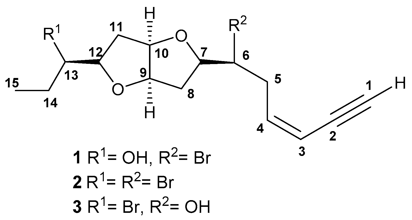
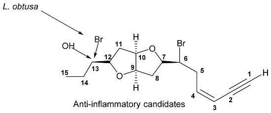
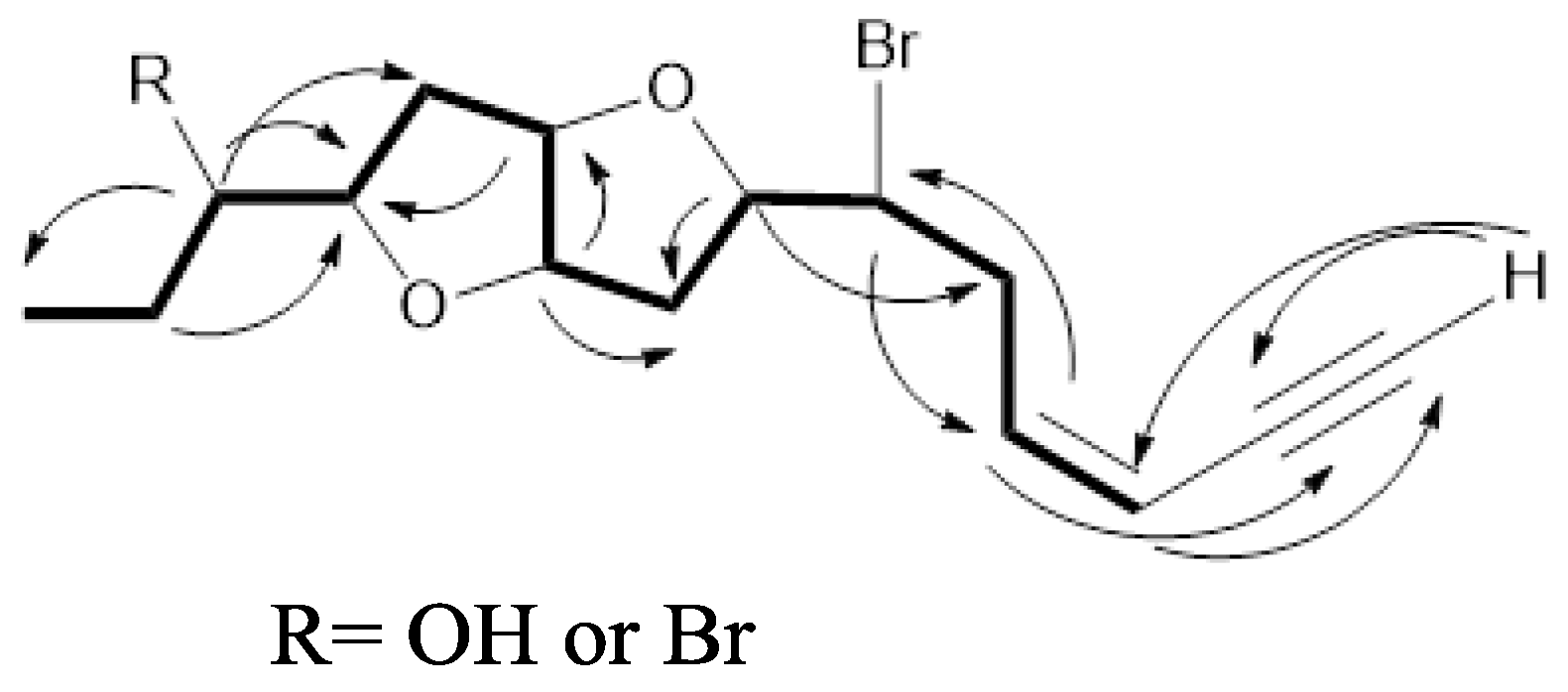
2. Results and Discussion

Compound 1 was isolated as a colorless oil, [ α ] D 22 = −18.0 (c 0.016, CH2Cl2). The molecular formula was determined as C15H21BrO3, employing positive mode HR-ESI-MS, requiring five degrees of unsaturation. EIMS showed a characteristic molecular-ion cluster at m/z 328 and 330 in 1:1 ratio, which clearly indicated the presence of one Br atom. The presence of terminal acetylenic and hydroxyl groups and a furan ring were evidenced from the infrared spectroscopy (IR) absorptions at υmax3292, 3481and 1252, and 1080 cm−1, respectively. The ultraviolet (UV) spectrum exhibited the presence of a conjugated enyne function absorbing at 223 nm. The 13C-NMR spectrum displayed 15 signals, categorized by a distortionless enhancement by polarization transfer (DEPT) experiment into one methyl, four methylene, ninemethine, and one quaternary carbons (Figure S1f–S1h. 1H and heteronuclear single quantum coherence (HSQC) NMR spectra led to the assignment of the following features: one acetylenic proton resonating at δHC 3.15 (d, J = 1.7 Hz)/82.8; two olefinic methine protons 6.18 (ddd, J = 11.1, 10.2, 7.7 Hz)/141.1 and 5.63 (ddd, J = 11.1, 3.4, 1.7 Hz)/111.0; one hydroxylated methine 3.54 (ddd, J = 11.1, 6.8, 4.3 Hz)/75.2; one halogenated methine 4.15 (ddd, J = 10.2, 8.5, 4.3 Hz)/55.7; four etheric methine protons 4.55 (ddd, J = 6.0, 4.3, 1.7 Hz)/84.3, 4.53 (ddd, J = 6.0, 4.3, 1.7 Hz)/85.0, 4.08 (dd, J = 14.5, 7.7 Hz)/83.6, and 3.88 (ddd, J = 14.7, 8.5, 6.8 Hz)/84.3, four methylenes assigned to carbons resonating at δC 36.8, 35.5, 34.9, and 26.5; and a tertiary methyl at δHC 1.00 (t, J = 6.8 Hz)/10.1 carbons (Figures S1a–S1e and S1l–S1n). Interpretation of the 1H-1H correlation spectroscopy (COSY) spectrum revealed the presence of one large spin sequence system from H-3 to H-15, as well as a long rang coupling of H-1 with H-3 (Figure 1), (Figure S1i–S1k). The presence of terminal conjugated enyne was proposed from a HMBC correlation from H-3 at δH 5.63 to C-1 (CH, 82.8), C-2 (C, 79.9) and C-4 (CH, 141.1). The presence of a OH function at C-12 was concluded from the HMBC correlation between both the Me protons at δH 1.0 (H-15) and the ether proton at 3.88 (H-12) with C-13 (CH, 75.2) (Figure S1i–S1k). The relative configuration of 1 was determined by a combination of data from the nuclear Overhauser effect spectroscopy (NOESY) spectrum and the coupling constant values (J). NOESY enhancements were observed between H-9 (δH 4.55) and H-10 (δH 4.53), H-7 (δH 4.08) and H-12 (δH 3.88) (Figure S1r), suggesting their co-facial orientation, Extensive comparison between the spectral data of 1 with those reported for laurenidificin (3, Figure 2) isolated from L. nidifica [15], revealed the similarity in gross carbon skeleton and orientation of Hα-9 (owing to the similar J values),with the difference of the switched positions of the Br atom and OH function. The trivial name isolaurenidificin was given to compound 1 (Figure 2).
Compound 2 was isolated as colorless oil, [ α ] D 22 = − 20.5 (c 0.005, CH2Cl2). The molecular formula was determined as C15H20Br2O2 employing positive mode HR-ESI-MS, requiring five degrees of unsaturation. EIMS showed a characteristic molecular-ion cluster at m/z 389, 391 and 393 in 1:2:1 ratio, which clearly indicated the presence of two Br atoms. The presence of a terminal acetylenic group and a furan ring were evidenced from the IR absorptions at υmax3294, 1236 and 1070 cm−1, respectively. The UV spectrum exhibited the presence of a conjugated enyne function absorbing at 222 nm. The 13C-NMR spectrum displayed 15 signals, categorized by DEPT experiment into one methyl, four methylene, nine methine, and one quaternary carbons (Figure S2f–S2h). 1H- and HSQC-NMR spectra led to the assignment of the following features:one acetylenic proton resonating at δHC 2.76 (d, J = 1.7 Hz)/82.8; two olefinic methine protons 5.85 (ddd, J = 11.9, 11.1, 6.8 Hz)/141.4 and 5.35 (ddd, J = 11.1, 2.6, 1.7 Hz)/110.6; two halogenated methines4.24 (ddd, J = 11.9, 9.4, 2.6 Hz)/61.4 and 3.83 (m)/55.0; four etheric methine protons 3.99 (ddd, J = 6.0, 3.4, 1.7 Hz)/84.9, 3.93 (ddd, J = 9.4, 8.5, 6.0 Hz)/83.6, 3.82 (m)/84.1, and 3.56 (ddd, J = 12.8, 7.7, 5.1 Hz)/82.7, four methylenes assigned to carbons resonating at δC 37.5, 36.9, 35.4, and 28.4; and a tertiary methyl at δHC 1.04 (t, J = 7.7 Hz)/11.2 carbons (Figures S2a–S2e and S2l–S2m). After examination of the1H-1HCOSY and HMBC correlation spectra (Figure 1), 2 is very similar to 1 with the exception of the appearance of halogenated methine proton signal in 2 instead of a hydroxyl methine proton signal in 1. The relative configuration of 2 was determined by the combination of both data from NOESY spectrum and the coupling constant values (J). Compound 2 was found also to be similar to 1, where signals due to H-7, H-9, H-10 and H-12 are correlated together, implying a co-facial orientation of these protons (Figure S2o–S2p). The trivial name brom laurenidificin was given to 2 (Figure 2).
As aforementioned the genus Laurencia generates unusual secondary metabolites. The vast majority of these compounds are halogenated diterpenes, sesquiterpenes and C15 non-terpenoids containing different functions, including acetylenic, vinyl acetylenic or allenic side chains. The diversity of their molecular structures enables them to exhibit different bioactivities [11,12,13,14]. Therefore, compounds 1 and 2 were evaluated against Artemia salina. The assay is a rapid and inexpensive screen for potential cytotoxicity [16]. This assay gave a preliminary overview of the cytotoxic activity of the tested compounds, and the toxicity of 1 and 2 was then further investigated in a dose-response study determine the IC50 values of compounds 1 and 2 against each cell type after a 24 h incubation period. The obtained IC50 of each cell line was then used to establish the time-course study of the apoptotic activity as shown in Figure 3. In general, cytotoxicity of 1 and 2 againstA549 lung carcinoma cells, HepG2 hepatocellular carcinoma cells, HCT116 colorectal carcinoma cells, MCF-7 breast cancer cells and PC-3 prostate cancer cells was weak, as indicated by the high IC50 values (>15 mM, Table 1). However, the IC50s of the tested compounds at 24 h of incubation was 14 µM for 1 and 11 µM for 2 versus dexamethasone 0.9 µM. These observations were further substantiated by assessing their effects on cellular apoptosis. Degradation of DNA into a specific fragmentation pattern is a characteristic feature of apoptosis. In contrast to the random fragmentation with necrosis, apoptosis-associated DNA fragmentation is characterized by cleavage of the DNA at regular intervals, visualized on agarose gel electrophoresis as a DNA ladder consisting of multimers of approximately 200 base pairs. Blood neutrophils as well as the five cell lines were prepared, cultured, and incubated for 24, 48 and 72 h in medium with and without the isolated compounds (Table 2). Both morphology and DNA fragmentation methods assessed the percentage of neutrophils apoptosis in each culture. The data in Figure 3 indicate a relatively potent apoptosis-inducing activity of both compounds 1 and 2 against peripheral blood neutrophils. However, the fraction of apoptotic tumor cells exposed to both compounds never exceeded 15% even at 72 h of incubation. It is well documented that cell death embraces apoptosis, necrosis, autophagy and others [17]. Thus, it can be concluded that other types of cell death cannot be excluded.

3. Experimental

3.1. General

Silica gel GF 254 (Merck, Darmstadt, Germany) was used for analytical thin layer chromatography (TLC). Preparative thin layer chromatography (PTLC) was performed on aluminum oxide plates (20 × 20 cm) of 250 µm thickness. Plates were sprayed with p-anisaldehyde-sulphuric acid reagent and heated at 100 °C for 1–2 min. for detection. Electron impact mass spectra were determined at 70 ev on a Kratos (Manchester, UK) MS-25 instrument. 1D and 2D NMR spectra were recorded by using a Bruker (Karlsruhe, Germany) AVANCE III WM 850 MHz spectrometer and 13C-NMR spectra were recorded at 212.5 MHz. Tetramethylsilane (TMS) was used as internal standard. The deuterated solvents deuterated chloroform (CDCl3) and hexadeuterated benzene (C6D6) were purchased from Sigma-Aldrich (Steinheim, Germany). Sulforhodamine-B was purchased from Sigma-Aldrich (St. Louis, MO, USA). RPMI-1640 medium, heat inactivated fetal bovine serum, streptomycin, penicillin and other cell culture materials were purchased from Invitrogen (Carlsbad, CA, USA). Other reagents were of the highest analytical grade.

3.2. Extraction and Isolation

Laurencia obtusa was collected in May 2016 from Salman Gulf, north of Jeddah, Saudi Arabia. The voucher sample (JAD 03060) was deposited at the Marine Chemistry Department, King Abdulaziz University, Jeddah, Saudi Arabia. It was dried then extracted with equal volumes of CH2Cl2 (dichloromethane)/MeOH (methanol). The residue (6 g) was subjected to column chromatography on aluminum oxide using gradient elution with n-hexane/diethyl ether (0–50% diethyl ether), followed by then n-hexane/ethyl acetate (10–50% ethyl acetate). Fractions of 25 mL were gathered and monitored using TLC. The promising fractions were further purified by PTLC and Sephadex LH-20 chromatography.

3.3. Spectral Data

Isolaurenidificin (1). The fraction eluted with 30% ethyl acetate was purified by preparative TLC system using n-hexane/ethylacetate (7:3). The green color zone with p-anisaldehyde-sulfuric acid was collected to provide 2.7 mg of colorless oil; Rf 0.20; [ α ] D 22 = −18.0 (c 0.016, CH2Cl2); UV (MeOH) λmax 223 nm; IR υmax 3481, 3292, 2924, 2854, 1729, 1663, 1461, 1252 and 1080 cm−1; electrospray ionization-high resolution mass spectrometry (ESI-HRMS) m/z 351.0559, 353.0538 [M + Na]+ (1:1) (calcd. for C15H2179BrO3Na, 351.0572; C15H2081BrO3Na, 351.0551; respectively); 1H-, 13C-NMR (CDCl3) δ ppm (Table 3).
Bromlaurenidificin (2). The fraction eluted with 15% diethyl ether was purified by preparative TLC system using n-hexane/diethyl ether (8.5:1.5). The greenish-gray color zone with p-anisaldehyde-sulfuric acid was collected to provide 1.1 mg of colorless oil; Rf 0.20; [ α ] D 22 = −20.5 (c 0.005, CH2Cl2); UV (MeOH) λmax 222 nm; IR νmax 3294, 2923, 2853, 1736, 1461, 1378, 1236 and 1070 cm−1; ESI-HRMS m/z 412.9728, 414.9707, 416.9687 [M + Na]+ (1:2:1) (calcd. for C15H2079Br2O2Na, 412.9716; C15H2079Br81BrO2Na, 414.9695; C15H2081Br2O2Na, 416.9673; respectively); 1H-, 13C-NMR (C6D6) δ ppm (Table 3).

3.4. Toxicity of the Isolated Compounds

3.4.1. Brine Shrimp Lethality (Artemia salina) Assay

A solution of sea water was made by dissolving 32.5 g (a natural blend of salts and trace element for sea water fish (Sera Company, AquaristikGmbh, Henisberg, Germany) in distilled water. Brine shrimp Artemia salina (Leach), eggs (ca. 1 mg) were placed in a hatching chamber (22 × 32 cm). The hatching chamber was kept under an inflorescent bulb for 48 h for the eggs to hatch into shrimp larvae (nauplii). One mg of each pure compound was dissolved in 5 mL of solvent in which it was soluble and from this stock solution was transferred to vials corresponding to (0.01 to 100 mM), respectively. Each dosage was tested in triplicate. Ten larvae (nauplii) of Artemia salina were transferred into each vial and the volume made into 5 mL with sea salt solution, immediately after adding the nauplii, 24 h later, the number of surviving shrimp at each dosage was counted B recorded. LC50 values were determined statistically [16].

3.4.2. Cytotoxicity Bioassay

Cell Culture

The culture medium for A549 lung carcinoma cells and PC-3 prostate cancer cells were Dulbecco’s Modified Eagle’s Medium (DMEM) formulated with high glucose, while Eagle Minimum Essential Medium (EMEM) was used for HepG2 hepatocellular carcinoma cells and MCF-7 Breast cancer cells. Finally, McCoy’s 5A medium, include 2 mM glutamine + 10% fetal bovine serum (FBS), was employed for HCT116 colorectal carcinoma cells. The cell lines were obtained from the National Cancer Institute (Cairo, Egypt) and maintained in Roswell Memorial Institute (RPMI)-1640 medium supplemented with 100 µg/mL of streptomycin, 100 units/ mL of penicillin and 10% of heat-inactivated fetal bovine serum (Invitrogen) in a humidified atmosphere containing 5% (v/v) CO2 at 37 °C.

Cytotoxicity Assay

Compounds 1 and 2 were tested against peripheral blood neutrophils, A549 lung carcinoma cells, HepG2 hepatocellular carcinoma cells, HCT116 colorectal carcinoma cells, MCF-7 breast cancer cells and PC-3 prostate cancer cells. The percentage of viability of cell was estimated by using doxorubicin as a positive standard anticancer drug. These assays had been performed according to the published protocols [18]. The final concentration of dimethylsulphoxide (DMSO) in each sample did not exceed 0.1% v/v. The cancer cells were batch cultured for 10 d, then seeded in 96 well plates of 10 × 103 cells/well in fresh complete growth medium in 96-well microtiter plastic plates at 37 °C for 24 h under 5% CO2 using a water jacketed carbon dioxide incubator. The medium was added and cells were incubated either alone (negative control) or with different concentrations of sample to give a range of concentrations (0.01 to 100 mM). Each cell type was suspended in the suitable medium, 1% antibiotic-antimycotic mixture (104 µg/mL potassium penicillin, 104 µg/mL streptomycin sulfate and 25 µg/mL amphotericin B and 1% l-glutamine in 96-well flat bottom micro-plates at 37 °C under 5% CO2. After 96 h of incubation, the medium was again aspirated, trays were inverted onto a pad of paper towels, the remaining cells rinsed carefully with medium, and fixed with 3.7% (v/v) formaldehyde in saline for at least 20 min. The fixed cells were rinsed with water, and examined. The cytotoxic activity was identified as confluent, relatively unaltered mono-layers of stained cells treated with compounds. The IC50 was calculated based on the 50% loss of monolayer.

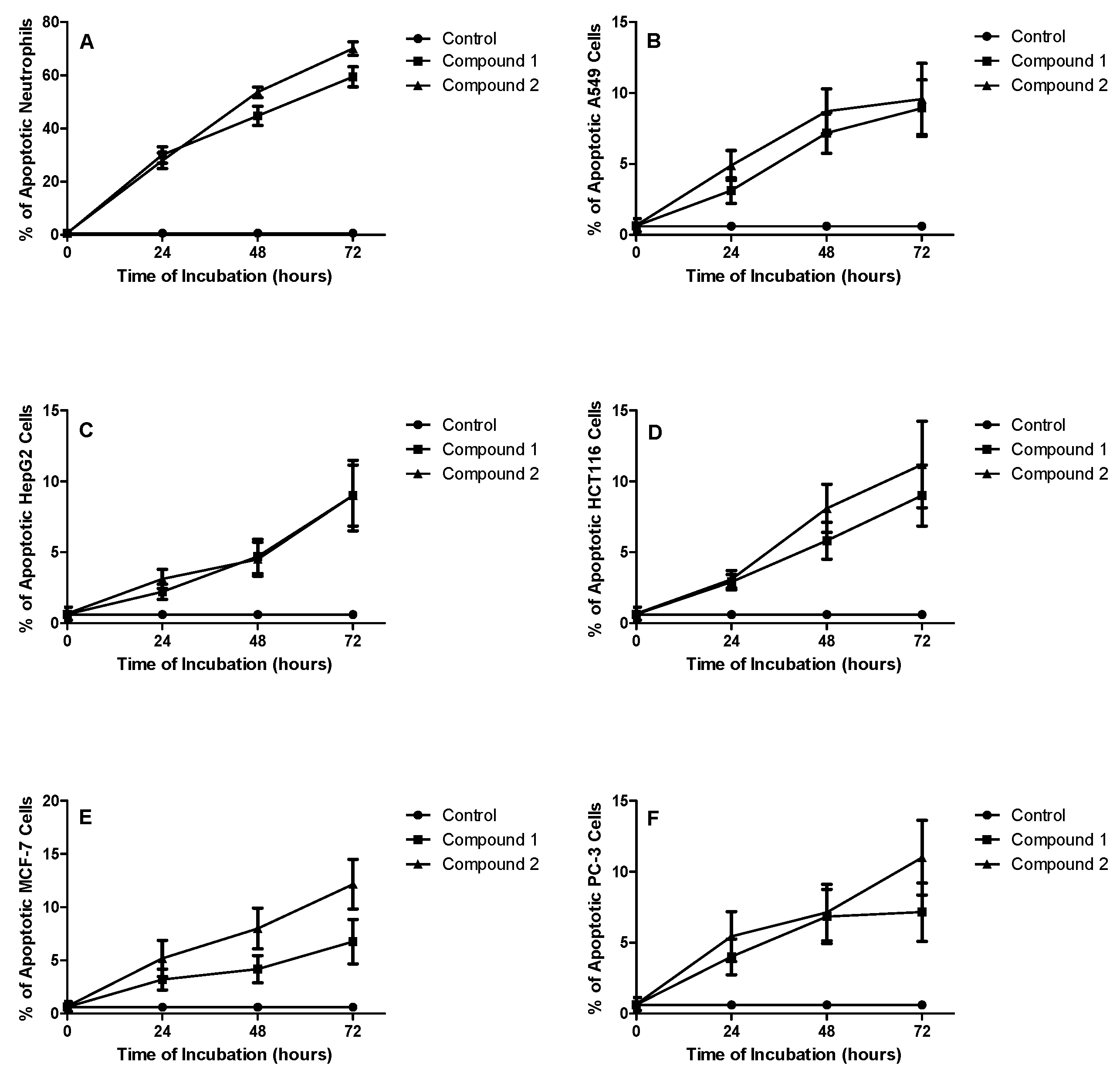
Apoptotic Effect on Neutrophils

Preparation of Blood Neutrophils: Neutrophils (>98% pure by May-Giemsa staining) were isolated from peripheral blood of normal healthy volunteer donors by a combination of dextran sedimentation and centrifugation through discontinuous plasma percoll gradients [19].

Culture of Neutrophils

Neutrophils were resuspended in an appropriate volume of RPMI 1640 medium with 10% autologous PRPDS and 100 μg/L of penicillin and streptomycin and divided into five equal volumes each put in culture tube. Cells were incubated (at 37 °C in a 5% carbon dioxide) as follows: (1) Only cells; (2) cells + DMSO at 0.01% v/v; (3) cells + each compound in DMSO at concentration of 50 mM/mL culture. The age of neutrophils in culture was calculated at the start of culture (zero time or base line), 24, 48, and 72 h [19].

Assessment of Cell Viability

At time 0 and then at subsequent times, cells were removed from culture and counted on a haemocytometer. Cell viability was determined by a Trypan Blue dye exclusion test; one volume of Trypan Blue (0.4% GiBCo, USA) was added to 5 volumes of cells at room temperature for 5 min. The IC50 values of isolated compounds were determined in comparison to dexamethasone [12,19].

4. Conclusions

This work reported two new hexahydrofuro[3,2-b]furan-based C15-acetogenins from an extract of the Red Sea red alga Laurencia obtusa. Both compounds showed weak cytotoxicity against A549, HepG-2, HCT116, MCF-7, and PC-3 cells. However, they exhibited a relatively potent cytotoxic effect against peripheral blood neutrophils with an IC50 of 14 µM for 1, and 11 µM for 2 versus 0.9 µM for dexamethasone at 24 h of exposure. This can be attributed partly to induction of apoptosis.

Supplementary Materials

The following are available online: Figure S1a–S1r: 1D and 2D-NMR spectra for Isolaurenidificin (1), Figure S2a–S2p: 1D and 2D-NMR spectra for Bromlaurenidificin (2).

Acknowledgments

The authors indebted to Kamal Al-Dahoudy, Faculty of Maritime Studies, King Abdulaziz University, for collection and identification of the alga sample. Also, they would like to thank King Fahd Center for Medical Research for giving them the opportunity to work in its central laboratory.

Author Contributions

Nahed O. Bawakid and Walied M. Alarif performed the experiments, analyzed the data, and wrote the paper; Hajer S. Alorfi and Mohamed A. Ghandourah helped perform some parts of the experiment; Khalid O. Al-Footy and Sultan S. Al-Lihaibi revised the paper; Najla A. Alburae assayed the bio-activities and interpreted the data. All authors read and approved the final manuscript.

Conflicts of Interest

The authors declare no conflict of interest.

References

  1. Suzuki, M.; Takahashi, Y.; Nakano, S.; Abe, T.; Masuda, M.; Ohnishi, T.; Noya, Y.; Seki, K. An experimental approach to study the biosynthesis of brominated metabolites by the red algal genus Laurencia. Phytochemistry 2009, 70, 1410–1415. [Google Scholar] [CrossRef] [PubMed]
  2. Masudai, M.; Abe, T.; Suzuki, T.; Suzuki, M. Morphological and chemotaxonomic studies on Laurencia composita and L. okamurae (Ceramiales, Rhodophyta). Phycologia 1996, 35, 550–562. [Google Scholar] [CrossRef]
  3. Guiry, M.D.; Guiry, G.M. Algae Base. World-Wide Electronic Publication, National University of Ireland: Galway, 2015. Available online: http://www.algaebase.org (accessed on 29 November 2015).
  4. Kurata, K.; Taniguchi, K.; Agatsuma, Y.; Suzuki, M. Diterpenoid feeding-deterrents from Laurencia saitoi. Phytochemistry 1998, 47, 363–369. [Google Scholar] [CrossRef]
  5. Paul, V.J.; Ritson-Williams, R.; Sharp, K. Marine chemical ecology in benthic environments. Nat. Prod. Rep. 2011, 28, 345–387. [Google Scholar] [CrossRef] [PubMed]
  6. Dias, D.A.; Urban, S. Phytochemical studies of the southern Australian marine alga, Laurenci aelata. Phytochemistry 2011, 72, 2081–2089. [Google Scholar] [CrossRef] [PubMed]
  7. König, G.M.; Wright, A.D. Laurencia rigida: Chemical investigations of its antifouling dichloromethane extract. J. Nat. Prod. 1997, 60, 967–970. [Google Scholar] [CrossRef]
  8. Davyt, D.; Fernandez, R.; Suescun, L.; Mombrú, A.W.; Saldaña, J.; Domínguez, L.; Coll, J.; Fujii, M.T.; Manta, E. New sesquiterpene derivatives from the red alga Laurencia scoparia. Isolation, structure determination, and anthelmintic activity. J. Nat. Prod. 2001, 64, 1552–1555. [Google Scholar] [CrossRef] [PubMed]
  9. Wanke, T.; Philippus, A.C.; Zatelli, G.A.; Vieira, L.F.O.; Lhullier, C.; Falkenberg, M. C15 acetogenins from the Laurencia complex: 50 years of research—An overview. Rev. Bras. Farmacogn. 2015, 25, 569–587. [Google Scholar] [CrossRef]
  10. Blunt, J.W.; Copp, B.R.; Hu, W.P.; Munro, M.H.; Northcote, P.T.; Prinset, M.P. Marine natural products. Nat. Prod. Rep. 2010, 27, 165–237. [Google Scholar] [CrossRef] [PubMed]
  11. Alarif, W.M.; Al-Lihaibi, S.S.; Ayyad, S.E.; Abdel-Rhman, M.H.; Badria, F.A. Laurene-type sesquiterpenes from the Red Sea red alga Laurencia obtusa as potential antitumor-antimicrobial agents. Eur. J. Med. Chem. 2012, 55, 462–466. [Google Scholar] [CrossRef] [PubMed]
  12. Alarif, W.M.; Al-Footy, K.O.; Zubair, M.S.; Halid, P.H.M.; Ghandourah, M.H.; Basaif, S.A.; Al-Lihaibi, S.S.; Ayyad, S.E.; Badria, F.A. The role of new eudesmane-type sesquiterpenoid and known eudesmane derivatives from the red alga Laurencia obtusa as potential antifungal-antitumour agents. Nat. Prod. Res. 2015, 30, 1150–1155. [Google Scholar] [CrossRef] [PubMed]
  13. Angawi, R.F.; Alarif, W.M.; Hamza, R.I.; Badria, F.A.; Ayyad, S.E. New cytotoxic laurene, cuparene and laurokamurene type-sesquiterpenes from the red alga Laurencia obtusa. Helv. Chem. Acta 2014, 97, 1388–1395. [Google Scholar] [CrossRef]
  14. Ayyad, S.E.; Al-Footy, K.O.; Alarif, W.M.; Sobahi, T.R.; Bassaif, S.A.; Makki, M.S.; Asiri, A.M.; Al Halwani, A.Y.; Badria, A.F.; Badria, F.A. Bioactive C-15 Acetogenins from the red alga Laurencia obtusa. Chem. Pharm. Bull. 2011, 59, 1294–1298. [Google Scholar] [CrossRef] [PubMed]
  15. Liu, X.; Li, X.M.; Li, C.S.; Ji, N.Y.; Wang, B.G. Laurenidificin, a new brominated C15-acetogenin from the marine red alga Laurencia nidifica. Chin. Chem. Lett. 2010, 21, 1213–1215. [Google Scholar] [CrossRef]
  16. Meyer, B.; Ferrigni, N.; Putnam, J.; Jacobsen, L.; Nichols, D.; McLaughlin, J. Brine shrimp: A convenient general bioassay for active plant constituents. Planta Med. 1982, 45, 31–34. [Google Scholar] [CrossRef] [PubMed]
  17. Kroemer, G.; Galluzzi, L.; Vandenabeele, P.; Abrams, J.; Alnemri, E.S.; Baehrecke, E.H.; Blagosklonny, M.V.; El-Deiry, W.S.; Golstein, P.; Green, D.R.; et al. Classification of cell death: Recommendations of the Nomenclature Committee on Cell Death 2009. Cell Death Differ. 2009, 16, 3–11. [Google Scholar] [CrossRef] [PubMed]
  18. Skehan, P.; Storeng, R.; Scudiero, D.; Monks, A.; McMahon, J. New colorimetric cytotoxicity assay for anticancer-drug screening. J. Natl. Cancer Inst. 1990, 82, 1107–1112. [Google Scholar] [CrossRef] [PubMed]
  19. Haslett, C.; Guthrie, L.A.; Kopaniak, M.M.; Johnston, R.B., Jr.; Henson, P.M. Modulation of multiple neutrophil functions by preparative methods or trace concentrations of bacterial lipopolysaccharide. Am. J. Pathol. 1985, 119, 101–110. [Google Scholar] [PubMed]
Sample Availability: Not available.
Figure 1. Selected COSY ( Molecules 22 00807 i001) and HMBC ( Molecules 22 00807 i002) correlations of 1 and 2.
Molecules 22 00807 g001
Figure 2. Chemical structures of compounds 13.
Molecules 22 00807 g002
Figure 3. Induction of apoptosis by compounds 1 and 2 in peripheral blood neutrophils (A); A549 lung carcinoma cells (B); HepG2 hepatocellular carcinoma cells (C); HCT116 colorectal carcinoma cells (D); MCF-7 Breast cancer cells (E) and PC-3 prostate cancer cells (F). Each compound was used at a concentration of 50 mM/L.
Molecules 22 00807 g003
Table 1. The cytotoxic activity (IC50) values of 1 and 2 against human cancer cells. *
CompoundIC50 (mM)
A 549HepG2HCT116MCF-7PC-3
115.615.1>2015.517.9
215.915.317.917.9>20
* A549 (lung carcinoma cells); HepG2 (hepatocellular carcinoma cells; HCT116 (colorectal carcinoma cells); MCF-7 (Breast cancer cells) and PC-3 (prostate cancer cells). All cells were incubated with compounds 1 or 2 for 72 h.
Table 2. Effect of the isolated compounds on apoptosis of peripheral blood neutrophils. *
Compound% Apoptotic Neutrophils (Mean ± S.D.)
0 h24 h48 h72 h
(Control)0.41 ± 0.010.41 ± 0.010.41 ± 0.010.41 ± 0.01
10.63 ± 0.5530.10 ± 3.1144.86 ± 3.5959.45 ± 3.76
20.80 ± 0.5627.91 ± 2.9353.62 ± 2.0170.13 ± 2.51
* Neutrophils were incubated with compounds 1 or 2 for the assigned time.
Table 3. 1H- and 13C-NMR spectral data for compounds 1 a and 2 b.
Position 1 2
δHMult. J in HzδCδHMult. J in HzδC
13.15d, J = 1.782.82.76d, J = 1.782.8
2- 79.9- 79.8
35.63ddd, J = 11.1, 3.4,1.7 1115.35ddd, J = 11.1, 2.6, 1.7110.6
46.18ddd, J = 11.1, 10.2, 7.7141.15.85ddd, J = 11.9, 11.1, 6.8141.4
52.9m35.52.83m35.4
2.96dddd, J = 15.3, 8.5, 7.0, 1.72.87m
64.15ddd, J = 10.2, 8.5, 4.355.73.83m55
74.08dd, J = 14.5, 7.783.63.56ddd, J = 12.8, 7.7,5.182.7
82.02m36.81.66ddd, J = 13.6, 6.8, 1.736.9
2.32ddd, J = 13.6, 7.7, 6.81.85ddd, J = 13.6, 6.0, 1.7
94.55ddd, J = 6.0, 4.3, 1.784.33.99ddd, J = 6.0, 3.4,1.784.9
104.53ddd, J = 6.0, 4.3, 1.7853.82m84.1
112.02m34.91.91ddd, J = 13.6, 7.7, 6.037.5
2.22ddd, J = 13.6, 7.7, 6.82.28dd, J = 13.6, 5.1
123.88ddd, J = 14.7, 8.5, 6.8 84.33.93ddd, J = 9.4, 8.5, 6.083.6
133.54ddd, J = 11.1, 6.8, 4.375.24.24ddd, J = 11.9, 9.4, 2.661.4
141.51m26.51.76ddq, J = 14.5, 9.4, 7.728.4
1.45m2.15dddd, J = 14.5, 10.2, 7.7, 2.6
151t, J = 6.810.11.04t, J = 7.711.2
a 1 was measured in CDCl3; b 2 was measured in C6D6.

