
Synthesis and Evaluation of Ciprofloxacin-Nitroxide Conjugates as Anti-Biofilm Agents

Abstract
:
1. Introduction
2. Results and Discussion
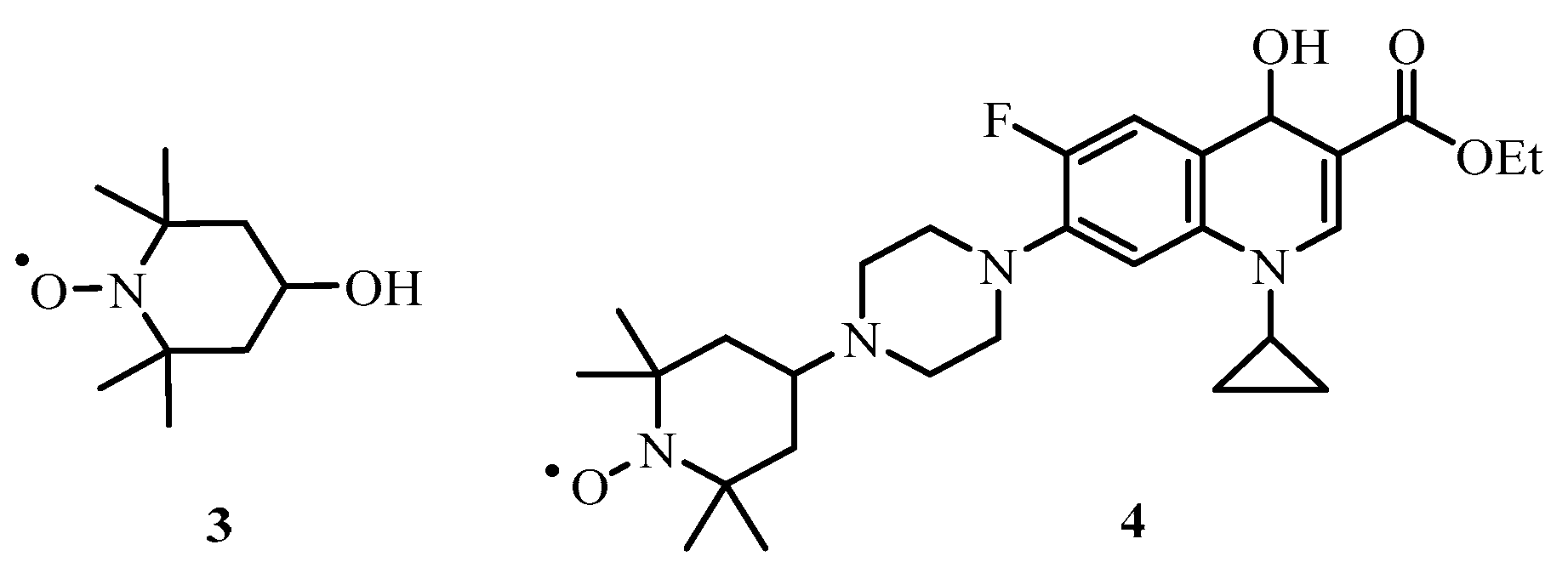
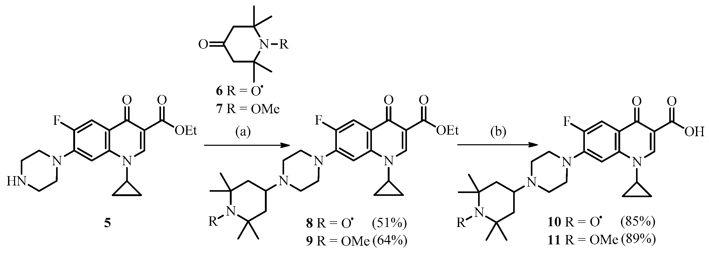
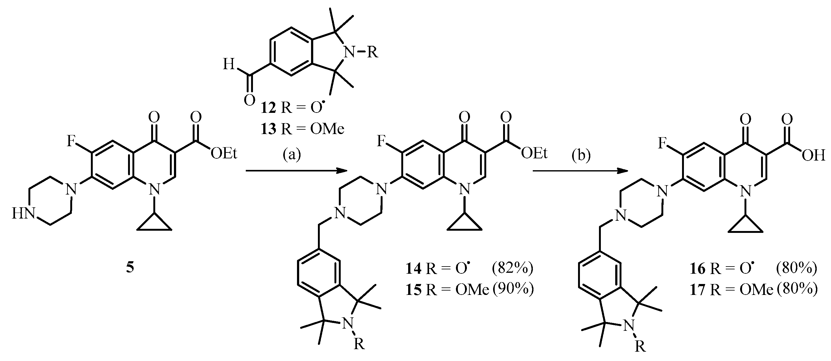
2.1. Chemistry
2.2. Biological Testing
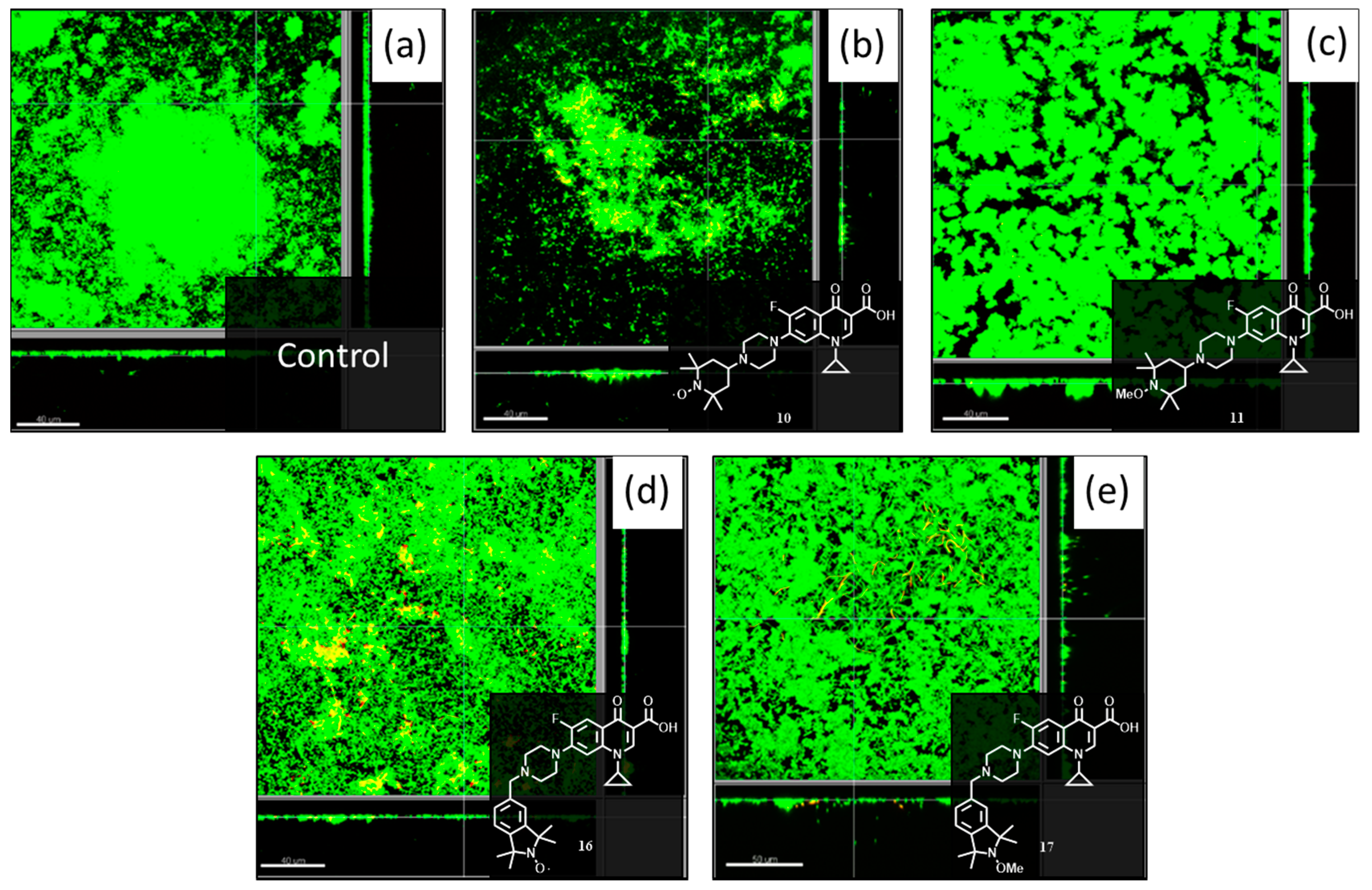
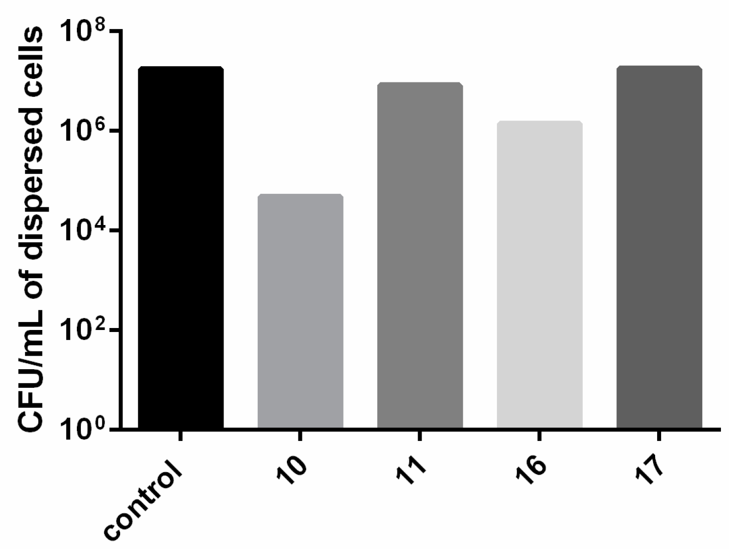
2.2.1. Evaluation of Compounds 10, 11, 16 and 17 at 20 μM in P. aeruginosa Biofilms
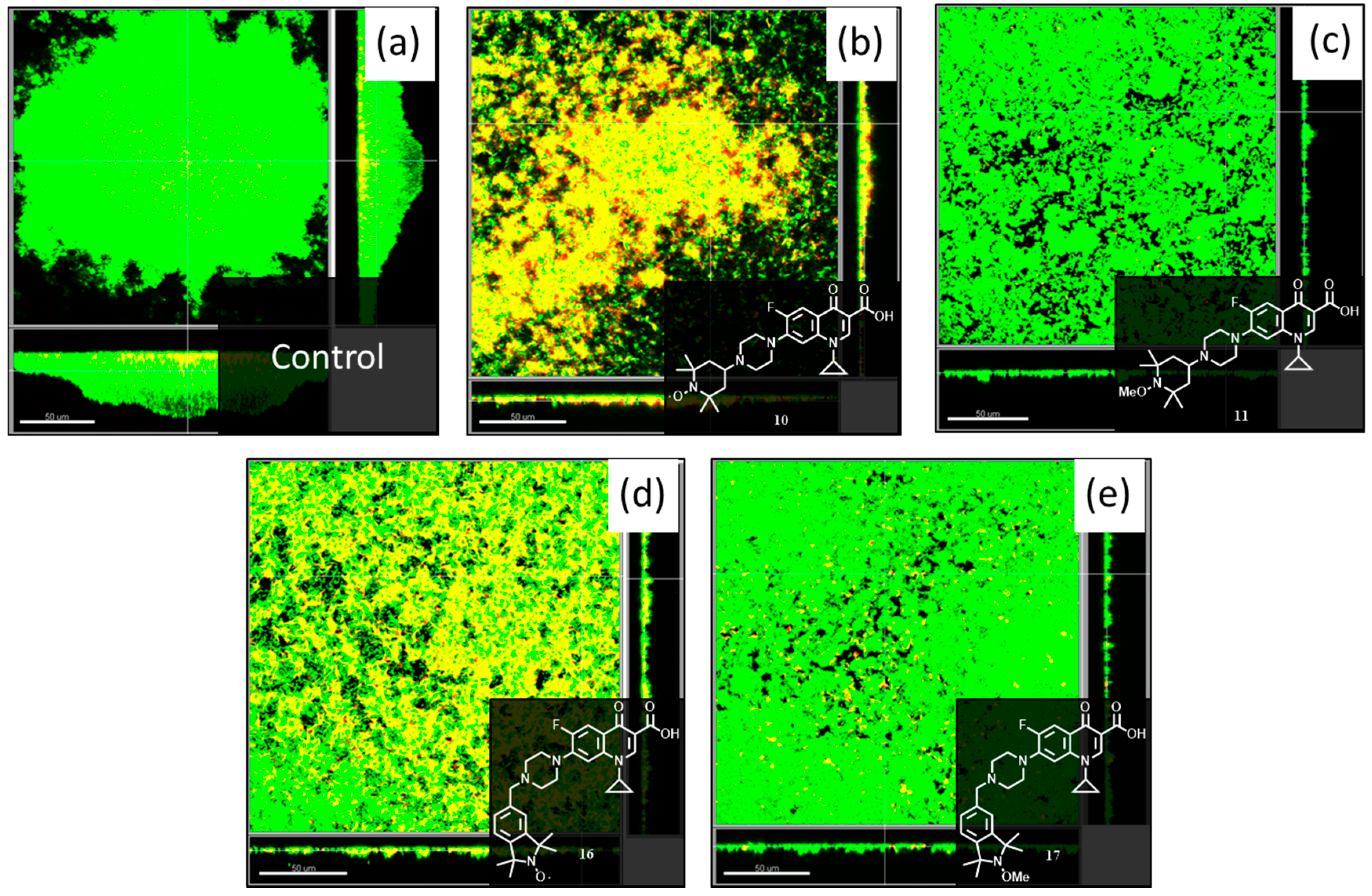
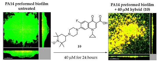
2.2.2. Evaluation of Compounds 10, 11, 16 and 17 at 40 μM in P. aeruginosa Biofilms
3. Experimental Section
3.1. General Procedures
3.2. Materials
3.3. Biofilm Dispersal Flow Cell Assays
3.4. General Procedure for Reductive Amination, Compounds 8, 9, 14 and 15
3.5. General Procedure for Ester Hydrolysis, Compounds 10, 11, 16 and 17
4. Conclusions
Supplementary Materials
Acknowledgments
Author Contributions
Conflicts of Interest
Abbreviations
| Cyclic di-GMP | bis-(3′-5′)-cyclic dimeric guanosine monophosphate |
| DCM | dichloromethane |
| DMSO | dimethyl sulfoxide |
| DNA | deoxyribonucleic acid |
| EPR | electron paramagnetic resonance |
| NMR | nuclear magnetic resonance |
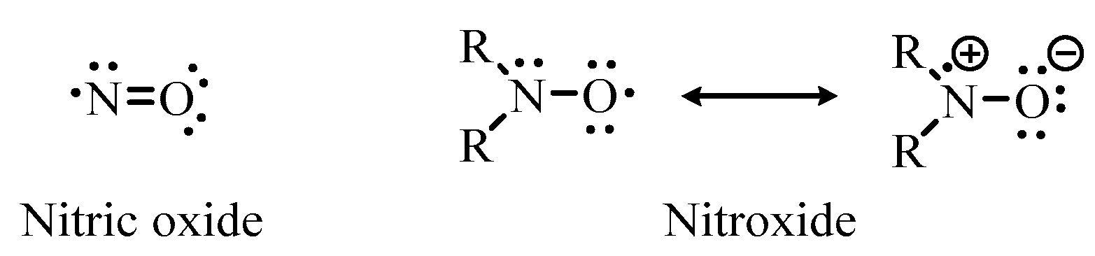
| NO | nitric oxide |
| THF | tetrahydrofuran |
| TLC | solid thin layer chromatography |
References
- Costerton, J.W.; Stewart, P.S.; Greenberg, E.P. Bacterial biofilms: A common cause of persistent infections. Science 1999, 284, 1318–1322. [Google Scholar] [CrossRef] [PubMed]
- Hall-Stoodley, L.; Costerton, J.W.; Stoodley, P. Bacterial biofilms: From the natural environment to infectious diseases. Nat. Rev. Microbiol. 2004, 2, 95–108. [Google Scholar] [CrossRef] [PubMed]
- Lynch, A.S.; Robertson, G.T. Bacterial and fungal biofilm infections. Annu. Rev. Med. 2008, 59, 415–428. [Google Scholar] [CrossRef] [PubMed]
- Vickery, K.; Hu, H.; Jacombs, A.S.; Bradshaw, D.A.; Deva, A.K. A review of bacterial biofilms and their role in device-associated infection. Healthcare Infection 2013, 18, 61–66. [Google Scholar] [CrossRef]
- Percival, S.L.; Hill, K.E.; Williams, D.W.; Hooper, S.J.; Thomas, D.W.; Costerton, J.W. A review of the scientific evidence for biofilms in wounds. Wound Repair Regen. 2012, 20, 647–657. [Google Scholar] [CrossRef] [PubMed]
- Davies, D. Understanding biofilm resistance to antibacterial agents. Nat. Rev. Drug Discovery 2003, 2, 114–122. [Google Scholar] [CrossRef] [PubMed]
- Costerton, J.W.; Cheng, K.J.; Geesey, G.G.; Ladd, T.I.; Nickel, J.C.; Dasgupta, M.; Marrie, T.J. Bacterial biofilms in nature and disease. Annu. Rev. Microbiol. 1987, 41, 435–464. [Google Scholar] [CrossRef] [PubMed]
- Lebeaux, D.; Ghigo, J.-M.; Beloin, C. Biofilm-related infections: Bridging the gap between clinical management and fundamental aspects of recalcitrance toward antibiotics. Microbiol. Mol. Biol. Rev. 2014, 78, 510–543. [Google Scholar] [CrossRef] [PubMed]
- Luppens, S.B.I.; Reij, M.W.; van der Heijden, R.W.L.; Rombouts, F.M.; Abee, T. Development of a standard test to assess the resistance of Staphylococcus aureus biofilm cells to disinfectants. Appl. Environ. Microbiol. 2002, 68, 4194–4200. [Google Scholar] [CrossRef] [PubMed]
- Stewart, P.S.; William Costerton, J. Antibiotic resistance of bacteria in biofilms. Lancet 2001, 358, 135–138. [Google Scholar] [CrossRef]
- McDougald, D.; Rice, S.A.; Barraud, N.; Steinberg, P.D.; Kjelleberg, S. Should we stay or should we go: Mechanisms and ecological consequences for biofilm dispersal. Nat. Rev. Microbiol. 2012, 10, 39–50. [Google Scholar] [CrossRef] [PubMed]
- Worthington, R.J.; Richards, J.J.; Melander, C. Non-microbicidal control of bacterial biofilms with small molecules. Anti-Infect. Agents 2014, 12, 120–138. [Google Scholar] [CrossRef]
- Bjarnsholt, T.; Ciofu, O.; Molin, S.; Givskov, M.; Hoiby, N. Applying insights from biofilm biology to drug development can a new approach be developed? Nat. Rev. Drug Discov. 2013, 12, 791–808. [Google Scholar] [CrossRef] [PubMed]
- Barraud, N.; Storey, M.V.; Moore, Z.P.; Webb, J.S.; Rice, S.A.; Kjelleberg, S. Nitric oxide-mediated dispersal in single- and multi-species biofilms of clinically and industrially relevant microorganisms. Microb. Biotechnol. 2009, 2, 370–378. [Google Scholar] [CrossRef] [PubMed]
- Schmidt, I.; Steenbakkers, P.J.M.; Op den Camp, H.J.M.; Schmidt, K.; Jetten, M.S.M. Physiologic and proteomic evidence for a role of nitric oxide in biofilm formation by Nitrosomonas europaea and other ammonia oxidizers. J. Bacteriol. 2004, 186, 2781–2788. [Google Scholar] [CrossRef] [PubMed]
- Barraud, N.; Hassett, D.J.; Hwang, S.-H.; Rice, S.A.; Kjelleberg, S.; Webb, J.S. Involvement of nitric oxide in biofilm dispersal of Pseudomonas aeruginosa. J. Bacteriol. 2006, 188, 7344–7353. [Google Scholar] [CrossRef] [PubMed]
- Cutruzzolà, F.; Frankenberg-Dinkel, N. Origin and impact of nitric oxide in Pseudomonas aeruginosa biofilms. J. Bacteriol 2016, 198, 55–65. [Google Scholar] [CrossRef] [PubMed]
- Chua, S.L.; Liu, Y.; Yam, J.K.H.; Chen, Y.; Vejborg, R.M.; Tan, B.G.C.; Kjelleberg, S.; Tolker-Nielsen, T.; Givskov, M.; Yang, L. Dispersed cells represent a distinct stage in the transition from bacterial biofilm to planktonic lifestyles. Nat. Commun. 2014, 5, 4462. [Google Scholar] [CrossRef] [PubMed]
- Liu, N.; Xu, Y.; Hossain, S.; Huang, N.; Coursolle, D.; Gralnick, J.A.; Boon, E.M. Nitric oxide regulation of cyclic di-gmp synthesis and hydrolysis in Shewanella woodyi. Biochemistry 2012, 51, 2087–2099. [Google Scholar] [CrossRef] [PubMed]
- Bill Cai, T.; Wang, P.G.; Holder, A.A. NO and NO donors. In Nitric Oxide Donors for Pharmaceutical and Biological Applications; Wang, P.G., Cai, T.B., Taniguchi, N., Eds.; Wiley-VCH: Weinheim, Germany, 2005; Volume 9, pp. 1–31. [Google Scholar]
- Arora, D.P.; Hossain, S.; Xu, Y.; Boon, E.M. Nitric oxide regulation of bacterial biofilms. Biochemistry 2015, 54, 3717–3728. [Google Scholar] [CrossRef] [PubMed]
- Barraud, N.; Kelso, M.J.; Rice, S.A.; Kjelleberg, S. Nitric oxide: A key mediator of biofilm dispersal with applications in infectious diseases. Curr. Pharm. Des. 2015, 21, 31–42. [Google Scholar] [CrossRef] [PubMed]
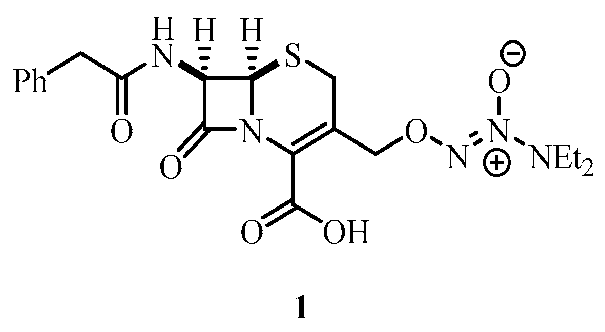
- Barraud, N.; Kardak, B.G.; Yepuri, N.R.; Howlin, R.P.; Webb, J.S.; Faust, S.N.; Kjelleberg, S.; Rice, S.A.; Kelso, M.J. Cephalosporin-3′-diazeniumdiolates: Targeted NO-donor prodrugs for dispersing bacterial biofilms. Angew. Chem. Int. Ed. Engl. 2012, 51, 9057–9060. [Google Scholar] [CrossRef] [PubMed]
- Wang, P.G.; Cai, T.B.; Taniguchi, N. Nitric Oxide Donors: For Pharmaceutical and Biological Applications; Wiley-VCH: Weinheim, Germany, 2005; Volume 9, p. 390. [Google Scholar]
- Likhtenshtein, G.I.; Yamauchi, J.; Nakatsuji, S.I.; Smirnov, A.I.; Tamura, R. Nitroxides; Applications in Chemistry, Biomedicine, and Materials Science; John Wiley & Sons: New York, NY, USA, 2008; p. 419. [Google Scholar]
- Lam, M.A.; Pattison, D.I.; Bottle, S.E.; Keddie, D.J.; Davies, M.J. Nitric oxide and nitroxides can act as efficient scavengers of protein-derived free radicals. Chem. Res. Toxicol. 2008, 21, 2111–2119. [Google Scholar] [CrossRef] [PubMed]
- De la Fuente-Núñez, C.; Reffuveille, F.; Fairfull-Smith, K.E.; Hancock, R.E.W. Effect of nitroxides on swarming motility and biofilm formation, multicellular behaviors in Pseudomonas aeruginosa. Antimicrob. Agents Chemother. 2013, 57, 4877–4881. [Google Scholar] [CrossRef] [PubMed]
- Alexander, S.-A.; Rouse, E.M.; White, J.M.; Tse, N.; Kyi, C.; Schiesser, C.H. Controlling biofilms oncultural materials: The role of 3-(dodecane-1-thiyl)-4-(hydroxymethyl)-2,2,5,5-tetramethyl-1-pyrrolinoxyl. Chem. Commun. 2015, 51, 3355–3358. [Google Scholar] [CrossRef] [PubMed]
- Alexander, S.-A.; Kyi, C.; Schiesser, C.H. Nitroxides as anti-biofilm compounds for the treatment of Pseudomonas aeruginosa and mixed-culture biofilms. Org. Biomol. Chem. 2015, 13, 4751–4759. [Google Scholar] [CrossRef] [PubMed]
- Gozdziewska, M.; Cichowicz, G.; Markowska, K.; Zawada, K.; Megiel, E. Nitroxide-coated silver nanoparticles: Synthesis, surface physicochemistry and antibacterial activity. RSC Adv. 2015, 5, 58403–58415. [Google Scholar] [CrossRef]
- Reffuveille, F.; de la Fuente-Núñez, C.; Fairfull-Smith, K.E.; Hancock, R.E.W. Potentiation of ciprofloxacin action against gram-negative bacterial biofilms by a nitroxide. Pathog. Dis. 2015, 73. [Google Scholar] [CrossRef] [PubMed]
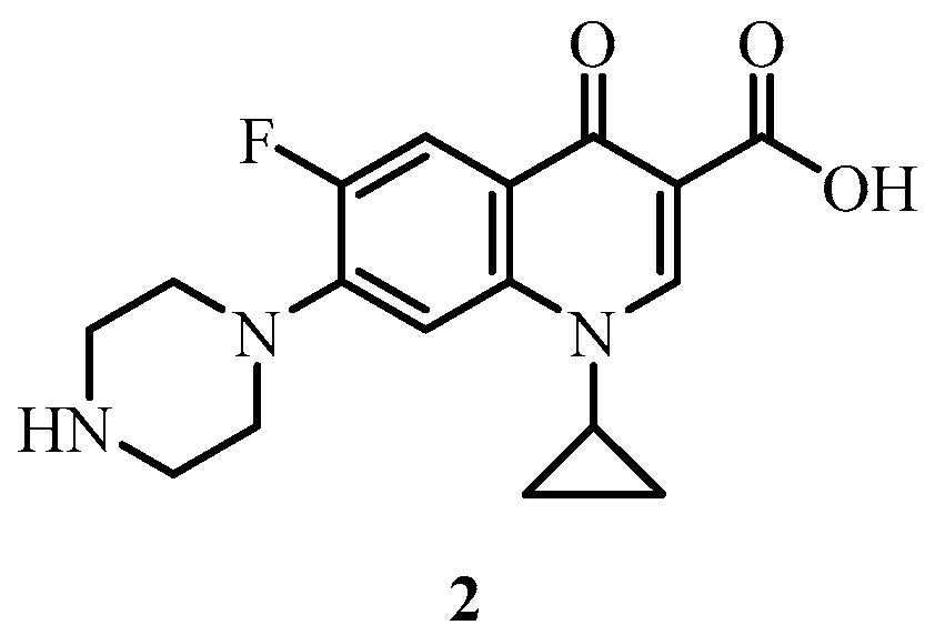
- Drlica, K.; Zhao, X. DNA gyrase, topoisomerase IV, and the 4-quinolones. Microbiol. Mol. Biol. Rev. 1997, 61, 377–392. [Google Scholar] [PubMed]
- Campoli-Richards, D.M.; Monk, J.P.; Price, A.; Benfield, P.; Todd, P.A.; Ward, A. Ciprofloxacin. A review of its antibacterial activity, pharmacokinetic properties and therapeutic use. Drugs 1988, 35, 373–447. [Google Scholar] [CrossRef] [PubMed]
- Zhi, C.; Long, Z.-Y.; Manikowski, A.; Comstock, J.; Xu, W.-C.; Brown, N.C.; Tarantino, P.M., Jr.; Holm, K.A.; Dix, E.J.; Wright, G.E.; et al. Hybrid antibacterials. DNA polymerase-topoisomerase inhibitors. J. Med. Chem. 2006, 49, 1455–1465. [Google Scholar] [CrossRef] [PubMed]
- Patel, R.V.; Park, S.W. Discovery of the highly potent fluoroquinolone-based benzothiazolyl-4-thiazolidinone hybrids as antibacterials. Chem. Biol. Drug Des. 2014, 84, 123–129. [Google Scholar] [CrossRef] [PubMed]
- Gootz, T.D.; Brighty, K.E. Fluoroquinolone antibacterials: SAR, mechanism of action, resistance, and clinical aspects. Med. Res. Rev. 1996, 16, 433–486. [Google Scholar] [CrossRef]
- Emami, S.; Shafiee, A.; Foroumadi, A. Structural features of new quinolones relationship to antibacterial activity against gram-positive bacteria. Mini-Rev. Med. Chem. 2006, 6, 375–386. [Google Scholar] [CrossRef] [PubMed]
- Shindikar, A.V.; Viswanathan, C.L. Novel fluoroquinolones: Design, synthesis, and in vivo activity in mice against Mycobacterium tuberculosis h 37 Rv. Bioorg. Med. Chem. Lett. 2005, 15, 1803–1806. [Google Scholar] [CrossRef] [PubMed]
- Sriram, D.; Aubry, A.; Yogeeswari, P.; Fisher, L.M. Gatifloxacin derivatives: Synthesis, antimycobacterial activities, and inhibition of Mycobacterium tuberculosis DNA gyrase. Bioorg. Med. Chem. Lett. 2006, 16, 2982–2985. [Google Scholar] [CrossRef] [PubMed]
- Dubar, F.; Anquetin, G.; Pradines, B.; Dive, D.; Khalife, J.; Biot, C. Enhancement of the antimalarial activity of ciprofloxacin using a double prodrug/bioorganometallic approach. J. Med. Chem. 2009, 52, 7954–7957. [Google Scholar] [CrossRef] [PubMed]
- Baxter, E.W.; Reitz, A.B. Reductive aminations of carbonyl compounds with borohydride and borane reducing agents. Org. React. 2002, 59, 711–714. [Google Scholar]
- Thomas, K.; Chalmers, B.A.; Fairfull-Smith, K.E.; Bottle, S.E. Approaches to the synthesis of a water-soluble carboxy nitroxide. Eur. J. Org. Chem. 2013, 2013, 853–857. [Google Scholar] [CrossRef]
- Volodarsky, L.B.; Reznikov, V.A.; Ovcharenko, V.I. Synthetic Chemistry Stable Nitroxides; CRC: Boca Raton, FL, USA, 1994; p. 240. [Google Scholar]
- Schmidt, M.; Harmuth, S.; Barth, E.R.; Wurm, E.; Fobbe, R.; Sickmann, A.; Krumm, C.; Tiller, J.C. Conjugation of ciprofloxacin with poly(2-oxazoline)s and polyethylene glycol via end groups. Bioconjugate Chem. 2015, 26, 1950–1962. [Google Scholar] [CrossRef] [PubMed]
- Brandt, S.D.; Moore, S.A.; Freeman, S.; Kanu, A.B. Characterization of the synthesis of N,N-dimethyltryptamine by reductive amination using gas chromatography ion trap mass spectrometry. Drug Test. Anal. 2010, 2, 330–338. [Google Scholar] [CrossRef] [PubMed]
- Abdel-Magid, A.F.; Carson, K.G.; Harris, B.D.; Maryanoff, C.A.; Shah, R.D. Reductive amination of aldehydes and ketones with sodium triacetoxyborohydride. Studies on direct and indirect reductive amination procedures. J. Org. Chem. 1996, 61, 3849–3862. [Google Scholar] [CrossRef] [PubMed]
- Borch, R.F.; Bernstein, M.D.; Durst, H.D. Cyanohydridoborate anion as a selective reducing agent. J. Am. Chem. Soc. 1971, 93, 2897–2904. [Google Scholar] [CrossRef]
- Lane, C.F. Sodium cyanoborohydride, a highly selective reducing agent for organic functional groups. Synthesis 1975, 3, 135–146. [Google Scholar] [CrossRef]
- Chalmers, B.A.; Morris, J.C.; Fairfull-Smith, K.E.; Grainger, R.S.; Bottle, S.E. A novel protecting group methodology for syntheses using nitroxides. Chem. Commun. 2013, 49, 10382–10384. [Google Scholar] [CrossRef] [PubMed]
- Keddie, D.J.; Johnson, T.E.; Arnold, D.P.; Bottle, S.E. Synthesis of profluorescent isoindoline nitroxides via palladium-catalyzed heck alkenylation. Org. Biomol. Chem. 2005, 3, 2593–2598. [Google Scholar] [CrossRef] [PubMed]
- Aubert, M.; Tirri, T.; Wilen, C.-E.; Francois-Heude, A.; Pfaendner, R.; Hoppe, H.; Roth, M. Versatile bis(1-alkoxy-2,2,6,6-tetramethylpiperidin-4-yl)-diazenes (azonors) and related structures and their utilization as flame retardants in polypropylene, low density polyethylene and high-impact polystyrene. Polym. Degrad. Stab. 2012, 97, 1438–1446. [Google Scholar] [CrossRef]
- Sample Availability: Samples of the compounds 10, 11, 16 and 17 are available from the authors.


| Compound | Remaining Biomass (%) | Dead Cells in Remaining Biomass (%) | Total Live Biofilm Biomass Eradication (%) |
|---|---|---|---|
| Control PA14 | -- | 4 | -- |
| 10 | 20 | 50 | 90 |
| 11 | 70 | 5 | 34 |
| 16 | 37 | 32 | 75 |
| 17 | 38 | 23 | 71 |
| Compound | Remaining Biomass (%) | Dead Cells in Remaining Biomass (%) | Total Live Biofilm Biomass Eradication (%) |
|---|---|---|---|
| Control PA14 | -- | 4 | -- |
| 10 | 59 | 91 | 95 |
| 11 | 65 | 7 | 40 |
| 16 | 52 | 64 | 81 |
| 17 | 52 | 27 | 62 |
© 2016 by the authors. Licensee MDPI, Basel, Switzerland. This article is an open access article distributed under the terms and conditions of the Creative Commons Attribution (CC-BY) license ( http://creativecommons.org/licenses/by/4.0/).
Share and Cite
Verderosa, A.D.; Mansour, S.C.; De la Fuente-Núñez, C.; Hancock, R.E.W.; Fairfull-Smith, K.E. Synthesis and Evaluation of Ciprofloxacin-Nitroxide Conjugates as Anti-Biofilm Agents. Molecules 2016, 21, 841. https://doi.org/10.3390/molecules21070841
Verderosa AD, Mansour SC, De la Fuente-Núñez C, Hancock REW, Fairfull-Smith KE. Synthesis and Evaluation of Ciprofloxacin-Nitroxide Conjugates as Anti-Biofilm Agents. Molecules. 2016; 21(7):841. https://doi.org/10.3390/molecules21070841
Chicago/Turabian StyleVerderosa, Anthony D., Sarah C. Mansour, César De la Fuente-Núñez, Robert E. W. Hancock, and Kathryn E. Fairfull-Smith. 2016. "Synthesis and Evaluation of Ciprofloxacin-Nitroxide Conjugates as Anti-Biofilm Agents" Molecules 21, no. 7: 841. https://doi.org/10.3390/molecules21070841
APA StyleVerderosa, A. D., Mansour, S. C., De la Fuente-Núñez, C., Hancock, R. E. W., & Fairfull-Smith, K. E. (2016). Synthesis and Evaluation of Ciprofloxacin-Nitroxide Conjugates as Anti-Biofilm Agents. Molecules, 21(7), 841. https://doi.org/10.3390/molecules21070841