Article

Metabolites of Siamenoside I and Their Distributions in Rats

1
Guangxi Key Laboratory of Functional Phytochemicals Research and Utilization, Guangxi Institute of Botany, Guangxi Zhuang Autonomous Region and Chinese Academy of Sciences, No. 85, Yanshan Road, Yanshan District, Guilin 541006, China
2
State Key Laboratory of Natural and Biomimetic Drugs, School of Pharmaceutical Sciences, Peking University, No. 38 Xueyuan Road, Beijing 100191, China
*
Authors to whom correspondence should be addressed.
Molecules 2016, 21(2), 176; https://doi.org/10.3390/molecules21020176
Submission received: 3 January 2016 / Revised: 25 January 2016 / Accepted: 27 January 2016 / Published: 30 January 2016
(This article belongs to the Section Metabolites)

Abstract

:
Siamenoside I is the sweetest mogroside that has several kinds of bioactivities, and it is also a constituent of Siraitiae Fructus, a fruit and herb in China. Hitherto the metabolism of siamenoside I in human or animals remains unclear. To reveal its metabolic pathways, a high-performance liquid chromatography-electrospray ionization-ion trap-time of flight-multistage mass spectrometry (HPLC-ESI-IT-TOF-MSn) method was used to profile and identify its metabolites in rats. Altogether, 86 new metabolites were identified or tentatively identified, and 23 of them were also new metabolites of mogrosides. In rats, siamenoside I was found to undergo deglycosylation, hydroxylation, dehydrogenation, deoxygenation, isomerization, and glycosylation reactions. Among them, deoxygenation, pentahydroxylation, and didehydrogenation were novel metabolic reactions of mogrosides. The distributions of siamenoside I and its 86 metabolites in rat organs were firstly reported, and they were mainly distributed to intestine, stomach, kidney, and brain. The most widely distributed metabolite was mogroside IIIE. In addition, eight metabolites were bioactive according to literature. These findings would help to understand the metabolism and effective forms of siamenoside I and other mogrosides in vivo.

1. Introduction

Mogrosides are a group of cucurbitane-type triterpenoid saponins which have the common aglycone of mogrol [1]. They are responsible for the sweet taste and bioactivities of Siraitiae Fructus (Luo Han Guo in Chinese, the ripe fruits of Siraitia grosvenorii), a traditional Chinese medicine and an edible fruit [2].
Siamenoside I is one of the mogrosides, which is firstly isolated from Siraitia siamensis (a Chinese folk medicine) [3] and then from Siraitia grosvenorii [4]. Its relative sweetness (0.01% solution) to 5% sucrose is determined to be 563, higher than the famous sweetener mogroside V, making it the sweetest cucurbitane glycoside [4].
Besides its intense sweet taste, siamenoside I also has several kinds of bioactivities. It can inhibit the induction of Epstein–Barr virus early antigen (EBV-EA) by 12-O-tetradecanoylphorbol-13-acetate (TPA) in Raji cells, which implies that it is a potential cancer chemopreventive agent [5]. It also inhibits two-stage carcinogenesis induced by 9,10-dimethyl-1,2-benzanthracene (DMBA) and TPA in mice [6]. Furthermore, it exhibits a maltase inhibitory effect with IC50 value of 10 mM, which is more potent than those of mogroside V and mogroside IV (IC50 of 14 mM and 12 mM, respectively) [7].
In order to clarify the action mechanisms of the beneficial effects of mogrosides and to develop them into new health foods or drugs or sweeteners, it is necessary to investigate their metabolism and disposition. Up to now, there are only three reports on the metabolism of mogrosides. The first is about the human intestinal microflora biotransformation of mogroside III [8]; the second is on the in vivo digestion, absorption and metabolism of 72% mogroside V in rats [9], and the third is our study on the in vitro, in vivo metabolism of mogroside V (purity >98%) and the distributions of its metabolites in rats [10]. We find that mogroside V can be metabolized to its secondary glycosides and the aglycone morgol, and then morgol is oxidized to lots of metabolites. However, there are no reports on the metabolism of siamenoside I so far.
Although the importance of studying drug distribution in various organs is well established in the drug development field, the studies on the distributions of metabolites of bioactive natural products are neglected. Since our previous research indicates that the metabolites of natural products distribute unevenly in different organs of rats, such as mogroside V [10] and (+)-catechin [11], we believe that revealing the distributions of a bioactive natural product and its metabolites can be helpful in understanding its target organ and organ-specific bioactivities.
Accordingly, in the present work, the metabolites of siamenoside I and the distributions of siamenoside I and its metabolites in rats were firstly investigated by high-performance liquid chromatography-electrospray ionization-ion trap-time of flight-multistage mass spectrometry (HPLC-ESI-IT-TOF-MSn). In total, 86 new metabolites of siamenoside I in rats were detected and identified or tentatively identified, and the metabolic pathways and in vivo processes of siamenoside I were proposed. Siamenoside I and its metabolites were mainly distributed to intestine, stomach, kidney, and brain, and mogroside IIIE was the most widely distributed metabolite.

2. Results

2.1. Profiling the Metabolites of Siamenoside I in Different Biosamples by HPLC-ESI-IT-TOF-MSn

Based on the strategy described in Section 4.7, 86 new metabolites (M1M86) of siamenoside I were detected altogether in different drug-containing samples by the HPLC-ESI-IT-TOF-MSn technique (Table 1, Table S1, Figure 1, and Figures S1–S25).
Eighty-three metabolites were detected in drug-containing feces; 19 metabolites were found in urine, and only two were detected in plasma.
As for different organs, 2, 7, 7, 3, 13, 21, 19, and 14 metabolites were detected in heart, liver, spleen, lungs, kidneys, stomach, intestine, and brain, respectively. Furthermore, no metabolites were detected in muscles.

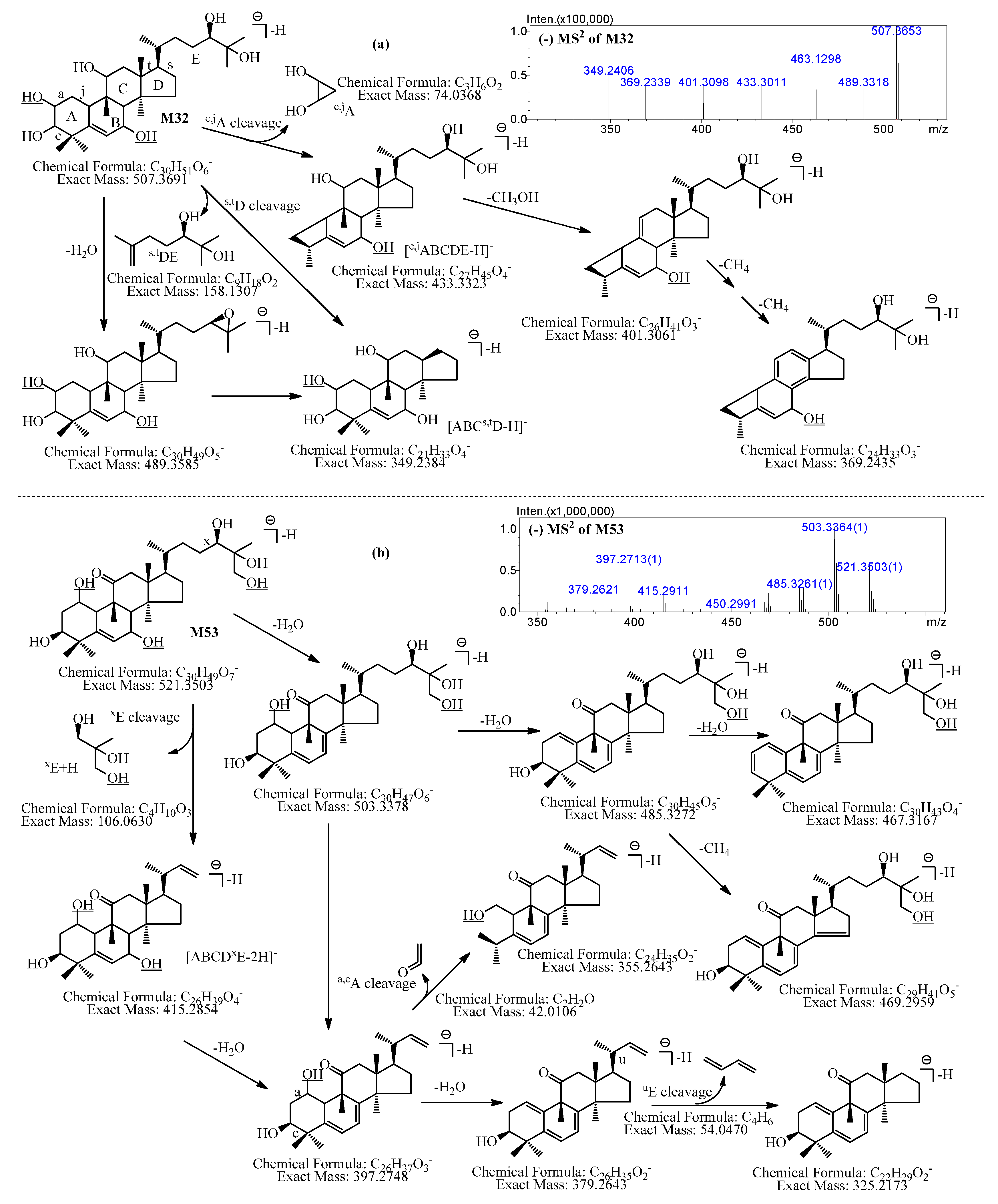
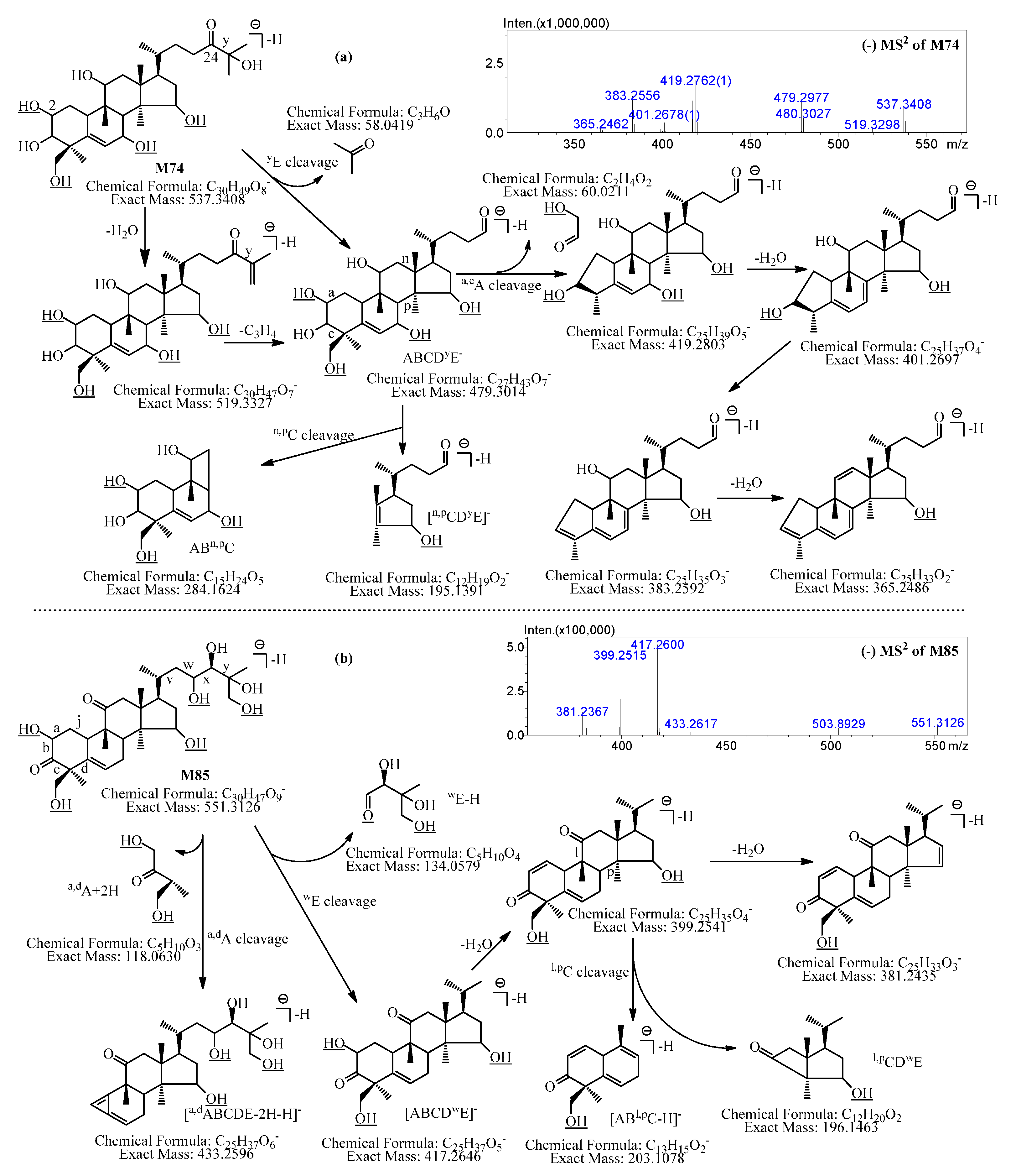
2.2. Identification of the Metabolites of Siamenoside I in Different Biosamples by HPLC-ESI-IT-TOF-MSn

Nine new metabolites of siamenoside I were unambiguously identified to be mogroside IVA (M3), mogroside IVE (M4), mogroside III (M8), mogroside IIIE (M9), mogroside IIIA1 (M10), mogroside IIE (M15), mogroside IIA2 (M17), 11-oxomogroside IIE (M20), and mogrol (M29) sequentially by comparison of their LC-MSn data to those of reference compounds.
The other 77 metabolites were tentatively identified by interpretation of their LC-MSn data and by comparison with literature.
These 86 new metabolites of siamenoside I can be classified into 24 classes according to their formative reactions and molecular formulae.
Table 1. LC-MS data of siamenoside I and its 86 metabolites formed in rats and their formative reactions.
No.tR (min)Meas. (Da)Pred.(Da)Err. (ppm)DBE 2FormulaIdentificationReactions
M0 124.9661169.59591169.59610.179C54H92O24siamenoside I
M125.6981285.64441285.64340.7810C60H102O29mogroside V isomer+Glc
M226.0431285.64411285.64340.5410C60H102O29mogroside V isomer+Glc
M3 125.3451169.59051169.5961−0.099C54H92O24mogroside IVAisomerization
M4 125.9601169.59231169.5961−3.259C54H92O24mogroside IVEisomerization
M526.6961169.59501169.5961−0.949C54H92O24mogroside IV isomerisomerization
M625.9231167.57301167.5804−2.2310C54H90O24dehydrogenated siamenoside I−2H
M725.9831153.59571153.6011−4.259C54H92O23deoxygenated siamenoside I−O
M8 126.8031007.54251007.5432−0.698C48H82O19mogroside III−Glc
M9 127.1731007.53871007.5432−4.478C48H82O19mogroside IIIE−Glc
M10 130.2171007.53761007.5432−5.568C48H82O19mogroside IIIA1−Glc
M1130.5251007.53951007.5432−3.678C48H82O19mogroside III isomer−Glc
M1230.9401007.53851007.5432−4.668C48H82O19mogroside III isomer−Glc
M1326.9271005.52691005.52760.709C48H80O19dehydrogenated mogroside III isomer−Glc − 2H
M1427.728991.5441991.5483−4.248C48H82O18deoxygenated mogroside III isomer−Glc − O
M15 129.365845.4881845.4904−2.727C42H72O14mogroside IIE−2Glc
M1630.648845.4944845.49044.737C42H72O14mogroside II isomer−2Glc
M17 131.775845.4910845.49040.717C42H72O14mogroside IIA2−2Glc
M1833.298845.4884845.4904−2.377C42H72O14mogroside II isomer−2Glc
M1933.905845.4918845.49041.667C42H72O14mogroside II isomer−2Glc
M20 129.908843.4737843.4748−1.308C42H70O1411-oxomogroside IIE−2Glc − 2H
M2133.604843.4726843.4748−2.618C42H70O14dehydrogenated mogroside II isomer−2Glc − 2H
M2234.028829.4946829.4955−1.087C42H72O13deoxygenated mogroside II isomer−2Glc − O
M2334.813827.4780827.4798−2.188C42H70O13dehydrogenated deoxygenated mogroside II isomer−2Glc − 2H − O
M2434.997683.4348683.4376−1.766C36H62O9mogroside IA1−3Glc
M2537.507683.4366683.43761.466C36H62O9mogroside IE1−3Glc
M2636.120681.4196681.4219−3.387C36H60O9dehydrogenated mogroside I isomer−3Glc − 2H
M2738.860681.4215681.4219−0.597C36H60O9dehydrogenated mogroside I isomer−3Glc − 2H
M2845.978521.3834521.3848−2.695C30H52O4mogrol isomer−4Glc
M29 146.467521.3838521.3848−1.925C30H52O4mogrol−4Glc
M3052.478519.3676519.3691−2.896C30H50O4dehydrogenated mogrol−4Glc − 2H
M3152.953519.3687519.3691−0.776C30H50O4dehydrogenated mogrol−4Glc − 2H
M3223.007553.3721553.3746−4.525C30H52O6dihydroxylated mogrol−4Glc + 2O
M3326.528553.3699553.3746−8.495C30H52O6dihydroxylated mogrol−4Glc + 2O
M3427.482553.3717553.3746−5.245C30H52O6dihydroxylated mogrol−4Glc + 2O
M3528.152553.3740553.3746−1.085C30H52O6dihydroxylated mogrol−4Glc + 2O
M3626.475551.3559551.3589−5.406C30H50O6dehydrogenated dihydroxylated mogrol−4Glc − 2H + 2O
M3727.050551.3557551.3589−5.806C30H50O6dehydrogenated dihydroxylated mogrol−4Glc − 2H + 2O
M3829.487551.3566551.3589−4.176C30H50O6dehydrogenated dihydroxylated mogrol−4Glc − 2H + 2O
M3930.887551.3558551.3589−5.626C30H50O6dehydrogenated dihydroxylated mogrol−4Glc − 2H + 2O
M4031.537551.3561551.3589−5.046C30H50O6dehydrogenated dihydroxylated mogrol−4Glc − 2H + 2O
M4133.122551.3549551.3589−7.256C30H50O6dehydrogenated dihydroxylated mogrol−4Glc − 2H + 2O
M4216.305569.3655569.3695−7.035C30H52O7trihydroxylated mogrol−4Glc + 3O
M4316.728569.3671569.3695−4.225C30H52O7trihydroxylated mogrol−4Glc + 3O
M4417.280569.3660569.3695−6.155C30H52O7trihydroxylated mogrol−4Glc + 3O
M4518.140569.3674569.3695−3.695C30H52O7trihydroxylated mogrol−4Glc + 3O
M4618.923569.3675569.3695−3.515C30H52O7trihydroxylated mogrol−4Glc + 3O
M4721.464569.3675569.3695−3.515C30H52O7trihydroxylated mogrol−4Glc + 3O
M4821.755569.3674569.3695−3.695C30H52O7trihydroxylated mogrol−4Glc + 3O
M4922.121569.3666569.3695−5.095C30H52O7trihydroxylated mogrol−4Glc + 3O
M5022.531569.3667569.3695−7.035C30H52O7trihydroxylated mogrol−4Glc + 3O
M5111.410567.3489567.3539−8.806C30H50O7dehydrogenated trihydroxylated mogrol−4Glc − 2H + 3O
M5218.482567.3463567.3539−13.46C30H50O7dehydrogenated trihydroxylated mogrol−4Glc − 2H + 3O
M5319.893567.3534567.3539−2.646C30H50O7dehydrogenated trihydroxylated mogrol−4Glc − 2H + 3O
M5420.977567.3494567.3539−0.886C30H50O7dehydrogenated trihydroxylated mogrol−4Glc − 2H + 3O
M5521.761567.3512567.3539−7.936C30H50O7dehydrogenated trihydroxylated mogrol−4Glc − 2H + 3O
M5622.365567.3507567.3539−4.766C30H50O7dehydrogenated trihydroxylated mogrol−4Glc − 2H + 3O
M5724.123567.3497567.3539−5.646C30H50O7dehydrogenated trihydroxylated mogrol−4Glc − 2H + 3O
M5824.478567.3508567.3539−7.236C30H50O7dehydrogenated trihydroxylated mogrol−4Glc − 2H + 3O
M5925.380567.3499567.3539−5.466C30H50O7dehydrogenated trihydroxylated mogrol−4Glc − 2H + 3O
M6027.050567.3498567.3539−7.056C30H50O7dehydrogenated trihydroxylated mogrol−4Glc − 2H + 3O
M6127.728567.3520567.3539−7.236C30H50O7dehydrogenated trihydroxylated mogrol−4Glc − 2H + 3O
M6224.412565.3351565.3382−5.487C30H48O7didehydrogenated trihydroxylated mogrol−4Glc − 4H + 3O
M6326.052565.3357565.3382−4.427C30H48O7didehydrogenated trihydroxylated mogrol−4Glc − 4H + 3O
M6428.810565.3368565.3382−2.487C30H48O7didehydrogenated trihydroxylated mogrol−4Glc − 4H + 3O
M6530.340565.3347565.3382−6.197C30H48O7didehydrogenated trihydroxylated mogrol−4Glc − 4H + 3O
M6613.872585.3613585.3644−5.305C30H52O8tetrahydroxylated mogrol−4Glc + 4O
M6714.242585.3601585.3644−7.355C30H52O8tetrahydroxylated mogrol−4Glc + 4O
M6814.603585.3608585.3644−6.155C30H52O8tetrahydroxylated mogrol−4Glc + 4O
M6918.307585.3603585.3644−4.955C30H52O8tetrahydroxylated mogrol−4Glc + 4O
M7019.408585.3613585.3644−5.305C30H52O8tetrahydroxylated mogrol−4Glc + 4O
M7115.573583.3444583.3488−7.546C30H50O8dehydrogenated tetrahydroxylated mogrol−4Glc − 2H + 4O
M7215.997583.3454583.3488−8.546C30H50O8dehydrogenated tetrahydroxylated mogrol−4Glc − 2H + 4O
M7320.492583.3454583.3488−6.006C30H50O8dehydrogenated tetrahydroxylated mogrol−4Glc − 2H + 4O
M7420.800583.3465583.3488−3.946C30H50O8dehydrogenated tetrahydroxylated mogrol−4Glc − 2H + 4O
M7521.453583.3465583.3488−3.946C30H50O8dehydrogenated tetrahydroxylated mogrol−4Glc − 2H + 4O
M7622.895583.3452583.3488−6.176C30H50O8dehydrogenated tetrahydroxylated mogrol−4Glc − 2H + 4O
M7724.710583.3447583.3488−7.036C30H50O8dehydrogenated tetrahydroxylated mogrol−4Glc − 2H + 4O
M7820.615581.3297581.3331−5.857C30H48O8didehydrogenated tetrahydroxylated mogrol−4Glc − 4H + 4O
M7921.815581.3290581.3331−7.057C30H48O8didehydrogenated tetrahydroxylated mogrol−4Glc − 4H + 4O
M8023.007581.3292581.3331−6.717C30H48O8didehydrogenated tetrahydroxylated mogrol−4Glc − 4H + 4O
M8123.433581.3306581.3331−4.037C30H48O8didehydrogenated tetrahydroxylated mogrol−4Glc − 4H + 4O
M8223.988581.3308581.3331−3.967C30H48O8didehydrogenated tetrahydroxylated mogrol−4Glc − 4H + 4O
M8325.682581.3304581.3331−4.647C30H48O8didehydrogenated tetrahydroxylated mogrol−4Glc − 4H + 4O
M8425.990581.3291581.3331−4.007C30H48O8didehydrogenated tetrahydroxylated mogrol−4Glc − 4H + 4O
M8517.707587.2977587.2992−2.557C30H48O9didehydrogenated pentahydroxylated mogrol−4Glc − 4H + 5O
M8618.607587.2983587.2992−1.537C30H48O9didehydrogenated pentahydroxylated mogrol−4Glc − 4H + 5O
1 Confirmed by comparison with reference compounds. 2 DBE, double bond equivalent.

2.2.1. Metabolites Formed by Monoglycosylation (M1, M2)

M1M2 showed [M + HCOOH − H] at m/z 1285.64, which indicated that their molecular formulae were C60H102O29. The formulae had an additional glucosyl (C6H10O5) than that of siamenoside I (C54H92O24). Hence, they were mogroside V isomers.
Figure 1. The proposed metabolic pathways of siamenoside I in rats.
Molecules 21 00176 g001

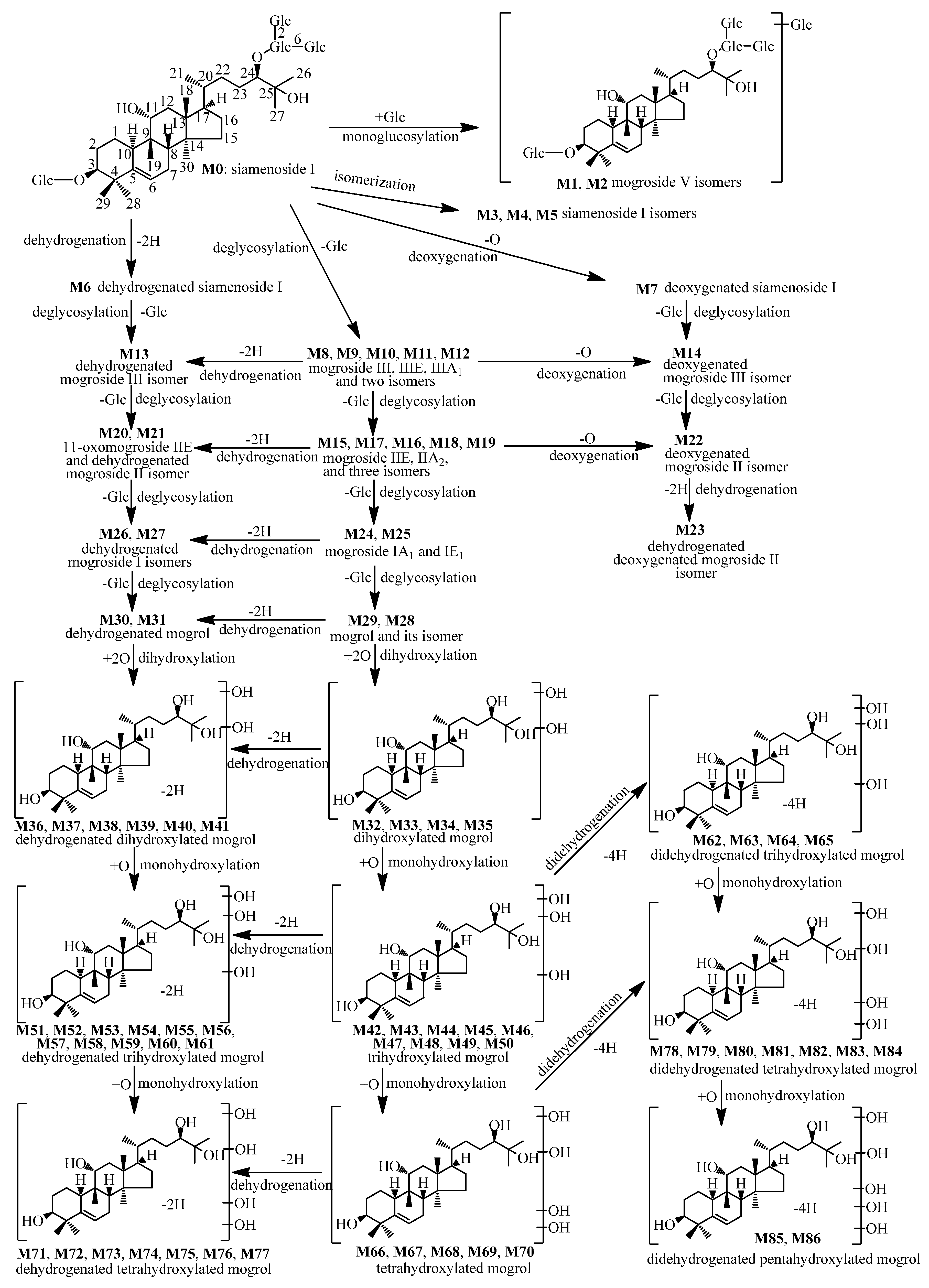
2.2.2. Metabolites Formed by Isomerization (M3M5)

M3M5 showed [M + HCOOH − H] at m/z 1169.59, indicating the molecular formula of C54H92O24, which was the same to siamenoside I. Hence, they were mogroside IV isomers, and M3M4 were further confirmed as mogroside IVA and mogroside IVE by comparison with reference compounds.

2.2.3. Metabolites Formed by Dehydrogenation (M6)

The molecular formula of M6 was predicted to be C54H90O24 based on its [M + HCOOH − H] at m/z 1167.5730, which was formed by loss of two hydrogen atoms from siamenoside I, thus M6 was tentatively identified as dehydrogenated siamenoside I.

2.2.4. Metabolites Formed by Deoxygenation (M7)

The molecular formula of M7 was C54H92O23 calculated from its [M + HCOOH − H] at m/z 1153.5957, which has one less oxygen atom than that of siamenoside I. Accordingly, it was tentatively identified as deoxygenated siamenoside I.

2.2.5. Metabolites Formed by Deglucosylation (M8M12)

M8M12 showed [M + HCOOH − H] at m/z 1007.54, implying their molecular formulae of C48H82O19. The formulae had one less glucosyl group (element composition: C6H10O5) than that of siamenoside I, so they were mogroside III isomers. In addition, M8M10 were unambiguously identified as mogroside III, mogroside IIIE, and mogroside IIIA1 by comparison with reference compounds.

2.2.6. Metabolites Formed by Deglucosylation and Dehydrogenation (M13)

M13 had the molecular formula of C48H80O19 predicted by its [M + HCOOH − H] at m/z 1005.5269. Compared with C48H82O19 of mogroside III isomers, it was tentatively identified as dehydrogenated mogroside III isomer. Moreover, in the MS2 of M13, [M − H − 2Glc] at m/z 797.4629 (C42H69O14), [M −H − 3Glc] at m/z 635.4090 (C36H59O9), [aglycon − H] at m/z 473.3623 (C30H49O4) were observed. Hence, M13 was a triglucoside of dehydrogenated mogrol.

2.2.7. Metabolites Formed by Deglucosylation and Deoxygenation (M14)

The molecular formula of M14 was calculated to be C48H82O18 by its [M + HCOOH − H] at m/z 991.5441, which had one less oxygen atom than C48H82O19 of mogroside III, so it was tentatively identified as deoxygenated mogroside III isomer.

2.2.8. Metabolites Formed By Dideglucosylation (M15M19)

The molecular formulae of M15M19 were determined to be C42H72O14 based on their [M + HCOOH − H] at m/z 845.49, which had one less glucosyl group (element composition: C6H10O5) than C48H82O19 of mogroside III, so they were mogroside II isomers. Furthermore, M15 and M17 were confirmed to be mogroside IIE and mogroside IIA2 by comparison with reference compounds.

2.2.9. Metabolites Formed by Dideglucosylation and Dehydrogenation (M20M21)

M20M21 showed [M + HCOOH − H] at m/z 843.47 in MS, suggesting their molecular formulae of C42H70O14. Additionally, they showed [aglycon − H] at m/z 473.3623 (C30H49O4) in MS2 spectra. Therefore, they were tentatively identified as dehydrogenated mogroside II isomer, i.e., diglucoside of dehydrogenated mogrol. Further, M20 was unambiguously identified as 11-oxomogroside IIE by comparison with reference compounds.

2.2.10. Metabolites Formed by Dideglucosylation and Deoxygenation (M22)

M22 was tentatively identified as deoxygenated mogroside II isomer, since its molecular formula was determined to be C42H72O13 by its [M + HCOOH − H] at m/z 829.4946, which had one less oxygen atom than C42H72O14 of mogroside II isomers. In addition, M22 showed [M − H] at m/z 783.4833 (C42H71O13), [M−H−C6H10O4 (deoxyhexosyl)] at m/z 637.4252 (C36H61O9), and [M−H−C6H10O4−Glc] at m/z 475.3742 (C30H51O4) in MS2 spectra, which indicated that the deoxygenation occurred in hexose and the aglycone was mogrol. Thus, M22 was identified as a deoxyhexosyl-glucosyl mogrol.

2.2.11. Metabolites Formed by Dideglucosylation, Dehydrogenation, and Deoxygenation (M23)

The molecular formula of M23 was determined to be C42H70O13 according to its [M + HCOOH − H] at m/z 827.4780, which lost two hydrogen atoms from C42H72O13 of M22. Consequently, M23 was tentatively identified as a dehydrogenated deoxygenated mogroside II isomer.

2.2.12. Metabolites Formed by Trideglucosylation (M24M25)

M24M25 showed [M + HCOOH − H] at m/z 683.43 in MS, and [M−H] at m/z 637.42 (C36H61O9), [aglycone−H] at m/z 475.37 (C30H51O4) in MS2 spectra, which implied that they were mogrol glucoside. By comparison with the LC-MSn data in literature [10], M24 and M25 were tentatively identified as mogroside IA1 and mogroside IE1, respectively.

2.2.13. Metabolites Formed by Trideglucosylation and Dehydrogenation (M26M27)

The molecular formulae of M26M27 were predicted to be C36H60O9 based on its [M + HCOOH − H] at m/z 681.42 in MS, which had two less hydrogen atoms than C36H62O9 of mogroside I isomers. In their negative ion (NI) MS2 spectra, [M − H] at m/z 635.41 (C36H59O9) and [aglycone−H] at m/z 473.36 (C30H49O4) were detected. As a result, M26M27 were tentatively identified as glucosides of dehydrogenated mogrol, i.e., dehydrogenated mogroside I isomers.

2.2.14. Metabolites Formed by Tetradeglucosylation (M28M29)

M29 was unambiguously identified as mogrol by comparison with reference compound. M28 had the same molecular formula to mogrol, which showed [M + HCOOH − H] at m/z 521.38 in MS, so it was a mogrol isomer.

2.2.15. Metabolites Formed by Tetradeglucosylation and Dehydrogenation (M30M31)

M30 and M31 had the molecular formulae of C30H50O4 predicted by their [M + HCOOH − H] at m/z 519.36, which had two less hydrogen atoms than C30H52O4 of mogrol, thus they were tentatively identified as dehydrogenated mogrols.

2.2.16. Metabolites Formed by Tetradeglucosylation and Dihydroxylation (M32M35)

M32M35 showed [M + HCOOH − H] at m/z 553.37, indicating the molecular formula of C30H52O6. Compared to C30H52O4 of mogrol, it had two more oxygen atoms. Accordingly, M32M35 were tentatively identified as dihydroxylated mogrols.
In addition, the possible hydroxylation sites of M32 can be deduced by its MS2 data and one possible structure of M32 is shown in Figure 2a. The nomenclature for the fragmentation pathways and fragment ions of cucurbitanes proposed by the authors [10] were used in this study.
In MS2 spectra of M32, m/z 433.3011 ([c,jABCDE − H], C27H45O4) generated by c,jA cleavage and m/z 349.2406 ([s,tDE − H], C21H33O4) generated by s,tD cleavage were observed, which indicated that one hydroxylation site was in c,jA, and the other was in c,jABCs,tD (Figure 2a).
Figure 2. The MS2 spectra, characteristic fragment ions, and proposed fragmentation pathways of M32 and M53. (a) M32; (b) M53.
Molecules 21 00176 g002

2.2.17. Metabolites Formed by Tetradeglucosylation, Dihydroxylation, and Dehydrogenation (M36M41)

The molecular formulae of M36M41 were calculated to be C30H50O6 based on their [M + HCOOH − H] at m/z 551.35. Compared to C30H52O6 of M32M35, it had two less hydrogen atoms. Accordingly, M36M41 were tentatively identified as dehydrogenated dihydroxylated mogrols.

2.2.18. Metabolites Formed by Tetradeglucosylation and Trihydroxylation (M42M50)

M42M50 had the molecular formulae of C30H52O7 predicted by their [M + HCOOH − H] at m/z 569.36. Compared to C30H52O6 of M32M35 (dihydroxylated mogrol), it had one more oxygen atom. Accordingly, M42M50 were tentatively identified as trihydroxylated mogrols.

2.2.19. Metabolites Formed by Tetradeglucosylation, Trihydroxylation, and Dehydrogenation (M51M61)

The molecular formulae of M51M61 were determined to be C30H50O7 on the basis of their [M + HCOOH − H] at m/z 567.35. In comparison with C30H52O7 of M42M50 (trihydroxylated mogrol), it had two less hydrogen atoms. As a result, M51M61 were tentatively identified as dehydrogenated trihydroxylated mogrols.
Further, the possible hydroxylation sites of M53 could be deduced by its MS2 data, and one possible structure of M53 is shown in Figure 2b.
M53 showed [M + HCOOH − H] at m/z 567.3524 in MS, and then it was fragmented into [M − H] at m/z 521.3207 (C30H49O7) in MS2 spectrum. The [M − H] was further cleaved into product ions at m/z 503.3364 (C30H47O6), 485.3261 (C30H45O5), and 467.3056 (C30H43O4) formed by sequential losses of H2O. It was also cleaved into product ion at m/z 415.2911 (C26H39O4) by losing C4H10O3 (xE+H), which indicated that one hydroxylation site was in xE. Besides, the characteristic fragment ions at m/z 397.2713 (C26H37O3), m/z 379.2621 (C26H35O2), m/z 355.2613 (C24H35O2), and m/z 325.2449 (C22H29O2) were observed in MS2, indicating that the other two hydroxylation sites should be located at a,cABCDuE (Figure 2b).

2.2.20. Metabolites Formed by Tetradeglucosylation, Trihydroxylation, and Didehydrogenation (M62M65)

M62-M65 had the molecular formulae of C30H48O7 predicted by their [M + HCOOH − H] at m/z 565.33, which had two less hydrogen atoms than C30H50O7 of M51M61. Accordingly, M62M65 were tentatively identified as didehydrogenated trihydroxylated mogrols.

2.2.21. Metabolites Formed by Tetradeglucosylation and Tetrahydroxylation (M66M70)

M66M70 showed [M + HCOOH − H] at m/z 585.36, indicating the molecular formula of C30H52O8. Compared with C30H52O4 of mogrol, their molecular formula had four more oxygen atoms. Therefore, they were tetrahydroxylated mogrols.

2.2.22. Metabolites Formed by Tetradeglucosylation, Tetrahydroxylation, and Dehydrogenation (M71M77)

The molecular formulae of M71M77 were determined to be C30H50O8 based on their [M + HCOOH − H] at m/z 583.34, which had two less hydrogen atoms than C30H52O8 of M66M70 (tetrahydroxylated mogrol). Accordingly, they were tentatively identified as dehydrogenated tetrahydroxylated mogrol.
Besides, the possible dehydrogenation and hydroxylation sites of M74 could be deduced by its MS2 and MS3 data, and one possible structure of M74 is shown in Figure 3a.
The characteristic product ions at m/z 479.2977 (C27H43O7, ABCDyE) produced by yE cleavage, m/z 419.2762 (C25H39O5) generated by a,cA cleavage, and m/z 195.1347 (C12H19O2) generated by n,pC cleavage indicated that the C24-hydroxyl group of M74 was dehydrogenated, and one of the four tetrahydroxylation sites was at C2, one was in n,pCDyE, and the other two were in ABn,pC (Figure 3a).
Figure 3. The MS2 spectra, characteristic fragment ions, and proposed fragmentation pathways of M74 and M85. (a) M74; (b) M85.
Molecules 21 00176 g003

2.2.23. Metabolites Formed by Tetradeglucosylation, Tetrahydroxylation, and Didehydrogenation (M78M84)

M78M84 showed [M + HCOOH − H] at m/z 581.33 in their MS, indicating their molecular formulae of C30H48O8, which had two less hydrogen atoms than C30H50O8 of M71M77. Therefore, M78M84 were tentatively identified to be didehydrogenated tetrahydroxylated mogrol.

2.2.24. Metabolites Formed by Tetradeglucosylation, Pentahydroxylation, and Didehydrogenation (M85M86)

M85M86 had the molecular formulae of C30H48O9 predicted by their [M + HCOOH − H] at m/z 587.29 in their MS, which had one more oxygen atom than C30H48O8 of M78M84. Accordingly, M85M86 were tentatively identified as didehydrogenated pentahydroxylated mogrol.
In addition, the possible dehydrogenation and hydroxylation sites of M85 could be deduced by its MS2 and MS3 data, and one possible structure of M85 is shown in Figure 3b.
M85 showed [M + Cl] at m/z 587.2977, which was fragmented into [M − H] at m/z 551.3126 (C30H47O9) in MS2 spectrum. The [M − H] was then fragmented into characteristic product ions at m/z 433.2617 (C25H37O6, [a,dABCDE − 2H − H]) and m/z 417.2600 (C25H37O5, [ABCDwE]) by a,dA cleavage and wE cleavage respectively, which implied that two of the five hydroxylation sites were in a,dA, other two were in wE. Furthermore, the ion at m/z 417.2600 (C25H37O5, [ABCDwE]) was cleaved into product ions at m/z 399.2515 (C25H35O4) and m/z 203.0987 (C13H15O2) by sequential loss of H2O and l,pCDwE (C12H20O2) in MS3 spectra, which indicated that the last of the five hydroxylation sites was in l,pCDwE.

2.3. Distribution of the Metabolites of Siamenoside I in Rats

The peak areas and distributions of siamenoside I and the 86 identified metabolites in different biological samples are shown in Table 2.
Table 2. Distribution of siamenoside I and its 86 metabolites in rat organs and their peak areas calculated from extracted ion chromatograms (EICs).
No.FecesUrinePlasmaHeart 1Liver 1Spleen 1Lung 1Kidney 1Stomach 1Intestine 1Brain 1MuscleTPA 2
M09,489,56145,194,235929,711655,7251,151,49115,473,8081,201,52471,162,19086,071,968
M11,144,6579,555,9002,001,7691,487,067680,78614,870,179
M215,997,951531,34516,529,296
M3760,580847,8691,307,6731,655,0972,264,310177,9567,013,485
M4545,5935,114,6662,993,9721,200,2342,991,3751,454,8472,022,51666,66516,389,868
M51,984,3202,244,0413,096,436301,4337,626,230
M61,370,70124,868,935522,375216,179460,4995,658,9082,342,2768,964,56844,404,441
M7151,4761,610,4851,761,961
M8686,710509,1059,795,2853,463,19114,454,291
M95,499,3804,564,759247,2453,396,21110,439,0991,349,085621,65417,082,22123,756,27315,351,6707,796,21190,103,808
M1025,071,25125,071,251
M116,584,3216,584,321
M127,864,5037,864,503
M135,673,582927,1521,104,6551,569,801126,7252,884,61510,255,235861,73123,403,496
M144,700,4981,594,5952,186,0938,481,186
M15122,469,0482,086,502752,0821,976,,0589,533,75634,065,738170,883,184
M1610,029,17910,029,179
M1714,873,4273,964,56918,837,996
M18131,377,6972,775,3952,221,8341,382,3136,051,761868,285144,677,285
M1945,423,120394,0658,102,455562,37054,482,010
M2050,669,383447,6581,165,19534,649,28486,931,520
M2150,269,416885,941128,120905,38552,188,862
M22162,025,962861,129162,887,091
M2329,484,81429,484,814
M24180,532,696352,905685,1281,649,227184,835,243
M25896,803,1694,472,84429,155,815486,61221,253,177297,909,9671,250,081,584
M2648,856,1081,376,742598,62150,831,471
M27790,013,5982,541,94018,180,494810,736,032
M2892,475,8131,666,7101,426,86795,569,390
M29646,804,735459,452313,54312,256,3091,839,703661,673,742
M3024,183,72824,183,728
M31324,786,3315,151,046329,937,377
M32390,217,566818,492391,036,058
M3346,223,58246,223,582
M34125,631,7232,735,1641,822,3251,674,281131,863,493
M35488,223,8801,363,3388,760,0944,522,744502,870,056
M3633,006,73933,006,739
M3723,418,33723,418,337
M3846,101,38846,101,388
M3927,487,97527,487,975
M4020,088,77320,088,773
M4127,408,60827,408,608
M4212,435,30112,435,301
M4377,259,58977,259,589
M44284,733,513284,733,513
M4544,372,83044,372,830
M4689,188,73189,188,731
M47108,520,771108,520,771
M4830986,85530,986,855
M4934,753,95234,753,952
M5053,158,41853,158,418
M5115,908,47015,908,470
M5230,236,80043,804,7982,369,28176,410,879
M53463,948,611463,948,611
M5421,448,93721,448,937
M5579,454,25879,454,258
M5679,252,36779,252,367
M5739,700,63639,700,636
M5886,848,22086,848,220
M5917,421,55817,421,558
M6021,097,29421,097,294
M6188,863,19688,863,196
M6238,437,24538,437,245
M63102,677,796102,677,796
M6435,880,11335,880,113
M6523,867,7871,449,63925,317,426
M6613,548,27013,548,270
M675,583,7665,583,766
M685,640,3115,640,311
M699,156,9849,156,984
M7010,312,53910,312,539
M7113,648,74213,648,742
M724,644,7574,644,757
M7354,507,90154,507,901
M7477,998,65877,998,658
M7539,341,20939,341,209
M7612,303,03912,303,039
M777,437,1477,437,147
M7830,479,61030,479,610
M7945,861,42345,861,423
M8018,882,76418,882,764
M81285,519,6821,729,889287,249,571
M8219,376,25319,376,253
M8336,030,99826,693,03262,724,030
M8414,114,95414,114,954
M8530,984,34627,085,01358,069,359
M8650,436,19050,436,190
TPA 27,540,531,449125,100,0731,699,3314,148,29350,488,31537,920,4851,620,283115,671,871147,536,576449,779,86753,283,70108,527,780,244
Sum 3831922773132119140
Peak Area (A)A ≥ 109109 >A ≥ 108108 > A ≥ 107107 > A ≥ 106106 > A ≥ 105104 ≤ A < 105A = 0
Color
1 These data are comparable. 2 Total peak areas. 3 The total number of metabolites detected.

3. Discussion

The metabolism of siamenoside I in rats was firstly investigated in the present work. In total, 86 new metabolites of siamenoside I were detected in different biological samples from rats, and nine of them were unambiguously identified by comparison with reference compounds, and the others were tentatively identified by careful interpretation of their LC-MSn data.

3.1. The Metabolic Pathways of Siamenoside I in Rats

Based on the structures of the metabolites (M1M86), the metabolic pathways of siamenoside I in rats are proposed and shown in Figure 1. From Figure 1, we can find that the metabolic reactions of siamenoside I include deglycosylation, hydroxylation, dehydrogenation, deoxygenation, isomerization, and glycosylation.
Most of the metabolic reactions of siamenoside I are the same to mogroside V [10]. However, there are also some differences. For example, methylation metabolites were not found in the metabolism of siamenoside I, and deoxygenation is found to be a novel metabolic reaction of mogrosides. Furthermore, pentahydroxylation, didehydrogenation are also found as novel metabolic reactions of mogrosides.
Among 86 metabolites, 63 metabolites (M6, M13, M20, M21, M26, M27, M30M86) are formed by oxidation reactions such as hydroxylation and dehydrogenation, and 79 metabolites (M8M86) are formed by deglycosylation, which indicate that deglycosylation and oxidation (hydroxylation, dehydrogenation) are the major metabolic reactions of siamenoside I.
Astonishingly, four metabolites (M7, M14, M22, and M23) are formed by reduction reaction (deoxygenation). The deoxygenation reaction might occur in hexose, not in aglycone, which is inferred from the identification of M22 (Section 2.2.10.).
Furthermore, 23 metabolites (M6, M7, M13, M14, M22, M23, M26-M28, M30, M62-M65, M78M86) are firstly reported as new metabolites of mogrosides.
These results suggest that siamenoside I has its own metabolism characteristics in comparison with mogroside V.

3.2. Distribution of the Metabolites of Siamenoside I in Rats

From Table 2, we could find that total peak areas of all detected compounds (siamenoside I and metabolites) in different rat organs are ranked as follows: intestines (449,779,867) > stomach (147,536,576) > kidneys (115,671,871) > brain (53,283,701) > liver (50,488,315) > spleen (37,920,485) > heart (4,148,293) > lungs (1,620,283). In addition, the total numbers of compounds (siamenoside I and metabolites) detected in different organs are in the order of stomach (22) > intestines (20) > brain (15) > kidneys (14) > liver (7) > spleen (7) > lungs (4) > heart (2). Therefore, siamenoside I and its metabolites are mainly distributed to the intestines, stomach, kidneys, and brain.
We also could find the specific metabolites detected in different biosamples. For example, 50 metabolites (M10-M12, M16, M23, M30, M33, M36M50, M53-M64, M66M80, M82, M84, M86) are only detected in feces; M2 is only detected in urine and the brain; M7 is only detected in urine and feces; M51 is only detected in kidneys; M85 is only detected in kidneys and feces; M17, M31, M32 are only detected in stomach and feces; M65 and M81 are only detected in intestines and feces; M83 is only detected in brain and feces. Besides, siamenoside I and 14 metabolites (M1M6, M9, M13, M18M19, M24, M26, M52, M83) are detected in the brain for the first time.
M9 (mogroside IIIE) is detected in all biosamples except muscle, indicating that it is the most widely distributed metabolite.

3.3. The Proposed in Vivo Process of Siamenoside I in Rats

From Table 2, we could find that M1M29 seem to be the main metabolites, and most of the other metabolites are only detected in feces. We think the reasons for this result may be: (1) the first pass effect of these metabolites may be very high, i.e., their hepatic extraction ratios are very high, which leads to their very low contents in plasma, organs, and urine samples. As a result, they are not easily detected in these samples; (2) these metabolites are mainly excreted into bile and then to feces, which make their contents in feces high and detectable.
Based on our research results and general metabolic knowledge, we hypothesize that the in vivo process of siamenoside I in rats may be as follows.
After oral administration, siamenoside I is degraded into its secondary glycosides (e.g., mogroside III, IIIE, IIIA1, IIE, IIA2, IA1, IE1, etc.) and its aglycone (mogrol) or dehydrogenated aglycone (dehydrogenated mogrol) by gastric juice, intestinal juice, intestinal enzymes, or intestinal microflora. Then, mogrol or dehydrogenated mogrol permeate across intestinal mucosa and enter the liver, where they undergo extensive oxidative metabolic reactions to form lots of hydroxylation and/or dehydrogenation metabolites. These polar oxidative metabolites may be largely excreted into the bile and then to the feces, and only a limited amount of them enter general circulation. Besides, some of them may also undergo hepatoenteral circulation and are absorbed into general circulation, and then distributed to different organs, and finally excreted into feces or/and urine.

3.4. Bioactivities of the Metabolites of Siamenoside I

On the basis of literature retrieval, eight metabolites (M3, mogroside IVA; M4, mogroside IVE; M8, mogroside III; M15, mogroside IIE; M24, mogroside IA1; M25, mogroside IE1; M29, mogrol; M9, mogroside IIIE) of siamenoside I can be regarded as bioactive metabolites.
Among them, seven metabolites (M3, M4, M8, M15, M24, M25, M29) are able to inhibit the induction of Epstein-Barr virus early antigen by 12-O-tetradecanoylphorbol-13-acetate in Raji cells [5]. M4 and M9 can inhibit maltase [7]. Hence, they might contribute to the bioactivities of siamenoside I.

4. Materials and Methods

4.1. Chemicals and Reagents

Siamenoside I, mogroside III, mogroside IIIE, mogroside IVE, mogroside V, mogroside IIE, 11-oxomogroside IIE, and mogrol (all purities >98%, determined by HPLC-DAD-ELSD) were isolated from the dried fruits of Siraitia grosvenorii and the 50% mogroside V enzymatic hydrolysate by the authors [12,13], and their structures were confirmed by spectral data (UV, IR, NMR and MS). Mogroside IVA, mogroside IIIA1 and mogroside IIA2 (all purities >98%, determined by HPLC-DAD-ELSD) were purchased from Chengdu MUST Bio-technology Co., Ltd. (Chengdu, Sichuan, China).
Ultra-pure water was prepared by a Millipore Milli-Q Integral 3 Ultrapure Water System (Billerica, MA, USA). Acetonitrile (HPLC grade) was bought from Fisher Chemicals Co. (Fairlawn, NJ, USA) and formic acid (HPLC grade) was purchased from Mreda Technology Inc. (Beijing, China).

4.2. Animals

Sprague-Dawley (SD) rats (male, 210 ± 20 g) were bought from the Experimental Animal Center of Peking University Health Science Center (Beijing, China). They were handled in agreement with the Guide for the Care and Use of Laboratory Animals of the US National Institutes of Health. All animal experiments were approved by the Biomedical Ethical Committee of Peking University (Approval No. LA2011-058).

4.3. Instruments

A Shimadzu LCMS-IT-TOF instrument was used to perform HPLC-ESI-IT-TOF-MSn analysis, which consists of a CBM-20A system controller, a DGU-20A3 degasser, two LC-20AD pumps, an SIL-20AC autosampler, a CTO-20A column oven, an SPD-M20A photodiode array (PDA) detector, an ESI ion source, and a hybrid IT-TOF mass spectrometer (Shimadzu, Kyoto, Japan).

4.4. Animal Experiments and Sample Collection

Eight rats were divided into two groups: two were blank group and the others were test group. Each rat was put into a clean metabolic cage (Suzhou Fengshi Laboratory Animal Equipment Co., Suzhou, Jiangsu, China) and given food and water ad libitum.
Because the research aim is to find the general/average differences between test group rats (drug-containing sample) and blank group rats (blank sample), the individual differences among the same group rats are not taken into consideration. Accordingly, all of the biosamples from each group were combined into one sample which was more representative than individual samples in the following sample collection processes.
The animal experiment lasted six days. The whole urine and feces of days 1–2 were collected as blank urine and feces samples, respectively. On days 3–5, the rats of test group were orally administrated with siamenoside I [50 mg/kg body weight, in normal saline (NS) solution] at 9:00, and all 72-h urine and feces were collected as drug-containing urine and feces samples, respectively. The rats of blank group were orally administrated with the same volume of NS. On day 6 at 9:00, the test and the blank group were treated with siamenoside I and NS again, respectively. After 1 h, blood sample was collected into a vacuum tube with sodium citrate as anticoagulant (Hebei Xinle Technology Co., Ltd., Shijiazhuang, Hebei, China) from rat heart under anesthesia. Then, the organs (heart, liver, spleen, lung, kidneys, stomach, small intestine, brain) and skeletal muscles of rats were collected and washed with NS, separately. All samples were kept at −80 °C before further pretreatment.

4.5. Sample Preparation

4.5.1. Blood Samples

The blood samples were centrifuged at 3000 rpm for 20 min at 4 °C, and the supernatant plasma samples were collected. Afterward, 8 mL methanol was added to 2 mL of plasma sample, and then was mixed and centrifuged at 9000 rpm for 30 min. The supernatant was collected and evaporated to dryness at 55 °C by a rotatory evaporator. The 15 mg residue was reconstituted in 1.00 mL methanol, filtrated through 0.45 μm filter membrane, and stored at 4 °C before analysis.

4.5.2. Urine Samples

Urine samples were filtered and then evaporated to dryness under vacuum at 55 °C by a rotatory evaporator. Subsequently, the residue was ultrasonically extracted with 10 mL methanol for 30 min, and the extract was then centrifuged at 9000 rpm for 30 min. The supernatant was transferred to another tube and evaporated to dryness at 55 °C by a rotatory evaporator. Next, a 100 mg residue was dissolved in 1.00 mL methanol, filtered through 0.45 μm membranes, and stored at 4 °C before analysis.

4.5.3. Feces Samples

Feces samples were dried at 55 °C and pulverized. Subsequently, the 10 g powder was ultrasonically extracted with 50 mL methanol for 30 min for three times, and the three supernatants were combined and evaporated to dryness. Next, the residue was mixed with 20 mL methanol and centrifuged at 9000 rpm for 30 min. The supernatant was evaporated to dryness again at 55 °C and the residue was collected. Then, 15 mg of residue was dissolved in 1.00 mL methanol, 0.45 μm membranes, and stored at 4 °C before analysis.

4.5.4. Organ and Skeletal Muscle Samples

Each organ was weighed, minced, and homogenized in 4 times volume (mL/g) of 4 °C NS by a homogenizer (Ultra-Turrax T8, Ika-Werke, Gmbh & Co. KG, Staufen, Germany). Then, 10 mL homogenate was mixed with 9 times volume (mL/mL) of acetonitrile, ultrasonically treated for 30 min, and centrifuged at 12,000 rpm for 30 min at 4 °C. Afterward, the supernatant was collected and evaporated to dryness at 50 °C. The residue was dissolved in 1.00 mL methanol, filtered through 0.45 μm filter membrane, and stored at 4 °C before analysis. The skeletal muscle samples were treated by the same method.

4.6. LC-MSn Conditions

The column used was Inertsil ODS-3 C18 (250 mm × 4.6 mm, 5 μm) (Shimadzu, Kotyo, Japan) protected with a Phenomenex Security Guard column (C18, 4 mm × 3.0 mm, 5.0 μm) (Phenomenex, Torrance, CA). The column temperature was 35 °C. The injection volume of all samples was 20 μL. The mobile phases were water-formic acid (100:0.1, v/v) (A) and acetonitrile (B).The flow rate was 1.0000 mL/min. A gradient elution program was used: 0.01–10.00 min, 10%–18% B; 10.00–20.00 min, 18%–28% B; 20.00–35.00 min, 28%–45% B; 35.00–60.00 min, 45%–90% B; 60.00–70.00 min, 90%–100% B; 70.00–80.00 min, 100% B. The UV spectrum was recorded from 195 nm to 400 nm.
The parameters of the ESI-IT-TOF-MSn instrument were: (1) flow rate: 0.2000 mL/min (split from HPLC effluent); (2) positive ion (PI) and negative ion (NI) alternate detection; (3) mass range: MS, m/z 300–2000; MS2 and MS3, m/z 50–2000; (4) temperature of heat block and curved desolvation line (CDL): 250 °C; (5) nebulizing nitrogen gas flow: 1.5 L/min; interface voltage: (+), 4.5 kV; (−), −3.5 kV; detector voltage: 1.70 kV; (6) ion accumulation time: MS, 30 ms; MS2 and MS3, 20 ms; relative collision-induced dissociation (CID) energy: 50%; (7) data-dependent MS2 and MS3 fragmentation; (8) All data were recorded and analyzed by LCMS solution Version 3.60, Formula Predictor Version 1.01, and Accurate Mass Calculator (Shimadzu, Kyoto, Japan). The mass range of 50–3000 Da was calibrated by a trifluoroacetic acid sodium solution (2.5 mM).

4.7. Strategy for Profiling and Identification of the Metabolites of Siamenoside I in Biosamples

The strategy [14] previously proposed by the authors was used to find and identify the metabolites of siamenoside I in the present study. In short, the base peak chromatograms (BPCs) of drug-containing samples and blank samples were thoroughly analyzed and compared. Meanwhile, the possible metabolites predicted by general metabolism rules were also screened and confirmed by comparing their corresponding extracted ion chromatograms (EICs). The metabolites were identified by comparison of their retention times and MSn data with those of reference compounds, or tentatively identified by interpretation of their MSn data.

4.8. Preliminary Evaluation of the Relative Contents of Siamenoside I and Its Metabolites in Biosamples

To preliminarily estimate the relative contents of siamenoside I and its metabolites in biosamples, the peak area of each metabolite calculated from its NI EIC was used. In the present study, only the data of different organ samples were comparable, since they were prepared and analyzed by the same method.

5. Conclusions

The metabolism of siamenoside I in rats was studied for the first time. In total, 86 new metabolites were detected. Nine of them were unambiguously identified by comparison with reference compounds, and the other 77 were tentatively identified by HPLC-ESI-IT-TOF-MSn technique. The metabolic pathways of siamenoside I in rats were proposed based on the structures of metabolites. The metabolic reactions of siamenoside I were found to be deglycosylation, hydroxylation, dehydrogenation, deoxygenation, isomerization, and glycosylation, among which deoxygenation, pentahydroxylation, and didehydrogenation were novel metabolic reactions of mogrosides. In addition, 23 metabolites were new metabolites of mogrosides. The distributions of siamenoside I and its 86 metabolites in rat organs were firstly reported, and they were mainly distributed to intestines, the stomach, kidneys, and the brain. Mogroside IIIE was the most widely distributed metabolite. Eight metabolites had bioactivities, indicating that they might contribute to the bioactivities of siamenoside I. These findings not only provide valuable information on the metabolism and disposition of siamenoside I and mogrosides in rats but also provide useful information on the chemical basis of the pharmacological effects of siamenoside I and mogrosides in vivo.

Supplementary Materials

The following are available online at www.mdpi.com/1420-3049/21/2/176, Figures S1–S25: EICs of 86 metabolites in drug-containing sample and blank sample, Table S1: Retention time (tR), LC-ESI-IT-TOF-MSn data, molecular formula, and identification of siamenoside I and its metabolites in different biosamples.

Acknowledgments

This study was financially supported by the National Natural Science Foundation of China (No. 81160392) and Guangxi Natural Science Foundation (2014GXNSFBA118049).

Author Contributions

F. Xu and D.-P. Li conceived and designed the experiments; X.-R. Yang, F. Xu, F.-L. Lu, and L. Wang performed the experiments; X.-R. Yang and F. Xu analyzed the data; D.-P. Li, Y.-L. Huang, G.-X. Liu, M.-Y. Shang, and S.-Q. Cai contributed reagents/materials/analysis tools; F. Xu, X.-R. Yang, and D.-P. Li wrote the paper.

Conflicts of Interest

The authors declare no conflict of interest. The founding sponsors had no role in the design of the study; in the collection, analyses, or interpretation of data; in the writing of the manuscript, and in the decision to publish the results.

Abbreviations

The following abbreviations are used in this manuscript:
EBV-EA
Epstein-Barr virus early antigen
TPA
12-O-tetradecanoylphorbol-13-acetate
DMBA
9,10-Dimethyl-1,2-benzanthracene
HPLC-ESI-IT-TOF-MSn
High-performance liquid chromatography-electrospray ionization-ion trap-time of flight-multistage mass spectrometry
Glc
Glucosyl group
NI
Negative ion
DAD-ELSD
Diode array detector coupled with evaporative light scattering detector
NS
Normal saline
BPC
Base peak chromatogram
EIC
Extracted ion chromatogram

References

  1. Li, C.; Lin, L.M.; Sui, F.; Wang, Z.M.; Huo, H.R.; Dai, L.; Jiang, T.L. Chemistry and pharmacology of Siraitia grosvenorii: A review. Chin. J. Nat. Med. 2014, 12, 89–102. [Google Scholar] [CrossRef]
  2. Jin, J.K.; Lee, J.H. Phytochemical and pharmacological aspects of Siraitia grosvenorii, luo han kuo. Orient. Pharm. Exp. Med. 2012, 12, 233–239. [Google Scholar] [CrossRef]
  3. Kasai, R.; Nie, R.L.; Nashi, K.; Ohtani, K.; Zhou, J.; Tao, G.D.; Tanaka, O. Sweet cucurbitane glycosides from fruits of Siraitia siamensis (chi-zi luo-han-guo), a Chinese folk medicine. Agric. Biol. Chem. 1989, 53, 3347–3349. [Google Scholar] [CrossRef]
  4. Matsumoto, K.; Kasai, R.; Ohtani, K.; Tanaka, O. Minor cucurbitane glycosides from fruits of Siraitia grosvenori (Cucurbitaceae). Chem. Pharm. Bull. 1990, 38, 2030–2032. [Google Scholar] [CrossRef]
  5. Ukiya, M.; Akihisa, T.; Tokuda, H.; Toriumi, M.; Mukainaka, T.; Banno, N.; Kimura, Y.; Hasegawa, J.; Nishino, H. Inhibitory effects of cucurbitane glycosides and other triterpenoids from the fruit of Momordica grosvenori on Epstein-Barr virus early antigen induced by tumor promoter 12-O-tetradecanoylphorbol-13-acetate. J. Agric. Food Chem. 2002, 50, 6710–6715. [Google Scholar] [CrossRef] [PubMed]
  6. Konoshima, T. Inhibitory effects of sweet glycosides from fruits of Siraitia grosvenori on two stage carcinogenesis. Food Style 21 2004, 8, 77–81. [Google Scholar]
  7. Suzuki, Y.A.; Murata, Y.; Inui, H.; Sugiura, M.; Nakano, Y. Triterpene glycosides of Siraitia grosvenori inhibit rat intestinal maltase and suppress the rise in blood glucose level after a single oral Administration of maltose in rats. J. Agric. Food Chem. 2005, 53, 2941–2946. [Google Scholar] [CrossRef] [PubMed]
  8. Yang, X.W.; Zhang, J.Y.; Xu, W. Biotransformation of mogroside III by human intestinal bacteria. J. Peking Univ. (Health Sci.) 2007, 39, 657–662. [Google Scholar]
  9. Murata, Y.; Ogawa, T.; Suzuki, Y.A.; Yoshikawa, S.; Inui, H.; Sugiura, M.; Nakano, Y. Digestion and absorption of Siraitia grosvenori triterpenoids in the rat. Biosci. Biotechnol. Biochem. 2010, 74, 673–676. [Google Scholar] [CrossRef] [PubMed]
  10. Xu, F.; Li, D.P.; Huang, Z.C.; Lu, F.L.; Wang, L.; Huang, Y.L.; Wang, R.F.; Liu, G.X.; Shang, M.Y.; Cai, S.Q. Exploring in vitro, in vivo metabolism of mogroside V and distribution of its metabolites in rats by HPLC-ESI-IT-TOF-MSn. J. Pharm. Biomed. Anal. 2015, 115, 418–430. [Google Scholar] [CrossRef] [PubMed]
  11. Liang, J.; Xu, F.; Zhang, Y.Z.; Zang, X.Y.; Wang, D.; Shang, M.Y.; Wang, X.; Chui, D.H.; Cai, S.Q. The profiling and identification of the metabolites of (+)-catechin and study on their distribution in rats by HPLC-DAD-ESI-IT-TOF-MSn technique. Biomed. Chromatogr. 2014, 28, 401–411. [Google Scholar] [CrossRef] [PubMed]
  12. Li, D.P.; Ikeda, T.; Nohara, T.; Liu, J.L.; Wen, Y.X.; Sakamoto, T.; Nonaka, G. Cucurbitane glycosides from unripe fruits of Siraitia grosvenori. Chem. Pharm. Bull. 2007, 55, 1082–1086. [Google Scholar] [CrossRef] [PubMed]
  13. Yang, X.R.; Lu, F.L.; Wang, L.; Chen, B.; Xu, F.; Cai, S.Q.; Li, D.P. Methodology of enzymic hydrolysis of fifty percent mogroside V. Guihaia 2015, 35, 812–816. [Google Scholar]
  14. Liang, J.; Xu, F.; Zhang, Y.Z.; Huang, S.; Zang, X.Y.; Zhao, X.; Zhang, L.; Shang, M.Y.; Yang, D.H.; Wang, X.; et al. The profiling and identification of the absorbed constituents and metabolites of Paeoniae Radix Rubra decoction in rat plasma and urine by the HPLC-DAD-ESI-IT-TOF-MSn technique: A novel strategy for the systematic screening and identification of absorbed constituents and metabolites from traditional Chinese medicines. J. Pharm. Biomed. Anal. 2013, 83, 108–121. [Google Scholar] [PubMed]
  • Sample Availability: Samples of the compounds siamenoside I, mogroside V are available from the authors.

