Review

Calcium-Based Biomineralization: A Smart Approach for the Design of Novel Multifunctional Hybrid Materials

Institute of Science and Technology for Ceramics-National Research Council (CNR), 48018 Faenza, Italy
*
Authors to whom correspondence should be addressed.
J. Compos. Sci. 2021, 5(10), 278; https://doi.org/10.3390/jcs5100278
Submission received: 23 August 2021 / Revised: 29 September 2021 / Accepted: 8 October 2021 / Published: 15 October 2021
(This article belongs to the Special Issue Bioceramic Composites)

Abstract

:
Biomineralization consists of a complex cascade of phenomena generating hybrid nano-structured materials based on organic (e.g., polymer) and inorganic (e.g., hydroxyapatite) components. Biomineralization is a biomimetic process useful to produce highly biomimetic and biocompatible materials resembling natural hard tissues such as bones and teeth. In detail, biomimetic materials, composed of hydroxyapatite nanoparticles (HA) nucleated on an organic matrix, show extremely versatile chemical compositions and physical properties, which can be controlled to address specific challenges. Indeed, different parameters, including (i) the partial substitution of mimetic doping ions within the HA lattice, (ii) the use of different organic matrices, and (iii) the choice of cross-linking processes, can be finely tuned. In the present review, we mainly focused on calcium biomineralization. Besides regenerative medicine, these multifunctional materials have been largely exploited for other applications including 3D printable materials and in vitro three-dimensional (3D) models for cancer studies and for drug testing. Additionally, biomineralized multifunctional nano-particles can be involved in applications ranging from nanomedicine as fully bioresorbable drug delivery systems to the development of innovative and eco-sustainable UV physical filters for skin protection from solar radiations.

1. Introduction

Biomineralization is a naturally occurring process in which organisms form minerals and consist in a complex cascade of phenomena generating hybrid nanostructured materials based on organic and inorganic matter [1,2,3]. These components are hierarchically organized from the nanoscale to the macroscopic scale to create a protective and/or load-bearing structure [4,5,6,7,8,9]. Resulting structures combine the hardness and pressure resistance, due to the inorganic phase, and elasticity and tensile strength, due to the organic one. Indeed, the inorganic phase helps to protect the living organisms (e.g., mollusk shells or crustacean exoskeleton) and to support organisms (e.g., bones, teeth, and coral skeleton) [10,11,12]. Due to the strict interaction between biomineralized crystals and organic matter, natural structures are usually very different to the synthetic ones. In detail, the high level of control over the composition, structure, size, and morphology of natural structures allows to create very fascinating properties that often overtake those of the synthetic analogues [13,14,15]. Organisms use macromolecules (e.g., collagen and chitin) to control the nucleation and growth of biominerals as well as crystalline form and shape of inorganic crystals in a process called molecular recognition [2,16,17].
Biomineralization can be subdivided in two main categories, namely biological induction and biological control. These processes differ for the fine regulation of size, shape and arrangement of resulting biominerals [2,8,18]. It is no surprise, then, that scientists are strongly intrigued by these processes that have become a source of inspiration for the development of highly organized materials with customized properties [19,20,21].

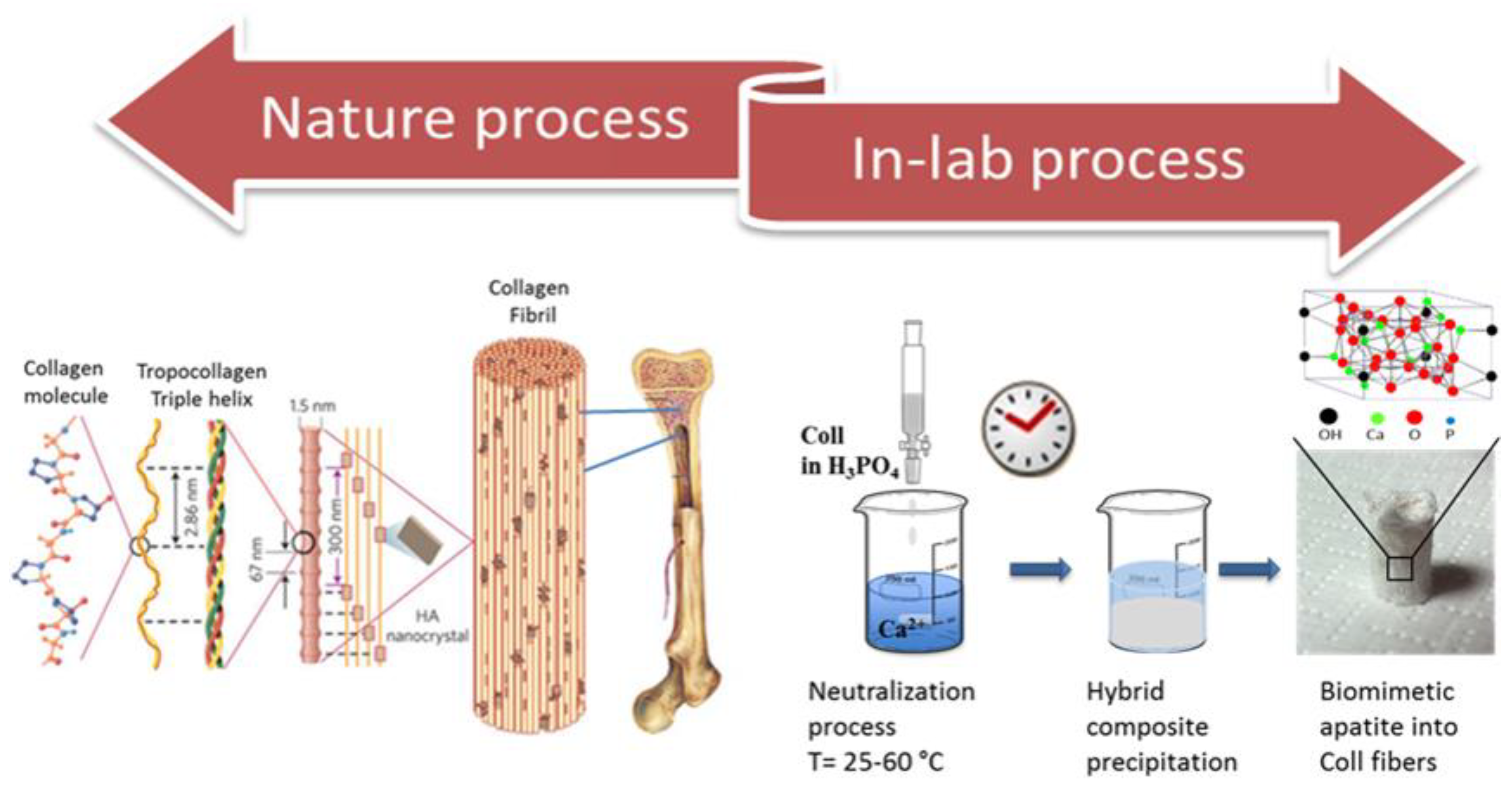
Mimicking Biomineralization in the Lab

Biominerals compared to natural or synthetic minerals often display excellent mechanical and other properties due to their multi-level order, hierarchically organized from the nanoscale to the microscale. For this reason, in the last decades, researchers have been trying to reproduce the calcium-based biomineralization processes in laboratory, inducing the heterogeneous nucleation of the inorganic phase into organic matrix through fine mechanisms driven by the organic matrix itself. The chemical and physical interaction between phases confers unique features to the resulting hybrid materials, in a similar way compared to what happens in the natural biomineralization process (Figure 1). These peculiar properties cannot be obtained through a simple mixing of the phases [22,23,24].
As a consequence, the biomineralization process study, together with other emerging technologies to synthesize new nanomaterials, has spread into many fields in our life such as mechanical, electrical [25,26], and environmental [27,28], as well as biomedical engineering [29,30].
In this review focused on the biomedical field, we aim to provide an overview of different materials mimicking the natural calcium-based biomineralization process to prove that, finely tuning some process variables, it is possible to design multifunctional materials. These materials can be exploited in several applications in order to obtain customized and precise medical tools [31,32,33,34]. We will first provide a brief overview of the biomineralization process: how it happens in nature, and how scientists have translated this natural process to an in-lab process. Taking into account the wide chosen topic, we decided to focus on calcium-based biomineralization, more specifically on different applications aside from bone regeneration, that were poorly or not considered in other reviews. Specifically, we will discuss biomimetic hybrid material features that can be obtained by modulating different process parameters, focusing on the materials chemical–physical and biological features which are essential to make them suitable for biomedical field. Finally, we will discuss the several applications of these materials besides tissue regeneration, such as their use for the creation of 3D cancer predictive models or drug testing, as well as on their use as innovative physical filters against solar radiations or as nano and micro drug delivery systems.

2. Features of Biomimetic and Hybrid Biomaterials

From its first understanding, calcium-based biomineralization process uniqueness has attracted high attention due to its applicability in many different fields, especially for bone tissue engineering and regenerative medicine as well as the mild conditions, i.e., physiological temperature, pressure, and pH, in which this process occurs. Thus, the first step has been trying to translate the natural process into an in-lab process, mimicking the formation of the natural hierarchically structured organic–inorganic composites. Natural mineralization is commonly divided into two different groups recognized as “biologically induced” and “biologically controlled”.
As reported by Weiner and Dove [35], the precipitation of minerals that occurs as result of interactions between biological components and the environment is termed “biologically induced” mineralization. In this situation, the chemical conditions of the environment, such as pH and CO2, indirectly favor the formation of a specific mineral type. The mineral composition varies consistently with the variation of the environments in which they form, resulting in different morphologies, water content, element composition, structure, and particle size.
In “biologically controlled” mineralization, the organism uses cells that actively take part in the nucleation, growth, morphology, and final location of the mineral that is deposited. While the degree of control varies across species, almost all controlled mineralization processes occur in an isolated environment. The result can be remarkably sophisticated, species-specific products that give the organism specialized biological functions. Calcium-based biomineralization can be described with subsequent stages, the first of which consist of inorganic molecules pre-assembling into ordered structures. Then, the molecular recognition among the organic and inorganic interfaces controls crystal nucleation and growth, allowing the formation of subunits. Finally, cells will take part in the process, forming biominerals with multilevel structure by assembling subunit minerals. All these steps are controlled by synergistic action of various environmental factors such as pH, temperature, and organic matrix chemistry [2]. Regarding the matrix, the interaction between inorganic and organic matrices leads the entire process, resulting in widely different results depending on the stereo-chemical and physical interaction occurring among the two components. This process is affected by different parameters; nevertheless, taking into account the influencing factors, the process can be transferred into an “in-lab” process and can even be controlled and directed towards the development of novel materials endowed with intriguing features. Speaking of organic matrices, it has been proven that the structural organization complexity highly affects the final biomineralization product, which might result in a three-dimensional scaffold in the case of highly organized matrices, or well as flakes or powders in the case of poorly organized matrices [36]. A clear example of this is given by the mineralization of collagen compared to gelatin. Collagen, with its multi-level organization, can undergo the biomineralization process without losing its structure, resulting in a three-dimensional scaffold with different properties depending on the biomineralization grade [10]. Gelatin, on the other hand, being a lower assembled polymer, can be exploited to form low structured biomineralized materials, composed of hybrid micro-flakes made of clusters of nano-particles [37].
In addition to collagen and gelatin, it is possible to find examples in the literature of mineralization with different polymer matrices as cellulose, chitosan, alginate, and silk (Table 1). Ahmed Salama has shown, for instance, how cellulose and its derivatives are promising candidates for developing and constructing smart organic/inorganic hybrid biomaterials through a calcium-based biomineralization process [38]. Cellulose/calcium phosphate hybrid materials, for example, combine the properties of both components, the functionality and flexibility of the cellulose with the heat resistance and stability of the inorganic material, to generate compounds that are being exploited for different applications such as bone regeneration, drug delivery vehicles, dental repair, and adsorption. Chitosan can be efficiently exploited to produce hybrid composites based on hydroxyapatite [39,40]. These hybrid composites are mainly devised for bone regeneration. Indeed, chitosan shares some peculiar features with collagen (e.g., a comparable role in the exo- vs. endo-skeleton and flexibility) [41]. Regarding alginate, a recent work [42]. reported the effect of alginate and its well-defined oligomers with defined structure on brushite nucleation and growth for the synthesis of hybrid materials, useful in bioactive agent delivery, wound healing, and tissue engineering. Growth experiments showed that molecular weight and additives of alginate affect the crystal growth rates and the growth mechanisms. Finally, Yang and collaborators [22] have described as silk fibroin as able to facilitate nucleation of the hydroxyapatite crystals through its molecular self-assembly, and to create efficient biomaterial with mechanical and functional properties for biomedical applications.
The ideal scaffold useful to obtain a robust in vivo affinity must be designed with the final purpose to resemble the 3D architecture, nanostructure, chemical composition and mechanical properties of the extracellular matrix (ECM) of the native tissue [21]. The scaffold must be composed of biocompatible materials able to guarantee cells viability, support of cellular functions and induction of molecular and mechanical signals without eliciting adverse effects on cells and, consequently, local or systemic undesired responses in the host [43,44]. Biomineralized substrates influence cells biochemistry by the exchange in proteins and ions that, subsequently, conditions the 3D microenvironment. This implies that proteins and ions composition (e.g., calcium and phosphate ratio, collagen, proteoglycans, etc.) of native tissue is a crucial parameter to be consider for scaffold design and synthesis [45]. If materials quickly degrade in vivo, the scaffold fails in its mechanical support role; conversely, an inflammatory response could be provoked by the foreign material if the scaffold has an excessively long biodegradation time. Therefore, a controlled biodegradability is crucial in scaffolds fabrication [46]. Overall, the microarchitecture of the support should be highly porous and interconnected to provide inwards diffusion of oxygen and nutrients and elimination of waste products in order to meet cellular requirements for adhesion, growth, differentiation and migration [47].
Porosity generally supports cell migration into the scaffold by promoting interaction between cells and the available surface area of the scaffold. Pores density and size influence cellular behavior in a inversely proportional way [48]; as pores size decreases, pores density increases as well as the surface area available on the scaffold for cells interactions. However, if pores are too small, cells are not able to penetrate and migrate inside the structure. Indeed, the pores dimension is able to affect vascularization of scaffolds. A different vascularization can consequently tune cells differentiation [49,50,51]. Consequently, scaffolds composition, but also scaffold porosity, need to be optimized on dependence of the tissue type to interact with; as example, for bone tissue engineering scaffolds need to contain a mixture of macro- and micro-pores that allow cells to grow in vivo, facilitating cell-material interaction and complete scaffold colonization, respectively [52].
Additionally, in vivo mechanical signals (Young’s modulus, compressive strength and fatigue strength mechanical forces) for cells must be replicated in the scaffold to induce the correct cellular differentiation pathway. Mechanical stiffness and porosity are often conflicting physical properties, as the first is inversely related to the other. Consequently, finding a good compromise between the scaffold properties for to promote the correct cellular activity and mechanical integrity is often a hard challenge [53]. The scaffold surface can be bio-decorated with specific proteins/biomolecules or biomotives to improve its outcomes on cell adhesion, proliferation, and differentiation. Tissue formation and ECM deposition are regulated and performed by a wide variety of biomolecules. For this reason, scaffolds can be enriched by growth factors during their production, assuming a significant role in tissue engineering applications [54,55,56], such as improving tissue formation or the reward of specific biomolecules in pathological conditions [21].
The set of all above-mentioned properties makes the scaffold bioactive, osteoconductive and osteoinductive in tissue engineering, regeneration and modelling of mineralized tissue (bones, tooth, tendons, and cartilage). Osteoconductive scaffold can support the ingrowth of cells into pores, channel or pipes, while osteoinduction refers to the activity of a contact or soluble material to induce the differentiation of pluripotent stem cells towards specific cells lineage. Achieving these demands, calcium-based biomineralization process represents an excellent biomimetic manufacturing strategy to obtain hierarchically designed scaffolds with appropriate mechanical stability, flexibility and a highly porous and interconnected structure [12,15,57].
Biomineralized nanomaterials represent also a promising tool for different clinical applications. Nanoparticles as drug carriers for drug delivery involves the linking of drug molecules to the nanomaterial to guarantee a controlled distribution and release rate of the drug [58,59]. In this case a good biological activity and biodegradability together with good stability under close physiological conditions are required. Most of the components of biomineralized nanomaterials (e.g., calcium phosphate, carbonate and iron oxide) are naturally present in the body and easily and normally metabolized and absorbed by it; (3) good biological activity. Biomineralized nanomaterials facilitate loading of active molecules into the mineral phase by the interaction between biominerals and biomolecules, making possible to use them for drugs and molecules loading to be used inseveral biomedical practices [1].
Table 1. Influence of physical chemical properties of hybrid materials on in vitro/in vivo behavior.
Physic-Chemical ParameterScaffoldNanosystems
BiocompatibilityAbsence of cytotoxicity [44];
Support and stimulation of cellular activity [43]
Absence of cytotoxicity
Support and stimulation of cellular activity [1]
BiodegradabilityControlled biodegradability [46]High bioabsorption and biodegradability
Absence of bioaccumulation of ions [1]
ArchitectureStability under physiological condition
Highly porous and interconnected [47]
Hierarchical design structure [9,57]
Stability under physiological conditions [1]
Porosity and pore sizeMixture of macro- and micro-porosity [48,49,50,51,52]/
Mechanical propertiesMechanical integrity [53]Stability under physiological conditions [1]
Surface propertiesSupport and stimulation of cellular activity
Tissue-specific functionalization [54,55,56]
Tissue-specific functionalization
Target-specific functionalization [1]
BioactivityOsteoinductive
Osteoconductive [6,9,57]
Controlled drug release and distribution [1,58,59]

3. Applications in Biomedical Field: Tissue Regeneration and Many More

Bone is the second most transplanted tissue with four million operations per year, using different bone alternatives worldwide [21,60]. Calcium-based biomineralization process is responsible for the formation of natural hard tissues such as bone and teeth [2,9,36,61] that are composed of extracellular matrix (ECM), several cells types, and water, which through a fascinating process are able to create a highly organized and hierarchically structured nanocomposite [13,36,62]. In detail, biomineralized ECM is mainly composed of plate-like nanocrystalline hydroxyapatite (the inorganic phase) and several organic components among which collagen is prevailing [63,64,65].
ECM highly influences adhesion, proliferation and differentiation of several cells types such as osteoblasts, bone lining cells, osteocytes as well as osteoclasts [65,66,67]. Several factors determine the fascinating and unique characteristic of bone, starting from composition arriving to structure and porosity, features essential for bone remodeling process. More precisely, bone tissue is characterized by a dynamic nature which allows to preserve its healthy condition, thanks to bone remodeling which consist in a dynamic and fine equilibrium between bone resorption by osteoclasts and bone formation by osteoblasts in response to biomechanical stimuli [68,69].
However, although this process preserve bone, its self-healing capacity is enough only to repair small bone damages. Subsequently, in case of extensive bone damage, bone substitutes are often required. For several decades, the gold standards were autografts or allografts [36,44]. In the last decade, instead, tissue engineering and regenerative medicine attention shifted towards development of biological replacements able to mimic and regenerate the tissues itself. In this perspective, scaffolds are becoming a fundamental tool for tissue engineering and regeneration, they indeed act as substrates able to structurally guide cells and provide anchorage sites, making possible to develop engineered structure made of a combination of materials and living cells. More precisely, the ideal bone scaffold is a transient implant that must create an adequate environment for cells to stay viable, attach, proliferate, differentiate and deposit ECM to replace the damaged or impaired one [70,71,72]. To do what describe above, scaffolds should resemble the natural bone tissue under different points of view, such as composition, structure, mechanical requirements and biological features. As consequence, to mimic the chemistry and structure of natural bone, biomineralization process is absolutely the most suitable in-lab process to produce biomimetic biomaterials, in which low-crystalline hydroxyapatite is mineralized on collagen fibrils, and highly porous scaffolds suitable to promote bone tissue regeneration [13,16,73].
Although this process has touched several fields of our life, biomedical engineering has surely received more specific attention, ending to be the most investigated. Nonetheless, recently, the attention has moved towards new advanced applications of mineralized materials. These applications include: (i) development of 3D predictive models for cancer study or drug testing, (ii) study of physical filters against solar radiation, (iii) creation of nano and micro controlled drug delivery systems. Additionally, the introduction of new fabrication technologies, e.g., 3D (bio) printing, are further extending the applications in other fields. These features will be discussed in the next sections.

3.1. Biomineralization, 3D Printing and 3D Bio-Printing

Biomineralization has been proven to be an incredible versatile process, exploitable for multiple purposes in different fields. The same could be stated for 3D printing, that with its simple basic principle paved the way for realization of incredibly ambitious and elaborated projects. The combination of these two elements have been giving rise, in the last decades, to incredible results.
The importance of scaffolds geometry at multiple level, from nano- to macro- scale, for cells attachment, spreading and viability has been so far well assessed and recognized. At the macro-scale, it is desirable for the scaffold to assume the defective part shape of the tissue or organ meant to be replaced or repaired, in order to help the neo tissue to organize into the required three-dimensional structure [74].
From the macro- point of view, especially for pursuing highly challenging goals, as it can be the regeneration of long bone defects, additive manufacturing has been attracting great interest. This, as it allows both a precise and controlled spatial-deposition of materials as well as their easy combination in complex multi-material structure, in addition to the possibility of creating shape-customized scaffold based on the anatomical site targeted [75].
If the scaffold shape and geometry play a crucial role at the macroscale, the same can be said for the composition of the material used as ink for the realization of the three-dimensional structure at the micro- and nanoscale
Speaking of bone regeneration, the key element lies in the mineral phase, in nature commonly produced by cells through the calcium-based biomineralization process.
Many approaches have been used through the years to mimic, reproduce, or induce this process in order to exploit its unique ability within the creation of new materials suitable for bone tissue regeneration. 3D printing tries to combine the ultimate technology with the ancient principle of biomimesis. One of the most used approaches has been the use of different mineral phases, such as hydroxyapatite (HA), within the composition of the ink or as post-process modification. This, with the intention to not only to emulate, but also to stimulate the biomineralization, triggers the process by making the cells perceive a suitably functionalized environment and transforming the material from just osteoconductive to osteoinductive (Figure 2) [20,76]. In order to stimulate bone regeneration, other biomineralized polymers, including silk, a well-known high strength polymer, were exploited as biocompatible filler for 3D printing [77].
An alternative approach was devised by Romanazzo and collaborators. According to this strategy, it is possible to promote the formation of mineralized constructs in a support bath containing live cells and microgels, mimicking the complex and hierarchical structure of native bone. In these conditions, cells were able to differentiate at the interface of the constructs, while remaining multipotent in the intervening spaces, opening the potential for fabricating gradient tissue structures and for future in situ fabrication of bone-like tissues [78].
In this context, it is easy to understand how coatings to improve prosthetic implants biomimetic features have been the first to arise, but in recent years this trend has reached upgraded levels. For instance, Park and co-workers reported an osteoinductive coating of polydopamine, biomineralized HA, and bone morphogenetic protein-2 of a 3D printed polycaprolactone (PCL) structure elegantly exploits the advantages of three different strategies to reach noteworthy results. A different approach, which departs from post-process modifications and moves to the inclusion of biomineralization boosting elements into the ink formulation, is proposed by Hernandez and collaborators. A complex 3D printed multi-material system made of PCL and HA-loaded hydrogel for long bone defect regeneration was developed. In this work, the PCL provided the appropriate mechanical support, while the hydrogel composition supports cells, promoting biomineralization [75]. Recently, more unconventional mineral phases, such as graphene oxide (GO) and black phosphorus (BP) have been used with the same purpose, with promising results [79].
Indeed, Yang and co-workers used an innovative combination of bio-glass and black phosphorus to devise three-dimensional therapeutic structures for localized photothermal osteosarcoma treatment and subsequent bone regeneration (Figure 3), profit from black phosphorus degradation to induce a phosphorus-driven in situ biomineralization [80].
A similar approach, but with different applications was exploited by Lin et al. They used a biomineralization inspired process to create a synthetic graphene composite with reshaping and self-healing features which can be used in a large variety of applications, including energy storage to actuators [81].

3.2. 3D Predictive Models: From Cancer Study to Drug Testing

The scientific community has recently focused on the design and bioengineering of innovative 3D culture systems able to overcome the well-recognized inadequacy of conventional bi-dimensional (2D) in vitro models in recapitulating the complexity of in vivo microenvironment [82,83,84]. 3D tools and technologies reproduce cellular heterogeneity, tissue-specific ECM, and biological interactions in a more biomimetic way, providing in vitro platforms which closely resemble native microenvironment. These biomimetic materials can be exploited for basic biological studies, drug screening, and reproduction of viable biological niches for in vivo transplantation. The biomineralization process is an excellent strategy for the development of advanced biomimetic models, including 3D biomaterials, cellular coatings, and nanoplatforms with flexibility, diversity, and utility of frameworks for a wide variety of applications [85,86].
Ye and co-workers [87] established a rapid biomimetic mineralization approach to obtain a 3D porous and mineralized hydroxyapatite/collagen composite scaffold for bone regeneration. By a custom synthesis process based on self-assembled collagen fibrils as fixed template, the authors created an in vitro 3D bone-like niche seeded with human Umbilical Cord Mesenchymal Stem Cells (hUCMSCs) with high cell viability, adhesion, proliferation, and differentiation into osteoblasts due to the mineralized scaffold. A rabbit femoral condyle defect model was tested to confirm the ability of the viable niche to facilitate bone regeneration and repair over a period of 6–12 weeks. The mineralized collagen scaffold seeded with hUCMSCs successfully promoted the healing of bone defect in vivo; as new bone tissue formed, the scaffold gradually degraded and was absorbed, confirming the promising use of the hUCMSCs-loaded bone-like niche for in vivo transplantation for bone tissue regeneration.
The same concept was exploited by Menale et al. [71] in 2019 for a cell-therapy based strategy. In this case, a biomineralized bone-like scaffold was used as a rationally designed device conceived to be seeded with cells and subsequently transplanted in vivo to restore or replace a missing function that cannot be completely renewed by only cells [88]. The authors used the scaffold as productive factory of bioactive soluble osteogenic Receptor Activator of Nuclear Factor k B Ligand (RANKL) directly secreted by seeded Mesenchymal Stem Cells (MSCs) on the 3D support [89,90]. The scaffold, obtained through direct nucleation of magnesium-doped hydroxyapatite (HA) nano-crystals on self-assembling collagen fibrils (MgHA/Coll) by a pH-driven biomineralization process, showed structural, compositional, and morphological similarities to the native bone ECM. The biomineralized scaffold guarantees the development of an in vitro, viable, bone-like niche able to compensate the RANKL factor deficit in Autosomal Recessive Osteopetrosis (RANKL-ARO) once transplanted in vivo due to the continuous secretion by MSCs; the MgHA/Coll scaffold promoted the differentiation of MSCs towards osteoclasts [91,92], helping to restore the physiological functions of bone cells in a RANKL-/- mice.
Recently, the same MgHA/Coll scaffold was used as bone-like ECM to be seeded with tumor spheroids, called sarcospheres, and parental cells of MG63 and SAOS-2 osteosarcoma cell lines as enriched Cancer Stem Cells (CSCs) models with the final purpose of obtaining a 3D in vitro CSC-niche of osteosarcoma (Figure 4) [73]. The material provided specific physical-chemical and biomechanical stimuli to the critical pluripotent stem cell population, giving birth to a 3D predictive in vitro model of CSC-niche of osteosarcoma with enhanced stemness and niche-related properties compared to those seeded with parental cells. Through an in-depth cellular and molecular characterization of sarcospheres, and an optimization of the scaffold resembling tumor ECM, the authors were able to provide a closely mimetic in vitro platform for tumor studies and CSC-specific drug screening [93].
A novel biomineralization-inspired cancer therapy has recently been developed as proof-of-concept of advanced nanotechnological therapy. Natural mineral accumulation is a significant biological process that, in abnormal cases, causes the excessive deposition of calcium ions in damaged or defective tissues, leading to common pathologies, such as kidney stones and vascular calcification [94]. The anomalous mineralization can be exploited as “biomimetic pathological mineralization” onto some tumor cells, such as human cervical cancer cell line (HeLa), which can selectively assimilate, folate, and concentrate calcium ions by the overexpression of folate receptor in cancer cells, creating a Cancer Cell-Targeting Calcification-based therapy (CCTC) as reported by Zhao and co-workers [95]. On this trajectory, a biomineralization-inspired drug free strategy can be used to promote cell death by creating a calcium phosphate (CaP) mineral cell coating that leads to the agglutination of tumor cell nuclei without inducing normal cell death [95]. This approach also showed promising results on metastasis, where the survival rate of pathological mice improved significantly (up to 80%) due to the suppression of the metastasis by selective calcification-based substitution of the tumor with curable sclerosis. However, the required concentration of calcium ions exceeds physiological levels, thus identifying an innovative biomineralization-inspired material able to specifically accumulate ions in the target tissue to facilitate calcium mineral nucleation is still a challenge that may be addressed, for example, by the exploitation of specific ligand/antigens interactions on cancer cell membranes [95]. Without a doubt, this concept can be used to eventually create in vitro 3D biomineralized-based scaffolds able to specifically induce tumor cell death.
Microcalcifications (MCs) also serve as diagnostic markers for breast cancer; breast cancer screenings (e.g., mammography) frequently rely on MCs, and their chemical composition (e.g., calcium phosphate, apatite, calcium oxalate, etc.) is associated with tumor malignancy [96,97]. However, due to the absence of sufficiently predictive 3D tumor models, little is known about how they form in the body, their effective role in cancer progression, or how cancer cells are involved in the mineralization process. Therefore, Vidavsky and co-workers [98] exploited the role of biological induction of biomineralization [99] for developing in vitro 3D model of breast tumor MCs to study the cellular pathways involved in MCs formation as a function of malignancy potential. Mammary multicellular spheroids were obtained by parent MCF10A benign human breast epithelial cell line, MCF10DCIS.com [100] and MCF10CA1a [101] which derived from MCF10A and possessed ductal carcinoma in situ (DCIS) and invasive tumors characteristics, respectively; together, these three cell lines allow the modeling of varying stages of breast cancer, ranging from non-malignant (MCF10A), pre-cancerous (MCF10DCIS.com), to invasive phenotype (MCF10CA1a) allowing us to investigate the correlation between cell phenotype and MCs formation. To ensure the physiological relevance of the model, the authors cultured cells in ultra-low attachment conditions with media that contained calcium, magnesium, and phosphate concentrations similar to the human body, but lacked any osteogenic agents in order to observe the real malignancy potential of spheroids just by the development of MCs. Obtained spheroids had diameters larger than 300 mm with low cell viability at the core due to limited diffusion of oxygen and nutrients (Figure 5A). Interestingly, no particles are observed in the MCF10A spheroids (Figure 5B–D). Moreover, apatite MCs were primarily detected within viable cell regions in the shells and their number and size increased with malignancy potential of the spheroids; conversely, alkaline phosphatase (ALP) decreased with malignancy potential, while osteopontin (OPN) increased. These findings support the induction of a mineralization pathway by cancer cells in a manner that is linked to their malignancy potential. This work offers an innovative exploitation of the mineralization process, which allows us to both create more reliable 3D stage-specific cancer models by inducing specific-MCs as indicators of malignancy potential and, consequently, use these platforms to deeply investigate cancer pathways.
In conclusion, few studies exploited biomineralization-inspired process for various useful biomedical applications, from cancer modelling [73] to diagnosis markers [98], showing the need to deeply investigate the potential used of this process independently from conventional applications.

3.3. Physical Filters against Solar Radiations

Sunlight is essential for our well-being; it is responsible for regulating our internal clock, metabolism, immune systems, and for vitamin D production, essential for healthy bones. Nevertheless, it is well-known that excessive exposure to the solar radiations can cause serious damage to human health [102]. In particular, UVA (320–400 nm) and UVB (290–320 nm) radiations are the main radiations to interact with the human body, and their hazard relies in their ability to generate reactive oxygen species (ROS), which cause skin photo-aging, sunburn, dermatitis, and can also evoke long-term health effects, such as malignant tumors [103,104]. In this regard, the use of sunscreens composed of effective UV filters as protective barriers, absorbing harmful UVA and UVB radiation, has become a very important topic on which research is paying an increasing attention. UV filters can be divided in two main classes, chemical or physical filters, but nowadays physical filters are considered more attractive for sunscreens. Indeed chemical filters, despite having different advantages [105], were proven to increase environment pollution [106], and resulted to be harmful for human health [107]. For these reasons, physical filters, especially titanium dioxide (TiO2) and zinc oxide (ZnO) [104], being able to shield the skin from both UVA and UVB radiation, are the most commonly used. However, these also have some important limitations, especially regarding their size and photocatalytic properties (Table 2). Indeed, to decrease the difficult spreading and whitening effect on the skin, the particle size is reduced to the nano-size range [108]. This entails that these particles are able to penetrate deep layers of skin, causing phototoxic reactions [109]. Furthermore, TiO2 is known for its high photocatalytic activity, generating reactive oxygen species (ROS), which can oxidize and degrade other ingredients in the formulation, raising safety concerns [110]. Finally, the main problem, relating all common UV-filters, is that, being mainly used on the beach, the components of the cream are often released in water, causing damage to the marine environment, coral blenching, and bioaccumulation in the fauna [111]. Considering all these issues, attention is shifting towards the development of effective and safer UV-physical filters for both humans and the environment.
Recently, Battistin and co-workers [112] reported a new class of UV-physical filters through the combination of a common physical filter, TiO2, and dihydroxyphenyl benzimidazole carboxylic acid (Oxisol) [113], an antioxidant molecule with booster effect. Boosters can be small molecules, polymers, or other particles, that act on the rheological properties of the formulation, but can also synergistically interact with the UV filters through antioxidant mechanisms or interfere with the electronic processes of UV radiations absorption. In particular, this work reported that Oxisol, functionalizing the surface of TiO2, is able to increase the UV-protection, and also to stabilize TiO2 nanoparticles, preventing their penetration to deeper skin layers. Furthermore, its booster activity, by means of antioxidant effects, allows a reduction in physical filter content in sunscreen formulation and a significant lowering of photocatalytic effect, typical of TiO2. Nevertheless, Oxisol is considered as a low eco-sustainable sunscreen product. Thus, alternative, safer, and eco-sustainable sunscreen products are currently under investigation.
In the last years, the sector of sunscreens shifted attention towards formulations containing calcium phosphates (CaPs), especially hydroxyapatite (HA), the main component of animal bones, due to their excellent biocompatibility, non-toxicity, and ability to partially absorb UV radiation (Table 3) [114]. In the literature, there are some works related to hydroxyapatite as physical filter; for instance, Rehab and collaborators [115] reported the synthesis of ascorbic acid-modified, nanosized HA, stabilized with polyvinylpyrrolidone (PVP), to act as a potential biocompatible and safe constituent of sunscreens. In detail, the incorporation of the antioxidant ascorbic acid (vitamin C) [116] in HA particles maximizes photoprotection against UV damage and removes reactive oxygen species (ROS), while PVP prevent nanoparticles aggregation avoiding their skin permeation.
Another type of HA-based sunscreen has been shown by Morsy and co-authors [117], who developed a multifunctional hydroxyapatite-chitosan (HA-chitosan) gel that works as a natural antibacterial sunscreen agent for skin care. Through the simple coprecipitation method, thus avoiding use of toxic or high-cost materials, nanosized HA particles trapped within the chitosan matrix were obtained. HA acts as a physical filter against solar radiation, while chitosan acts as polymer matrix, able to avoid the agglomeration of particles and to prevent skin penetration. Additionally, chitosan acts as a natural antimicrobial agent, preventing skin wound infections caused by excessive sun exposure. Both works [115,117] focused on the intrinsic photoprotective capability of hydroxyapatite, combining it with other compounds to improve its absorption range in the UV region and to bypass the main drawbacks related to this kind of material, such as nano-size and whitening effect.
However, several studies on HA [118,119] showed its lattice has the particular ability to be modified through the doping with ions (such as Mg2+, Sr2+, CO3, Fe2+/3+, Zn2+, and Ti4+), thus making it a multifunctional product, adaptable according to the requests. Due to this, in the specific case of sunscreens, some works have reported that doping HA with appropriate ions can lead to an increase in the value of protection factor without necessarily having to combine other external components. In 2010, de Araujo et al. [120,121] developed, through a chemical precipitation process, four different hydroxyapatites doped with Cr3+, Fe3+, Zn2+, and Mn2+ ions, having better absorption properties than pure HA in the UV region. Mostly, iron and manganese-doped HA showed the best absorption features in the UV range, necessary to be an effective sunscreen, without creating problems of toxicity or photocatalytic effect. Inspired by the previously published results [120,121], another work [122] has reported an iron-doped HA-based material containing both Fe ions (Fe2+/Fe3+) substituted into the hydroxyapatite lattice and iron oxide in hematite (α-Fe2O3) form, successfully developed from waste fish bones with a simple treatment. This was the first time an HA-based sunscreen has been synthetized, formulated in cream, and validated as proof of concept. In detail, the introduction of iron ions in the HA lattice allowed an increase in the absorption range in the UV-region, creating an effective physical filter, no photoreactivity, and a potential safe option for cream formulation, starting from waste by-products with several environmental benefits. Although iron is able to improve the photoprotective abilities of hydroxyapatite, several studies reported that titanium has a greater shielding power [123,124].
On the other hand, taking into account the photocatalytic problems associated with the use of titanium dioxide within sunscreens, some recent works shifted attention towards titanium as Ti4+ ions, developing titanium-doped hydroxyapatite. Yasukawa and Tamura [125] were the first to demonstrate the effective protection from solar radiations of titanium-hydroxyapatite suspensions combined with cerium ions (TiCeHA). In particular, it was revealed that the Ti4+ ions and Ce3+ ions absorbed another range of UV: UVB and UVA, respectively. Therefore, the simultaneous use of these ions further enhances the UV absorptive ability and by changing their contents in TiCeHA it is possible to create a physical filter suitable for shielding from UVA and/or UVB. Given the potential of these compounds, it would be interesting to evaluate the development of a new UV-physical filter composed of titanium-doped hydroxyapatite and biopolymers obtained by a nature inspired calcium-based biomineralization process [126]. Considering the problems associated with “classic” commercial sunscreen, having a physical filer not only able to shield solar radiations, but also safe for the human body and eco-friendly, could be the solution to overcome the main UV-filters drawbacks.
Table 2. Advantages and drawbacks of some physical filters.
Physical FilterAdvantagesDrawbacks
TiO2-Oxiol [112]
  • Booster UV-shield
  • Stabilize nanoparticles
  • Antioxidant effect
  • Low ecosustainable
  • Photocatalytic effect
  • No biocompatible
HA-Ascorbic Acid [115]
  • High UV-shield
  • No toxic residual (ROS)
  • Biocompatible
  • Low chemical stability
HA-Chitosan [117]
  • Antibacterial activity
  • Low-cost material
  • Biocompatible
  • Eco-sustainable
  • Low UV-shield
ions-doped HA (Cr3+, Fe3+, Zn2+, Mn2+, Ti4+) [120,121,125]
  • Biocompatible
  • High UV-shield
  • Eco-friendly
  • No photocatalytic effect
  • Stabilize nanoparticles
Table 3. Differences between chemical, physical and hydroxyapatite (HA)-based physical filters.
Chemical FilterPhysical FilterHA-Based Physical Filter
  • Absorb UV-rays [105];
  • Not degradable [106];
  • Lypophilic [107];
  • Partial penetration of UV-rays in the skin [107];
  • Harmful for the environment [106].
  • Reflect UV-rays [104];
  • Not degradable [106];
  • Cause whitening effect [108];
  • Avoid penetration of UV-rays [104];
  • Photocatalytic effect [110];
  • Nanoparticles penetration [109];
  • Harmful for the environment [111].
  • Absorb and reflect UV-rays [114];
  • Biodegradable [126];
  • Avoid whitening effect [127];
  • Ecosustainable [114];
  • No-photocatalytic effect [127];
  • Avoid particles penetration [127].

3.4. Nano and Micro Drug Delivery Systems

Bioceramics are widely used as components of implants for bone and teeth restoration. Nowadays the advanced processing techniques and the new synthesis strategies allow the incorporation of drugs, bioactive molecules, or cells within them or on their functionalized surfaces. In this regard, bioceramics and biomineralized materials can be exploited as drug delivery or controlled release in several applications, such as nanomedicine, wound healing, and bone regeneration [128,129].
Local antibiotic release is a promising and effective procedure for delivering drugs at the implantation site. With this strategy, antibiotic was loaded on a scaffold in order to both promote bone regeneration and to prevent common bacterial infections happening after surgery. In this way, scaffolds act as carriers for local antibiotic release to avoid following implant removal due to osteomyelitis (Figure 6) [130]. Different drugs can be loaded into the scaffolds, including anticancer drugs. For instance antitumoral drug-loaded scaffolds can be used to restore large bone defects after tumor extirpation, resulting in tumor inhibition with low levels of systemic toxicity [131,132,133,134].
Often, in orthopedic, maxillofacial, and dental surgery, whether the defect size is complex or irregular, bioceramic beads are used to induce bone tissue regeneration [135,136,137]. In the last decades, some research has been focused on the possibility, not only to promote tissue regeneration due to bioceramics beads, but also to modify the functionalization of them with several active molecules such as antibiotics, anticancer agents, and osteogenic agents to act themselves as drug delivery vehicles [129,138,139,140].
Ceramic component contributes to the mechanical stability and bioactivity of the structure; however, its adsorption of drugs often is featured by weak bonds leading to an initial burst release [140,141,142]. To overcome this issue, polymers can be added, forming a composite material endowed with a fine chemical and physical control of the drug adsorption and release [143,144]. These polymeric and bioceramic phases can be used as separated phases or as a single mixed phase. For example, hydrogel/bioceramic core-shell beads can be developed, by means of concentric nozzles or microfluidics exploiting both advantages of the two phases; polymers can preserve the drug, avoiding an initial excessive release, whereas ceramics contribute to the mechanical stability and bioactivity of the structure for a synergic and effective loading and sustained release of proteins [145], or drugs [146] (Figure 7), as well as cells [147].
For example, Raja and co-workers fabricated a multifunctional core-shell bead structure featured by a hydrogel shell composed of alginate including cells around a ceramic core made of α-tricalcium phosphate (α-TCP) loaded with Quercitin dihydrate, a well-known phytochemical used for the treatment of osteoporosis [129]. The core-shell beads, immersed in PBS, lead to the formation of bone-like low-crystalline apatite from α-TCP that provides structural integrity to the bead, along with a surface for the growth of cells embedded in the hydrogel shell. Researchers demonstrated a slow release of quercetin throughout the entire 120 days testing period, together with the formation of a homogenous cell layer on the ceramics structure, due to cells loaded into the hydrogel. Finally, they showed that in the region in which hydrogel and ceramics are strictly in contact, cell growth was specifically increased, highlighting the potential of the core-shell model for further material–cells interaction study [129]. This kind of composite belongs to promising class of materials able to load different types of drugs and cells to produce highly biofunctional beads, which provide an effective bone substitute for both drug delivery and bone tissue regeneration [8,147,148].
Moreover, calcium-based biomineralization paves the way to promising and very interesting materials where ceramics composites are nucleated onto the organic phase, as previously described, creating a single and very reactive phase that combines the advantages of the two phases. Furthermore, thanks to the possibility of introducing doping ions into apatite lattice, the resulting phase will be featured by different and new functionalities, in addition to those normally occurring in vivo, such as high bioactivity, biocompatibility, and biodegradability. Doping with magnesium or Zn ions, for example, is possible to confer antimicrobic properties essential to prevent bacterial infection or in wound healing [149,150].
The encapsulation of antibiotics in nanocarriers such as bioceramics allows the elimination of microorganisms by releasing a high antibiotic dose at a target site before the development of resistance [151]. Furthermore, many researchers demonstrated that ions present in hydroxyapatite can promote the antibacterial activity of the device. For instance, Ain and co-workers demonstrated that vancomycin-loaded HA had a slower release in comparison with pure vancomycin and also an enhanced antibacterial activity due to the presence of ions in the HA structure [140].
On the other hand, doping with Fe ions results in an interesting superparamagnetic apatite phase, able to be exploited in diagnostic field as a contrast agent or therapeutic field, due to the possibility to move it by an external magnetic field or to release drugs by means of magneto-shaking [4,139,152,153].
Concerning that point, Patricio and collaborators have developed a bio-hybrid microsphere obtained through the biomineralization of iron-doped hydroxyapatite (FeHA) within an organic matrix. In this case, the organic matrix is an animal-free recombinant peptide based on human type I collagen (RCP) enriched with RGD motif. The resulting material is bioresorbable, biocompatible, and can enhance cell adhesion. Through the fine tuning of the emulsification process, the resulting hybrid microsphere is endowed with a monomodal size dispersion, low crystallinity, and superparamagnetic properties typical of FeHA [4,23,154,155,156].
The resulting microspheres displayed excellent osteogenic ability with human mesenchymal stem cells, and were able to provide a slow release of recombinant human bone morphogenetic protein-2 (rhBMP-2) within 14 days. Furthermore, the release profile can be finely tuned by application of pulsed electromagnetic field, thus highlighting the potential of remote controlling the bioactivity of the new micro-devices, an interesting feature for their application in precisely designed and personalized therapies.
To conclude, the administration of therapeutic agents is still a major concern of medicine, as the systemic dose prescribed needs to be high to ensure the suitable dose in the target area, causing several collateral effects. The synergy between bioceramics and drugs therapy has paved the way to several possibilities, especially in bone pathologies, anticancer therapy, and heart diseases [157,158,159].

4. Conclusions and Future Perspectives

Biomimetic approaches are very promising for the design of advanced and multifunctional materials. The application of self-organization has wide potential for the tailoring structure, composition, properties, and function of materials from nano- to macroscale. Additionally, the calcium-based biomineralization process can be finely tuned by changing the environmental conditions (e.g., pH), doping ions, and organic network. Biomineralized materials can be tailored to address specific issues, including devising of materials for regenerative medicine, as well as 3D predictive models and development of drug delivery systems. Furthermore, these hybrid materials display an excellent resource to devise physical filters able to prevent UV-light-induced danger.
We believe this review will point out the future development of calcium-based biomineralization process for the creation of materials in several applications. Indeed, some issues need to be addressed, including the industrial production scale up and the sustainability—both economic and environmental—of the production.

Author Contributions

Writing—original draft preparation and editing, E.C., M.M. (Margherita Montanari), C.A., G.B. and F.F.; writing—review, M.M. (Monica Montesi), S.P., M.S. and A.T. All authors have read and agreed to the published version of the manuscript.

Funding

This research received no external funding.

Conflicts of Interest

The authors declare no conflict of interest.

References

  1. Wang, W.; Liu, X.; Zheng, X.; Jin, H.J.; Li, X. Biomineralization: An Opportunity and Challenge of Nanoparticle Drug Delivery Systems for Cancer Therapy. Adv. Heal. Mater. 2020, 9, e2001117. [Google Scholar] [CrossRef]
  2. Chen, Y.; Feng, Y.; Deveaux, J.G.; Masoud, M.A.; Chandra, F.S.; Chen, H.; Zhang, D.; Feng, L. Biomineralization Forming Process and Bio-inspired Nanomaterials for Biomedical Application: A Review. Minerals 2019, 9, 68. [Google Scholar] [CrossRef] [Green Version]
  3. Tampieri, A.; Ruffini, A.; Ballardini, A.; Montesi, M.; Panseri, S.; Salamanna, F.; Fini, M.; Sprio, S. Heterogeneous chemistry in the 3-D state: An original approach to generate bioactive, mechanically-competent bone scaffolds. Biomater. Sci. 2018, 7, 307–321. [Google Scholar] [CrossRef]
  4. Campodoni, E.; Patricio, T.; Montesi, M.; Tampieri, A.; Sandri, M.; Sprio, S. Biomineralization Process Generating Hybrid Nano- and Micro-Carriers; Woodhead Publishing: Sawston, UK, 2018; pp. 19–42. [Google Scholar] [CrossRef]
  5. Tampieri, A.; D’Alessandro, T.; Sandri, M.; Sprio, S.; Landi, E.; Bertinetti, L.; Panseri, S.; Pepponi, G.; Goettlicher, J.; Bañobre-López, M.; et al. Intrinsic magnetism and hyperthermia in bioactive Fe-doped hydroxyapatite. Acta Biomater. 2012, 8, 843–851. [Google Scholar] [CrossRef]
  6. Luz, G.; Mano, J. Nanoscale Design in Biomineralization for Developing New Biomaterials for Bone Tissue Engineering (BTE); Woodhead Publishing: Sawston, UK, 2014; pp. 153–195. [Google Scholar] [CrossRef]
  7. Luz, G.; Mano, J.F. Mineralized structures in nature: Examples and inspirations for the design of new composite materials and biomaterials. Compos. Sci. Technol. 2010, 70, 1777–1788. [Google Scholar] [CrossRef] [Green Version]
  8. Preti, L.; Lambiase, B.; Campodoni, E.; Sandri, M.; Ruffini, A.; Pugno, N.; Tampieri, A.; Sprio, S. Nature-Inspired Processes and Structures: New Paradigms to Develop Highly Bioactive Devices for Hard Tissue Regeneration; IntechOpen: London, UK, 2019. [Google Scholar] [CrossRef] [Green Version]
  9. Elisabetta, C.; Maria, D.S.; Manuela, M.; Margherita, M.; Monica, M.; Silvia, P.; Simone, S.; Anna, T.; Monica, S. Biomimetic Approaches for the Design and Development of Multifunctional Bioresorbable Layered Scaffolds for Dental Regeneration; CRC Press: Boca Raton, FL, USA, 2020; pp. 104–119. [Google Scholar] [CrossRef]
  10. Tampieri, A.; Sandri, M.; Landi, E.; Pressato, D.; Francioli, S.; Quarto, R.; Martin, I. Design of graded biomimetic osteochondral composite scaffolds. Biomaterials 2008, 29, 3539–3546. [Google Scholar] [CrossRef] [Green Version]
  11. Gómez-Morales, J.; Iafisco, M.; Delgado-López, J.M.; Sarda, S.; Drouet, C. Progress on the preparation of nanocrystalline apatites and surface characterization: Overview of fundamental and applied aspects. Prog. Cryst. Growth Charact. Mater. 2013, 59, 1–46. [Google Scholar] [CrossRef] [Green Version]
  12. Pereira, D.D.M.; Habibovic, P. Biomineralization-Inspired Material Design for Bone Regeneration. Adv. Health Mater. 2018, 7, e1800700. [Google Scholar] [CrossRef] [PubMed]
  13. Thula, T.T.; Rodriguez, D.E.; Lee, M.H.; Pendi, L.; Podschun, J.; Gower, L.B. In vitro mineralization of dense collagen substrates: A biomimetic approach toward the development of bone-graft materials. Acta Biomater. 2011, 7, 3158–3169. [Google Scholar] [CrossRef] [PubMed] [Green Version]
  14. Ravindran, S.; George, A.; Tooth, B.; Genetics, D. Engineering Mineralized and Load Bearing Tissues; Springer International Publishing: Berlin/Heidelberg, Germany, 2015; Volume 881, pp. 129–142. [Google Scholar] [CrossRef] [Green Version]
  15. Tampieri, A.; Sprio, S.; Sandri, M.; Valentini, F. Mimicking natural bio-mineralization processes: A new tool for osteochondral scaffold development. Trends Biotechnol. 2011, 29, 526–535. [Google Scholar] [CrossRef]
  16. Nudelman, F.; Sonmezler, E.; Bomans, P.H.H.; de With, G.; Sommerdijk, N.A.J.M. Stabilization of amorphous calcium carbonate by controlling its particle size. Nanoscale 2010, 2, 2436–2439. [Google Scholar] [CrossRef]
  17. Walker, J.M.; Marzec, B.; Nudelman, F. Solid-State Transformation of Amorphous Calcium Carbonate to Aragonite Captured by CryoTEM. Angew. Chem. 2017, 129, 11902–11905. [Google Scholar] [CrossRef] [Green Version]
  18. Dellaquila, A.; Campodoni, E.; Tampieri, A.; Sandri, M. Overcoming the Design Challenge in 3D Biomimetic Hybrid Scaffolds for Bone and Osteochondral Regeneration by Factorial Design. Front. Bioeng. Biotechnol. 2020, 8, 743. [Google Scholar] [CrossRef]
  19. Bharadwaz, A.; Jayasuriya, A.C. Recent trends in the application of widely used natural and synthetic polymer nanocomposites in bone tissue regeneration. Mater. Sci. Eng. C 2020, 110, 110698. [Google Scholar] [CrossRef] [PubMed]
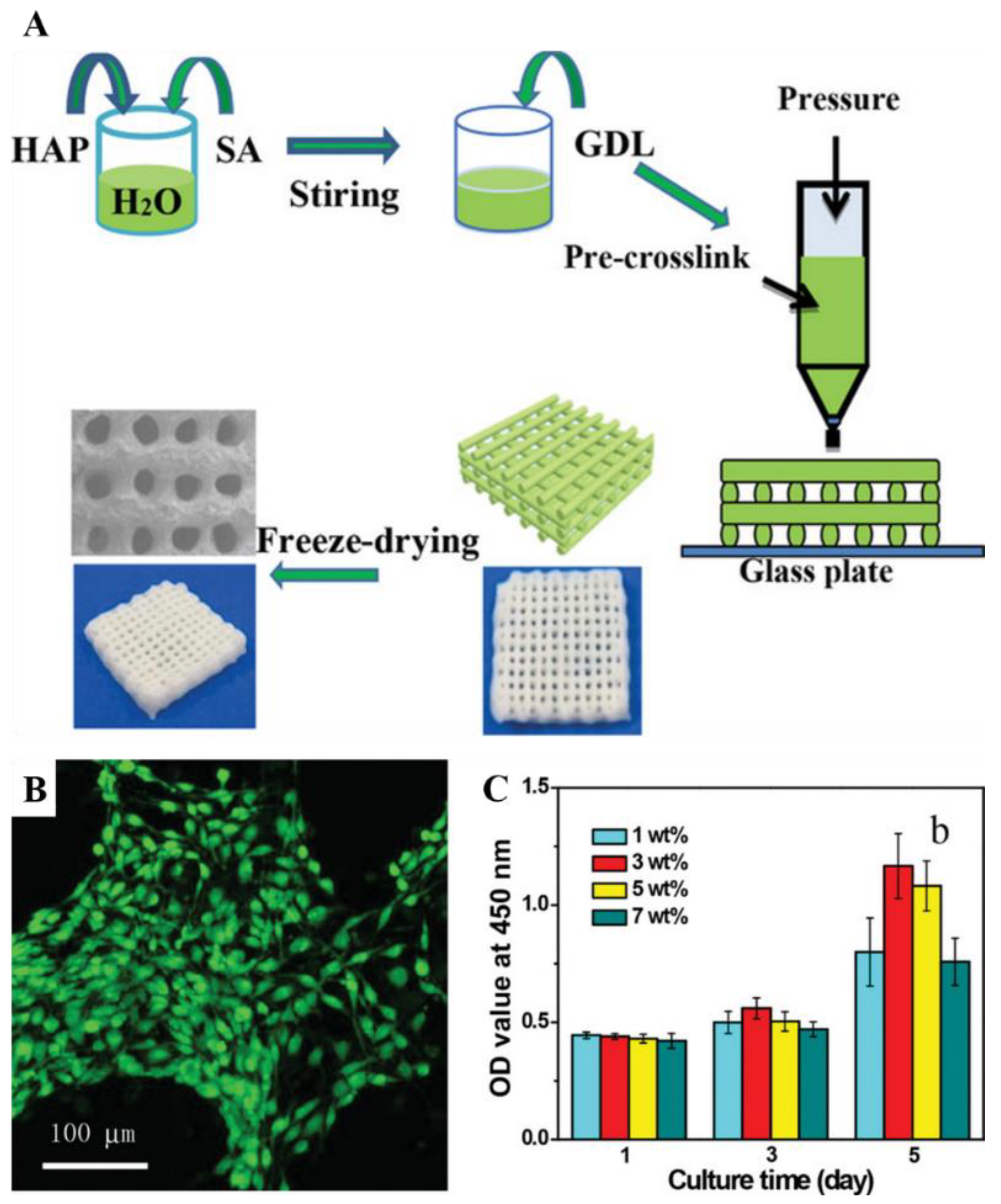
  20. Liu, S.; Hu, Y.; Zhang, J.; Bao, S.; Xian, L.; Dong, X.; Zheng, W.; Li, Y.; Gao, H.; Zhou, W. Bioactive and Biocompatible Macroporous Scaffolds with Tunable Performances Prepared Based on 3D Printing of the Pre-Crosslinked Sodium Alginate/Hydroxyapatite Hydrogel Ink. Macromol. Mater. Eng. 2019, 304, 1800698. [Google Scholar] [CrossRef]
  21. Turnbull, G.; Clarke, J.; Picard, F.; Riches, P.; Jia, L.; Han, F.; Li, B.; Shu, W. 3D bioactive composite scaffolds for bone tissue engineering. Bioact. Mater. 2017, 3, 278–314. [Google Scholar] [CrossRef] [PubMed] [Green Version]
  22. Yang, M.; He, W.; Shuai, Y.; Min, S.; Zhu, L. Nucleation of hydroxyapatite crystals by self-assembled Bombyx mori silk fibroin. J. Polym. Sci. Part B Polym. Phys. 2013, 51, 742–748. [Google Scholar] [CrossRef]
  23. Patrício, T.M.F.; Panseri, S.; Sandri, M.; Tampieri, A.; Sprio, S. New bioactive bone-like microspheres with intrinsic magnetic properties obtained by bio-inspired mineralisation process. Mater. Sci. Eng. C 2017, 77, 613–623. [Google Scholar] [CrossRef] [PubMed]
  24. Chatzipanagis, K.; Baumann, C.G.; Sandri, M.; Sprio, S.; Tampieri, A.; Kröger, R. In situ mechanical and molecular investigations of collagen/apatite biomimetic composites combining Raman spectroscopy and stress-strain analysis. Acta Biomater. 2016, 46, 278–285. [Google Scholar] [CrossRef] [PubMed] [Green Version]
  25. Yan, Y.; Shi, X.; Miao, M.; He, T.; Dong, Z.H.; Zhan, K.; Yang, J.H.; Zhao, B.; Xia, B.Y. Bio-inspired design of hierarchical FeP nanostructure arrays for the hydrogen evolution reaction. Nano Res. 2017, 11, 3537–3547. [Google Scholar] [CrossRef]
  26. Hou, Y.-K.; Pan, G.-L.; Sun, Y.-Y.; Gao, X.-P. LiMn0.8Fe0.2PO4/Carbon Nanospheres@Graphene Nanoribbons Prepared by the Biomineralization Process as the Cathode for Lithium-Ion Batteries. ACS Appl. Mater. Interfaces 2018, 10, 16500–16510. [Google Scholar] [CrossRef]
  27. Wu, M.; Chen, K.; Yang, S.; Wang, Z.; Huang, P.-H.; Mai, J.; Li, Z.-Y.; Huang, T.J. High-throughput cell focusing and separation via acoustofluidic tweezers. Lab Chip 2018, 18, 3003–3010. [Google Scholar] [CrossRef]
  28. Zhang, W.; Zhou, S.; Sun, J.; Meng, X.; Luo, J.; Zhou, D.; Crittenden, J.C. Impact of Chloride Ions on UV/H2O2 and UV/Persulfate Advanced Oxidation Processes. Environ. Sci. Technol. 2018, 52, 7380–7389. [Google Scholar] [CrossRef]
  29. Chen, W.; Wang, G.; Tang, R. Nanomodification of living organisms by biomimetic mineralization. Nano Res. 2014, 7, 1404–1428. [Google Scholar] [CrossRef]
  30. Walsh, P.J.; Fee, K.; Clarke, S.A.; Julius, M.L.; Buchanan, F.J. Blueprints for the Next Generation of Bioinspired and Biomimetic Mineralised Composites for Bone Regeneration. Mar. Drugs 2018, 16, 288. [Google Scholar] [CrossRef] [Green Version]
  31. Osorio, R.; Alfonso-Rodríguez, C.A.; Osorio, E.; Medina-Castillo, A.L.; Alaminos, M.; Toledano-Osorio, M.; Toledano, M. Novel potential scaffold for periodontal tissue engineering. Clin. Oral Investig. 2017, 21, 2695–2707. [Google Scholar] [CrossRef] [PubMed]
  32. Sandri, M.; Filardo, G.; Kon, E.; Panseri, S.; Montesi, M.; Iafisco, M.; Savini, E.; Sprio, S.; Cunha, C.; Giavaresi, G.; et al. Fabrication and Pilot In Vivo Study of a Collagen-BDDGE-Elastin Core-Shell Scaffold for Tendon Regeneration. Front. Bioeng. Biotechnol. 2016, 4. [Google Scholar] [CrossRef] [PubMed] [Green Version]
  33. Abdulghani, S.; Mitchell, G.R. Biomaterials for In Situ Tissue Regeneration: A Review. Biomolecules 2019, 9, 750. [Google Scholar] [CrossRef] [PubMed] [Green Version]
  34. Kaczmarek, B.; Sionkowska, A.; Kozlowska, J.; Osyczka, A. New composite materials prepared by calcium phosphate precipitation in chitosan/collagen/hyaluronic acid sponge cross-linked by EDC/NHS. Int. J. Biol. Macromol. 2018, 107, 247–253. [Google Scholar] [CrossRef]
  35. Weiner, S.; Dove, P.M. An Overview of Biomineralization Processes and the Problem of the Vital Effect. Biomineralization 2003, 54, 1–30. [Google Scholar] [CrossRef]
  36. Zhong, L.; Qu, Y.; Shi, K.; Chu, B.; Lei, M.; Huang, K.; Gu, Y.; Qian, Z. Biomineralized polymer matrix composites for bone tissue repair: A review. Sci. China Ser. B Chem. 2018, 61, 1553–1567. [Google Scholar] [CrossRef]
  37. Campodoni, E.; Dozio, S.M.; Panseri, S.; Montesi, M.; Tampieri, A.; Sandri, M. Mimicking Natural Microenvironments: Design of 3D-Aligned Hybrid Scaffold for Dentin Regeneration. Front. Bioeng. Biotechnol. 2020, 8, 836. [Google Scholar] [CrossRef]
  38. Salama, A. Cellulose/calcium phosphate hybrids: New materials for biomedical and environmental applications. Int. J. Biol. Macromol. 2019, 127, 606–617. [Google Scholar] [CrossRef]
  39. Wang, Q.; Tang, Y.; Ke, Q.; Yin, W.; Zhang, C.; Guo, Y.; Guan, J. Magnetic lanthanum-doped hydroxyapatite/chitosan scaffolds with endogenous stem cell-recruiting and immunomodulatory properties for bone regeneration. J. Mater. Chem. B 2020, 8, 5280–5292. [Google Scholar] [CrossRef]
  40. Tang, Y.-Q.; Wang, Q.-Y.; Ke, Q.-F.; Zhang, C.-Q.; Guan, J.-J.; Guo, Y.-P. Mineralization of ytterbium-doped hydroxyapatite nanorod arrays in magnetic chitosan scaffolds improves osteogenic and angiogenic abilities for bone defect healing. Chem. Eng. J. 2020, 387, 124166. [Google Scholar] [CrossRef]
  41. Furlani, F.; Marfoglia, A.; Marsich, E.; Donati, I.; Sacco, P. Strain Hardening in Highly Acetylated Chitosan Gels. Biomacromolecules 2021, 22, 2902–2909. [Google Scholar] [CrossRef]
  42. Ucar, S.; Bjørnøy, S.H.; Bassett, D.C.; Strand, B.L.; Sikorski, P.; Andreassen, J.-P. Nucleation and Growth of Brushite in the Presence of Alginate. Cryst. Growth Des. 2015, 15, 5397–5405. [Google Scholar] [CrossRef]
  43. Lloyd, A.W. Interfacial bioengineering to enhance surface biocompatibility. Med. Device Technol. 2002, 13, 18–21. [Google Scholar] [PubMed]
  44. Finkemeier, C.G. Bone-grafting and bone-graft substitutes. J. Bone Jt. Surg. 2002, 84, 454–464. [Google Scholar] [CrossRef] [PubMed]
  45. Barrère, F.; van Blitterswijk, C.A.; de Groot, K. Bone regeneration: Molecular and cellular interactions with calcium phosphate ceramics. Int. J. Nanomed. 2006, 1, 317. [Google Scholar]
  46. Mountziaris, P.M.; Mikos, A.G. Modulation of the Inflammatory Response for Enhanced Bone Tissue Regeneration. Tissue Eng. Part B Rev. 2008, 14, 179–186. [Google Scholar] [CrossRef]
  47. Cukierman, E.; Pankov, R.; Stevens, D.R.; Yamada, K.M. Taking Cell-Matrix Adhesions to the Third Dimension. Science 2001, 294, 1708–1712. [Google Scholar] [CrossRef] [PubMed]
  48. O’Brien, F.; Harley, B.; Yannas, I.; Gibson, L. The effect of pore size on cell adhesion in collagen-GAG scaffolds. Biomaterials 2005, 26, 433–441. [Google Scholar] [CrossRef] [PubMed]
  49. Tsuruga, E.; Takita, H.; Itoh, H.; Wakisaka, Y.; Kuboki, Y. Pore Size of Porous Hydroxyapatite as the Cell-Substratum Controls BMP-Induced Osteogenesis. J. Biochem. 1997, 121, 317–324. [Google Scholar] [CrossRef] [PubMed] [Green Version]
  50. Ishida, H.; Haniu, H.; Takeuchi, A.; Ueda, K.; Sano, M.; Tanaka, M.; Takizawa, T.; Sobajima, A.; Kamanaka, T.; Saito, N. In Vitro and In Vivo Evaluation of Starfish Bone-Derived β-Tricalcium Phosphate as a Bone Substitute Material. Materials 2019, 12, 1881. [Google Scholar] [CrossRef] [Green Version]
  51. Habibovic, P.; Yuan, H.; van der Valk, C.M.; Meijer, G.; van Blitterswijk, C.; de Groot, K. 3D microenvironment as essential element for osteoinduction by biomaterials. Biomaterials 2005, 26, 3565–3575. [Google Scholar] [CrossRef]
  52. Whang, K.; Healy, K.E.; Elenz, D.R.; Nam, E.K.; Tsai, D.C.; Thomas, C.H.; Nuber, G.W.; Glorieux, F.H.; Travers, R.; Sprague, S.M. Engineering Bone Regeneration with Bioabsorbable Scaffolds with Novel Microarchitecture. Tissue Eng. 1999, 5, 35–51. [Google Scholar] [CrossRef]
  53. Boccaccio, A.; Ballini, A.; Pappalettere, C.; Tullo, D.; Cantore, S.; Desiate, A. Finite Element Method (FEM), Mechanobiology and Biomimetic Scaffolds in Bone Tissue Engineering. Int. J. Biol. Sci. 2011, 7, 112–132. [Google Scholar] [CrossRef]
  54. Cipitria, A.; Wagermaier, W.; Zaslansky, P.; Schell, H.; Reichert, J.; Fratzl, P.; Hutmacher, D.; Duda, G. BMP delivery complements the guiding effect of scaffold architecture without altering bone microstructure in critical-sized long bone defects: A multiscale analysis. Acta Biomater. 2015, 23, 282–294. [Google Scholar] [CrossRef] [Green Version]
  55. Kaigler, D.; Wang, Z.; Horger, K.; Mooney, D.J.; Krebsbach, P.H. VEGF Scaffolds Enhance Angiogenesis and Bone Regeneration in Irradiated Osseous Defects. J. Bone Miner. Res. 2006, 21, 735–744. [Google Scholar] [CrossRef] [Green Version]
  56. Dimitriou, R.; Tsiridis, E.; Giannoudis, P.V. Current concepts of molecular aspects of bone healing. Injury 2005, 36, 1392–1404. [Google Scholar] [CrossRef]
  57. Tampieri, A.; Landi, E.; Valentini, F.; Sandri, M.; D’Alessandro, T.; Dediu, V.; Marcacci, M. A conceptually new type of bio-hybrid scaffold for bone regeneration. Nanotechnology 2010, 22, 015104. [Google Scholar] [CrossRef] [PubMed]
  58. Parveen, S.; Misra, R.; Sahoo, S.K. Nanoparticles: A boon to drug delivery, therapeutics, diagnostics and imaging. Nanomed. Nanotechnol. Biol. Med. 2011, 8, 147–166. [Google Scholar] [CrossRef] [PubMed]
  59. Cho, K.; Wang, X.; Nie, S.; Chen, Z.; Shin, D.M. Therapeutic Nanoparticles for Drug Delivery in Cancer. Clin. Cancer Res. 2008, 14, 1310–1316. [Google Scholar] [CrossRef] [Green Version]
  60. Papakostidis, C.; Kanakaris, N.K.; Pretel, J.; Faour, O.; Morell, D.J.; Giannoudis, P.V. Prevalence of complications of open tibial shaft fractures stratified as per the Gustilo-Anderson classification. Injury 2011, 42, 1408–1415. [Google Scholar] [CrossRef]
  61. Sprio, S.; Campodoni, E.; Sandri, M.; Preti, L.; Keppler, T.; Müller, F.A.; Pugno, N.M.; Tampieri, A. A Graded Multifunctional Hybrid Scaffold with Superparamagnetic Ability for Periodontal Regeneration. Int. J. Mol. Sci. 2018, 19, 3604. [Google Scholar] [CrossRef] [PubMed] [Green Version]
  62. Hutchens, S.A.; Benson, R.S.; Evans, B.R.; O’Neill, H.; Rawn, C.J. Biomimetic synthesis of calcium-deficient hydroxyapatite in a natural hydrogel. Biomaterials 2006, 27, 4661–4670. [Google Scholar] [CrossRef]
  63. Yue, K.; de Santiago, G.T.; Alvarez, M.M.; Tamayol, A.; Annabi, N.; Khademhosseini, A. Synthesis, properties, and biomedical applications of gelatin methacryloyl (GelMA) hydrogels. Biomaterials 2015, 73, 254–271. [Google Scholar] [CrossRef] [Green Version]
  64. Alfano, M.; Nebuloni, M.; Allevi, R.; Zerbi, P.; Longhi, E.; Lucianò, R.; Locatelli, I.; Pecoraro, A.; Indrieri, M.; Speziali, C.; et al. Linearized texture of three-dimensional extracellular matrix is mandatory for bladder cancer cell invasion. Sci. Rep. 2016, 6, 36128. [Google Scholar] [CrossRef]
  65. Murphy, C.A.; Costa, J.; Correia, J.S.; Oliveira, J.M.; Reis, R.L.; Collins, M. Biopolymers and polymers in the search of alternative treatments for meniscal regeneration: State of the art and future trends. Appl. Mater. Today 2018, 12, 51–71. [Google Scholar] [CrossRef]
  66. Hao, Y.; Song, J.; Ravikrishnan, A.; Dicker, K.T.; Fowler, E.W.; Zerdoum, A.B.; Li, Y.; Zhang, H.; Rajasekaran, A.K.; Fox, J.M.; et al. Rapid Bioorthogonal Chemistry Enables in Situ Modulation of the Stem Cell Behavior in 3D without External Triggers. ACS Appl. Mater. Interfaces 2018, 10, 26016–26027. [Google Scholar] [CrossRef]
  67. Li, W.; Xu, R.; Huang, J.; Bao, X.; Zhao, B. Treatment of rabbit growth plate injuries with oriented ECM scaffold and autologous BMSCs. Sci. Rep. 2017, 7, 44140. [Google Scholar] [CrossRef] [PubMed] [Green Version]
  68. Florencio-Silva, R.; Sasso, G.R.D.S.; Sasso-Cerri, E.; Simões, M.D.J.; Cerri, P. Biology of Bone Tissue: Structure, Function, and Factors That Influence Bone Cells. BioMed Res. Int. 2015, 2015, 421746. [Google Scholar] [CrossRef] [Green Version]
  69. Seeman, E. Bone Modeling and Remodeling. Crit. Rev. Eukaryot. Gene Expr. 2009, 19, 219–233. [Google Scholar] [CrossRef]
  70. Bello, A.B.; Kim, D.; Kim, D.; Park, H.; Lee, S.-H. Engineering and Functionalization of Gelatin Biomaterials: From Cell Culture to Medical Applications. Tissue Eng. Part B Rev. 2020, 26, 164–180. [Google Scholar] [CrossRef] [Green Version]
  71. Menale, C.; Campodoni, E.; Palagano, E.; Mantero, S.; Erreni, M.; Inforzato, A.; Fontana, E.; Schena, F.; Hof, R.V.; Sandri, M.; et al. Mesenchymal Stromal Cell-Seeded Biomimetic Scaffolds as a Factory of Soluble RANKL in Rankl-Deficient Osteopetrosis. STEM CELLS Transl. Med. 2018, 8, 22–34. [Google Scholar] [CrossRef] [PubMed]
  72. Chocholata, P.; Kulda, V.; Dvorakova, J.; Dobra, J.K.; Babuska, V. Biological Evaluation of Polyvinyl Alcohol Hydrogels Enriched by Hyaluronic Acid and Hydroxyapatite. Int. J. Mol. Sci. 2020, 21, 5719. [Google Scholar] [CrossRef] [PubMed]
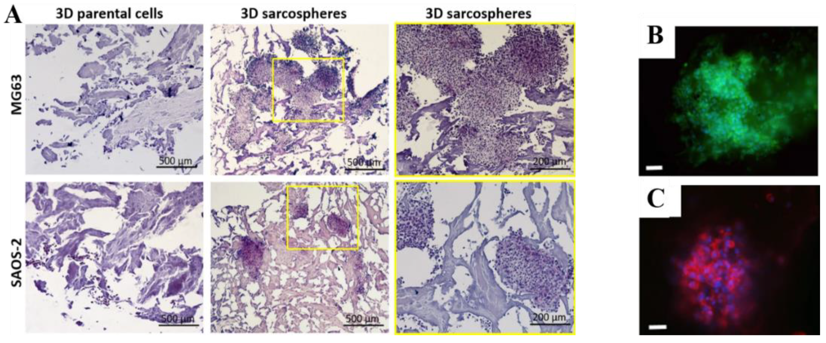
  73. Bassi, G.; Panseri, S.; Dozio, S.M.; Sandri, M.; Campodoni, E.; Dapporto, M.; Sprio, S.; Tampieri, A.; Montesi, M. Scaffold-based 3D cellular models mimicking the heterogeneity of osteosarcoma stem cell niche. Sci. Rep. 2020, 10, 1–12. [Google Scholar] [CrossRef]
  74. Wang, P.; Hu, J.; Ma, P.X. The engineering of patient-specific, anatomically shaped, digits. Biomaterials 2009, 30, 2735–2740. [Google Scholar] [CrossRef] [PubMed] [Green Version]
  75. Hernandez, I.; Kumar, A.; Joddar, B. A Bioactive Hydrogel and 3D Printed Polycaprolactone System for Bone Tissue Engineering. Gels 2017, 3, 26. [Google Scholar] [CrossRef] [PubMed] [Green Version]
  76. Hu, Y.; Wang, J.; Li, X.; Hu, X.; Zhou, W.; Dong, X.; Wang, C.; Yang, Z.; Binks, B.P. Facile preparation of bioactive nanoparticle/poly(ε-caprolactone) hierarchical porous scaffolds via 3D printing of high internal phase Pickering emulsions. J. Colloid Interface Sci. 2019, 545, 104–115. [Google Scholar] [CrossRef]
  77. Huang, T.; Fan, C.; Zhu, M.; Zhu, Y.; Zhang, W.; Li, L. 3D-printed scaffolds of biomineralized hydroxyapatite nanocomposite on silk fibroin for improving bone regeneration. Appl. Surf. Sci. 2019, 467–468, 345–353. [Google Scholar] [CrossRef]
  78. Romanazzo, S.; Molley, T.G.; Nemec, S.; Lin, K.; Sheikh, R.; Gooding, J.J.; Wan, B.; Li, Q.; Kilian, K.A.; Roohani, I. Synthetic Bone-Like Structures Through Omnidirectional Ceramic Bioprinting in Cell Suspensions. Adv. Funct. Mater. 2021, 31. [Google Scholar] [CrossRef]
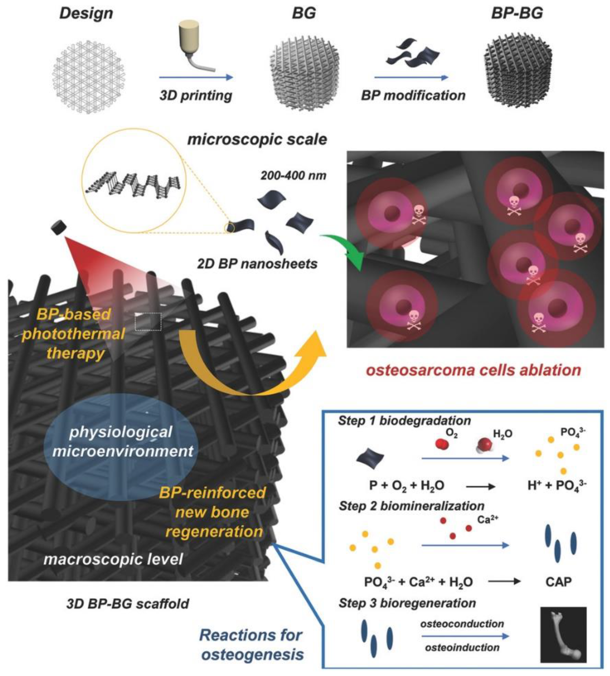
  79. Liu, X.; Miller, A.L.; Park, S.; George, M.; Waletzki, B.E.; Xu, H.; Terzic, A.; Lu, L. Two-Dimensional Black Phosphorus and Graphene Oxide Nanosheets Synergistically Enhance Cell Proliferation and Osteogenesis on 3D Printed Scaffolds. ACS Appl. Mater. Interfaces 2019, 11, 23558–23572. [Google Scholar] [CrossRef] [PubMed]
  80. Yang, B.; Yin, J.; Chen, Y.; Pan, S.; Yao, H.; Gao, Y.; Shi, J. 2D-Black-Phosphorus-Reinforced 3D-Printed Scaffolds: A Stepwise Countermeasure for Osteosarcoma. Adv. Mater. 2018, 30. [Google Scholar] [CrossRef]
  81. Lin, S.; Zhong, Y.; Zhao, X.; Sawada, T.; Li, X.; Lei, W.; Wang, M.; Serizawa, T.; Zhu, H. Synthetic Multifunctional Graphene Composites with Reshaping and Self-Healing Features via a Facile Biomineralization-Inspired Process. Adv. Mater. 2018, 30, e1803004. [Google Scholar] [CrossRef]
  82. Unger, C.; Kramer, N.; Walzl, A.; Scherzer, M.; Hengstschläger, M.; Dolznig, H. Modeling human carcinomas: Physiologically relevant 3D models to improve anti-cancer drug development. Adv. Drug Deliv. Rev. 2014, 79–80, 50–67. [Google Scholar] [CrossRef]
  83. Antoni, D.; Burckel, H.; Josset, E.; Noel, G. Three-Dimensional Cell Culture: A Breakthrough In Vivo. Int. J. Mol. Sci. 2015, 16, 5517–5527. [Google Scholar] [CrossRef]
  84. Borella, G.; Da Ros, A.; Borile, G.; Porcù, E.; Tregnago, C.; Benetton, M.; Marchetti, A.; Bisio, V.; Montini, B.; Michielotto, B.; et al. Targeting mesenchymal stromal cells plasticity to reroute acute myeloid leukemia course. Blood 2021. [Google Scholar] [CrossRef]
  85. Bhattacharya, P.; Du, D.; Lin, Y. Bioinspired nanoscale materials for biomedical and energy applications. J. R. Soc. Interface 2014, 11, 20131067. [Google Scholar] [CrossRef] [Green Version]
  86. Lopa, S.; Madry, H. Bioinspired Scaffolds for Osteochondral Regeneration. Tissue Eng. Part A 2014, 20, 2052–2076. [Google Scholar] [CrossRef] [PubMed]
  87. Ye, B.; Luo, X.; Li, Z.; Zhuang, C.; Li, L.; Lu, L.; Ding, S.; Tian, J.; Zhou, C. Rapid biomimetic mineralization of collagen fibrils and combining with human umbilical cord mesenchymal stem cells for bone defects healing. Mater. Sci. Eng. C 2016, 68, 43–51. [Google Scholar] [CrossRef] [PubMed]
  88. Rebelo, M.A.; Alves, T.F.R.; De Lima, R.; Oliveira, J.M.; Vila, M.M.D.C.; Balcão, V.; Severino, P.; Chaud, M.V.; Oliveira, J.M., Jr. Scaffolds and tissue regeneration: An overview of the functional properties of selected organic tissues. J. Biomed. Mater. Res. Part B: Appl. Biomater. 2015, 104, 1483–1494. [Google Scholar] [CrossRef]
  89. Yagüe, M.F.; Abbah, S.; McNamara, L.; Zeugolis, D.I.; Pandit, A.; Biggs, M.J. Biomimetic approaches in bone tissue engineering: Integrating biological and physicomechanical strategies. Adv. Drug Deliv. Rev. 2014, 84, 1–29. [Google Scholar] [CrossRef]
  90. Mravic, M.; Péault, B.; James, A.W. Current Trends in Bone Tissue Engineering. BioMed Res. Int. 2014, 2014, 865270. [Google Scholar] [CrossRef]
  91. Yousefi, A.-M.; James, P.F.; Akbarzadeh, R.; Subramanian, A.; Flavin, C.; Oudadesse, H. Prospect of Stem Cells in Bone Tissue Engineering: A Review. Stem Cells Int. 2016, 2016, 6180487. [Google Scholar] [CrossRef] [Green Version]
  92. Murphy, M.B.; Moncivais, K.; Caplan, A. Mesenchymal stem cells: Environmentally responsive therapeutics for regenerative medicine. Exp. Mol. Med. 2013, 45, e54. [Google Scholar] [CrossRef] [Green Version]
  93. Lei, B.; Wang, L.; Chen, X.; Chae, S.-K. Biomimetic and molecular level-based silicate bioactive glass–gelatin hybrid implants for loading-bearing bone fixation and repair. J. Mater. Chem. B 2013, 1, 5153–5162. [Google Scholar] [CrossRef]
  94. Gashti, M.P.; Stir, M.; Bourquin, M.; Hulliger, J. Mineralization of Calcium Phosphate Crystals in Starch Template Inducing a Brushite Kidney Stone Biomimetic Composite. Cryst. Growth Des. 2013, 13, 2166–2173. [Google Scholar] [CrossRef]
  95. Zhao, R.; Wang, B.; Yang, X.; Xiao, Y.; Wang, X.; Shao, C.; Tang, R. A Drug-Free Tumor Therapy Strategy: Cancer-Cell-Targeting Calcification. Angew. Chem. Int. Ed. 2016, 55, 5225–5229. [Google Scholar] [CrossRef] [PubMed]
  96. Wang, Z.; Hauser, N.; Singer, G.; Trippel, M.; Kubik-Huch, R.A.; Schneider, C.; Stampanoni, M. Non-invasive classification of microcalcifications with phase-contrast X-ray mammography. Nat. Commun. 2014, 5, 3797. [Google Scholar] [CrossRef]
  97. Varsano, N.; Dadosh, T.; Kapishnikov, S.; Pereiro, E.; Shimoni, E.; Jin, X.; Kruth, H.S.; Leiserowitz, L.; Addadi, L. Development of Correlative Cryo-soft X-ray Tomography and Stochastic Reconstruction Microscopy. A Study of Cholesterol Crystal Early Formation in Cells. J. Am. Chem. Soc. 2016, 138, 14931–14940. [Google Scholar] [CrossRef]
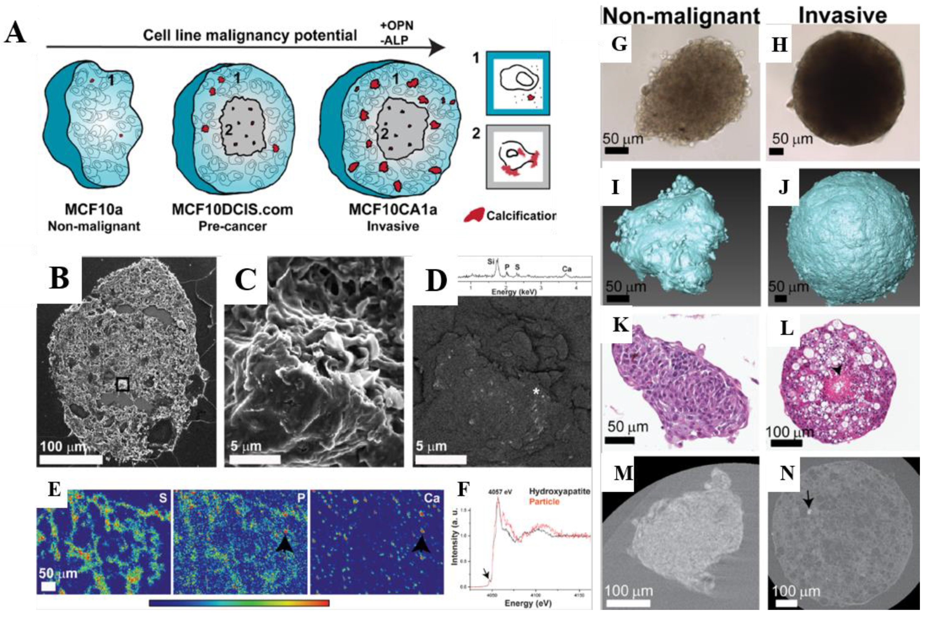
  98. Vidavsky, N.; Kunitake, J.A.; Chiou, A.E.; Northrup, P.; Porri, T.J.; Ling, L.; Fischbach, C.; Estroff, L.A. Studying biomineralization pathways in a 3D culture model of breast cancer microcalcifications. Biomaterials 2018, 179, 71–82. [Google Scholar] [CrossRef]
  99. Chen, H.; Fu, S.; Fu, L.; Yang, H.; Chen, D. Simple Synthesis and Characterization of Hexagonal and Ordered Al–MCM–41 from Natural Perlite. Minerals 2019, 9, 264. [Google Scholar] [CrossRef] [Green Version]
  100. Miller, F.R.; Santner, S.J.; Tait, L.; Dawson, P.J. MCF10DCIS.com xenograft model of human comedo ductal carcinoma in situ. J. Natl. Cancer Inst. 2000, 92, 1185–1186. [Google Scholar] [CrossRef] [PubMed] [Green Version]
  101. Santner, S.J.; Dawson, P.J.; Tait, L.; Soule, H.D.; Eliason, J.; Mohamed, A.N.; Wolman, S.R.; Heppner, G.H.; Miller, F.R. Malignant MCF10CA1 Cell Lines Derived from Premalignant Human Breast Epithelial MCF10AT Cells. Breast Cancer Res. Treat. 2001, 65, 101–110. [Google Scholar] [CrossRef] [PubMed]
  102. Palm, M.D.; O’Donoghue, M.N. Update on photoprotection. Dermatol. Ther. 2007, 20, 360–376. [Google Scholar] [CrossRef] [PubMed]
  103. Smaoui, S.; Ben Hlima, H.; Ben Chobba, I.; Kadri, A. Development and stability studies of sunscreen cream formulations containing three photo-protective filters. Arab. J. Chem. 2017, 10, S1216–S1222. [Google Scholar] [CrossRef] [Green Version]
  104. Serpone, N.; Dondi, D.; Albini, A. Inorganic and organic UV filters: Their role and efficacy in sunscreens and suncare products. Inorganica Chim. Acta 2007, 360, 794–802. [Google Scholar] [CrossRef]
  105. Antoniou, C.; Kosmadaki, M.; Stratigos, A.; Katsambas, A. Sunscreens—What’s important to know. J. Eur. Acad. Dermatol. Venereol. 2008, 22, 1110–1119. [Google Scholar] [CrossRef]
  106. Zhong, X.; Downs, C.A.; Li, Y.; Zhang, Z.; Li, Y.; Liu, B.; Gao, H.; Li, Q. Comparison of toxicological effects of oxybenzone, avobenzone, octocrylene, and octinoxate sunscreen ingredients on cucumber plants (Cucumis sativus L.). Sci. Total Environ. 2020, 714, 136879. [Google Scholar] [CrossRef]
  107. Hiller, J.; Klotz, K.; Meyer, S.; Uter, W.; Hof, K.; Greiner, A.; Göen, T.; Drexler, H. Systemic availability of lipophilic organic UV filters through dermal sunscreen exposure. Environ. Int. 2019, 132, 105068. [Google Scholar] [CrossRef]
  108. Monteiro-Riviere, N.A.; Wiench, K.; Landsiedel, R.; Schulte, S.; Inman, A.O.; Riviere, J.E. Safety Evaluation of Sunscreen Formulations Containing Titanium Dioxide and Zinc Oxide Nanoparticles in UVB Sunburned Skin: An In Vitro and In Vivo Study. Toxicol. Sci. 2011, 123, 264–280. [Google Scholar] [CrossRef] [Green Version]
  109. Schulz, J.; Hohenberg, H.; Pflücker, F.; Gärtner, E.; Will, T.; Pfeiffer, S.; Wepf, R.; Wendel, V.; Gers-Barlag, H.; Wittern, K.-P. Distribution of sunscreens on skin. Adv. Drug Deliv. Rev. 2002, 54, S157–S163. [Google Scholar] [CrossRef]
  110. Shao, Y.; Schlossman, D. Effect of particle size on performance of physical sunscreen formulas. In Proceedings of the PCIA Conference, Shanghai, China, March 1999; pp. 1–9. [Google Scholar]
  111. Levine, A. Sunscreen use and awareness of chemical toxicity among beach goers in Hawaii prior to a ban on the sale of sunscreens containing ingredients found to be toxic to coral reef ecosystems. Mar. Policy 2020, 117, 103875. [Google Scholar] [CrossRef]
  112. Battistin, M.; Dissette, V.; Bonetto, A.; Durini, E.; Manfredini, S.; Marcomini, A.; Casagrande, E.; Brunetta, A.; Ziosi, P.; Molesini, S.; et al. A New Approach to UV Protection by Direct Surface Functionalization of TiO2 with the Antioxidant Polyphenol Dihydroxyphenyl Benzimidazole Carboxylic Acid. Nanomaterials 2020, 10, 231. [Google Scholar] [CrossRef] [PubMed] [Green Version]
  113. Bino, A.; Baldisserotto, A.; Scalambra, E.; Dissette, V.; Vedaldi, D.E.; Salvador, A.; Durini, E.; Manfredini, S.; Vertuani, S. Design, synthesis and biological evaluation of novel hydroxy-phenyl-1H-benzimidazoles as radical scavengers and UV-protective agents. J. Enzym. Inhib. Med. Chem. 2017, 32, 527–537. [Google Scholar] [CrossRef]
  114. Hossan, J.; Gafur, M.A.; Kadir, M.R.; Mainul, M. Preparation and Characterization of Gelatin- Hydroxyapatite Composite for Bone Tissue Engineering. Int. J. Eng. Technol. 2014, 57, 113–122. [Google Scholar]
  115. Amin, R.M.; Elfeky, S.; Verwanger, T.; Krammer, B. A new biocompatible nanocomposite as a promising constituent of sunscreens. Mater. Sci. Eng. C 2016, 63, 46–51. [Google Scholar] [CrossRef] [PubMed]
  116. Lin, J.-Y.; Selim, M.; Shea, C.R.; Grichnik, J.M.; Omar, M.M.; Monteiro-Riviere, N.; Pinnell, S.R. UV photoprotection by combination topical antioxidants vitamin C and vitamin E. J. Am. Acad. Dermatol. 2003, 48, 866–874. [Google Scholar] [CrossRef] [PubMed] [Green Version]
  117. Morsy, R.; Ali, S.S.; El-Shetehy, M. Development of hydroxyapatite-chitosan gel sunscreen combating clinical multidrug-resistant bacteria. J. Mol. Struct. 2017, 1143, 251–258. [Google Scholar] [CrossRef]
  118. Landi, E.; Tampieri, A.; Mattioli-Belmonte, M.; Celotti, G.; Sandri, M.; Gigante, A.; Fava, P.; Biagini, G. Biomimetic Mg- and Mg,CO3-substituted hydroxyapatites: Synthesis characterization and in vitro behaviour. J. Eur. Ceram. Soc. 2006, 26, 2593–2601. [Google Scholar] [CrossRef]
  119. Landi, E.; Tampieri, A.; Celotti, G.; Sprio, S.; Sandri, M.; Logroscino, G. Sr-substituted hydroxyapatites for osteoporotic bone replacement. Acta Biomater. 2007, 3, 961–969. [Google Scholar] [CrossRef]
  120. de Araujo, T.; de Souza, S.; Miyakawa, W.; de Sousa, E. Phosphates nanoparticles doped with zinc and manganese for sunscreens. Mater. Chem. Phys. 2010, 124, 1071–1076. [Google Scholar] [CrossRef]
  121. De Araujo, T.S.; De Souza, S.O.; De Sousa, E.M.B. Effect of Zn2+, Fe3+ and Cr3+ addition to hydroxyapatite for its application as an active constituent of sunscreens. J. Physics: Conf. Ser. 2010, 249. [Google Scholar] [CrossRef]
  122. Piccirillo, C.; Rocha, C.; Tobaldi, D.M.; Pullar, R.C.; Labrincha, J.A.; Ferreira, M.O.; Castro, P.M.L.; Pintado, M.M.E. A hydroxyapatite–Fe2O3 based material of natural origin as an active sunscreen filter. J. Mater. Chem. B 2014, 2, 5999–6009. [Google Scholar] [CrossRef]
  123. Popov, A.; Priezzhev, A.V.; Lademann, J.; Myllylä, R. Alteration of skin light-scattering and absorption properties by application of sunscreen nanoparticles: A Monte Carlo study. J. Quant. Spectrosc. Radiat. Transf. 2011, 112, 1891–1897. [Google Scholar] [CrossRef]
  124. Morlando, A.; Cardillo, D.; Devers, T.; Konstantinov, K. Titanium doped tin dioxide as potential UV filter with low photocatalytic activity for sunscreen products. Mater. Lett. 2016, 171, 289–292. [Google Scholar] [CrossRef]
  125. Yasukawa, A.; Tamura, J. Preparation and structure of titanium-cerium-calcium hydroxyapatite particles and their ultraviolet protecting ability. Colloids Surf. A Physicochem. Eng. Asp. 2020, 609, 125705. [Google Scholar] [CrossRef]
  126. Tampieri, A.; Sandri, M.; Sprio, S. Physical Solar Filter Consisting of Substituted Hydroxyapatite in An Organic Matrix. US 10,813,856 B2, 27 October 2020. [Google Scholar]
  127. Burnett, M.E.; Wang, S.Q. Current sunscreen controversies: A critical review. Photodermatol. Photoimmunol. Photomed. 2011, 27, 58–67. [Google Scholar] [CrossRef] [PubMed]
  128. Manzano, M.; Lamberti, G.; Galdi, I.; Vallet-Regí, M. Anti-Osteoporotic Drug Release from Ordered Mesoporous Bioceramics: Experiments and Modeling. AAPS PharmSciTech 2011, 12, 1193–1199. [Google Scholar] [CrossRef] [Green Version]
  129. Raja, N.; Park, H.; Choi, Y.-J.; Yun, H.-S. Multifunctional Calcium-Deficient Hydroxyl Apatite–Alginate Core–Shell-Structured Bone Substitutes as Cell and Drug Delivery Vehicles for Bone Tissue Regeneration. ACS Biomater. Sci. Eng. 2021, 7, 1123–1133. [Google Scholar] [CrossRef]
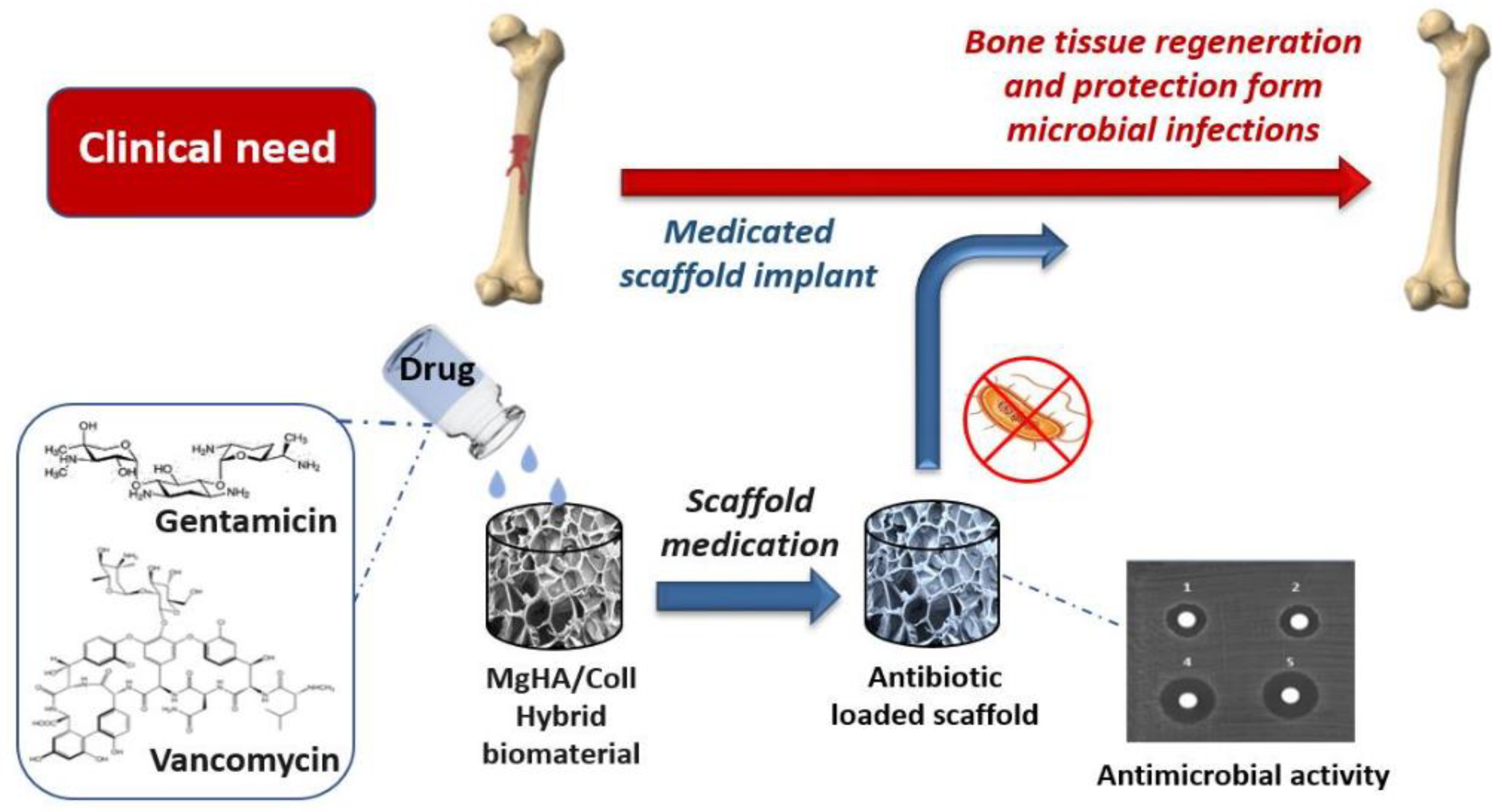
  130. Mulazzi, M.; Campodoni, E.; Bassi, G.; Montesi, M.; Panseri, S.; Bonvicini, F.; Gentilomi, G.A.; Tampieri, A.; Sandri, M. Medicated Hydroxyapatite/Collagen Hybrid Scaffolds for Bone Regeneration and Local Antimicrobial Therapy to Prevent Bone Infections. Pharmaceutics 2021, 13, 1090. [Google Scholar] [CrossRef] [PubMed]
  131. Barroug, A.; Glimcher, M.J. Hydroxyapatite crystals as a local delivery system for cisplatin: Adsorption and release of cisplatin in vitro. J. Orthop. Res. 2002, 20, 274–280. [Google Scholar] [CrossRef]
  132. Barroug, A.; Kuhn, L.T.; Gerstenfeld, L.C.; Glimcher, M.J. Interactions of cisplatin with calcium phosphate nanoparticles: In vitro controlled adsorption and release. J. Orthop. Res. 2004, 22, 703–708. [Google Scholar] [CrossRef]
  133. Chen, Q.; Liang, J.; Wang, S.; Wang, D.; Wang, R. An asymmetric approach toward chiral multicyclic spirooxindoles from isothiocyanato oxindoles and unsaturated pyrazolones by a chiral tertiary amine thiourea catalyst. Chem. Commun. 2013, 49, 1657–1659. [Google Scholar] [CrossRef]
  134. Srivastava, P.; Hira, S.K.; Srivastava, D.N.N.; Singh, V.K.; Gupta, U.; Singh, R.; Singh, R.A.; Manna, P.P. ATP-Decorated Mesoporous Silica for Biomineralization of Calcium Carbonate and P2 Purinergic Receptor-Mediated Antitumor Activity against Aggressive Lymphoma. ACS Appl. Mater. Interfaces 2018, 10, 6917–6929. [Google Scholar] [CrossRef]
  135. Descamps, M.; Duhoo, T.; Monchau, F.; Lu, J.; Hardouin, P.; Hornez, J.; Leriche, A. Manufacture of macroporous β-tricalcium phosphate bioceramics. J. Eur. Ceram. Soc. 2008, 28, 149–157. [Google Scholar] [CrossRef]
  136. Okazaki, Y.; Abe, Y.; Yasuda, K.; Hiasa, K.; Hirata, I. Osteoclast Response to Bioactive Surface Modification of Hydroxyapatite. Open J. Stomatol. 2014, 04, 340–344. [Google Scholar] [CrossRef] [Green Version]
  137. Pokhrel, S. Hydroxyapatite: Preparation, Properties and Its Biomedical Applications. Adv. Chem. Eng. Sci. 2018, 08, 225–240. [Google Scholar] [CrossRef] [Green Version]
  138. Iafisco, M.; Sandri, M.; Panseri, S.; Delgado-López, J.M.; Gómez-Morales, J.; Tampieri, A. Magnetic Bioactive and Biodegradable Hollow Fe-Doped Hydroxyapatite Coated Poly(l-lactic) Acid Micro-nanospheres. Chem. Mater. 2013, 25, 2610–2617. [Google Scholar] [CrossRef]
  139. Iafisco, M.; Drouet, C.; Adamiano, A.; Pascaud, P.; Montesi, M.; Panseri, S.; Sarda, S.; Tampieri, A. Superparamagnetic iron-doped nanocrystalline apatite as a delivery system for doxorubicin. J. Mater. Chem. B 2015, 4, 57–70. [Google Scholar] [CrossRef] [PubMed] [Green Version]
  140. Ain, Q.-U.; Munir, H.; Jelani, F.; Anjum, F.; Bilal, M. Antibacterial potential of biomaterial derived nanoparticles for drug delivery application. Mater. Res. Express 2019, 6, 125426. [Google Scholar] [CrossRef]
  141. Caplin, J.D.; García, A.J. Implantable antimicrobial biomaterials for local drug delivery in bone infection models. Acta Biomater. 2019, 93, 2–11. [Google Scholar] [CrossRef] [PubMed]
  142. Mondal, S.; Pal, U. 3D hydroxyapatite scaffold for bone regeneration and local drug delivery applications. J. Drug Deliv. Sci. Technol. 2019, 53, 101131. [Google Scholar] [CrossRef]
  143. Dorati, R.; De Trizio, A.; Genta, I.; Merelli, A.; Modena, T.; Conti, B. Formulation and in vitro characterization of a composite biodegradable scaffold as antibiotic delivery system and regenerative device for bone. J. Drug Deliv. Sci. Technol. 2016, 35, 124–133. [Google Scholar] [CrossRef]
  144. El-Husseiny, M.; Patel, S.; Macfarlane, R.J.; Haddad, F.S. Biodegradable antibiotic delivery systems. J. Bone Jt. Surg. 2011, 93, 151–157. [Google Scholar] [CrossRef] [Green Version]
  145. Wu, C.; Fan, W.; Gelinsky, M.; Xiao, Y.; Chang, J.; Friis, T.; Cuniberti, G. In situ preparation and protein delivery of silicate–alginate composite microspheres with core-shell structure. J. R. Soc. Interface 2011, 8, 1804–1814. [Google Scholar] [CrossRef] [Green Version]
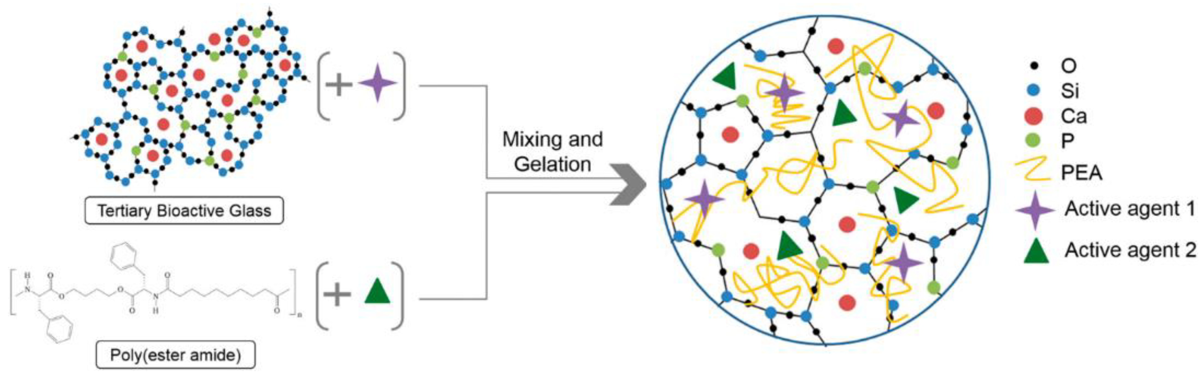
  146. Aslankoohi, N.; Mequanint, K. Intrinsically fluorescent bioactive glass-poly(ester amide) hybrid microparticles for dual drug delivery and bone repair. Mater. Sci. Eng. C 2021, 128, 112288. [Google Scholar] [CrossRef]
  147. Etter, J.N.; Karasinski, M.; Ware, J.; Oldinski, R.A. Dual-crosslinked homogeneous alginate microspheres for mesenchymal stem cell encapsulation. J. Mater. Sci. Mater. Med. 2018, 29, 143. [Google Scholar] [CrossRef]
  148. Radwan, N.H.; Nasr, M.; Ishak, R.A.; Awad, G.A. Moxifloxacin-loaded in situ synthesized Bioceramic/Poly(L-lactide-co-ε-caprolactone) composite scaffolds for treatment of osteomyelitis and orthopedic regeneration. Int. J. Pharm. 2021, 602, 120662. [Google Scholar] [CrossRef]
  149. Alshemary, A.Z.; Akram, M.; Goh, Y.-F.; Tariq, U.; Butt, F.K.; Abdolahi, A.; Hussain, R. Synthesis, characterization, in vitro bioactivity and antimicrobial activity of magnesium and nickel doped silicate hydroxyapatite. Ceram. Int. 2015, 41, 11886–11898. [Google Scholar] [CrossRef]
  150. Ballardini, A.; Montesi, M.; Panseri, S.; Vandini, A.; Balboni, P.G.; Tampieri, A.; Sprio, S. New hydroxyapatite nanophases with enhanced osteogenic and anti-bacterial activity. J. Biomed. Mater. Res. Part A 2017, 106, 521–530. [Google Scholar] [CrossRef]
  151. Huh, A.J.; Kwon, Y.J. “Nanoantibiotics”: A new paradigm for treating infectious diseases using nanomaterials in the antibiotics resistant era. J. Control. Release 2011, 156, 128–145. [Google Scholar] [CrossRef]
  152. Sprio, S.; Sandri, M.; Iafisco, M.; Panseri, S.; Montesi, M.; Ruffini, A.; Adamiano, A.; Ballardini, A.; Tampieri, A.B.A.A. Nature-Inspired Nanotechnology and Smart Magnetic Activation: Two Groundbreaking Approaches toward a New Generation of Biomaterials for Hard Tissue Regeneration; IntechOpen: London, UK, 2016. [Google Scholar] [CrossRef] [Green Version]
  153. Campodoni, E.; Adamiano, A.; Dozio, S.M.; Panseri, S.; Montesi, M.; Sprio, S.; Tampieri, A.; Sandri, M. Development of innovative hybrid and intrinsically magnetic nanobeads as a drug delivery system. Nanomedicine 2016, 11, 2119–2130. [Google Scholar] [CrossRef] [PubMed]
  154. Patrício, T.M.F.; Panseri, S.; Montesi, M.; Iafisco, M.; Sandri, M.; Tampieri, A.; Sprio, S. Superparamagnetic hybrid microspheres affecting osteoblasts behaviour. Mater. Sci. Eng. C 2018, 96, 234–247. [Google Scholar] [CrossRef] [PubMed]
  155. Patrício, T.M.F.; Mumcuoglu, D.; Montesi, M.; Panseri, S.; Witte-Bouma, J.; Garcia, S.F.; Sandri, M.; Tampieri, A.; Farrell, E.; Sprio, S. Bio-inspired polymeric iron-doped hydroxyapatite microspheres as a tunable carrier of rhBMP-2. Mater. Sci. Eng. C 2020, 119, 111410. [Google Scholar] [CrossRef] [PubMed]
  156. Tatiana, F.P.; Simone, S.; Monica, S.; Natalia, G.; Monica, M.; Silvia, P.; Bas, K.; Anna, T. Bio-inspired superparamagnetic microspheres for bone tissue engineering applications. Front. Bioeng. Biotechnol. 2016, 4. [Google Scholar] [CrossRef]
  157. Miragoli, M.; Ceriotti, P.; Iafisco, M.; Vacchiano, M.; Salvarani, N.; Alogna, A.; Carullo, P.; Ramirez-Rodríguez, G.B.; Patrício, T.; Degli Esposti, L.; et al. Inhalation of peptide-loaded nanoparticles improves heart failure. Sci. Transl. Med. 2018, 10, eaan6205. [Google Scholar] [CrossRef] [Green Version]
  158. Degli Esposti, L.; Carella, F.; Adamiano, A.; Tampieri, A.; Iafisco, M. Calcium phosphate-based nanosystems for advanced targeted nanomedicine. Drug Dev. Ind. Pharm. 2018, 44, 1223–1238. [Google Scholar] [CrossRef]
  159. Barbanente, A.; Nadar, R.A.; Degli Esposti, L.; Palazzo, B.; Iafisco, M.; Beucken, J.J.J.P.V.D.; Leeuwenburgh, S.C.G.; Margiotta, N. Platinum-loaded, selenium-doped hydroxyapatite nanoparticles selectively reduce proliferation of prostate and breast cancer cells co-cultured in the presence of stem cells. J. Mater. Chem. B 2020, 8, 2792–2804. [Google Scholar] [CrossRef]
Figure 1. Schematic illustration of the naturally occurring structure of bone microstructure and the synthetic approach used to reproduce it. More in detail, in the natural occurring structure of bone, collagen fibers are organized in a triple helix, forming tropocollagen fibrils; these fibrils are tightly tied and reinforced by hydroxyapatite crystals; the organization and association of these fibrils confers peculiar structural and mechanical properties to the bone. Within the lab process, a collagen acidic solution containing phosphate ions (e.g., phosphoric acid) is dropwise added and mixed to a basic solution containing calcium ions (e.g., calcium hydroxide), promoting the formation of nano-hydroxyapatite crystals within the collagen fibers. Reproduced from “Biomineralization process generating hybrid nano-and micro-carriers” by E. Campodoni et al., 2018, Core-Shell Nanostructures for Drug Delivery and Theranostics: Challenges, Strategies, and Prospects for Novel Carrier Systems, 19–24, (doi:10.1016/C2016-0-03458-7) (Under a Creative Commons Attribution 4.0 International License).
Jcs 05 00278 g001
Figure 2. (A) Schematic illustration of the 3D printing of hydrogels based on sodium alginate and hydroxyapatite (SA/HAP). In this case, alginate and hydroxyapatite were solubilized in water and extensively mixed together. Glucono-delta-lactone (GDL), a controlled acidifying agent able to promote partial calcium release from hydroxyapatite, was then added to the alginate mixture (pre-crosslinking). The resulting mixture was then extruded. The progressive calcium release was able to promote the gelation of alginate, and thus the formation of hybrid hydrogels based on alginate and hydroxyapatite. These hydrogels were freeze-dried obtaining scaffolds. (B) Confocal Laser Scanning Microscopy (CLSM) micrographs of fluorophore labelled Bone Marrow Stem Cells (BMSCs) after 5 days of culture on the same scaffolds; (C) BMSCs proliferation at different timeframes (1, 3, and 5 days of culture) on the porous scaffolds with different HAP amount. Reproduced with permission from Bioactive and Biocompatible Macroporous Scaffolds with Tunable Performances Prepared Based on 3D Printing of the Pre-Crosslinked Sodium Alginate/Hydroxyapatite Hydrogel Ink by S. Liu et al., 2019, Macromolecular Materials and Engineering, 304 (4), 11 (doi:10.1002/mame.201800698). Copyright 2019 by John Wiley and Sons.
Jcs 05 00278 g002
Figure 3. Schematic illustration of scaffolds fabrication based on black phosphorous/bio-glass (BP-BG) and devised for the elimination of osteosarcoma and the subsequent osteogenesis. These scaffolds were produced by 3D printing a black phosphorus and bio-glass mixture (BP-BG). These scaffolds can be exploited to promote osteosarcoma cells ablation upon light exposure. Subsequently, the scaffolds, due to their osteoconductive and osteoinductive properties, can be degraded into the main components of bone, promoting biomineralization and formation of new bone. Reproduced with permission from 2D-Black-Phosphorus-Reinforced 3D-Printed Scaffolds: A Stepwise Countermeasure for Osteosarcoma by Shi et al., 2018, Advanced Materials, 30 (10), 12 [80]. Copyright 2018 by John Wiley and Sons.
Jcs 05 00278 g003
Figure 4. Panel of figures of in vitro 3D model of Cancer Stem Cells (CSCs)-niche of osteosarcoma from [73]. (Under a Creative Commons Attribution 4.0 International License) using biomineralized scaffolds based on collagen and magnesium-doped hydroxyapatite (MgHA/Coll scaffold) as bone-like ExtraCellular Matrix (ECM). (A) Histological analysis after Haematoxylin–Eosin (H&E) staining of the MgHA/Coll scaffold seeded with both cellular phenotypes, parental and spheroidal, of MG63 and SAOS-2 osteosarcoma cell lines. The morphological features and the interaction behavior of the sarcospheres and parental cells with the scaffold is shown, with image enlargements of 200 µm on the right of the figure. (B,C) Immunofluorescence analysis of the 3D MgHA/Coll models with sarcospheres. Representative image of OCT-4 immunolocalization in SAOS-2 sarcospheres in image (B); scale bar 50 µm. SOX-2 immunolocalization in MG63 sarcospheres in image (C); scale bar 25 µm. Blue DAPI: cell nuclei; green: OCT-4; and red: SOX-2.
Jcs 05 00278 g004
Figure 5. Panel of figures of in vitro 3D culture model of breast cancer microcalcifications from [98]. (A) A schematic description of the proposed mineralization pathways in the 3D in vitro breast cancer model of various tumor stages; while OPN expression levels increase, ALP expression levels decrease with an increase in malignancy potential of cell line. Viable cell region in light blue, necrotic core in gray, and calcification in red. (BD) Mineralized particles in pre-cancerous MCF10DCIS.com spheroids core by SEM magnified section in images; EDS spectrum in image C of asterisk-marked area showing the presence of calcium (Ca), phosphorus (P), and sulfur (S). SRF maps of a spheroid section showing S, P, and Ca distribution in image (E). Ca K-edge XANES of the particle marked in (E) and a hydroxyapatite standard in image (F). (GN) Characterization of non-malignant MCF10A (G,I,K,M) and invasive MCF10CA1a (H,J,L,N) spheroids at day 13 of culture; light microscope (G,H), 3D reconstructed volumes of spheroids (I,J), H&E histological staining (K,L), and nanoCT data stained with iodine of the spheroids cross section (M,N). All the figures of this panel are reproduced with permission from Studying biomineralization pathways in a 3D culture model of breast cancer microcalcifications by Vidavsky et al., 2018, Biomaterials, 179, 12 (doi:10.1016/j.biomaterials.2018.06.030). Copyright 2018 by Elsevier.
Jcs 05 00278 g005
Figure 6. Schematic illustration of the loading of antibiotics within hybrid scaffolds based on collagen and magnesium-doped hydroxyapatite. Two different antibiotics, namely gentamicin and vancomycin, were introduced during biomineralization of collagen. Antibiotics proved to be tightly associated within the biomineralized scaffolds. These scaffolds were able to provide a piecemeal release of antibiotics, avoiding microbial colonization (and therefore avoiding infections) and simultaneously promoting bone tissue regeneration. The figure is reproduced from Medicated Hydroxyapatite/Collagen Hybrid Scaffolds for Bone Regeneration and Local Antimicrobial Therapy to Prevent Bone Infections by M. Mulazzi et al., 2021, Pharmaceutics, 13 (7), 1090 (doi:10.3390/pharmaceutics13071090) (Under a Creative Commons Attribution 4.0 International License).
Jcs 05 00278 g006
Figure 7. Schematic illustration of the two-stage synthesis approach of hybrid microparticles based on bioactive glass and poly(ester amide) (PEA). The singular components were mixed with an active agent. Subsequently the two mixtures were mixed together, and gelation was promoted, forming hybrid microparticles loaded with two different drugs. The figure is reproduced with permission from Intrinsically fluorescent bioactive glass-poly(ester amide) hybrid microparticles for dual drug delivery and bone repair by Aslankoohia and Mequanint et al., 2021, Materials Science and Engineering: C, 128, 112288 (doi:10.1016/j.msec.2021.112288). Copyright 2021 by Elsevier.
Jcs 05 00278 g007
Publisher’s Note: MDPI stays neutral with regard to jurisdictional claims in published maps and institutional affiliations.

