Abstract
Biomarkers to discriminate the main pathologies underlying frontotemporal lobar degeneration (FTLD-Tau, FTLD-TDP) are lacking. Our previous FTLD cerebrospinal fluid (CSF) proteome study revealed that sex hormone-binding globulin (SHBG) was specifically increased in FTLD-Tau patients. Here we investigated the potential of CSF SHBG as a novel biomarker discriminating the main FTLD pathological subtypes. SHBG was measured in CSF samples from patients with FTLD-Tau (n = 23), FTLD-TDP (n = 29) and controls (n = 33) using an automated electro-chemiluminescent immunoassay. Differences in CSF SHBG levels across groups, as well as its association with CSF YKL40, pTau181/total-Tau ratio and cognitive function were analyzed. CSF SHBG did not differ across groups, though a trend towards elevated levels in FTLD-Tau cases compared to FTLD-TDP and controls was observed. CSF SHBG levels were not associated with either CSF YKL40 or the p/tTau ratio. They, however, inversely correlated with the MMSE score (r = −0.307, p = 0.011), an association likely driven by the FTLD-Tau group (r FTLD-Tau = −0.38; r FTLD-TDP = −0.02). CSF SHBG is not a suitable biomarker to discriminate FTLD-Tau from FTLD-TDP.
1. Introduction
Frontotemporal lobar degeneration (FTLD) covers a spectrum of highly heterogeneous disorders from a clinical, genetic and pathological perspective [1]. The majority of FTLD cases are neuropathologically characterized by either the presence of aggregates of proteins tau (FTLD-Tau) or TDP43 (FTLD-TDP), which likely requires distinct pharmacological therapy. However, the clinical presentation of these pathological subtypes is heterogeneous and overlapping, making this diagnostic subtyping at present difficult. Thus, there is a strong unmet need to identify body-fluid-based biomarkers discriminating FTLD pathological subtypes.
Most biomarker studies performed to date have analyzed pathologically heterogeneous populations [2]. The few studies analyzing antemortem cerebrospinal fluid (CSF) with known underlying neuropathology have revealed several candidate biomarkers (e.g., p/tTau ratio) [2,3,4]. However, their specificity is still not optimal and most of the identified markers are awaiting further validation [2]. Our previous unbiased proteomics study revealed lower levels of sex hormone-binding globulin (SHBG) in the CSF of FTLD-Tau cases compared to FTLD-TDP or non-demented controls [4], suggesting that SHGB could be a potential biomarker for FTLD related tauopathies. SHBG is an androgen transport protein, and thus regulates the bioavailability of sex hormones. Despite their role on FTLD remains unknown, sex hormones have been associated to cognitive function, neurodegeneration and, more specifically, to Tau proteostasis [5,6]. In this study, we aimed to validate whether CSF SHBG is a useful biomarker for discriminating the main FTLD pathological subtypes in an independent cohort using an automated electro-chemiluminescent immunoassay (ECLIA), a serum test utilised in routine diagnostics. Next, the relationship of SHBG with cognitive function and other CSF markers known to be changed in FTLD or between FTLD subtypes (i.e., YKL40 and the ratio of phosphorylated Tau at 181 to total Tau (p/tTau)) was evaluated.
2. Methods
2.1. Humans CSF Samples
The total cohort (n = 85) included ante-mortem CSF samples from 52 FTLD-confirmed patients and 33 cognitive unimpaired individuals that were used as controls. FTLD cases with tau neuropathology (FTLD-Tau, n = 23) were selected based on autopsy (n = 10; including 2 cases with progressive supranuclear palsy (PSP) and 2 cases with corticobasal degeneration CBD tauopathy), and the presence of mutations in the MAPT gene predictive of Tau proteinopathy (n = 8) [1]. Diagnostic groups were enriched with patients with FTD clinical syndromes that reflect high correlation with a specific neuropathology. Thus, the FTLD-Tau group was enriched with non-autopsied patients with clinical PSP (n = 5), highly-associated to Tau pathology [7]. FTLD patients with an underlying TDP43 pathology (FTLD-TDP, n = 29) were selected based on autopsy (n = 13) or the presence of mutations predictive of TDP43 proteopathy (9 C9orf72 and 3 GRN mutation carriers) [1]. The FTLD-TDP group was enriched with CSF from non-autopsied patients with clinical FTD and amyotrophic lateral sclerosis (FTD-ALS, n = 4), which is associated with underlying TDP43 pathology [1]. CSF samples were selected from the Amsterdam Dementia cohort (24 non-demented controls, 6 FTLD-Tau, 12, FTLD-TDP), the Center for Neurodegenerative Disease Research at the University of Pennsylvania (8 controls, 7 FTLD-Tau and 15 FTLD-TDP), Erasmus Medical Center (1 control, 6 FTLD-Tau and 2 FTLD-TDP) and Emory University (4 FTLD-Tau). CSF was collected by lumbar puncture, and processed and stored in agreement with the JPND BIOMARKAPD guidelines [8].
All participants underwent standard neurological and cognitive assessments and diagnosis was assigned according to consensus criteria [7,9,10]. Mini-Mental State Examination (MMSE) was used as a measure of global cognition. The control group were cognitively unimpaired individuals or individuals labeled during a multidisciplinary consensus meeting as subjective cognitive decline when they presented with subjective cognitive complaints, but objective cognitive and laboratory investigations were not abnormal. Demographic and CSF biomarkers data are summarized in Table 1. The biobanks were approved by the Institutional Ethical Review Boards of each center. Informed consent was obtained from all subjects.

Table 1.
Demographic characteristics.
2.2. CSF Biomarker Analysis
CSF SHBG concentration was determined using the automated ECLIA serum SHBG test (cobas e 601, Roche, Basel, Switzerland) following manufacturer’s recommendations. We first evaluated and validated the performance of the assay for CSF analysis using CSF pools (Supplementary Figure S1). Dilution-linearity showed good recovery until 1:3 dilution. Spike-recovery analysis showed optimal recovery rates. Intra- and inter-coefficient of variation (CV) were 2.3% and 8.3% respectively. As CSF samples included in this study had undergone between 1–3 freeze–thaw cycles, we additionally analyzed the effects of repeated freeze–thawing on SHBG levels. We observed that SHBG levels remained stable for up to 8 freeze–thaw cycles. Samples were measured in duplicate in two different batches. Three bridging samples were used for reference sample normalization to control for potential batch effects.
Levels of YKL40 were available for a subset of samples (n = 30) from a previous study [11] (MicroVue YKL-40 ELISA, Quidel, San Diego, CA, USA). CSF pTau181 and total tau are analyzed locally as part of the routine diagnostic procedure using commercially available kits (VUmc: hTAUAg, phospho-Tau(181P); Fujirebio, Ghent, Belgium; Penn and Emory: Luminex xMAP INNO-BIA AlzBio3; Austin, TX, USA).
2.3. Statistical Analysis
Statistical analyses were performed in SPSS version 27 (IBM Corp., Armonk, NY, USA) and graphs were drafted with GraphPad Prism 9 (GraphPad, La Jolla, CA, USA). Between-group analyses of demographic variables were performed using the Student’s t-test or Pearson’s chi-square test after normalizing skewed data using two-step transformation. Regression and Spearman correlation analyses were performed to analyze the relationship between SHBG, age and sex. Differences in SHBG concentration between clinical groups were evaluated by general linear modeling using normalized values and including age as covariate. Diagnostic performance was evaluated using receiver operator characteristic (ROC) and areas under the curve (AUC). Partial non-parametric correlation analyses were performed to analyze the association between CSF SHBG with CSF YKL40, p/tTau ratio and MMSE score including age as covariates. Passing-Babock transformation formulas were calculated based on individuals with both Luminex and Innotest values for tTau and pTau to estimate the equivalent Innotest values for those samples measured with Luminex platform only.
3. Results
Analysis of demographic characteristics showed that sex did not differ between FTLD and controls. However, nominal significance indicated a higher number of females in the FTLD-Tau group and higher number of males in the FTLD-TDP group. FTLD and its pathological subtypes had lower MMSE than controls. FTLD patients had lower CSF p/tTau ratio and higher YKL40 levels compared to controls. CSF p/tTau ratio was lower in FTLD-TDP compared to both FTLD-Tau and controls (Table 1).
No difference in CSF SHBG levels was observed between females and males (Figure 1A), neither after stratifying for diagnosis type. CSF SHBG levels were positively associated with age (r = 0.3, p = 0.006). Stratification into clinical groups showed that this correlation was significant in the FTLD group only (Spearman-rho = 0.35, p = 0.01, Figure 1B). CSF SHBG was comparable between the different diagnostic groups (Figure 1C), though a tendency towards elevated levels of CSF SHBG in FTLD-Tau compared to FTLD-TDP and controls was detected (p = 0.051 and 0.066, Figure 1C). Exploratory graphical evaluation within FTLD subgroups suggests that CSF SHBG was not associated to any specific subgroup within each FTLD subtype (i.e., FTD clinical syndrome, specific mutation or autopsy phenotype; Figure 1C). CSF SHBG could not discriminate FTLD-Tau from FTLD-TDP (AUC: 0.64, p > 0.05, Figure 1D), and did not provide any additional diagnostic value when combined with p/tTau ratio (p > 0.05).
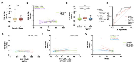
Figure 1.
CSF SHBG levels of FTLD of patients with known underlying neuropathology. (A) Box-dot plots depict the CSF SHBG levels between males and females. (B) Spearman correlation analysis depicts an association between SHBG and age, which was mainly driven by the FTLD group (purple). Correlation coefficient and p value are presented in inserts. Regression line and 95% confident intervals for FTLD (purple) and controls (green) are included. (C) Box-dot plots depict the CSF SHBG levels between controls, FTLD-Tau and FTLD-TDP. Specific symbols depict patient subgroups within each pathological subtype. Exact p values after post-hoc analysis are included. (D) Receiver operator characteristic (ROC) curve depicting the predictive performance of CSF SHBG (red) or CSF p/tTau ratio (black) discriminating FTLD-Tau from FTLD-TDP subtypes. The exact area under the curve (AUC) and p value are presented in inserts. (E–G) Scatter plots depict the correlation between CSF SHBG with (E) CSF YKL40, (F) p/tTau ratio and (G) MMSE score. Correlation coefficient and p value are presented in inserts. Regression line and 95% confident intervals are included. The different diagnostic groups are depicted by colors. CN, controls; FTLD, Frontotemporal lobar degeneration; PSP, progressive supranuclear palsy; FTD-ALS, Frontotemporal dementia—Amyotrophic lateral sclerosis; ns, non-significant.
We next investigated the relationship of CSF SHBG with other FTLD CSF markers and with cognitive function. CSF SHBG did not correlate with YKL40 or the p/Tau ratio (Figure 1E–G). However, we detected an inverse correlation between CSF SHBG and MMSE (r = −0.31, p = 0,01; Figure 1G). Stratification into pathological subtypes suggest that such association was likely driven by the FTLD-Tau group (r = −0.38, p > 0.05, Figure 1G).
4. Discussion
This study reveals that CSF SHBG, as measured by an automated ECLIA, did not differ across pathological groups, though a trend towards increased levels of CSF SHBG was observed in FTLD-Tau cases compared to FTLD-TDP and controls. CSF SHBG levels were inversely associated with cognitive function.
The increasing trend of CSF SHBG levels in FTLD-Tau patients detected in this study may become significant when analyzing larger data sets. However, the CSF SHBG overlap between FTLD-Tau and FTLD-TDP will likely still be too high to be a useful discriminatory marker for FTLD pathological subtypes. This is further supported by the low and variable discriminative performance of SHBG detected in this study. The results of this study counters our previous unbiased proteomic findings in which CSF SHBG was decreased in FTLD-Tau patients compared to both controls and FTLD-TDP [4]. Translation of proteomic findings into high-throughput immunoassays for routine analysis is a major hurdle in the development of optimal biofluid-based biomarkers and highlights the importance of subsequent validation studies [12]. Our previous mass-spectrometry results detected SHBG peptides covering the complete protein sequence, and thus it seems unlikely that the divergent findings might be explained by the detection of different protein sequences or isoforms through the different technologies. Despite the fact that we analyzed pathologically-confirmed FTLD cases in both studies, each FTLD pathological subtype is highly heterogeneous and can cover a wide range of different pathological phenotypes [1]. Importantly, it is still not clear whether sporadic and familial cases, or even the different mutations with converging proteopathies (e.g., GRN or C9orf72), share the same pathophysiological processes, which can ultimately impact the body-fluid biochemical profiles [13,14,15,16,17]. This study included a considerable amount of autopsy-confirmed FTLD-Tau cases and several patients harboring C9orf72 mutation or with FTLD-ALS, which were not analyzed in our previous study. There was, however, no apparent pattern associated with the specific clinic-pathologic and genetic subgroups, suggesting that within group heterogeneity may not explain the contrasting findings. The FTLD-Tau group of the previous study was younger than the patients included in this study. Considering CSF SHBG increases with age, demographic differences may partly explain the discrepancies observed. Different disease stages at time of sample collection could also contribute to the difference in findings. Analyzing the effect of the disease stage on FTLD biomarkers is challenging, especially considering there is not yet an established definition of the prodromal phase of FTLD [18]. It is also worth noting that similar inconsistencies on SHBG levels across studies analyzing blood from Alzheimer’s disease (AD) patients have also been observed [19,20,21,22].
Despite serum SHBG levels being often elevated in females [19,23], we did not observe such changes in CSF, which may indicate that sex related SHBG differences might be more pronounced in blood than in CSF [16]. The lack of association of CSF SHBG with CSF YKL-40 or p/tTau ratio suggests that SHBG is not related to neuroinflammatory processes or Tau proteostasis.
We observed that CSF SHBG was inversely associated, albeit weakly, with cognitive function, which is in line with previous findings showing higher levels of serum SHBG associated with risk of dementia or cognitive decline [19,24]. Interestingly, such association was likely driven by the FTLD-Tau, which showed a correlation coefficient considerably stronger than the FTLD-TDP group (−0.38 vs. −0.02). Previous studies have shown that not only SHBG but also other hormones (e.g., estrogen, free thyroxin or thyroid-stimulating hormone) are associated with MMSE [22,23], suggesting that changes in the whole hypothalamic-pituitary (HP) axes may influence memory function and the risk of dementia or AD [24,25]. This is supported by different neuropathological and imaging studies, highlighting the importance of HP dysfunction (e.g., body weight, circadian rhythm, sleep) in AD pathophysiology [25]. The association of CSF SHBG with MMSE detected in this study suggests that such processes might be also important in FTLD-Tau. Indeed, hypothalamic alterations have already been identified in the behavioral form of FTD [26], in which a more prominent hypothalamic proteopathy was observed in FTLD-Tau than in FTLD-TDP cases [27].
Despite the use of well-characterized samples from cases with confirmed FTLD diagnosis is a strength of this study, we did not have access to samples from patients with an underlying FUS pathology, who account for approximately 5% of all FTLD cases [1]. We acknowledge that the sample size to perform additional subgroup analysis within each pathological subtypes (e.g., sporadic, specific underlying mutation) is still limited. Such a type of sub-analysis requires the inclusion of large set of samples with post-mortem information and deep phenotyping, underpinning the need for high-collaborative multicenter studies and the extension and development of patients’ cohorts within the FTLD field. Yet, the sample size of pathologically-confirmed FTLD cases is still comparable or even larger to the ones used in previous pathological FTLD biomarker studies, including ours [3,4,11]. In addition, we still do not know whether the disease stage at time of collection contributes to the difference in findings on SHBG observed across studies. The incorporation of the newly-developed psychometric measures to characterize the different FTLD symptomatic stages (e.g., FTLD-CDR, MIR) may facilitate validation of FTLD biomarkers [18,28]. Studies on larger datasets are still needed, as they will allow more complex statistical modelling that can account for the heterogeneity of disease in more detail to resolve these issues.
In conclusion, our results indicate that, despite the fact that CSF SHBG tends to be increased FTLD-Tau, there is high overlap between controls and FTLD pathological subtypes. We showed that CSF SHBG is not a suitable biomarker to discriminate FTLD pathological subtypes using the automated immunoassay employed in this study, and thus, the quest for novel body-fluid based biomarkers deciphering FTLD pathological complexity will continue.
Supplementary Materials
The following are available online at https://www.mdpi.com/article/10.3390/biom11101484/s1, Figure S1: SHBG ECLIA can optimally measure SHBG concentration in CSF.
Author Contributions
Conceptualization, C.E.T.; methodology, M.d.C., H.A.M.T., L.V., F.M., A.C.H. and C.E.T.; formal analysis, M.d.C.; resources, Y.A.L.P., A.C.-P., D.J.I., M.G., W.T.H., L.H.M., J.v.S., A.C.H. and C.E.T.; writing—original draft preparation, M.d.C.; writing—review and editing, M.d.C., Y.A.L.P., A.C.-P., D.J.I., M.G., H.A.M.T., W.T.H., L.H.M., J.v.S., L.V., F.M., A.C.H. and C.E.T.; supervision, M.d.C. and C.E.T.; project administration, M.d.C., F.M., A.C.H. and C.E.T.; funding acquisition, C.E.T. All authors have read and agreed to the published version of the manuscript.
Funding
M.d.C. is supported by the attraction talent fellowship of Comunidad de Madrid (2018-T2/BMD-11885) and San Pablo CEU University. D.J.I. is supported by NIH grants P01-AG066597, P30-AG010124. Roche Diagnostics International Ltd. supported the measurements and analysis of CSF SHBG and the APC.
Institutional Review Board Statement
The study was conducted according to the guidelines of the Declaration of Helsinki, and approved by the Institutional Review Board (or Ethics Committee) of VU medical center, the Center for Neurodegenerative Disease Research at the University of Pennsylvania, Erasmus Medical Center and Emory University.
Informed Consent Statement
Informed consent was obtained from all subjects involved in the study.
Conflicts of Interest
The authors declare no conflict of interest.
References
- Irwin, D.J.; Cairns, N.J.; Grossman, M.; McMillan, C.T.; Lee, E.B.; Van Deerlin, V.M.; Lee, V.M.-Y.; Trojanowski, J.Q. Frontotemporal lobar degeneration: Defining phenotypic diversity through personalized medicine. Acta Neuropathol. 2014, 129, 469–491. [Google Scholar] [CrossRef] [PubMed]
- Swift, I.J.; Sogorb-Esteve, A.; Heller, C.; Synofzik, M.; Otto, M.; Graff, C.; Galimberti, D.; Todd, E.; Heslegrave, A.J.; Van Der Ende, E.L.; et al. Fluid biomarkers in frontotemporal dementia: Past, present and future. J. Neurol. Neurosurg. Psychiatry 2020, 92, 204–215. [Google Scholar] [CrossRef] [PubMed]
- Hu, W.T.; Watts, K.; Grossman, M.; Glass, J.; James, J.; Hales, C.; Shelnutt, M.; Van Deerlin, V.; John, Q.; Levey, A.I. Reduced CSF p-Tau 181 to Tau ratio is a biomarker for FTLD-TDP Study Sponsorship: Author Disclosure. Neurology 2013, 81, 1945–1952. [Google Scholar] [CrossRef] [PubMed] [Green Version]
- Teunissen, C.E.; Elias, N.; Koel-Simmelink, M.J.A.; Durieux-Lu, S.; Malekzadeh, A.; Pham, T.V.; Beccari, T.; Meeter, L.H.H.; Dopper, E.G.P.; van Swieten, J.C.; et al. Novel diagnostic cerebrospinal fluid protein biomarkers for pathologic subtypes of frontotemporal dementia identified by proteomics. Alzheimer’s Dement. Diagn. Assess. Dis. Monit. 2016, 2, 86–94. [Google Scholar] [CrossRef] [Green Version]
- Dye, R.V.; Miller, K.J.; Singer, E.J.; Levine, A.J. Hormone replacement therapy and risk for neurodegenerative diseases. Int. J. Alzheimer’s Dis. 2012, 2012, 258454. [Google Scholar] [CrossRef] [Green Version]
- Hansberg-Pastor, V.; González-Arenas, A.; Piña-Medina, A.G.; Camacho-Arroyo, I. Sex hormones regulate cytoskeletal proteins involved in brain plasticity. Front. Psychiatry 2015, 6, 165. [Google Scholar] [CrossRef] [Green Version]
- Höglinger, G.U.; Respondek, G.; Stamelou, M.; Kurz, C.; Josephs, K.A.; Lang, A.E.; Mollenhauer, B.; Müller, U.; Nilsson, C.; Whitwell, J.L.; et al. Clinical diagnosis of progressive supranuclear palsy: The movement disorder society criteria. Mov. Disord. 2017, 32, 853–864. [Google Scholar] [CrossRef]
- Del Campo, M.; Mollenhauer, B.; Bertolotto, A.; Engelborghs, S.; Hampel, H.; Simonsen, A.H.; Kapaki, E.; Kruse, N.; Le Bastard, N.; Lehmann, S.; et al. Recommendations to standardize preanalytical confounding factors in Alzheimer’s and Parkinson’s disease cerebrospinal fluid biomarkers: An update. Biomark. Med. 2012, 6, 419–430. [Google Scholar] [CrossRef]
- Rascovsky, K.; Hodges, J.R.; Knopman, D.; Mendez, M.F.; Kramer, J.H.; Neuhaus, J.; van Swieten, J.C.; Seelaar, H.; Dopper, E.G.P.; Onyike, C.U.; et al. Sensitivity of revised diagnostic criteria for the behavioural variant of frontotemporal dementia. Brain 2011, 134, 2456–2477. [Google Scholar] [CrossRef]
- Litvan, I.; Agid, Y.; Calne, D.; Campbell, G.; Dubois, B.; Duvoisin, R.C.; Goetz, C.G.; Golbe, L.I.; Grafman, J.; Growdon, J.H.; et al. Clinical research criteria for the diagnosis of progressive supranuclear palsy (Steele-Richardson-Olszewski syndrome): Report of the NINDS-SPSP international workshop. Neurology 1996, 47, 1–9. [Google Scholar] [CrossRef]
- del Campo, M.; Galimberti, D.; Elias, N.; Boonkamp, L.; Pijnenburg, Y.A.; van Swieten, J.; Watts, K.; Paciotti, S.; Beccari, T.; Hu, W.; et al. Novel CSF biomarkers to discriminate FTLD and its pathological subtypes. Ann. Clin. Transl. Neurol. 2018, 5, 1163–1175. [Google Scholar] [CrossRef]
- Del Campo, M.; Jongbloed, W.; Twaalfhoven, H.A.; Veerhuis, R.; Blankenstein, M.A.; Teunissen, C.E. Facilitating the Validation of Novel Protein Biomarkers for Dementia: An Optimal Workflow for the Development of Sandwich Immunoassays. Front. Neurol. 2015, 6, 1–10. [Google Scholar] [CrossRef] [Green Version]
- Irwin, D.J.; Lleó, A.; Xie, S.X.; McMillan, C.T.; Wolk, D.A.; Lee, E.B.; Van Deerlin, V.M.; Shaw, L.M.; Trojanowski, J.Q.; Grossman, M. Ante mortem cerebrospinal fluid tau levels correlate with postmortem tau pathology in frontotemporal lobar degeneration. Ann. Neurol. 2017, 82, 247–258. [Google Scholar] [CrossRef]
- Lleó, A.; Irwin, D.J.; Illán-Gala, I.; McMillan, C.T.; Wolk, D.A.; Lee, E.B.; Van Deerlin, V.M.; Shaw, L.M.; Trojanowski, J.Q.; Grossman, M. A 2-Step Cerebrospinal Algorithm for the Selection of Frontotemporal Lobar Degeneration Subtypes. JAMA Neurol. 2018, 75, 738–745. [Google Scholar] [CrossRef] [Green Version]
- Heller, C.; Foiani, M.S.; Moore, K.; Convery, R.; Bocchetta, M.; Neason, M.; Cash, D.M.; Thomas, D.; Greaves, C.V.; Woollacott, I.O.C.; et al. Plasma glial fibrillary acidic protein is raised in progranulin-associated frontotemporal dementia. J. Neurol. Neurosurg. Psychiatry 2020, 91, 263–270. [Google Scholar] [CrossRef] [PubMed] [Green Version]
- Cook, C.N.; Wu, Y.; Odeh, H.M.; Gendron, T.F.; Jansen-West, K.; Del Rosso, G.; Yue, M.; Jiang, P.; Gomes, E.; Tong, J.; et al. C9orf72 poly(GR) aggregation induces TDP-43 proteinopathy. Sci. Transl. Med. 2020, 12. [Google Scholar] [CrossRef]
- Zhang, J.; Velmeshev, D.; Hashimoto, K.; Huang, Y.H.; Hofmann, J.W.; Shi, X.; Chen, J.; Leidal, A.M.; Dishart, J.G.; Cahill, M.K.; et al. Neurotoxic microglia promote TDP-43 proteinopathy in progranulin deficiency. Nature 2020, 588, 459–465. [Google Scholar] [CrossRef] [PubMed]
- Boxer, A.L.; Gold, M.; Feldman, H.; Boeve, B.F.; Dickinson, S.L.-J.; Fillit, H.; Ho, C.; Paul, R.; Pearlman, R.; Sutherland, M.; et al. New directions in clinical trials for frontotemporal lobar degeneration: Methods and outcome measures. Alzheimer’s Dement. 2020, 16, 131–143. [Google Scholar] [CrossRef] [PubMed]
- Xu, W.; Su, B.J.; Shen, X.N.; Bi, Y.L.; Tan, C.C.; Li, J.Q.; Cao, X.P.; Dong, Q.; Tan, L.; Yu, J.T. Plasma sex hormone-binding globulin predicts neurodegeneration and clinical progression in prodromal Alzheimer’s disease. Aging 2020, 12, 14528–14541. [Google Scholar] [CrossRef] [PubMed]
- Hoskin, E.K.; Tang, M.X.; Manly, J.J.; Mayeux, R. Elevated sex-hormone binding globulin in elderly women with Alzheimer’s disease. Neurobiol. Aging 2004, 25, 141–147. [Google Scholar] [CrossRef]
- Xu, J.; Xia, L.-L.; Song, N.; Chen, S.-D.; Wang, G. Testosterone, Estradiol, and Sex Hormone-Binding Globulin in Alzheimer’s Disease: A Meta-Analysis. Curr. Alzheimer Res. 2015, 13, 215–222. [Google Scholar] [CrossRef] [PubMed]
- Johansson, P.; Johansson, J.O.; Labrie, F.; Mattsson, N.; Hansson, O.; Blennow, K.; Zetterberg, H.; Wallin, A.; Ohlsson, C.; Svensson, J. Mild dementia is associated with increased adrenal secretion of cortisol and precursor sex steroids in women. Clin. Endocrinol. 2011, 75, 301–308. [Google Scholar] [CrossRef] [PubMed]
- Lin, K.A.; Rundel, C.; Doraiswamy, P.M. Serum SHBG Levels are not Associated with Longitudinal Cognitive Decline in Mild Cognitive Impairment. J. Alzheimer’s Dis. 2017, 55, 1123–1130. [Google Scholar] [CrossRef] [PubMed]
- Ge, Y.J.; Xu, W.; Tan, C.C.; Tan, L. Blood-based biomarkers in hypothalamic-pituitary axes for the risk of dementia or cognitive decline: A systematic review and meta-analysis. Aging 2020, 12, 20350–20365. [Google Scholar] [CrossRef] [PubMed]
- Ishii, M.; Iadecola, C. Metabolic and Non-Cognitive Manifestations of Alzheimers Disease: The Hypothalamus as Both Culprit and Target of Pathology. Cell Metab. 2015, 22, 761–776. [Google Scholar] [CrossRef] [PubMed] [Green Version]
- Vercruysse, P.; Vieau, D.; Blum, D.; Petersén, Å.; Dupuis, L. Hypothalamic alterations in neurodegenerative diseases and their relation to abnormal energy metabolism. Front. Mol. Neurosci. 2018, 11, 2. [Google Scholar] [CrossRef] [Green Version]
- Piguet, O.; Petersén, Å.; Yin Ka Lam, B.; Gabery, S.; Murphy, K.; Hodges, J.R.; Halliday, G.M. Eating and hypothalamus changes in behavioral-variant frontotemporal dementia. Ann. Neurol. 2011, 69, 312–319. [Google Scholar] [CrossRef]
- Miyagawa, T.; Brushaber, D.; Syrjanen, J.; Kremers, W.; Fields, J.; Forsberg, L.K.; Heuer, H.W.; Knopman, D.; Kornak, J.; Boxer, A.; et al. Use of the CDR® plus NACC FTLD in mild FTLD: Data from the ARTFL/LEFFTDS consortium. Alzheimer’s Dement. 2020, 16, 79–90. [Google Scholar] [CrossRef]
Publisher’s Note: MDPI stays neutral with regard to jurisdictional claims in published maps and institutional affiliations. |
© 2021 by the authors. Licensee MDPI, Basel, Switzerland. This article is an open access article distributed under the terms and conditions of the Creative Commons Attribution (CC BY) license (https://creativecommons.org/licenses/by/4.0/).