


PromptSeg: An End-to-End Universal Medical Image Segmentation Method via Visual Prompts †

Abstract
1. Introduction
- We introduce prompt learning into medical image segmentation, creating a novel end-to-end universal segmentation method.
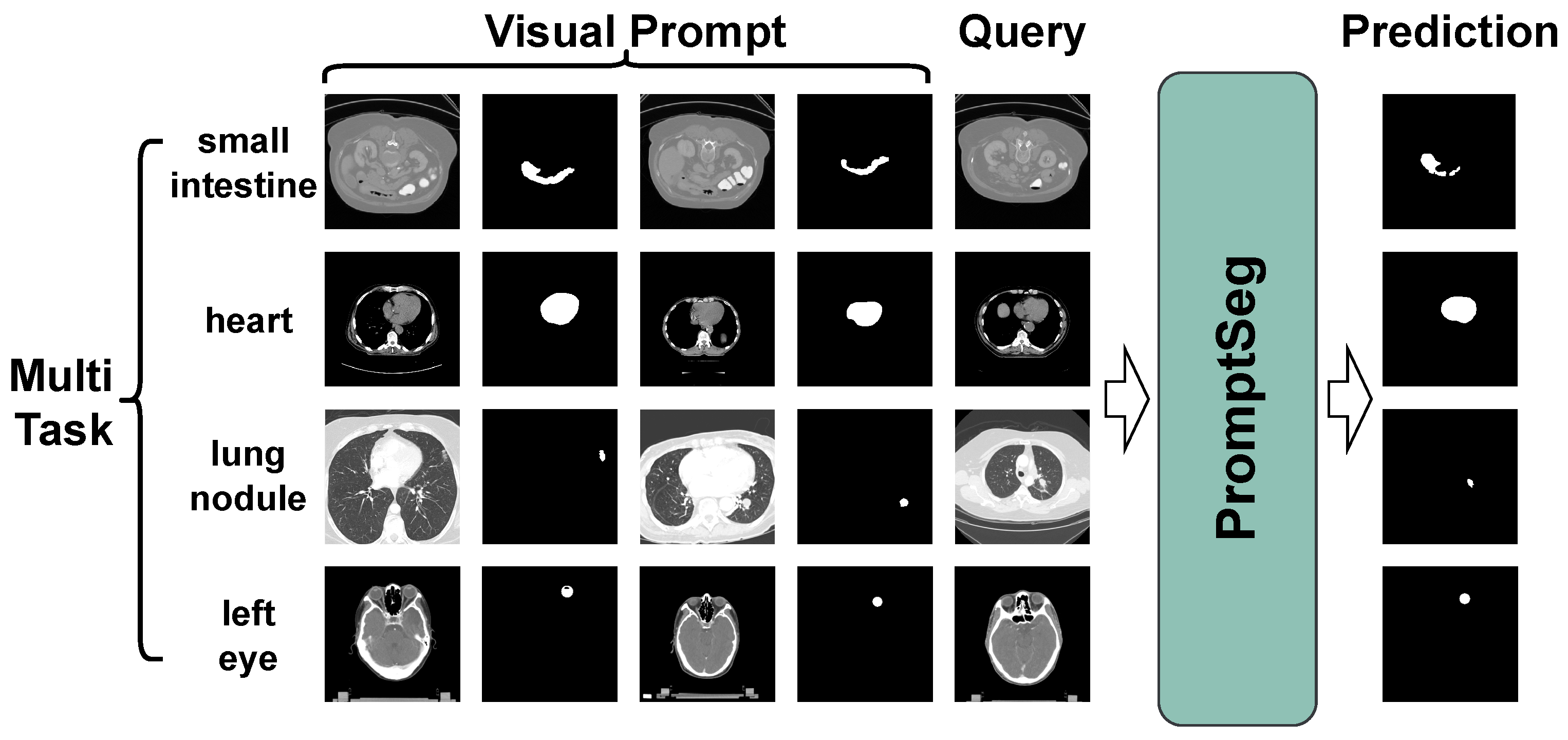
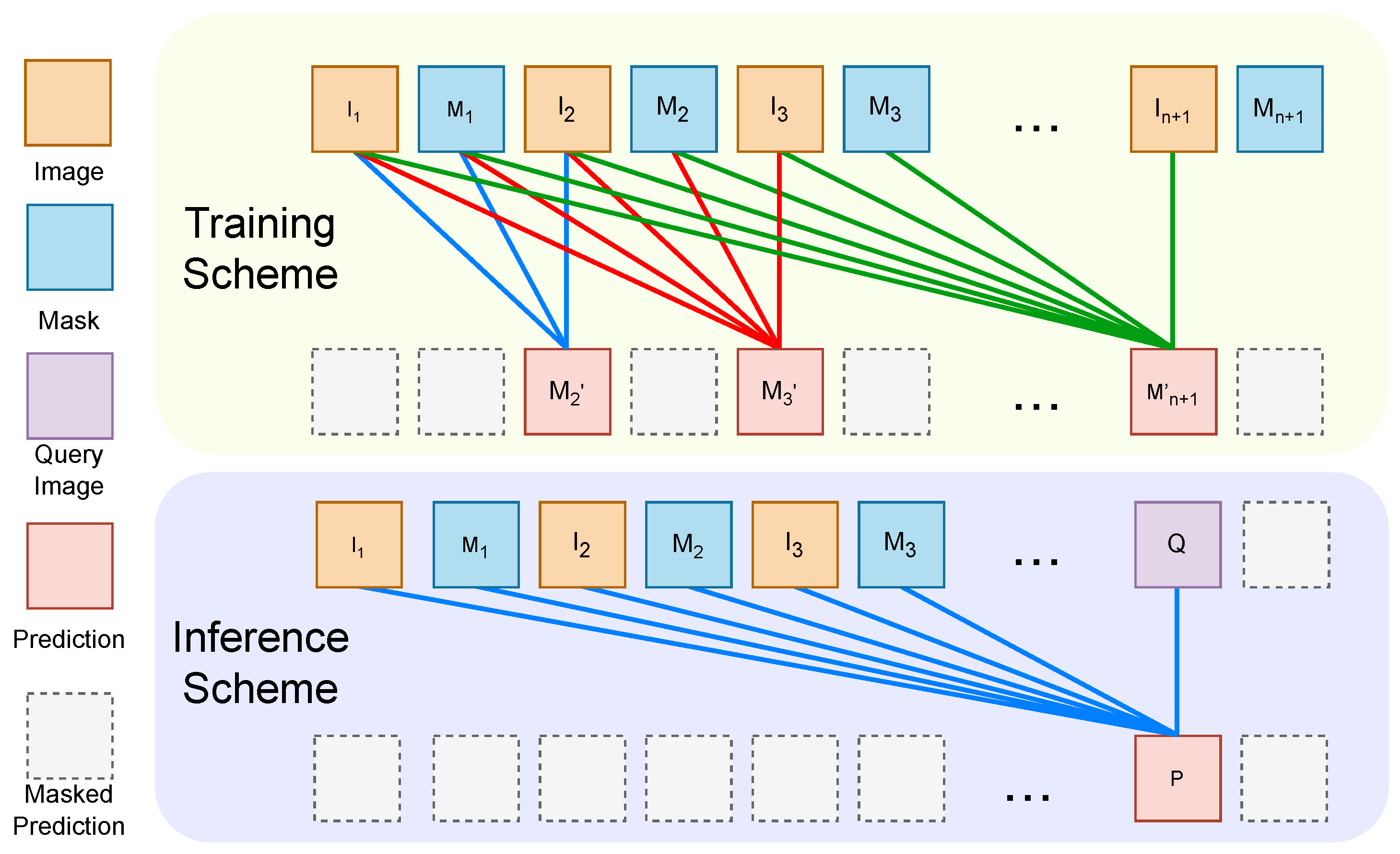
- We propose PromptSeg, a task-agnostic framework that employs an image-level auto-regressive model to segment query images through next-image prediction, using a handful of image–mask pairs with the same segmentation class as visual prompts to indicate the task. Furthermore, we aggregate existing open-source datasets to construct a large-scale, multi-source medical segmentation dataset for training PromptSeg.
- Extensive experiments on multiple open-source datasets demonstrate that PromptSeg outperforms existing few-shot methods in terms of segmentation accuracy and generalization capability on unseen datasets and targets, accompanied by remarkable scalability.
2. Related Work
2.1. Few-Shot Medical Image Segmentation
2.2. Prompt-Based In-Context Learning
2.3. Large Vision Model
3. Methods
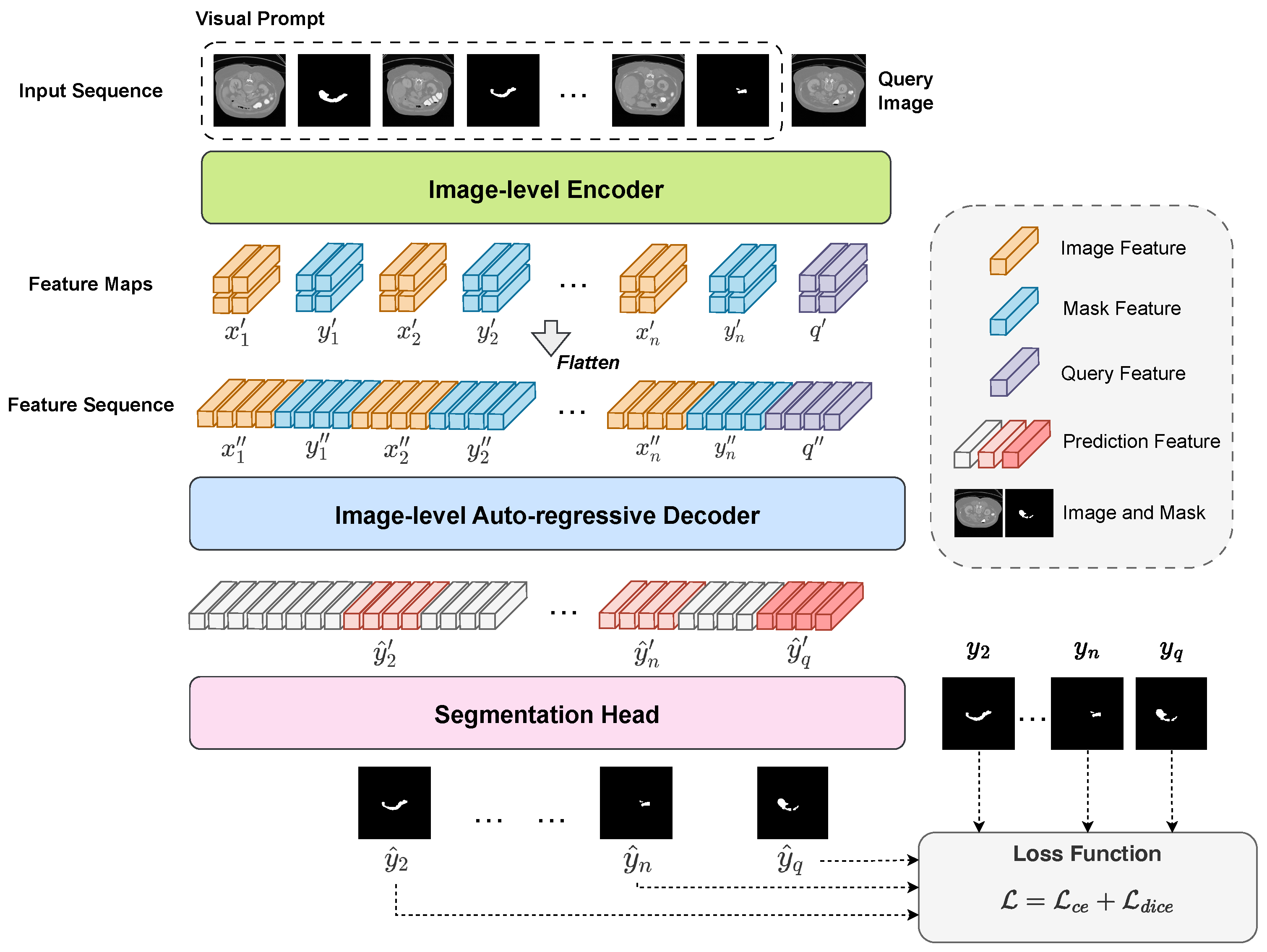
3.1. Overview of PromptSeg
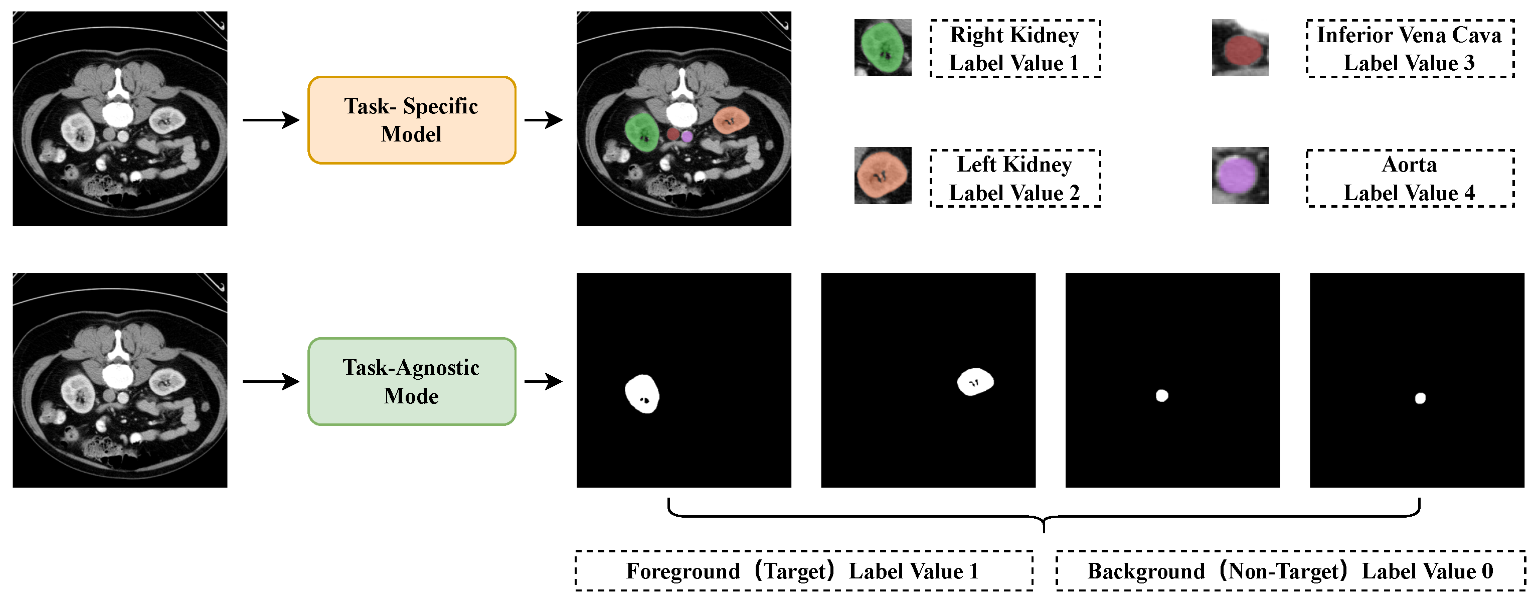
3.2. General Segmentation Task with Visual Prompt
3.3. Window Attention Based Encoder with Information Bottleneck
3.4. Image-Level Auto-Regressive Decoder
4. Experiments
4.1. Dataset and Metrics
4.2. Implementation Details
4.3. Comparative Experiments on CT Modality
4.4. Comparative Experiments on MRI Modality
5. Discussion
5.1. Ablation Study
5.2. Prompt Following Study
5.3. Scaling Study
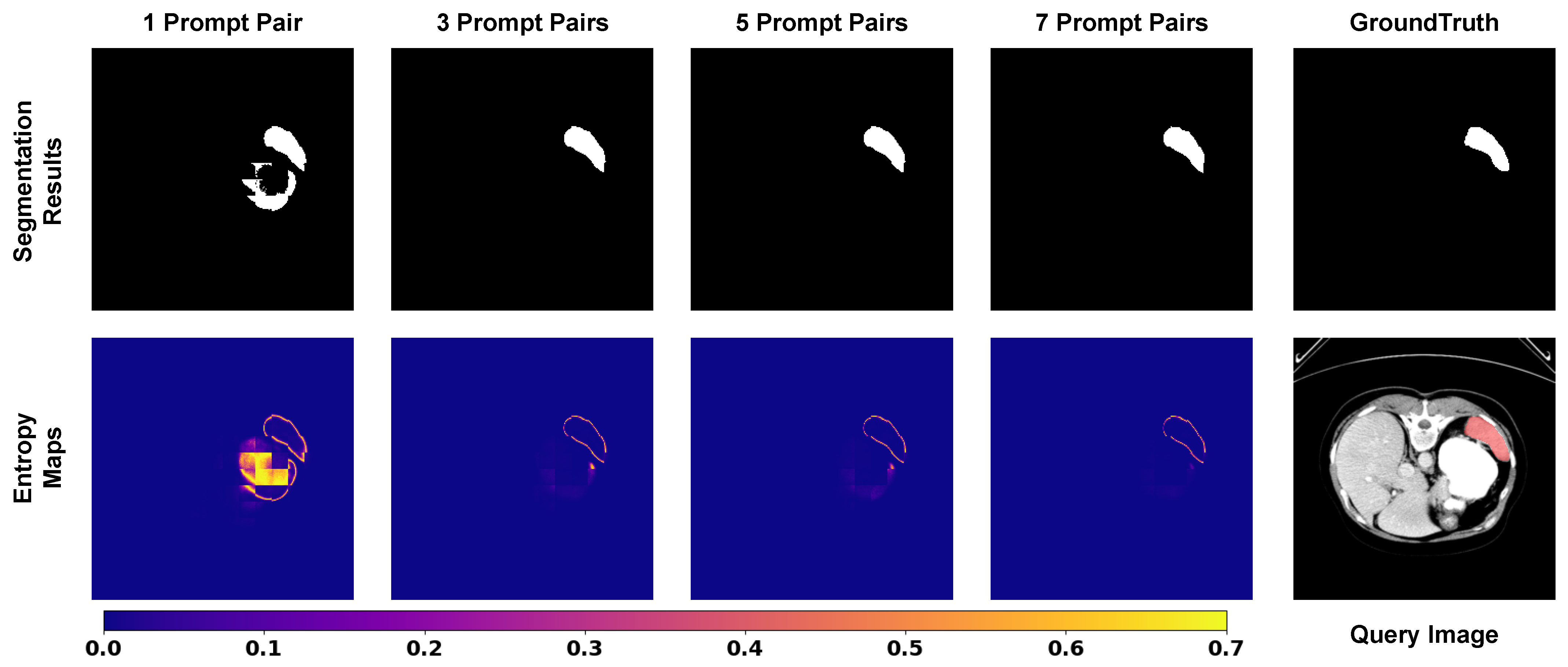
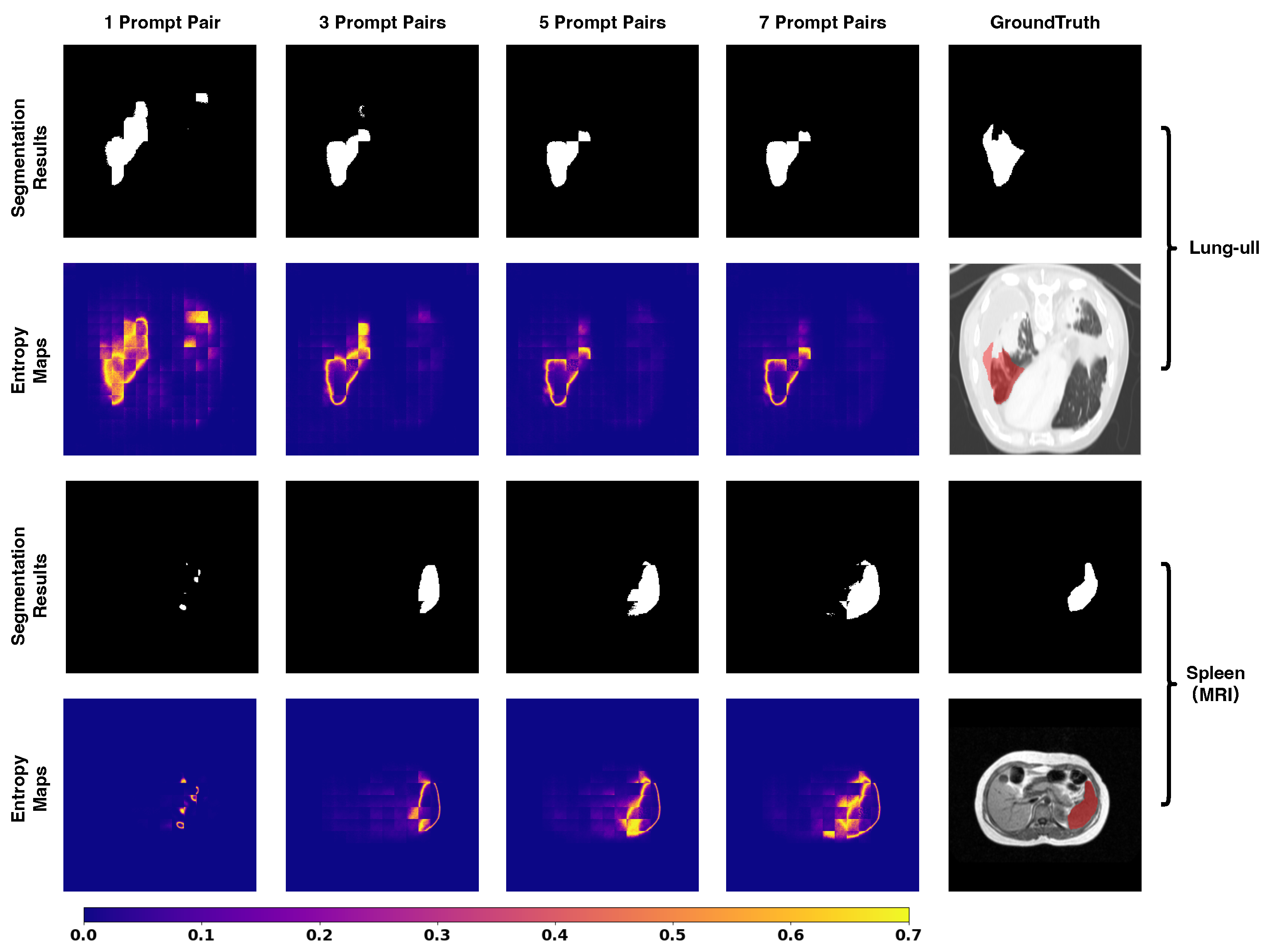
5.4. Entropy Dynamics and Uncertainty Quantification
5.5. Limitations
6. Conclusions
Author Contributions
Funding
Institutional Review Board Statement
Data Availability Statement
Acknowledgments
Conflicts of Interest
References
- Kamnitsas, K.; Ferrante, E.; Parisot, S.; Ledig, C.; Nori, A.V.; Criminisi, A.; Rueckert, D.; Glocker, B. DeepMedic for Brain Tumor Segmentation. In Proceedings of the Brainlesion: Glioma, Multiple Sclerosis, Stroke and Traumatic Brain Injuries: Second International Workshop, BrainLes 2016, with the Challenges on BRATS, ISLES and mTOP 2016, Held in Conjunction with MICCAI 2016, Athens, Greece, 17 October 2016; Revised Selected Papers 2; Springer: Berlin/Heidelberg, Germany, 2016; pp. 138–149. [Google Scholar]
- Isensee, F.; Jaeger, P.F.; Kohl, S.A.; Petersen, J.; Maier-Hein, K.H. nnU-Net: A Self-configuring Method for Deep Learning-based Biomedical Image Segmentation. Nat. Methods 2021, 18, 203–211. [Google Scholar] [CrossRef] [Scilit]
- Ronneberger, O.; Fischer, P.; Brox, T. U-net: Convolutional Networks for Biomedical Image Segmentation. In Proceedings of the Medical Image Computing and Computer-Assisted Intervention–MICCAI 2015: 18th International Conference, Munich, Germany, 5–9 October 2015; Proceedings, Part III 18; Springer: Berlin/Heidelberg, Germany, 2015; pp. 234–241. [Google Scholar]
- Chen, J.; Lu, Y.; Yu, Q.; Luo, X.; Adeli, E.; Wang, Y.; Lu, L.; Yuille, A.L.; Zhou, Y. Transunet: Transformers Make Strong Encoders for Medical Image Segmentation. arXiv 2021, arXiv:2102.04306. [Google Scholar] [CrossRef] [Scilit]
- Huang, G.; Liu, Z.; Van Der Maaten, L.; Weinberger, K.Q. Densely Connected Convolutional Networks. In Proceedings of the IEEE Conference on Computer Vision and Pattern Recognition, Honolulu, HI, USA, 21–26 July 2017; pp. 4700–4708. [Google Scholar]
- Shi, J.; Kan, H.; Ruan, S.; Zhu, Z.; Zhao, M.; Qiao, L.; Wang, Z.; An, H.; Xue, X. H-DenseFormer: An Efficient Hybrid Densely Connected Transformer for Multimodal Tumor Segmentation. In Proceedings of the International Conference on Medical Image Computing and Computer-Assisted Intervention; Springer: Berlin/Heidelberg, Germany, 2023; pp. 692–702. [Google Scholar]
- Sharma, N.; Aggarwal, L.M. Automated Medical Image Segmentation Techniques. J. Med. Phys. 2010, 35, 3. [Google Scholar] [CrossRef] [Scilit] [PubMed]
- Sun, B.; Feng, J.; Saenko, K. Return of Frustratingly Easy Domain Adaptation. In Proceedings of the AAAI Conference on Artificial Intelligence, Phoenix, AZ, USA, 12–17 February 2016; Volume 30. [Google Scholar]
- Zhou, Z.; Sodha, V.; Pang, J.; Gotway, M.B.; Liang, J. Models Genesis. Med. Image Anal. 2021, 67, 101840. [Google Scholar] [CrossRef] [Scilit] [PubMed]
- Alzubaidi, L.; Al-Amidie, M.; Al-Asadi, A.; Humaidi, A.J.; Al-Shamma, O.; Fadhel, M.A.; Zhang, J.; Santamaría, J.; Duan, Y. Novel Transfer Learning Approach for Medical Imaging with Limited Labeled Data. Cancers 2021, 13, 1590. [Google Scholar] [CrossRef] [Scilit] [PubMed]
- Ghafoorian, M.; Mehrtash, A.; Kapur, T.; Karssemeijer, N.; Marchiori, E.; Pesteie, M.; Guttmann, C.R.; de Leeuw, F.E.; Tempany, C.M.; Van Ginneken, B.; et al. Transfer Learning for Domain Adaptation in MRI: Application in Brain Lesion Segmentation. In Proceedings of the Medical Image Computing and Computer Assisted Intervention- MICCAI 2017: 20th International Conference, Quebec City, QC, Canada, 11–13 September 2017; Proceedings, Part III 20; Springer: Berlin/Heidelberg, Germany, 2017; pp. 516–524. [Google Scholar]
- Raghu, M.; Zhang, C.; Kleinberg, J.; Bengio, S. Transfusion: Understanding Transfer Learning for Medical Imaging. Adv. Neural Inf. Process. Syst. 2019, 32. [Google Scholar] [CrossRef] [Scilit]
- Shie, C.K.; Chuang, C.H.; Chou, C.N.; Wu, M.H.; Chang, E.Y. Transfer Representation Learning for Medical Image Analysis. In Proceedings of the 2015 37th Annual International Conference of the IEEE Engineering in Medicine and Biology Society (EMBC); IEEE: New York, NY, USA, 2015; pp. 711–714. [Google Scholar]
- Deng, J.; Dong, W.; Socher, R.; Li, L.J.; Li, K.; Fei-Fei, L. Imagenet: A Large-scale Hierarchical Image Database. In Proceedings of the 2009 IEEE Conference on Computer Vision and Pattern Recognition; IEEE: New York, NY, USA, 2009; pp. 248–255. [Google Scholar]
- Lin, T.Y.; Maire, M.; Belongie, S.; Hays, J.; Perona, P.; Ramanan, D.; Dollár, P.; Zitnick, C.L. Microsoft Coco: Common Objects in Context. In Proceedings of the Computer Vision–ECCV 2014: 13th European Conference, Zurich, Switzerland, 6–12 September 2014; Proceedings, Part V 13; Springer: Berlin/Heidelberg, Germany, 2014; pp. 740–755. [Google Scholar]
- Ouyang, C.; Biffi, C.; Chen, C.; Kart, T.; Qiu, H.; Rueckert, D. Self-supervision with Superpixels: Training Few-shot Medical Image Segmentation Without Annotation. In Proceedings of the Computer Vision–ECCV 2020: 16th European Conference, Glasgow, UK, 23–28 August 2020; Proceedings, Part XXIX 16; Springer: Berlin/Heidelberg, Germany, 2020; pp. 762–780. [Google Scholar]
- Lin, Y.; Chen, Y.; Cheng, K.T.; Chen, H. Few Shot Medical Image Segmentation with Cross Attention Transformer. arXiv 2023, arXiv:2303.13867. [Google Scholar] [CrossRef] [Scilit]
- Zhu, Y.; Wang, S.; Xin, T.; Zhang, H. Few-Shot Medical Image Segmentation via a Region-Enhanced Prototypical Transformer. In Proceedings of the International Conference on Medical Image Computing and Computer-Assisted Intervention; Springer: Berlin/Heidelberg, Germany, 2023; pp. 271–280. [Google Scholar]
- Butoi, V.I.; Ortiz, J.J.G.; Ma, T.; Sabuncu, M.R.; Guttag, J.; Dalca, A.V. Universeg: Universal Medical Image Segmentation. In Proceedings of the IEEE/CVF International Conference on Computer Vision, Paris, France, 2–3 October 2023; pp. 21438–21451. [Google Scholar]
- Brown, T.; Mann, B.; Ryder, N.; Subbiah, M.; Kaplan, J.D.; Dhariwal, P.; Neelakantan, A.; Shyam, P.; Sastry, G.; Askell, A.; et al. Language Models are Few-shot Learners. Adv. Neural Inf. Process. Syst. 2020, 33, 1877–1901. [Google Scholar]
- Radford, A.; Wu, J.; Child, R.; Luan, D.; Amodei, D.; Sutskever, I. Language Models are Unsupervised Multitask Learners. OpenAI Blog 2019, 1, 9. [Google Scholar]
- Bar, A.; Gandelsman, Y.; Darrell, T.; Globerson, A.; Efros, A. Visual Prompting via Image Inpainting. Adv. Neural Inf. Process. Syst. 2022, 35, 25005–25017. [Google Scholar]
- Bai, Y.; Geng, X.; Mangalam, K.; Bar, A.; Yuille, A.; Darrell, T.; Malik, J.; Efros, A.A. Sequential Modeling Enables Scalable Learning for Large Vision Models. arXiv 2023, arXiv:2312.00785. [Google Scholar] [CrossRef] [Scilit]
- Dong, Q.; Li, L.; Dai, D.; Zheng, C.; Wu, Z.; Chang, B.; Sun, X.; Xu, J.; Sui, Z. A Survey on In-context Learning. arXiv 2022, arXiv:2301.00234. [Google Scholar]
- Alayrac, J.B.; Donahue, J.; Luc, P.; Miech, A.; Barr, I.; Hasson, Y.; Lenc, K.; Mensch, A.; Millican, K.; Reynolds, M.; et al. Flamingo: A Visual Language Model for Few-shot Learning. Adv. Neural Inf. Process. Syst. 2022, 35, 23716–23736. [Google Scholar]
- Kirillov, A.; Mintun, E.; Ravi, N.; Mao, H.; Rolland, C.; Gustafson, L.; Xiao, T.; Whitehead, S.; Berg, A.C.; Lo, W.Y.; et al. Segment Anything. arXiv 2023, arXiv:2304.02643. [Google Scholar]
- Wang, X.; Wang, W.; Cao, Y.; Shen, C.; Huang, T. Images Speak in Images: A Generalist Painter for In-context Visual Learning. In Proceedings of the IEEE/CVF Conference on Computer Vision and Pattern Recognition, Vancouver, BC, Canada, 17–24 June 2023; pp. 6830–6839. [Google Scholar]
- Wang, X.; Zhang, X.; Cao, Y.; Wang, W.; Shen, C.; Huang, T. Seggpt: Segmenting Everything in Context. arXiv 2023, arXiv:2304.03284. [Google Scholar] [CrossRef] [Scilit]
- Esser, P.; Rombach, R.; Ommer, B. Taming Transformers for High-resolution Image Synthesis. In Proceedings of the IEEE/CVF Conference on Computer Vision and Pattern Recognition, Nashville, TN, USA, 20–25 June 2021; pp. 12873–12883. [Google Scholar]
- Dosovitskiy, A.; Beyer, L.; Kolesnikov, A.; Weissenborn, D.; Zhai, X.; Unterthiner, T.; Dehghani, M.; Minderer, M.; Heigold, G.; Gelly, S.; et al. An Image is Worth 16x16 Words: Transformers for Image Recognition at Scale. arXiv 2020, arXiv:2010.11929. [Google Scholar]
- Lambert, Z.; Petitjean, C.; Dubray, B.; Kuan, S. Segthor: Segmentation of Thoracic Organs at Risk in CT Images. In Proceedings of the 2020 Tenth International Conference on Image Processing Theory, Tools and Applications (IPTA); IEEE: New York, NY, USA, 2020; pp. 1–6. [Google Scholar]
- Landman, B.; Xu, Z.; Igelsias, J.; Styner, M.; Langerak, T.; Klein, A. Miccai Multi-atlas Labeling Beyond the Cranial Vault–Workshop and Challenge. In Proceedings of the MICCAI Multi-Atlas Labeling Beyond Cranial Vault—Workshop Challenge, Munich, Germany, 5–9 October 2015; Volume 5, p. 12. [Google Scholar]
- Kavur, A.E.; Gezer, N.S.; Barış, M.; Aslan, S.; Conze, P.H.; Groza, V.; Pham, D.D.; Chatterjee, S.; Ernst, P.; Özkan, S.; et al. CHAOS Challenge-Combined (CT-MR) Healthy Abdominal Organ Segmentation. Med. Image Anal. 2021, 69, 101950. [Google Scholar] [CrossRef] [Scilit] [PubMed]
- Wasserthal, J.; Breit, H.C.; Meyer, M.T.; Pradella, M.; Hinck, D.; Sauter, A.W.; Heye, T.; Boll, D.T.; Cyriac, J.; Yang, S.; et al. TotalSegmentator: Robust Segmentation of 104 Anatomic Structures in CT Images. Radiol. Artif. Intell. 2023, 5, e230024. [Google Scholar] [CrossRef] [Scilit] [PubMed]
- D’Antonoli, T.A.; Berger, L.K.; Indrakanti, A.K.; Vishwanathan, N.; Weiß, J.; Jung, M.; Berkarda, Z.; Rau, A.; Reisert, M.; Küstner, T.; et al. TotalSegmentator MRI: Robust Sequence-independent Segmentation of Multiple Anatomic Structures in MRI. arXiv 2024, arXiv:2405.19492. [Google Scholar] [CrossRef] [Scilit]
- Antonelli, M.; Reinke, A.; Bakas, S.; Farahani, K.; Kopp-Schneider, A.; Landman, B.A.; Litjens, G.; Menze, B.; Ronneberger, O.; Summers, R.M.; et al. The Medical Segmentation Decathlon. Nat. Commun. 2022, 13, 4128. [Google Scholar] [CrossRef] [Scilit]
- Sekuboyina, A.; Husseini, M.E.; Bayat, A.; Löffler, M.; Liebl, H.; Li, H.; Tetteh, G.; Kukačka, J.; Payer, C.; Štern, D.; et al. VerSe: A Vertebrae Labelling and Segmentation Benchmark for Multi-detector CT Images. Med. Image Anal. 2021, 73, 102166. [Google Scholar] [CrossRef] [Scilit]
- Ji, Y.; Bai, H.; Ge, C.; Yang, J.; Zhu, Y.; Zhang, R.; Li, Z.; Zhang, L.; Ma, W.; Wan, X.; et al. Amos: A Large-scale Abdominal Multi-organ Benchmark for Versatile Medical Image Segmentation. Adv. Neural Inf. Process. Syst. 2022, 35, 36722–36732. [Google Scholar]
- Armato, S.G., III; McLennan, G.; Bidaut, L.; McNitt-Gray, M.F.; Meyer, C.R.; Reeves, A.P.; Zhao, B.; Aberle, D.R.; Henschke, C.I.; Hoffman, E.A.; et al. The Lung Image Database Consortium (LIDC) and Image Database Resource Initiative (IDRI): A Completed Reference Database of Lung Nodules on CT Scans. Med. Phys. 2011, 38, 915–931. [Google Scholar] [CrossRef] [Scilit]
- Shi, J. StructSeg2019 GTV Segmentation. Available online: https://ieee-dataport.org/documents/structseg2019-gtv-segmentation (accessed on 24 December 2023).
- An, P.; Xu, S.; Harmon, S.A.; Turkbey, E.B.; Sanford, T.H.; Amalou, A.; Kassin, M.; Varble, N.; Blain, M.; Anderson, V.; et al. CT Images in COVID-19. Available online: https://www.cancerimagingarchive.net/collection/ct-images-in-covid-19/ (accessed on 24 December 2023).
- Zhao, M.; Zhu, Z.; Shi, J.; Wang, Z.; Chen, J.; An, H.; Yan, B. PromptSeg: Learning to Segment Medical Image via Visual Prompts. In Proceedings of the ICASSP 2025—2025 IEEE International Conference on Acoustics, Speech and Signal Processing (ICASSP), Hyderabad, India, 6–11 April 2025; pp. 1–5. [Google Scholar] [CrossRef] [Scilit]




| Dataset | Modality | Samples | Classes | Train | Valid | Test |
|---|---|---|---|---|---|---|
| Public Datasets | ||||||
| TotalSegmentatorV2 [34] | CT | 1228 | 113 | 982 | 246 | ∖ |
| TotalSegmentatorV2 [34] | CT | 1057 | 4 | ∖ | ∖ | 1057 |
| TotalSegmentatorMRI (Abdominal) [35] | MRI | 51 | 13 | 41 | 10 | ∖ |
| CHAOS [33] | MRI | 20 | 4 | ∖ | ∖ | 20 |
| MSD Heart [36] | CT | 20 | 1 | 16 | 4 | ∖ |
| MSD Lung [36] | CT | 63 | 1 | 50 | 13 | ∖ |
| MSD Pancreas [36] | CT | 281 | 2 | 224 | 57 | ∖ |
| MSD HepaticVessel [36] | MRI | 303 | 2 | 242 | 61 | ∖ |
| MSD Spleen [36] | CT | 41 | 1 | 32 | 9 | ∖ |
| MSD Colon [36] | CT | 126 | 1 | 100 | 26 | ∖ |
| VerSe [37] | CT | 374 | 28 | 300 | 74 | ∖ |
| AMOS-CT [38] | CT | 300 | 15 | 240 | 60 | ∖ |
| AMOS-MRI [38] | MRI | 60 | 13 | 48 | 12 | ∖ |
| LIDC [39] | CT | 726 | 1 | 580 | 146 | ∖ |
| StructSeg Thor [40] | CT | 40 | 6 | 32 | 8 | ∖ |
| StructSeg HaN [40] | CT | 40 | 22 | 32 | 8 | ∖ |
| StructSeg ThorGTV [40] | CT | 40 | 1 | 32 | 8 | ∖ |
| StructSeg HaNGTV [40] | CT∖MRI | 40 | 1 | 32 | 8 | ∖ |
| Covid-Seg [41] | CT | 199 | 1 | 159 | 40 | ∖ |
| SegThor [31] | CT | 40 | 4 | ∖ | ∖ | 40 |
| BTCV [32] | CT | 30 | 13 | ∖ | ∖ | 30 |
| Private Datasets | ||||||
| Abdominal OAR | CT | 39 | 8 | ∖ | ∖ | 39 |
| Stomach OAR | CT | 36 | 9 | 28 | 8 | ∖ |
| Lung OAR | CT | 90 | 4 | 72 | 18 | ∖ |
| Lung Tumor | CT | 80 | 1 | 64 | 16 | ∖ |
| Augmentation | Description |
|---|---|
| Flip Intensities | for all images (query and prompts) |
| Flip Labels | for all masks |
| Horizontal Flip | Flip all images and masks horizontally |
| Vertical Flip | Flip all images and masks vertically |
| Sobel-Edge Label | Apply a Sobel filter to each mask |
| Affine Shift | Affine shift for all images and masks |
| Brightness Contrast Change | Change brightness and contrast for all images |
| Elastic Warp | Elastic deformable warp for all images and masks |
| Gaussian Blur | Add Gaussian blur for all images |
| Gaussian Noise | Add Gaussian noise for all images |
| Sharpness Change | Change sharpness for all images |
| Method | Unseen Datasets | ||
|---|---|---|---|
| SegThor | BTCV | Abdominal OAR | |
| Few-Shot Model | |||
| ALPNet [16] | 36.39 ± 0.08 | 44.49 ± 0.10 | 74.09 ± 0.05 |
| CATNet [17] | 22.59 ± 0.22 | 24.88 ± 0.17 | 43.60 ± 0.07 |
| RPT [18] | 32.84 ± 0.11 | 50.05 ± 0.32 | 75.59 ± 0.13 |
| Universal Model | |||
| UniverSeg [19] | 55.31 ± 0.53 | 54.26 ± 0.37 | 74.97 ± 0.87 |
| PromptSeg | 55.72 ± 0.28 | 63.57 ± 0.25 | 84.70 ± 0.41 |
| Method | Unseen Targets | |||
|---|---|---|---|---|
| Lung-ulr | Lung-llr | Lung-ull | Lung-lll | |
| Few-Shot Model | ||||
| ALPNet [16] | 70.30 ± 1.25 | 63.36 ± 0.11 | 64.19 ± 0.52 | 57.43 ± 0.50 |
| CATNet [17] | 26.33 ± 1.02 | 20.16 ± 0.66 | 16.85 ± 0.75 | 15.55 ± 0.70 |
| RPT [18] | 63.06 ± 0.54 | 55.98 ± 0.19 | 57.95 ± 0.47 | 52.10 ± 0.35 |
| Universal Model | ||||
| UniverSeg [19] | 63.31 ± 0.20 | 63.21 ± 0.35 | 60.67 ± 0.22 | 58.72 ± 0.29 |
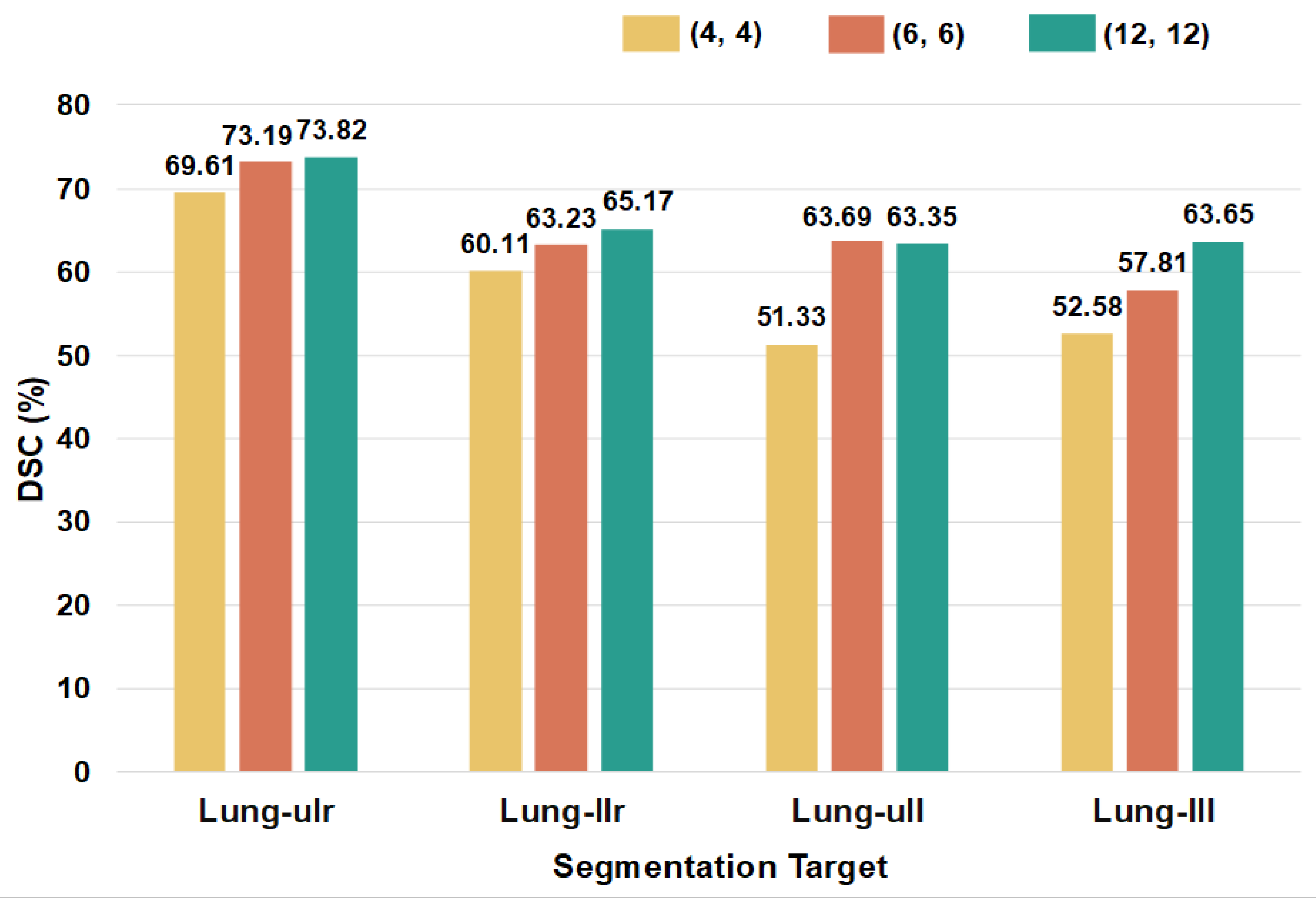
| PromptSeg | 73.82 ± 0.45 | 65.17 ± 0.24 | 63.35 ± 0.25 | 63.65 ± 0.09 |
| Method | Segmentation Targets (CHAOS) | Average | |||
|---|---|---|---|---|---|
| Liver | Right Kidney | Spleen | Left Kidney | ||
| Few-Shot Model | |||||
| ALPNet [16] | 78.98 ± 0.53 | 56.41 ± 0.36 | 59.74 ± 0.92 | 59.10 ± 0.65 | 63.56 ± 0.33 |
| CATNet [17] | 50.43 ± 0.09 | 13.13 ± 0.32 | 15.40 ± 0.13 | 12.63 ± 0.12 | 22.90 ± 0.07 |
| RPT [18] | 59.83 ± 0.17 | 62.81 ± 1.54 | 21.53 ± 0.43 | 54.61 ± 5.00 | 49.70 ± 1.38 |
| Universal Model | |||||
| UniverSeg [19] | 73.69 ± 0.82 | 68.04 ± 1.20 | 62.62 ± 1.17 | 70.53 ± 1.23 | 68.73 ± 0.35 |
| PromptSeg | 83.94 ± 0.96 | 73.48 ± 1.63 | 59.67 ± 1.65 | 63.86 ± 2.39 | 70.24 ± 0.81 |
| Model Variant | Abdominal OAR | SegThor [31] | BTCV [32] | TotalSeg- mentatorV2 [34] |
|---|---|---|---|---|
| w/o Bottleneck Adapter | 72.33 | 28.66 | 48.63 | 57.51 |
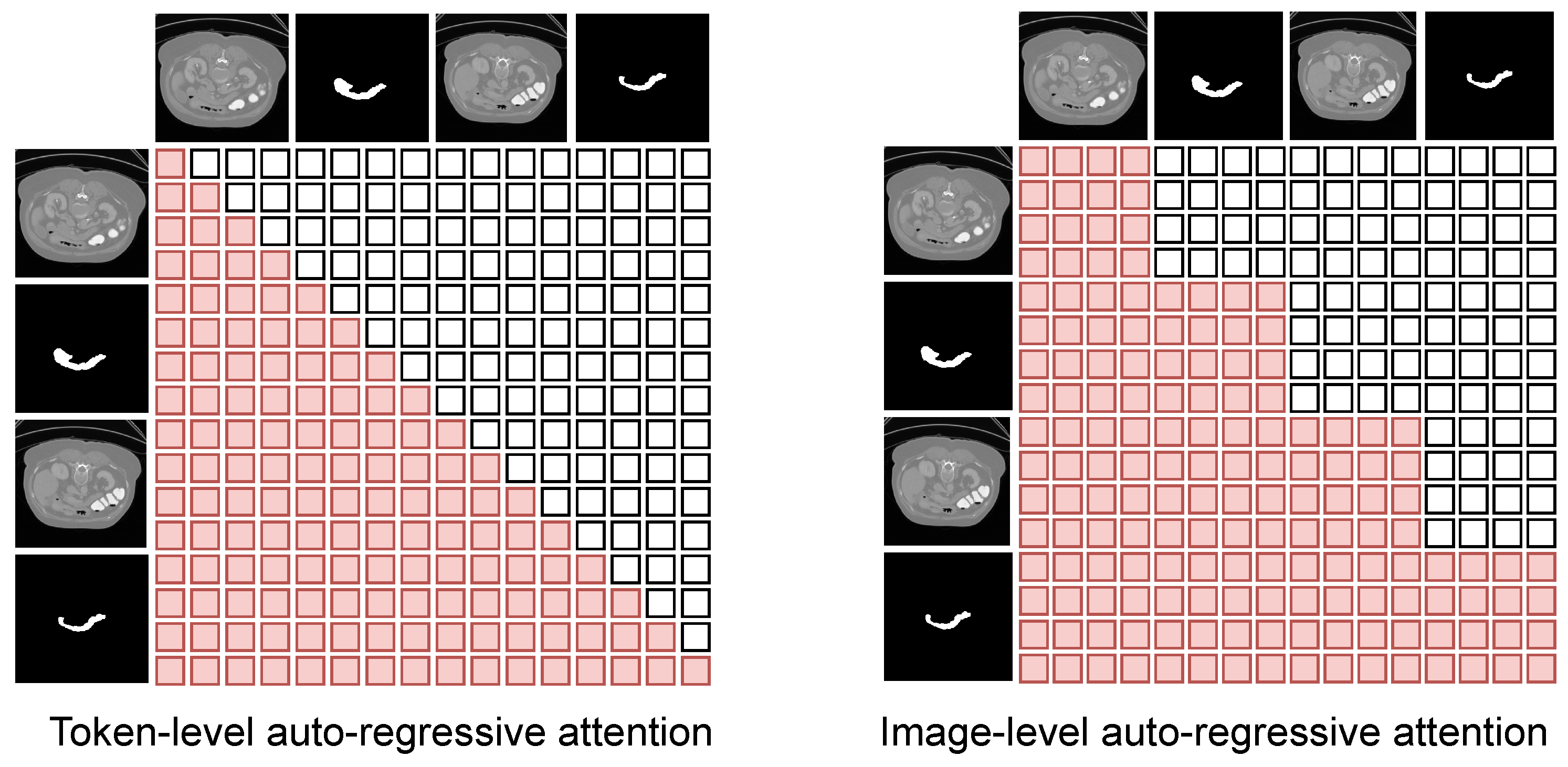
| w/ Token-level Mask | 82.38 | 54.99 | 63.27 | 61.37 |
| PromptSeg | 84.70 | 55.72 | 63.57 | 66.50 |
| Pairs | SegThor [31] | BTCV [32] | Abdominal OAR | TotalSeg- mentatorV2 [34] |
|---|---|---|---|---|
| 1 | 51.25 | 58.05 | 80.29 | 13.50 |
| 3 | 53.65 | 63.26 | 83.02 | 46.40 |
| 7 | 55.72 | 63.57 | 84.70 | 66.50 |
| 11 | 61.29 | 66.54 | 86.58 | 65.41 |
| Pairs | SegThor [31] | BTCV [32] | Abdominal OAR | TotalSeg- mentatorV2 [34] |
|---|---|---|---|---|
| 3 | 53.40 | 61.72 | 83.34 | 59.70 |
| 4 | 54.29 | 62.55 | 84.11 | 62.60 |
| 5 | 54.86 | 63.31 | 84.37 | 64.37 |
| 6 | 55.36 | 63.47 | 84.64 | 65.60 |
| 7 | 55.72 | 63.57 | 84.70 | 66.50 |
| Number of Prompts | 1 | 3 | 5 | 7 |
|---|---|---|---|---|
| Average Entropy (↓) | 0.008435 | 0.005974 | 0.005682 | 0.005652 |
| Prompts (n) | Params (M) | FLOPs (G) | Memory (MB) | Inference (ms/Slice) |
|---|---|---|---|---|
| 1 | 358.90 | 218.83 | 2219.00 | 13.83 |
| 3 | 358.90 | 714.48 | 2428.14 | 18.22 |
| 7 | 358.90 | 1937.69 | 3452.65 | 42.90 |
Disclaimer/Publisher’s Note: The statements, opinions and data contained in all publications are solely those of the individual author(s) and contributor(s) and not of MDPI and/or the editor(s). MDPI and/or the editor(s) disclaim responsibility for any injury to people or property resulting from any ideas, methods, instructions or products referred to in the content. |
© 2026 by the authors. Licensee MDPI, Basel, Switzerland. This article is an open access article distributed under the terms and conditions of the Creative Commons Attribution (CC BY) license.
Share and Cite
Zhao, M.; Wang, B.; Shi, J.; An, H. PromptSeg: An End-to-End Universal Medical Image Segmentation Method via Visual Prompts. Entropy 2026, 28, 342. https://doi.org/10.3390/e28030342
Zhao M, Wang B, Shi J, An H. PromptSeg: An End-to-End Universal Medical Image Segmentation Method via Visual Prompts. Entropy. 2026; 28(3):342. https://doi.org/10.3390/e28030342
Chicago/Turabian StyleZhao, Minfan, Bingxun Wang, Jun Shi, and Hong An. 2026. "PromptSeg: An End-to-End Universal Medical Image Segmentation Method via Visual Prompts" Entropy 28, no. 3: 342. https://doi.org/10.3390/e28030342
APA StyleZhao, M., Wang, B., Shi, J., & An, H. (2026). PromptSeg: An End-to-End Universal Medical Image Segmentation Method via Visual Prompts. Entropy, 28(3), 342. https://doi.org/10.3390/e28030342