Review

Glyphosate’s Suppression of Cytochrome P450 Enzymes and Amino Acid Biosynthesis by the Gut Microbiome: Pathways to Modern Diseases †

1
Independent Scientist and Consultant, Deerfield, NH 03037, USA
2
Computer Science and Artificial Intelligence Laboratory, MIT, Cambridge, MA 02139, USA
*
Author to whom correspondence should be addressed.
Note added by the Publisher: The editors of the journal have been alerted to concerns over potential bias in opinions and bias in the choice of citation sources used in this article. We note that the authors stand by the content as published. Since the nature of the claims against the paper concern speculation and opinion, and not fraud or academic misconduct, the editors would like to issue an Expression of Concern to make readers aware that the approach to collating literature citations for this article was likely not systematic and may not reflect the spectrum of opinions on the issues covered by the article. Please refer to our policy regarding possibly controversial articles.
Entropy 2013, 15(4), 1416-1463; https://doi.org/10.3390/e15041416
Submission received: 15 January 2013 / Revised: 10 April 2013 / Accepted: 10 April 2013 / Published: 18 April 2013
(This article belongs to the Special Issue Biosemiotic Entropy: Disorder, Disease, and Mortality)

Abstract

:
Glyphosate, the active ingredient in Roundup®, is the most popular herbicide used worldwide. The industry asserts it is minimally toxic to humans, but here we argue otherwise. Residues are found in the main foods of the Western diet, comprised primarily of sugar, corn, soy and wheat. Glyphosate's inhibition of cytochrome P450 (CYP) enzymes is an overlooked component of its toxicity to mammals. CYP enzymes play crucial roles in biology, one of which is to detoxify xenobiotics. Thus, glyphosate enhances the damaging effects of other food borne chemical residues and environmental toxins. Negative impact on the body is insidious and manifests slowly over time as inflammation damages cellular systems throughout the body. Here, we show how interference with CYP enzymes acts synergistically with disruption of the biosynthesis of aromatic amino acids by gut bacteria, as well as impairment in serum sulfate transport. Consequences are most of the diseases and conditions associated with a Western diet, which include gastrointestinal disorders, obesity, diabetes, heart disease, depression, autism, infertility, cancer and Alzheimer’s disease. We explain the documented effects of glyphosate and its ability to induce disease, and we show that glyphosate is the “textbook example” of exogenous semiotic entropy: the disruption of homeostasis by environmental toxins.

1. Introduction

The foodstuffs of the Western diet, primarily grown by industrial agriculture, are increasingly being produced using a two-part system of engineered plant seeds and toxic chemical application. Novel bacterial genes are incorporated through genetic engineering, and toxic chemical residues are readily taken up by the engineered plants. Research indicates that the new bacterial RNA and DNA present in genetically engineered plants, providing chemical herbicide resistance and other traits, have not yet fully understood biological effects. This paper however, will only examine the effects of the chemical glyphosate, the most popular herbicide on the planet.
Glyphosate (N-phosphonomethylglycine), the active ingredient in the herbicide Roundup®, is the main herbicide in use today in the United States, and increasingly throughout the World, in agriculture and in lawn maintenance, especially now that the patent has expired. 80% of genetically modified crops, particularly corn, soy, canola, cotton, sugar beets and most recently alfalfa, are specifically targeted towards the introduction of genes resistant to glyphosate, the so-called “Roundup Ready® feature” In humans, only small amounts (~2%) of ingested glyphosate are metabolized to aminomethylphosphonic acid (AMPA), and the rest enters the blood stream and is eventually eliminated through the urine [1]. Studies have shown sharp increases in glyphosate contamination in streams in the Midwestern United States following the mid 1990s, pointing to its increasing role as the herbicide of choice in agriculture [2]. A now common practice of crop desiccation through herbicide administration shortly before the harvest assures an increased glyphosate presence in food sources as well [3,4,5]. The industry asserts that glyphosate is nearly nontoxic to mammals [6,7], and therefore it is not a problem if glyphosate is ingested in food sources. Acutely, it is claimed to be less toxic than aspirin [1,6]. As a consequence, measurement of its presence in food is practically nonexistent. A vocal minority of experts believes that glyphosate may instead be much more toxic than is claimed, although the effects are only apparent after a considerable time lapse. Thus, while short-term studies in rodents have shown no apparent toxicity [8], studies involving life-long exposure in rodents have demonstrated liver and kidney dysfunction and a greatly increased risk of cancer, with shortened lifespan [9].
Glyphosate’s claimed mechanism of action in plants is the disruption of the shikimate pathway, which is involved with the synthesis of the essential aromatic amino acids, phenylalanine, tyrosine, and tryptophan [10]. The currently accepted dogma is that glyphosate is not harmful to humans or to any mammals because the shikimate pathway is absent in all animals. However, this pathway is present in gut bacteria, which play an important and heretofore largely overlooked role in human physiology [11,12,13,14] through an integrated biosemiotic relationship with the human host. In addition to aiding digestion, the gut microbiota synthesize vitamins, detoxify xenobiotics, and participitate in immune system homeostasis and gastrointestinal tract permeability [14]. Furthermore, dietary factors modulate the microbial composition of the gut [15]. The incidence of inflammatory bowel diseases such as juvenile onset Crohn’s disease has increased substantially in the last decade in Western Europe [16] and the United States [17]. It is reasonable to suspect that glyphosate’s impact on gut bacteria may be contributing to these diseases and conditions.
However, the fact that female rats are highly susceptible to mammary tumors following chronic exposure to glyphosate [9] suggests that there may be something else going on. Our systematic search of the literature has led us to the realization that many of the health problems that appear to be associated with a Western diet could be explained by biological disruptions that have already been attributed to glyphosate. These include digestive issues, obesity, autism, Alzheimer’s disease, depression, Parkinson’s disease, liver diseases, and cancer, among others. While many other environmental toxins obviously also contribute to these diseases and conditions, we believe that glyphosate may be the most significant environmental toxin, mainly because it is pervasive and it is often handled carelessly due to its perceived nontoxicity. In this paper, we will develop the argument that the recent alarming increase in all of these health issues can be traced back to a combination of gut dysbiosis, impaired sulfate transport, and suppression of the activity of the various members of the cytochrome P450 (CYP) family of enzymes. We have found clear evidence that glyphosate disrupts gut bacteria and suppresses the CYP enzyme class. The connection to sulfate transport is more indirect, but justifiable from basic principles of biophysics.
In the remainder of this paper, we will first provide evidence from the literature that explains some of the ways in which glyphosate adversely affects plants, microbes, amphibians and mammals. Section 3 will discuss the role that gut dysbiosis, arguably resulting from glyphosate exposure, plays in inflammatory bowel disease and its relationship to autism. Section 4 argues that the excess synthesis of phenolic compounds associated with glyphosate exposure represents a strategy to compensate for impairments in the transport of free sulfate. Section 5 will provide evidence that glyphosate inhibits CYP enzymes. Section 6 explains how obesity can arise from depletion of serum tryptophan due to its sequestering by macrophages responding to inflammation. Section 7 shows how extreme tryptophan depletion can lead to impaired nutrient absorption and anorexia nervosa. Section 8 provides a brief review of all the roles played by CYP enzymes in metabolism. Section 9 discusses a likely consequence to glyphosate’s disruption of the CYP-analog enzyme, endothelial nitric oxide synthase (eNOS). Section 10 shows how glyphosate’s effects could plausibly lead to brain-related disorders such as autism, dementia, depression, and Parkinson’s disease. Section 11 mentions several other health factors that can potentially be linked to glyphosate, including reproductive issues and cancer. Section 12 discusses the available evidence that glyphosate is contaminating our food supplies, especially in recent years. Following a discussion section, we sum up our findings with a brief conclusion.

2. Glyphosate’s Pathological Effects: Controlled Studies

It is well established that glyphosate, a member of the general class of organophosphates, inhibits the enzyme 5-enolpyruvylshikimic acid-3-phosphate synthase (EPSP synthase), the rate-limiting step in the synthesis of aromatic amino acids in the shikimate pathway in plants [18]. This pathway, while not present in mammals, is present in algae, Archaea, bacteria, fungi, and prokaryotes, and unicellular eukaryotic organisms [19]. Indeed, corn and soy crops have both been shown to accumulate excess shikimate in response to glyphosate exposure [20]. However, a study comparing glyphosate-tolerant and glyphosate-sensitive carrot cell lines identified several pathologies beyond the inhibition of aromatic amino acids following glyphosate exposure [21]. It was determined that, in addition to abnormally low levels of tryptophan, phenylalanine and tyrosine, the glyphosate-sensitive cells also had 50 to 65% reduced levels of serine, glycine and methionine. The reduction in methionine can have many adverse consequences, as methionine is an essential sulfur-containing amino acid that has to be supplied from the diet. In addition, there was evidence of excess ammonia in the glyphosate-sensitive but not the glyphosate-adapted cells. Both cell types readily absorbed glyphosate from the medium, with a rapid linear uptake observed during the first eight hours following exposure. This demonstrates that glyphosate would be present in food sources derived from glyphosate-exposed plants.
The excess ammonia observed in glyphosate-treated plants could be due to increased activity of phenylalanine ammonia lyase (PAL), an enzyme found in plants, animals, and microbes, that catalyzes the reaction that converts phenylalanine to trans-cinnamate, releasing ammonia [22]. In studies on transgenic tobacco, it was demonstrated that a decrease in the aromatic amino acid pool sizes (a direct consequence of glyphosate exposure) results in an enhancement of metabolic flux through the shikimate pathway, which leads to a rise in PAL activity as well as a doubling of the levels of chlorogenic acid, a polyphenolic compound related to cinnamate [23]. It has been proposed that glyphosate achieves part if not all of its growth-retardation effects on plants through induction of PAL activity [24]. The growth disruption could be due either to toxicity of the derived phenolic compounds [25] or to direct toxicity of the ammonia. A study of olive trees showed that there is a direct relationship between the total phenol concentration and PAL activity, suggesting that PAL is a major producer of phenolic compounds [26]. Glyphosate has been shown to increase PAL activity in both soybean seedlings [27] and in corn [28].
Under stress-inducing environments, the secondary metabolites derived from certain protein synthesis pathways become disproportionately important, and enzyme regulation induces dramatic shifts in the production of the amino acids versus the secondary metabolites. A study comparing glyphosate exposure with aromatic protein deprivation in plants found several effects in common, but there was a striking anomaly for glyphosate in that it caused a 20-fold increase in the synthesis of the rate-limiting enzyme for a pathway leading to flavonoid synthesis, as a side branch of the tryptophan synthesis pathway [29]. More generally, there is substantial evidence that glyphosate induces the synthesis of monophenolic compounds as well as the polyphenolic flavonoids, in both plants [30] and microbes [31], with concurrent depletion of aromatic amino acid supplies. When carrots are exposed to high doses of glyphosate, they produce significant amounts of various phenolic compounds as well as shikimic acid [32]. The significance of this will become apparent later on in Section 4 on sulfate transport. Elevated amounts of shikimate-derived benzoic acids such as protocatechuate and gallate are also found in plants exposed to glyphosate [29]. Strains of nitrogen-fixing bacteria in the soil produce hydroxybenzoic acids in the presence of glyphosate [31]. This digression towards the competing pathways to produce phenolic and benzoic acid compounds may well explain the suppression of aromatic amino acid synthesis by glyphosate.
Even Roundup Ready® crops typically experience slowed growth following glyphosate ap- plications, and this has been attributed to glyphosate’s role as a chelator of micronutrients. In early work, glyphosate was shown to interfere with the uptake of the divalent cations, calcium and magnesium, through soybean roots [33]. Glyphosate severely reduced calcium content in the mitochondria of both root and leaf cells. Since magnesium was also affected, but potassium was not, the authors suggested that this property might hold for all divalent cations. More recent greenhouse experiments demonstrated that glyphosate application to the root system decreased the levels of calcium, magnesium, iron and manganese in the seeds of the plants [34]. It was proposed that glyphosate binds to and immobilizes all of these divalent micronutrients, impairing their uptake by the plant. These glyphosate-induced deficiencies would carry over to the food supply, leading to deficiencies in these nutrients in humans who consume foods derived from glyphosate-exposed crops.
Evidence of disruption of gut bacteria by glyphosate is available for both cattle and poultry. It has recently been proposed that glyphosate may be a significant factor in the observed increased risk to Clostridium botulinum infection in cattle in Germany over the past ten to fifteen years [35]. Glyphosate's demonstrated toxicity to Enterococcus spp. leads to an imbalance in the gut favoring overgrowth of the toxic Clostridium species. Glyphosate has been shown to have remarkable adverse effects on the gut biota in poultry [36], by reducing the number of beneficial bacteria and increasing the number of pathogenic bacteria in the gut. Highly pathogenic strains of Salmonella and Clostridium were found to be highly resistant to glyphosate, whereas beneficial bacteria such as Enterococcus, Bacillus and Lactobacillus were found to be especially susceptible. Due to the antagonistic effect of the common beneficial bacterium Enterococcus spp. on Clostridia, toxicity of glyphosate to E. spp could lead to overgrowth of Clostridia and resulting pathologies.
Pseudomonas spp. is an opportunistic pathogen and an antibiotic-resistant Gram-negative bacterium that has been shown to be able to break down glyphosate to produce usable phosphate and carbon for amino acid synthesis, but a toxic by-product of the reaction is formaldehyde [37], which is neurotoxic, and low levels of formaldehyde can induce amyloid-like misfolding of tau protein in neurons, forming protein aggregates similar to those observed in association with Alzheimer's disease [38].
A recent genome-wide study of the effect of glyphosate on E. coli revealed metabolic starvation, energy drain, and other effects involving genes that are poorly understood [39], in addition to suppression of the shikimate pathway. For example, half of the eight genes encoding ATP synthase were downregulated, suggesting an impairment in mitochondrial ATP synthesis. At the same time, genes involved in importing sugars were upregulated, which suggests a switch to anaerobic fermentation, producing pyruvate (a much less efficient solution) rather than oxidizing glucose for full breakdown to carbon dioxide and water. A switch to anaerobic metabolism is also suggested from a study showing that, in soil treated with glyphosate, the total count of fungi was significantly increased, while oxygen consumption was significantly inhibited [40].
Research conducted by exposing an outdoor aquatic mesocosm (approximating natural conditions) to two pesticides and two herbicides revealed a unique effect (among the four toxins studied) of the herbicide, glyphosate, to destroy tadpoles. Following only a two-week exposure period, two species of tadpoles were completely eliminated and a third one was nearly exterminated, resulting in a 70% decline in the species richness of tadpoles [41]. Other experiments on bullfrog tadpoles showed that prior glyphosate exposure reduced the survival rates of tadpoles exposed to the fungal pathogen, Batrachochytrium dendrobatidis (Bd). [42]. It is thus conceivable that glyphosate may be instrumental in the worldwide decimation of frogs currently taking place [43]. This also suggests that glyphosate disrupts embryonic development, a topic to which we will return later.
An insidious issue with glyphosate is that its toxic effects on mammals take considerable time to be overtly manifested. Studies on Wistar rats exposed to the highest levels of glyphosate allowed in water for human consumption for 30 or 90 days showed enhanced lipid peroxidation and glutathione peroxidase activity, indicators of oxidative stress [44]. A long-term study conducted on rats showed remarkable pathologies that became apparent only after the three-month period that is usually allotted for toxicity trials. In this experiment, rats were monitored over their entire lifespan, while being fed either genetically modified (GM) or non-GM maize that had been optionally treated with Roundup® [9]. The rats that were chronically exposed to Roundup® developed several pathologies over the course of their lifespan, including large mammary tumors in the females and gastrointestinal, liver and kidney pathologies, especially in the males. The males developed both skin and liver carcinomas. Premature death in the treated male rats was mostly due to severe hepatorenal insufficiencies. Other researchers have shown that oral exposure to glyphosate in drinking water can induce DNA damage to mouse cells drawn from blood and liver [45].
Researchers have discovered that Roundup® is sometimes much more toxic than glyphosate by itself, and this discrepancy can be explained by the fact that Roundup® includes a surfactant which greatly enhances cytotoxic effects of glyphosate [46]. Specifically, the surfactant, TN-20, commonly found in glyphosate-based herbicides, was studied for its effect on glyphosate toxicity to rat cells in vitro. The results showed that the combination of the surfactant and glyphosate led to mitochondrial damage, apoptosis, and necrosis, under conditions where neither substance working alone achieved this effect. It was proposed that TN-20 disrupts the integrity of the cellular barrier to glyphosate uptake.
A study on three microorganisms commonly used as starters in dairy technologies demonstrated that Roundup®, but not glyphosate, inhibited microbial growth at lower concentrations than those recommended in agriculture [47]. This result illustrates an amplified effect of glyphosate's toxicity through the adjuvants found in Roundup®. The authors also suggested that a recent loss of microbiodiversity in raw milk may be explained through the same toxic mechanisms.
In humans, a prolonged accidental skin exposure to a glyphosate-surfactant herbicide has been shown to produce local swelling, bullae, and exuding wounds, followed by osteopenia, neurological impairment, and reduced nerve conduction [48]. Similarly oral exposure to glyphosate produces chemical burns and ulceration of the oral cavity [49].

3. Gut Dysbiosis, Autism and Colitis

It is now well established that autism spectrum disorder (ASD) is associated with dysbiosis in the gut [50], and, indeed, this is viewed by many as an important contributor to ASD [51]. An increase in short chain fatty acids and ammonia in the gut has been found in association with autism [52,53]. Since these are by-products of anaerobic fermentation, this suggests an overgrowth of anaerobic bacteria such as Clostridia, Bacteriodetes, and Desulfovibrio. Clostridia have indeed been found in excess in the feces of autistic children [54]. By-products of fermentation by anaerobes, such as phenols, amines, ammonia, and hydrogen sulfide, can be toxic to the large bowel [1,8]. A strong link between autism and hepatic encephalitis has been identified [55], where the key underlying pathology may be excess ammonia in the blood stream. Ammonia plays an important role in the etiology of hepatic encephalopathy associated with both acute and chronic liver dysfunction [56,57]. The source of the ammonia is believed to be intestinal bacteria, including those in both the small and large intestine [58]. Impaired liver function prevents detoxification of ammonia via the urea pathway. Thus, the increased activity of PAL induced by glyphosate [27,28] could play a role in creating a hyperammonemic environment in the gut and initiating subsequent pathology.
Indeed, there is now evidence that gut microbes can produce ammonia from phenylalanine via PAL [59]. A unique mouse phenotype has recently been identified that is defined by the behavior of its gut bacteria [60], and the authors suggest that this phenotype can be explained through increased metabolism of phenylalanine via the PAL pathway. Furthermore, this unique phenotype is also associated with excess synthesis of p-cresol, via a pathway involved in tyrosine breakdown. These authors go on to propose that the known sulfate deficiency associated with autism [61,62] may be explained by the depletion of sulfate through sulfation of p-cresol produced from tyrosine by Clostridium difficile in the gut [63,64], in order to detoxify it. As we will explain in the next section, we believe that, in fact, p-cresol and other phenolic compounds are part of the solution rather than the cause, with respect to impaired sulfate transport.
C. difficile is a well-established causal factor in colitis [65]. The incidence of C. difficile-associated disease has increased significantly in North America in recent years, and research into the association of this increase with inflammatory bowel disease has borne fruit [66]. In an observational study involving patients in a hospital in Wisconsin between 2000 and 2005, it was shown that C. difficile infection was almost nonexistent in patients with inflammatory bowel disease prior to 2003, but the rate grew from 4% to 7% to 16% in 2003, 2004, and 2005. One hypothesis presented was antibiotic use disrupting the beneficial gut bacteria, but it is conceivable that increased exposure to glyphosate is contributing to this increase.
A higher level of p-cresol in the urine has been associated with lower residual sulfonation [67] and with autism [68]. p-Cresol, formed via anaerobic metabolism of tyrosine by bacteria such as C. difficile [64], is a highly toxic carcinogen, which also causes adverse effects on the central nervous system, the cardiovascular system, lungs, kidney and liver [69]. A recent paper found that formula-fed infants had an overrepresentation of C. difficile in the gut bacteria [70]. In a case-control study, children with autism were found to be significantly more likely to have been formula-fed rather than breast-fed [71]. The study did not distinguish between organic and non-organic formula, but one can surmise that non-organic soy formula might be contaminated with glyphosate, and this could be a contributing factor to both the autism and the C. difficile. Urinary bacterial metabolites of phenylalanine, such as benzoic and phenylacetic acids, and of tyrosine (p-hydroxybenzoic acid and p-hydroxyphenylacetic acid) have been found to be elevated in association with several different diseases reflecting impaired intestinal resorption, including coeliac disease, cystic fibrosis, and unclassified diarrhoea [72]. It was proposed that these metabolites were produced by the gut bacteria. High concentrations of an abnormal phenylalanine metabolite have been found in the urine of people with autism and schizophrenia, up to 300x normal adult values, which is likely due to multiple species of anaerobic bacteria in the Clostridium genus [73]. Others have detected abnormally high concentrations of hippurate in the urine in association with autism [74]. Hippurate is a liver metabolite of benzoic acid [75]. Thus a variety of different compounds representing a deflection of aromatic amino acid synthesis towards oxidized benzene derivatives have been found in association with various digestive disorders and neurological disorders.
Studies have convincingly shown an inflammatory mucosal immunopathology in children with regressive autism characterized by infiltration of intestinal epithelial lymphocytes [76]. The infiltration of immune system cells like lymyphocytes and eosinophils is a direct response to the impaired barrier function. As will be seen in the next section, we propose that this dysbiosis is caused principally by impaired sulfate supply to the mucosa, and that the toxic phenolic compounds both assist in correcting this deficiency and induce inflammatory responses due to their oxidizing effects.

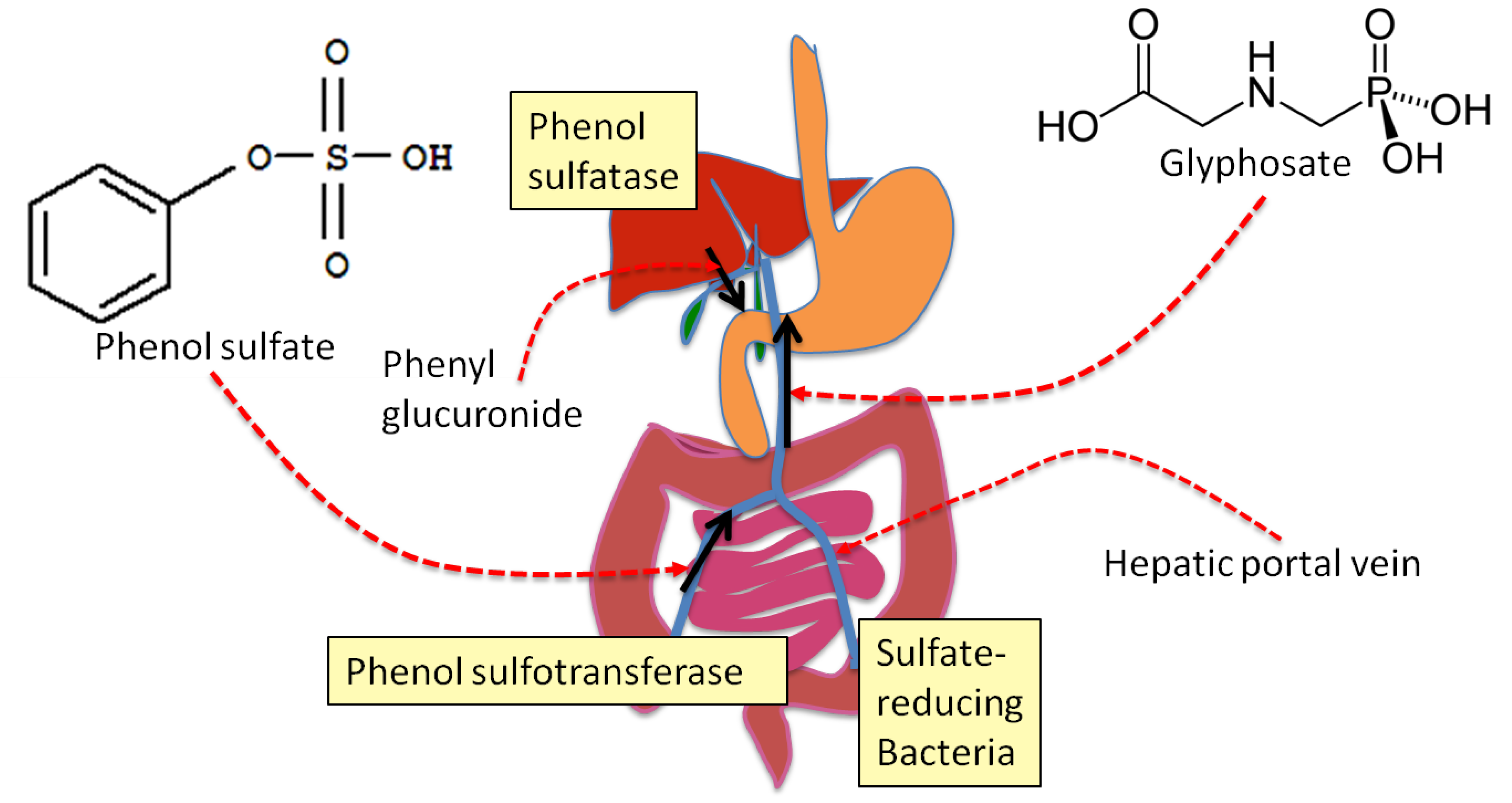
4. Sulfate Transport Impairment and Phenol Synthesis

Autism is a disorder involving impaired social skills and neurodevelopmental delay that has reached epidemic proportions in recent years, with one in 50 children born in the United States today now classified on the autism spectrum, according to the U.S. Centers for Disease Control and Prevention. Impaired sulfur oxidation and low levels of serum sulfate have been established in association with autism since 1990, as evidenced by the following quote from [77]: “These results indicate that there may be a fault either in manufacture of sulphate or that sulphate is being used up dramatically on an unknown toxic substance these children may be producing” (p. 198).
In this section, we develop a novel hypothesis for the effect of glyphosate on aromatic amino acids in plants and microbes. Our arguments depend upon the observation that glyphosate, a short carbon-nitrogen chain with a carbonyl group and a phosphate group, is a strong anionic kosmotrope, since both carbonate and phosphate have this property. Sulfate is also a kosmotrope, whereas nitrate is a chaotrope. Kosmotropes and chaotropes represent opposite extremes on the Hofmeister series [78,79], where kosmotropes tend to structure the water surrounding them and to desolubilize proteins, whereas chaotropes destructure the water and solubilize proteins. Studies on fatalities due to acute over-exposure to glyphosate reveal hemodynamic disturbances, including intravascular disseminated coagulation (DIC) and multiple organ failure, associated with high serum concentrations of glyphosate (over 800 mg/L) [80]. We suspect this has to do with glyphosate's effect as a potent kosmotrope, causing a "salting out" of blood proteins and resultant coagulation and a “no-flow” situation [81].
Molecules with a carbon ring and an available attachment site for sulfate (e.g., phenolic compounds) are attractive for the purpose of transporting sulfate through the bloodstream when the kosmotropic load is elevated. Phenolic compounds like p-cresol can be readily sulfated in the gut, and this provides an opportunity to transport sulfate through the hepatic portal vein in the presence of glyphosate. The ring supports a charge distribution that diffuses the negative charge and suppresses the water-structuring properties of sulfate, thus preventing the vascular disturbances. A single phenol can perform this feat multiple times, as the sulfate can be attached to the phenol in the colon via a phenol sulfotransferase, and the liver utilizes sulfatases and sulfotransferases to transfer the sulfate moiety from the phenol to an available substrate, typically a xenobiotic or a sterol [82]. Thus, phenols could be responsible for supplying the sulfate critically needed to detoxify xenobiotics and bile acids and to produce various sterol sulfates, as well as supplying sulfate to the pancreas to be incorporated into mucopolysaccharides being released into the gut along with proteases by acinar cells [83].
In this scenario, the glyphosate itself, due to its kosmotropic properties, is disrupting the transport of free sulfate, and therefore the aromatic amino acids are oxidized into various phenolic compounds in order to compensate for this problem. Unfortunately, once they are unsulfated, the phenols become toxic, as they will react destructively with phospholipids and DNA through one-electron transfer [84].
Although flavonoids are generally considered to be beneficial to health, the biological mechanisms behind their benefit are not yet established. In [85], it is stated that “the potential role of microbial metabolism in the gastrointestinal tract is often overlooked”. These authors propose that monophenolic derivatives are likely produced through ring fission of flavonoids by the gut microflora in the colon. Thus, flavonoids can promote sulfate transport to the liver via this process. Furthermore, flavonoids themselves can be both glucuronylated and sulfated [85,86], especially at the 4'-OH position [87], so they could serve directly as sulfate transporters without being broken down. In fact, we hypothesize that their role in sulfate transport is the reason for their abundant synthesis in plants in the presence of glyphosate at the expense of tryptophan [29]. Since they are less toxic than monophenols, they become attractive for sulfate transport in the presence of glyphosate.
Figure 1. Schematic of cyclical process that could be utilized to transport sulfate from the gut to the liver in the face of glyphosate contamination in the hepatic portal vein. Phenolic compounds derived from aromatic amino acids would be cycled back and forth between the gut and the liver, sulfated during transport from the gut to the liver and glucuronylated during transport back from the liver to the gut. Ultimately, a sulfate reducing bacterium could metabolize the phenol, consuming sulfate.
Entropy 15 01416 g001
The fact that glyphosate suppresses both alkaline and acid phosphatase activity in in vitro assays [88] as well as extracellular alkaline phosphatase synthesis in algae [89] suggests that phosphate faces the same problem as sulfate in plants, in the presence of glyphosate, and hence enzymatic activity that produces free phosphate is suppressed. It is interesting to note that autism is associated with elevated serum levels of pyridoxal phosphate (vitamin B6) even in the absence of supplements [90]. Despite this, supplemental B6 has been shown to alleviate symptoms of autism [91,92]. We hypothesize that vitamin B6 is exploited to transport phosphate safely in the presence of glyphosate. The pyridoxal ring distributes the negative charge on the phosphate anion in the same way that phenols distribute the charge on sulfate, thus allowing phosphate to be transported in a non-kosmotropic form.
Glyphosate’s kosmotropic effects can be counteracted through buffering of chaotropes in the blood, and this could be a factor in the increased levels of both ammonia [93] and various oxides of nitrogen, including nitric oxide, nitrite, and nitrate [94,95,96] observed in association with autism.
Thus, autism is associated with dysbiosis in the gut [50,51], along with impaired sulfate metabolism and a significantly reduced level of free sulfate in the blood stream (only one-third of the normal level) [63,97,98,99,100,101], excess production of nitric oxide [94,95,96], overgrowth of phenol-producing bacteria like C. difficile [101], and increased urinary levels of the toxic phenol, p-cresol [68]. Autism is also associated with a decreased ability to sulfate and hence detoxify acetaminophen, which aligns with insufficient sulfate bioavailability. A genetic defect in the phenol sulfotransferase gene is associated with autism [77]: this enzyme becomes more essential in the context of glyphosate contamination. All of these observations can potentially be explained by the effects of glyphosate on the gut bacteria and on the blood stream.
Both colitis and Crohn’s disease are associated with sulfate depletion in the gut [102], which could be caused by the impaired sulfate transport problem induced by glyphosate exposure. An overgrowth of the sulfur-reducing bacterium, Desulfovibrio, has been found in association with autism [103]. Sulfate-reducing bacteria can utilize aliphatic and aromatic hydrocarbons as electron donors, and therefore they can play an important role in detoxifying toxic phenolic compounds [104,105,106,107,108]. Thus, the presence of Desulfovibrio in the gut may serve a dual purpose by metabolizing phenolic compounds while also disposing of free sulfate, which could be problematic if allowed to enter the blood stream in the presence of glyphosate. Thus, we hypothesize that, in the context of glyphosate in the vasculature, aromatic amino acids are diverted into phenolic compounds which can safely transport sulfate from the gut to the liver. The liver can then transfer the sulfate to another metabolite, such as a steroid, and then ship the phenol back to the digestive system for another round via the bile acids following glucuronidation [108]. Possibly after multiple rounds, the phenol is finally metabolized by a sulfate-reducing bacterium in the colon. This idea is schematized in Figure 1.

5. Evidence that Glyphosate Inhibits CYP Enzymes

The evidence that glyphosate inhibits CYP enzymes comes from several directions. There are studies showing inhibition of aromatase, a CYP enzyme that converts testosterone to estrogen, and studies showing enhancement of retinoic acid, which could be achieved by suppressing the CYP enzyme involved in its catabolism. Finally, there are studies that directly show inhibition of detoxifying CYP enzymes in both plants and animals.
Two studies point to a disruption of aromatase activity [109,110]. In [109], as little as 10 ppm. of glyphosate disrupted aromatase activity in human liver HepG2 cells, a well-established cell line to study xenobiotic toxicity. In [110], it was shown that aromatase activity is disrupted in human placental cells at a concentration 100 times lower than that recommended in agricultural use. Furthermore, even small amounts of the adjuvants present in Roundup® could substantially enhance this effect of glyphosate, probably by enhancing the ease with which it gains access to the membrane-bound protein. In experiments with oyster larvae, Roundup® was found to be toxic at less than 1/20 the concentration of glyphosate needed to induce toxicity, thus exhibiting the enormous enhancing effect of Roundup®'s adjuvants [111].
Retinoic acid plays a key role in embryonic development, where its tightly-regulated concentration levels impact developmental stages [112]. Due to reports of neural defects and craniofacial malformations in children born in regions where glyphosate-based herbicides are used, a group of researchers investigated the effects of low doses of glyphosate (1/5,000 dilutions of a commercial glyphosate-based herbicide) in development of African clawed frog embryos and chick embryos [113]. The treated embryos were highly abnormal: the frog embryos developed into tadpoles with cranial deformities, and microcephaly was observed in the chick embryos. They traced this effect to an increase in endogenous retinoic acid (RA) activity, and showed that cotreatment with an RA antagonist prevented the deformities.
This increase in RA activity can be explained via inhibition of a CYP enzyme. A novel member of the CYP family has been discovered which is induced by retinoic acid and involved in its catabolism [114,115]. It is present in mammalian embryos and in the brain. Thus, if this enzyme is suppressed by glyphosate, it would explain the observed effect that glyphosate enhances levels of retinoic acid in embryonic development.
A study conducted in 1998 demonstrated that glyphosate inhibits cytochrome P450 enzymes in plants [116]. CYP71s are a class of CYP enzymes which play a role in detoxification of benzene compounds. An inhibitory effect on CYP71B1l extracted from the plant, Thlaspi arvensae, was demonstrated through an experiment involving a reconstituted system containing E. coli bacterial membranes expressing a fusion protein of CYP71B fused with a cytochrome P450 reductase. The fusion protein was assayed for activity level in hydrolyzing a benzo(a)pyrene, in the presence of various concentrations of glyphosate. At 15 microM concentration of glyphosate, enzyme activity was reduced by a factor of four, and by 35 microM concentration enzyme activity was completely eliminated. The mechanism of inhibition involved binding of the nitrogen group in glyphosate to the haem pocket in the enzyme.
A more compelling study demonstrating an effect in mammals as well as in plants involved giving rats glyphosate intragastrically for two weeks [117]. A decrease in the hepatic level of cytochrome P450 activity was observed. As we will see later, CYP enzymes play many important roles in the liver. It is plausible that glyphosate could serve as a source for carcinogenic nitrosamine exposure in humans, leading to hepatic carcinoma. N-nitrosylation of glyphosate occurs in soils treated with sodium nitrite [118], and plant uptake of the nitrosylated product has been demonstrated [119]. Preneoplastic and neoplastic lesions in the liver of female Wistar rats exposed to carcinogenic nitrosamines showed reduced levels of several CYP enzymes involved with detoxification of xenobiotics, including NADPH-cytochrome P450 reductase and various glutathione transferases [120]. Hence this becomes a plausible mechanism by which glyphosate might reduce the bioavailability of CYP enzymes in the liver.
Glyphosate is an organophosphate. Inhibition of CYP enzyme activity in human hepatic cells is a well-established property of organophosphates commonly used as pesticides [121]. In [122], it was demonstrated that organophosphates upregulate the nuclear receptor, constitutive androstane receptor (CAR), a key regulator of CYP activity. This resulted in increased synthesis of CYP2 mRNA, which they proposed may be a compensation for inhibition of CYP enzyme activity by the toxin. CYP2 plays an important role in detoxifying xenobiotics [123].
Beginning in around 2006, an alarming die-off of honeybees became apparent in the United States, and researchers are still struggling to understand what is causing this die-off [124]. Since the application of glyphosate also reached record levels that year, and has continued to increase since then, with no abatement in the bee colony collapse disorder, glyphosate could be playing a role in the bees' plight. While correlation does not necessary imply causation, there are strong reasons why glyphosate might interfere with bees' resistance to other environmental toxins. At first glance, pesticides might be more highly suspect, since bees are, after all, an insect. However, honeybees have an innate resistance to most pesticides, which unfortunately depends upon several CYP enzymes. For example, metabolic detoxification mediated by CYPs contributes significantly to honey bee tolerance of pyrethroid insecticides [125]. Thus, the fact that glyphosate disrupts CYP enzymes would suggest that exposure to glyphosate would leave bees especially vulnerable to pesticides in their environment, resulting in a synergistic effect. A 2005 study in Alberta (Canada) revealed a reduced wild bee abundance and highly-correlated reduced pollination in GM canola compared with organically grown canola [126], with Roundup-treated non-GM canola coming in at an intermediate level. A study comparing bees exposed to glyphosate and/or Roundup® against a control population demonstrated a significantly higher mortality rate in the glyphosate-exposed bees (p < 0.001) [127].  Neonicotinoids such as imidacloprid and clothianidin can kill bees, and have been implicated in colony collapse disorder [128]. However, this toxic effect is likely synergistic in combination with glyphosate, as would occur with bees ingesting herbicide-contaminated pollen. Glyphosate is an organophosphate, and a study of human self-poisoning has demonstrated that organophosphate ingestion synergistically greatly enhances the toxicity of ingested neonicotinoids [129].

6. The Path to Obesity

Having established a plausible mechanism whereby glyphosate’s effects on gut bacteria would lead to depleted sulfate supplies in the gut with resulting inflammatory bowel disease, we now turn our attention towards the likely consequences of the resulting “leaky gut syndrome.” It has been proposed that the exponential increase in the production of synthetic organic and inorganic chemicals may be causal in the current world-wide obesity epidemic, due to alterations in body chemistry that promote weight gain [130]. These chemicals are better known for causing weight loss at high exposure levels, and this apparent paradox can be explained with respect to glyphosate, by invoking its known effect of depleting tryptophan in plants and microbes. Its effect on CYP enzymes in the liver will compound the problem, due to the impaired ability to detoxify synthetic chemicals, which are increasingly present in the environment. In this section we will explain how glyphosate’s depletion of tryptophan bioavailability can lead to obesity, and in Section 6 we will provide evidence that extreme depletion of tryptophan in the absence of obesity can cause severe impairment of the intestinal barriers, resulting in weight loss and anorexia, due to an inability to transport critical micronutrients across the damaged gut barrier.
Tryptophan is an essential amino acid, meaning that mammalian cells cannot synthesize it. Serum tryptophan depletion leads to serotonin and melatonin depletion in the brain [131]. Since serotonin (derived from tryptophan) is a potent appetite suppressant [132], it follows that serotonin deficiency would lead to overeating and obesity. As we have seen, tryptophan supplies could be depleted both in plant-based food sources and through impaired tryptophan synthesis by gut bacteria as direct effects of glyphosate. The observed 20-fold increase in the synthesis of tryptophan-derived polyphenolic flavonoids in the context of glyphosate provides strong evidence of impaired tryptophan synthesis [29].
Tryptophan has several important roles in the body. Ordinarily, dietary tryptophan (aside from its role as an essential amino acid in protein synthesis) is taken up by the liver and either fully metabolized to produce ATP or processed through the enzymatic action of tryptophan dioxygenase (TDO) and indole amine dioxygenase (IDO), via a pathway involving kynurenine and quinolinate as intermediaries to produce NAD+, an essential cofactor in ATP synthesis and DNA repair [133] (see Figure 2). Any tryptophan not taken up by the liver circulates in the blood, and is transported across the blood brain barrier (BBB). It becomes the (sole) precursor to the synthesis of the neurotransmitter serotonin and the hormone melatonin [131]. A low ratio of tryptophan to competing proteins in the blood stream leads to reduced transport of tryptophan across the BBB and subsequent impaired serotonin and melatonin synthesis in the brain. Thus, low serum tryptophan levels translates into a tendency towards weight gain due to suppressed serotonin signaling [132].
Figure 2. Illustration of tryptophan pathways in the body and the adverse effect of glyphosate on tryptophan bioavailability. IDO: indole amine dioxygenase; TDO: Tryptophan dioxygenase; G: glyphosate.
Entropy 15 01416 g002
However, under inflammatory conditions, and in response to pathogenic stimuli such as the lipopolysaccharide (LPS) in bacterial cell walls, tryptophan is converted into kynurenine by lymphoid tissues at the site of inflammation [134] and stockpiled by in situ macrophages and neutrophils [135,136,137] as kynurenine. Therefore, it is expected that inflammation in the gut would lead directly to serum tryptophan depletion, thus further reducing the bioavailability of tryptophan to the liver. There are several reasons why macrophages need to sequester kynurenine, the most important of which is likely to be the assurance of a localized resource to regenerate NAD+ following its depletion through the synthesis of poly-ADP ribose by poly(ADP-ribose)polymerase (PARP) [138,139,140]. Poly-ADP ribose plays an important role in the DNA repair mechanisms that are required following DNA damage, induced by the reactive oxygen and nitrogen species (ROS and RNS) released by macrophages to fight infection – superoxide, nitric oxide, and their reaction product, peroxynitrite. Superoxide is induced from oxygen in the artery wall by transfer of an electron from cytosolic NADPH to oxygen, and its synthesis is essential for killing invasive pathogens, but collateral exposure also leads to tissue damage.
Both the inflammatory cytokine interferon-γ (IFN-γ) and superoxide itself induce IDO synthesis, and IDO detoxifies superoxide by using it to break the pyrrole ring in tryptophan [141]. The DNA in the cell nucleus is highly vulnerable to superoxide exposure, which can lead to strand breaks. The synthesis of kynurenine from tryptophan by IDO results in replenishing the supply of NAD+ and NADP+, which has been depleted due to the activities of PARP as part of the DNA repair process.
Studies have confirmed that serum tryptophan levels are low in association with obesity [142,143]. In [143], plasma tryptophan levels were monitored several times over the course of a twenty-four hour period, and it was confirmed that serum tryptophan levels were chronically depressed, and the levels of other competing large neutral amino acids were elevated, in obese subjects compared to controls. This pathology persisted even after weight reduction through intense dieting.
A recent experiment involving transferring a strain of endotoxin-producing bacteria from the gut of an obese human to the sterile gut of germ-free mice demonstrated the dramatic obesogenic effect that over-production of endotoxin by gut bacteria can have [144]. These mice became obese over a 16-week trial period, when concurrently placed on a high-fat diet, and the obesity was associated with a low-grade chronic inflammatory state. Control germ-free mice on the same diet but without the infective agent did not become obese. It was hypothesized that chylomicrons produced for fat transport became a vehicle for endotoxin delivery to blood serum and subsequently to the liver and body fat stores, since inflammatory cytokines were found predominantly in the liver and epididymal fat pad rather than in the ilium. Since glyphosate induces a shift in gut bacteria towards endotoxin-producers, this effect can conceivably explain the association of a high-fat diet with obesity [145].
The obesity epidemic began in the United States in 1975, simultaneous with the introduction of glyphosate into the food chain, and it has steadily escalated in step with increased usage of glyphosate in agriculture (see Figure 1 in [146]). While it is common knowledge that Americans are continuing to grow more and more obese with each passing year [147,148], there may be less awareness that obesity aligns with glyphosate usage elsewhere in the world [149]. For example, South Africa arguably has the highest obesity rates in all of Africa [150], and it is also the African country that has most heavily embraced glyphosate usage since the 1970’s and has freely adopted genetically modified crops with little regulation [151,152]. According to World Health Organization statistics [153], only 2.7% of adults in the United Kingdom were obese in 1972, a number that rose to 25.8% in 1999. Today, two thirds of U.K. citizens are either overweight or obese.

7. The Path to Inflammatory Bowel Disease and Anorexia Nervosa

We have seen how obesity can develop following the depletion of tryptophan through its diversion into polyphenolic flavonoids as well as aggressive uptake into macrophages, to provide assurance of DNA repair mechanisms in the face of excess ROS and RNS. Subsequent impaired serotonin synthesis stimulates overeating behaviors. Here, we argue that severe tryptophan deficiency without sufficient fat stores to harbor toxins and supply sterol sulfates can result in an inability to control microbial invasion as a consequence of impaired release of antimicrobial peptides. This can lead, paradoxically, to anorexia nervosa, resulting in a highly inflamed digestive system, pathogenic penetration through leaky intestinal epithelium, uncontrollable diarrhea, and subsequent anorexia.
Obesity offers protection against gastrointestinal inflammation, in part because the endotoxin can be stored in adipose tissue, sparing the gut barrier from inflammatory damage. However, a more important factor may be the ability of adipose tissue to directly supply sulfated steroids. The sulfotransferase that sulfates serotonin, thus inactivating it, is found in many tissues, including brain, heart, liver, lung, kidney and spleen [154]. Insufficient sulfate supply would likely compromise this function, leading to poor serotonin regulation. There is an interesting connection between levels of serotonin and sterol sulfates in the blood serum. DHEA sulfate is the most prominent sterol sulfate in the serum besides cholesterol sulfate [155]. Patients with autism have anomalously low DHEA sulfate levels along with anomalously low serotonin [156]. Serum DHEA sulfate levels are inversely associated with visceral fat [157], and DHEA sulfate supplements can induce weight loss in morbidly obese postmenopausal women [158]. We hypothesize that DHEA sulfate levels are a hormonal signal of sulfate bioavailability, and low bioavailability leads to low serotonin which induces overeating, in order to produce visceral fat. Visceral fat is a source of estrone sulfate [159], which, we hypothesize, may compensate for some deficiencies in DHEA sulfate and alleviate the burden on the adrenal glands to produce sterol sulfates. This would also reduce the demand on phenols to transport sulfate and therefore alleviate the inflammatory gut disorder, restoring homeostasis.
An important study elucidating the processes leading to inflammatory bowel disorder was conducted on male Ace2 knockout mice (Ace2−/y) [13]. Ace2 induces expression of the tryptophan transporter in the gut epithelium. Thus, these mice suffered from severe tryptophan deficiency. They responded to dextran sodium sulfate exposure with a much more severe colitis attack than their control littermates, leading to enhanced infiltration of inflammatory cells, increased intestinal bleeding, severe diarrhea, and weight loss. A series of further experiments revealed that a similar response could be provoked in the control mice by providing them with a diet that was specifically deficient in tryptophan. It was confirmed that the acute response was associated with impaired synthesis of antimicrobial peptides by macrophages, mediated by impaired mTOR (mammalian target of rapamycin) signaling. It is conceivable that the severe deficiency in tryptophan led to restricted protein synthesis in macrophages, preventing the synthesis of the antimicrobial peptide. Furthermore, the distribution of gut bacteria was profoundly affected by the Ace2−/y phenotype and by tryptophan deprivation.
Thus, severe tryptophan deficiency, as might be induced by glyphosate’s interference with tryptophan synthesis in plants and microbes, can lead to an extreme inflammatory bowel disease that would severely impair the ability to absorb nutrients through the gut, due to inflammation, bleeding and diarrhea. This could easily explain the alarming increases that have been seen recently in inflammatory bowel diseases [16,17,160].

8. Cytochrome P450 Enzymes

The cytochrome P450 (CYP) enzymes are a diverse, ancient class of enzymes that date back to three billion years ago, and play an important role in plant, animal, and microbial biology [161]. These enzymes participate in oxidation, peroxidation and reduction of compounds ranging from pharmaceutical drugs to environmental chemicals to endogenous bioactive molecules [123]. There are at least 18 distinct CYP families in humans, which are classified as a series of numerical “CYP” classes. In humans, CYP1, CYP2, CYP3, and CYP4 P450 enzymes in the liver are essential for detoxification of many xenobiotics [162]. Members of the CYP5 and CYP7 classes are essential for the formation of water-soluble bile acids from cholesterol in the liver. Bile acids act as powerful detergents to aid in the digestion of fats, and also provide a pathway for disposal of oxysterols. A loss-of-function mutation in CYP7B1 leads to liver failure in children, associated with high serum levels of oxysterols [163].
Both cholesterol and vitamin D3 synthesis and degradation depend upon various CYP enzymes. At least seven CYP enzymes have a role in converting acetate into sterols. Lanosterol 14α-desmethylase (CYP51A1) is pivotal in cholesterol synthesis. Two CYP enzymes in the liver catalyze 25- hydroxylation of vitamin D3 to its active form, and two other CYP enzymes catalyze the breakdown of vitamin D3 in the liver [164,165].
There is a growing epidemic of vitamin D deficiency in the United States. In a study on serum 25-hydroxyvitamin D levels obtained from the National Health and Nutrition Examination Survey (NHANES) data, it was found that vitamin D3 levels fell sharply in the interval from 2001 to 2004 compared to the interval from 1988 through 1994 [166]. While this problem is in part due to overaggressive sun avoidance practices, glyphosate’s interference with CYP proteins may play a role in disrupting vitamin D3 activation in the liver.
Several CYP enzymes participate in steroid synthesis. Cytochrome P450 oxidoreductase deficiency (POR) is a newly described disorder of steroidogenesis [167]. Five crucial lipid hormones, aldosterone, androstenedione, cortisol, corticosterone and dehydroepiandrosterone (DHEA), are produced in the adrenal glands, testes and ovaries, and in the adrenal cortex. All steroid hormones are produced from cholesterol by these CYP enzymes, contained within the inner mitochondrial membrane. The lipophilic nature of these steroids allows them to diffuse across the lipid bilayers. CYP19A1 (aromatase), whose inhibition has been confirmed in association with glyphosate [109,110] converts androgenic precursors into estrogen. Suppressed aromatase synthesis has been found in the brain in association with autism [168], leading to the “super-male” profile associated with this condition [169].
CYP26A1 catabolizes retinoic acid; hence, its suppression would lead to excess retinoic acid bioavailability. CYP26A1 is induced by retinoic acid during neural differentiation, and its action leads to the degradation of retinoic acid, a necessary step towards maturation of the developing neurons [114]. The aryl hydrocarbon receptor (Ahr) gene induces CYP1B expression, leading to degradation of retinoic acid. Ahr-knockout mice accumulate excess retinoic acid in the liver [170]. Thus, if liver CYP1B expression were disrupted by glyphosate, it would lead to excess retinoic acid. Retinoic acid suppresses the synthesis of cholesterol sulfate, a crucial step in bile acid synthesis [171]; thus, excess retinoic acid in the liver should lead to impaired synthesis of bile acids and impaired fat metabolism.
Mutations in CYP7A1 are associated with high serum LDL and high hepatic cholesterol content, along with deficient bile acid excretion [172]. Human CYP7B1 mutations lead to both defects in bile acid synthesis and spastic paraplegia, involving impaired myelin sheath in the spinal cord and uncontrolled movement disorders. The drug, clopidogrel (Plavix), administered to suppress life-threatening stent thrombosis following cardiovascular surgery, depends upon a liver CYP enzyme, CYP2C19, to transform it into an activated metabolite. Patients with a loss-of-function mutation in this CYP enzyme have significant risk of an adverse event following surgery [173,174].
Glyphosate from food sources or as a contaminant in water would be likely to reach the liver in high concentrations through direct transport from the digestive system via the hepatic portal vein. It could be anticipated that glyphosate would disrupt many of the diverse CYP enzymes that are bioactive in the liver, involved in cholesterol synthesis and metabolism, vitamin D3 synthesis and metabolism, the detoxification of xenobiotics, and regulation of retinoic acid.
Glyphosate would also be expected to travel throughout the blood stream, disrupting any CYP enzymes it comes in contact with. Of particular concern are the two that regulate blood clotting (thromboxane A2 synthase: CYP5A1) and hemorrhaging (prostacyclin synthase: CYP8A1). CYP5A1 stimulates platelet aggregation, whereas CYP8A1 inhibits platelet aggregation. The elderly often face instabilities in hemorrhaging and clotting leading to Disseminated Intravascular Coagulation (DIC) and life-threatening destabilization of the blood [175], which could be due to impaired function of these two enzymes.

9. Glyphosate’s Potential Role in eNOS Dysfunction

Thus far, we have developed a plausible argument for how glyphosate could disrupt gut microbiota, leading to inflammation, depletion of tryptophan, and subsequent obesity, or, in the extreme case, anorexia nervosa. We have also discussed the many roles of CYP enzymes, and proposed that glyphosate’s interference with CYP expression could lead to many pathologies that are commonly occurring today, such as vitamin D3 deficiency and abnormal blood clotting.
Endothelial nitric oxide synthase (eNOS) is an orphan member of the CYP family. It is present in endothelial cells that synthesize nitric oxide (NO), where it induces vessel relaxation and therefore enhanced blood flow [176]. Both eNOS and CYP enzymes are heme-thiolate proteins with the same redox partner, a diflavoprotein reductase. However, eNOS, unlike the other CYP enzymes, requires tetrahydrobiopterin (BH4) as a cofactor for the synthesis of NO, and no other member of the CYP family is capable of synthesizing NO.
It has recently been proposed that eNOS is a dual-purpose enzyme, producing NO when it is bound to calmodulin in the cytoplasm, and producing sulfate when it is bound to caveolin at the plasma membrane [177]. While no other CYP enzyme produces NO, this class is known to oxidize sulfur [178], an important aspect of their ability to detoxify sulfur-containing drugs. Red blood cells (RBCs) contain membrane-bound eNOS, and this has presented a puzzle to researchers, because the synthesis of NO by RBCs would be counterproductive, due to its high reactivity with hemoglobin to form a nitrosylated compound that is impaired in oxygen transport. Indeed, RBCs have mechanisms to maintain a very low concentration of the substrate L-arginine. However, it is highly plausible that RBCs use their eNOS to produce sulfate, which can then be combined with cholesterol to form cholesterol sulfate, known to be present in large amounts in RBC plasma membranes, where it has a stabilizing effect.
A significant adverse effect of glyphosate is its hypothesized disruption of sulfate synthesis by eNOS in the endothelium. This effect contributes to the inflammation already present due to the escape of pathogenic bacteria through the impaired gut barrier. In fact, the two effects are synergistic, because the sulfate depletion incurred by eNOS dysfunction further compromises the gut barrier, where sulfate deficiencies due to transport problems are already present. Due to its homology with the CYP enzymes, eNOS is predicted to be susceptible to disruption by glyphosate, but only in its sulfate-synthesis function. The result will be endothelial damage due to superoxide exposure, along with sulfate deficiency. We hypothesize that such disruption is a significant heretofore overlooked component of glyphosate’s toxicity in mammals.
If, as proposed in [177], RBCs use eNOS to produce sulfate, then the sulfate can be combined with cholesterol to produce cholesterol sulfate, which, unlike cholesterol itself, is amphiphilic. RBCs are well positioned to deliver both cholesterol and sulfate to the tissues, supplying them with these essential nutrients. In [177], it was further proposed that endothelial cells produce sulfate catalyzed by eNOS, using superoxide as the oxidizing agent, a reaction that is catalyzed by sunlight exposure, and that the sulfate serves to replenish sulfate supplies to the glycocalyx, which is constructed from highly sulfated proteoglycans. Accumulation of sulfate deficiencies in the endothelial glycocalyx contributes significantly to vascular dysfunction [179]. Colitis is less prevalent in areas with a sunny climate [180], suggesting that sunlight improves intestinal health by increasing sulfate supply.
Ingested glyphosate readily enters the vasculature, and hence membrane bound eNOS in RBCs and the endothelial wall is vulnerable to the disabling effects of glyphosate on the P450 active site. This, over time, would result in cholesterol and sulfate deficiencies, manifested as multiple disease states. It would also explain the pathology where eNOS synthesizes superoxide in an “uncoupled” mode [181], a pathology that has been proposed as a major source of inflammatory ROS and subsequent endothelial dysfunction. We hypothesize that the superoxide is prevented from oxidizing sulfur by the glyphosate, and thus becomes a destructive agent in the artery wall.

9.1. Lysosomal Dysfunction

In [177], it was proposed that lysosomal dysfunction could be predicted to follow long-term impairment of eNOS’ sulfate synthesis. Lysosomes, the “digestive system” of the cell, require substantial membrane cholesterol both to prevent hydrogen ion leaks and to protect membrane lipids from oxidative damage. Lysosomes also depend upon internalized sulfate, derived from heparan sulfate proteoglycans (HSPGs), to catalyze hydrolytic enzymes. Severe neurological dysfunction associated with lysosomal storage diseases involving impaired heparan sulfate homeostasis attest to the importance of sulfate in lysosomal function [182].
It has become increasingly apparent that lysosomal dysfunction is a major factor in Alzheimer’s disease and Parkinson’s disease [183], as well as in cardiovascular disease [184] and heart failure [185]. Mitochondria are ordinarily constantly broken down and renewed by lysosomal processes, and, when these become impaired, large aged mitochondria become a source of reactive oxygen species that contribute significantly to neuronal damage. Cardiomyocytes, like neurons, are long-lived postmitotic cells that are especially susceptible to lysosomal disrepair [186].

9.2. Tetrahydrobiopterin

The research literature has identified the cofactor tetrahydrobiopterin (BH4) as a significant player in eNOS function [187,188]. BH4 shifts the heme iron in eNOS to a high spin state, as well as increasing arginine binding, thus catalyzing the synthesis of NO by eNOS [187]. The synthesis of BH4 from its substrate GTP is induced by IFN-γ, which, in turn, is induced by bacterial lipopolysaccharides (LPS) [189]. Thus, a bacterial infection will induce NO synthesis by eNOS. However, an excess of exogenous NO (as might be expected to occur through iNOS synthesis of NO during a bacterial infection) causes a decrease in NO synthesis by eNOS with a simultaneous increase in superoxide synthesis, an effect that can lead to severe hypertension in infants with congenital heart disease treated with inhaled NO [187]. Superoxide’s reaction with NO to produce the highly toxic peroxynitrite (ONOO−), a potent bacteriocidal agent, is likely a critical factor. The subsequent oxidation of BH4 disrupts its ability to act as a cofactor [188], and causes “eNOS uncoupling,” leading to superoxide synthesis in a highly disruptive feedback loop.
We hypothesize that glyphosate’s nitrosylation of the active P450 site has derailed eNOS’ ability to synthesize sulfate in a contained environment at the caveolar sites in the membrane, thus requiring an alternative method to synthesize sulfate that exposes the cell to ROS. This method, as previously described in [177,190], involves the oxidation of homocysteine thiolactone, catalyzed by ascorbic acid (vitamin C) and retinoic acid (vitamin A). Since glyphosate enhances the bioavailability of retinoic acid through its suppression of the CYP enzyme that metabolizes it [115], this will help to promote the alternative reaction leading to sulfate synthesis in the artery wall from homocysteine thiolactone, but also requiring the inflammatory agent, superoxide, which over time destroys the artery wall, leading to endothelial dysfunction and cardiovascular disease.
Elevated serum homocysteine is a strong risk factor in cardiovascular disease [191], in heart failure [192], in dementia [193], and in kidney failure [194,195]. We propose that sulfur-containing amino acids are deflected towards homocysteine synthesis in order to supply substrate for the critically-needed sulfate synthesis from superoxide in the artery wall. This also explains both the inflammation in the artery wall associated with atherosclerosis [196] and the deficiency in methionine associated with glyphosate, due to its depletion through its role as a substrate for homocysteine synthesis.

10. Involvement of the Brain

Impairment in the homeostasis of serotonin, an important neurotransmitter that regulates mood, appetite and sleep, has been linked to depression [197], autism [198], and Alzheimer’s disease [199,200], as well as obesity [132]. We have already seen how glyphosate’s induction of tryptophan-derived flavonoids and tryptophan’s incorporation into macrophages as kynurenine via IPO can explain reduced brain serotonin levels. Vitamin D3 deficiency can also contribute to mood disorders, and is hypothesized to be a key factor in the syndrome, Seasonal Affective Disorder (SAD), manifested as depressed mood specifically during the winter months [201]. Excess ammonia and zinc deficiency are also implicated in neuronal disorders, particularly Alzheimer’s disease, attention deficit hyperactivity disorder (ADHD), and autism. DNA methylation impairment is a factor in neuronal diseases, and glyphosate’s depletion of methionine could contribute to this defect. Below, we elaborate on the effects of serotonin depletion, excess ammonia, zinc depletion, and methylation impairments on disorders of the brain. We conclude this section with specific mention of a possible role for glyphosate in two other diseases of the brain: multiple sclerosis and Parkinson’s disease.

10.1. Serotonin, Mood Disorders, and Autism

Defects in serotonin transport are associated with a wide range of mood disorders. Major depression is accompanied by immune system activation, and the term “inflammatory and neurodegenerative (I&ND) hypothesis” has been used to describe this complex [202]. A demonstrated increased production of cytokines and immunoglobulins against bacterial-derived lipopolysaccharides points to increased gut permeability as a feature in depression [203]. Patients with depression and sleep disorders exhibit significantly lower serum levels of tryptophan along with serum markers of inflammation such as IL-6 and IL-8 [204]. Selective serotonin reuptake inhibitors (SSRI’s) are a popular class of drugs to treat depression: they work by impairing serotonin reuptake in the synapse, effectively increasing its bioavailability for neuronal signaling. This strongly suggests that insufficient serotonin in the synapse could be a factor in depression. In fact, dietary tryptophan depletion leads to relapse in recovering depressed patients [197].
Defects in the serotonin transporter gene, 5-HTT, have been associated with antisocial personality disorder and violent behavior [205]. There has been a marked increase in the rate of irrational school-associated violent deaths in the United States since 1990 [206], and glyphosate may play a role in this pattern through depletion of serotonin bioavailability. Disturbances in serotonin function in the brain are known factors in impulsive aggression, violence, and criminal behavior [207]. Farmers in India experienced anomalously high suicide rates following adoption of Western agricultural methods based on extensive use of Roundup® [208]. While an explanation based on economic stress has been proposed, suicide victims in general have low serotonin levels in the brain [209], so it is conceivable that serotonin suppression via depletion of tryptophan by glyphosate played a role in the suicides among farmers in India.
Genetic mutations in serotonin transporter genes have been found in association with both obsessive compulsive disorder and autism [210]. A study comparing 40 children with idiopathic infantile autism with normal controls showed a significantly lower serum ratio of tryptophan to large neutral amino acids [211]. 35% of the children with autism had a ratio that was at least two standard deviations below the mean value from the control group. It has been shown that dietary tryptophan depletion exacerbates anxiety and repetitive ritualistic behaviors in autistic subjects [198], an effect that was surmised to be due to impaired serotonin synthesis. Researchers have studied a mouse model of a defective serotonin transporter gene which results in a decrease in the bioavailability of serotonin for neuronal signaling in the brain, and have shown that the genetically modified mice exhibit autism-like behaviors [212]. This strongly suggests that impaired serotonin supply in the brain is a feature of autism.
Melatonin, produced from serotonin, is secreted by the pineal gland, primarily at night, and is a potent antioxidant and regulator of redox reactions [213,214]. Its neuroprotective role in aging and many neurodegenerative conditions such as Alzheimer’s disease and Parkinson’s disease is most likely due to its antioxidant effects [215,216,217,218]. Thus, it is anticipated that glyphosate would lead to impaired antioxidant protection, due to the suppression of melatonin synthesis, following the depletion of tryptophan as substrate, as previously discussed. Since melatonin is also a regulator of the wake/sleep cycle, impaired melatonin supply will lead to sleep disorders.

10.2. Ammonia, Autism and Alzheimer’s Disease.

As stated previously, glyphosate enhances ammonia synthesis in susceptible plants via activation of the enzyme PAL [22], and gut microbes could produce excess ammonia through enhanced PAL synthesis under the influence of glyphosate. A parallel between autism and hepatic encephalitis has been made, emphasizing the role that ammonia plays as a toxin in the brain in both cases [219,220]. Ammonia has also been proposed to play a critical role in the etiology of Alzheimer’s disease [221]. Thus, excess ammonia synthesis by gut bacteria under the influence of glyphosate could be a factor in both autism and Alzheimer’s disease.

10.3. A Role for Zinc Deficiency.

Zinc deficiency is a major problem worldwide, particularly in the developing world, where limited access to zinc-rich foods such as shellfish and excess dietary exposure to phytates both contribute to the problem [222]. Phytates, found in many nuts and grains, bind to dietary minerals and interfere with their absorption. Lactobacilli and other beneficial gut bacteria produce the enzyme phytase, which catalyses the release of phosphate from phytates and improves the intestinal absorption of important minerals such as iron and zinc [223]. Because glyphosate reduces the number of these types of bacteria in the gut, it should enhance the chelating potential of phytates. This is likely a protective measure to avoid excess bioavailability of free phosphate, which is problematic in transport in the presence of glyphosate. Glyphosate’s known ability to itself chelate divalent cations is likely a factor as well. Zinc deficiency increases the risk of diarrhea, pneumonia and malaria in infants and young children.
Zinc is the most abundant trace metal in the brain [224]. Most of the amyloid-β degrading enzymes are zinc metalloproteases, and zinc is also critical in the nonamyloidogenic processing of the amyloid precursor protein. Hence, zinc deficiency in the brain would be expected to lead to the build-up of amyloid-β, a key factor in the development of Alzheimer’s disease. Zinc deficiency has also been implicated in autism [225] and ADHD [226,227]. Zinc is released into the synapse along with the neurotransmitter glutamate, and it is required for memory function and the maintenance of synaptic health as we age [228]. In [225], anomalously low zinc levels in hair analyses were found in children on the autism spectrum. In [226], zinc sulfate supplements improved ADHD symptoms, an effect that could be attributed to the supply of sulfate as well as zinc.
In [229], it was proposed that zinc deficiency along with excess exposure to copper may be a key factor in Alzheimer’s disease. A study conducted in South Africa revealed that zinc supplementation was not effective in raising plasma levels of zinc in zinc-deficient Alzheimer’s patients unless both vitamin A and vitamin D were simultaneously supplemented [230]. Hence, vitamin D3 deficiency (which could be caused by glyphosate’s impairment of liver CYP enzymes) may interfere with zinc absorption, further depleting the supplies to the tissues.

10.4. Methylation Impairment

Methylation impairment has been observed in association with autism [231] and Alzheimer’s disease [232], and this is caused by an inadequate supply of the substrate, methionine. While human cells are unable to synthesize methionine, it can be synthesized by many enteric bacteria, for example from cysteine via the transsulfuration pathway or through de novo synthesis from inorganic sulfur [233]. Glyphosate has been shown to significantly impair methionine synthesis in plants [21], and it may therefore be anticipated that it would have a similar effect in gut bacteria, which could then impair methionine bioavailability in humans. A further factor is the depletion of methionine through its deflection towards the transsulfuration pathway, as a precursor for homocysteine, which will be consumed to supply sulfate to the endothelial wall when eNOS’ sulfate synthesis is impaired. Since methionine is the source of methyl groups in methylation pathways, this effect of glyphosate could contribute directly to methylation impairment.

10.5. Molecular Mimicry and Multiple Sclerosis

An increased incidence of inflammatory bowel disease has been found in association with multiple sclerosis (MS) [234]. This could be explained by the hypothesis that gut bacteria leaking into the vasculature cause an immune reaction, and that molecular mimicry leads to an autoimmune disorder resulting in destruction of the myelin sheath. A systematic search comparing reported sequences from all known human bacterial and viral agents against three known encephalitogenic peptides identified matching mimics predominantly in gut bacteria [235]. This could explain why no infective agents have been found in association with MS, but would also suggest that the recent increase in the incidence of MS [236] may be traced to inflammatory bowel disease, and, hence, to glyphosate.

10.6. Dopamine and Parkinson’s Disease

Since dopamine is synthesized from tyrosine and its precursor phenylalanine, tyrosine and phenylalanine depletion by glyphosate in both plants and microbes would be expected to reduce their bioavailability in the diet. It has been demonstrated that dietary reductions of phenylalanine and tyrosine induce reduced dopamine concentrations in the brain [237]. Impaired dopaminergic signaling in the substantia nigra is a key feature of Parkinson’s disease, and Parkinson’s risk has been associated with exposure to various pesticides, including the herbicide paraquat [238], although, to our knowledge, glyphosate has not yet been studied in this respect. However, exposure of C. elegans to glyphosate led to pathology in the nervous system in the region analogous to the nigrostriatal dopamine system associated with Parkinson’s disease [239].
Sulfate deficiency in the brain has been associated with Parkinson’s disease, as well as Alzheimer’s disease and Amyotrophic Lateral Sclerosis (ALS) [240]. We have argued above that glyphosate disrupts sulfate transport from the gut to the liver, and may interfere with sulfate synthesis by eNOS in the arterial wall and in RBCs and platelets, leading over time to severe sulfate deficiency throughout all the tissues. This would further impact these devastating diseases of the elderly, all of which are currently on the rise.

11. Other Adverse Health Effects

In this section, we will briefly mention several other pathologies in which we suspect that glyphosate may play a role in the observed increases in incidences in recent times. These include liver disease, cancer, cachexia, and developmental and fertility problems.

11.1. Liver Disease

Cytokines like TNF-α have been identified as a key factor in fatty liver disease, which has emerged as a growing public health problem worldwide [241]. In the extreme case, liver pathology develops into nonalcoholic steatohepatitis (NASH), which can lead to cirrhosis and liver failure. Cytokines induce inflammation which damages the liver. TNF-α inhibits insulin signaling [242,243], and cytokines can induce hepatic lipid overloading as well as liver fibrosis. Glyphosate’s role in inducing cytokines has already been developed in this paper. Obesity is associated with an increased expression of membrane-associated TNF-α in adipose tissue [244].

11.2. Development and Fertility

Cholesterol sulfate plays an essential role in fertilization [245] and zinc is essential to the male reproductive system [246], with a high concentration found in semen. Thus, the likely reduction in the bioavailability of these two nutrients due to effects of glyphosate could be contributory to infertility problems. Furthermore, glyphosate’s suppression of CYP protein activity would be expected to disrupt steroidogenesis. Inflammation leads to excess ROS and RNS exposure, which can damage DNA during cell replication, thus disrupting embryonic development. Glyphosate is capable of crossing the placental barrier [247]. Preeclampsia, a life-threatening condition for both the mother and the fetus that develops during the third trimester, is on the rise in America, and it has been proposed that this may be due to impaired sulfate supply [248], directly attributable to glyphosate exposure. For all of these reasons, glyphosate exposure would lead to infertility problems.
According to the World Bank, the fertility rate in Argentina peaked at 3.39 in 1978, and has been declining steadily since then. The rate of decline accelerated during the last five years of the twentieth century. Social pressures certainly explain some of the drop in birth rate, but it is possible that environmental factors, such as glyphosate, also play a role. 1994 was the year that the FDA authorized the sale of Roundup Ready® (RR) soybeans in the North American market [249], and Argentina followed suit two years later. “After they were authorized in 1996, RR soybeans spread through Argentina at an absolutely unprecedented speed in the history of agriculture: an average of more than two million acres a year.” [249]. Argentina now exports 90% of its soybeans, which have become a monoculture crop and a cash cow.
The fertility rate in Brazil has also dropped dramatically over the past several decades from six children per woman on average to fewer than two, now lower than that of the United States. Brazil is the second largest producer and exporter of soybeans in the world behind Argentina, and it has embraced genetically modified soybeans engineered to be glyphosate-tolerant as a means to increase production since the mid 1990’s. A rapidly evolving glyphosate-resistant weed population in Brazil due to genetically engineered glyphosate-tolerant crops is leading to increased use of glyphosate in recent years [250], the same time period in which a rapid drop in birth rates was observed. A steady increase in the rate of preterm births in Brazil over the past two decades has been noted, although the cause remains elusive. For instance, the rate increased from 6% in 1982 to 15% in 2004 in the town of Pelotas [251]. It is conceivable that increased exposure to glyphosate is contributing to this problem. This idea is in line with a study of an Ontario farm population, which revealed that glyphosate exposure any time during pregnancy was associated with a statistically significant increased risk of a late-pregnancy spontaneous abortion [252].
The birth rates in Western Europe have been declining for decades, with Germany’s rate now being 1.36 children per woman. Birth rates have also been declining in the U.S since 2007, and are now at 1.9 children per woman, according to the 2011 government statistics [253].
Testicular Leydig cells produce testosterone, and thus play a crucial role in male reproductive function. The protein StAR (steroidogenic acute regulatory protein) mediates the rate-limiting step in steroidogenesis. In a recent in vitro study of a mouse tumor Leydig cell line, Roundup was shown to disrupt StAR expression, thus interfering with testosterone synthesis [254]. It was shown that Roundup® interferes with testosterone synthesis even at very low environmental doses, and higher doses were associated with necrosis and apoptosis in rat testicular cells.
In [255], the in vitro effects of several different pesticides and herbicides on the synthesis of progesterone in testicular Leydig cells were investigated. Comparing eight different pesticides (Ammo®, Banvel®, Cotoran®, Cyclone®, Dual®, Fusilade® and Roundup®), it was found that, among these eight, Roundup® uniquely disrupted the cells’ ability to produce progesterone, reducing synthesis levels by up to 94% in a dose-dependent manner, without reducing total protein synthesis.
For steroidogenesis, in addition to StAR, the side chain cleavage enzyme (P450scc) is required as well. The authors in [255] found that Roundup® inhibited both P450scc activity and StAR activity. Through formal measurements, it was determined that Roundup® reduced StAR protein levels by 90%, and inhibited P450scc activity by 71%. Glyphosate acting alone did not decrease steroidogenesis, suggesting that one or more of the adjuvants in Roundup® work in concert with glyphosate to suppress synthesis levels, e.g., by enabling glyphosate entry into the cell through a surfactant effect or perhaps acting on their own to inhibit synthesis. StAR plays an important role in steroid production not only in the reproductive organs but also in the adrenal glands. Thus, Roundup® exposure would be expected to adversely affect fertility and impair the synthesis of glucocorticoids and mineralocorticoids in the adrenal glands.
Sea urchins are a popular model for studying mitosis in development. During mitosis, DNA damage or replication errors (for example due to excess exposure to ROS and RNS) leads to cell cycle arrest at certain “checkpoints” in G1, S, or G2 phase [256]. Cyclin-dependent protein kinases (CDKs) are important regulators of these checkpoints, signaling the “go-ahead” to transition to the next phase. Glyphosate in combination with the adjuvants in Roundup® experimentally induced a cell cycle delay in the transition from G2 to M phase in sea urchin embryos [257,258]. CDK1, acting on cyclin B, universally regulates the M-phase of the cell cycle, and Roundup® was shown to delay activation of CDK1/cyclin B via tyrosine 15 dephosphorylation in vivo, the likely means by which it interferes with cell cycle progression.

11.3. Cancer

While glyphosate is not generally believed to be a carcinogen, a study on a population of professional pesticide applicators who were occupationally exposed to glyphosate revealed a substantial increased risk to multiple myeloma [259]. Myeloma has been associated with agents that cause DNA damage [260], and DNA damage is a known consequence of chronic exposure to inflammatory agents, which, we have argued, are induced by glyphosate acting on the gut bacteria and suppressing CYP activity. Depleted supply of tryptophan as a substrate for poly-ADP ribose also contributes to DNA damage.
Multiple myeloma accounts for around 15% of all lymphatohematopoietic cancers and around 2% of all cancer deaths each year in the United States [261]. Symptoms include bone destruction, hypercalcemia, anemia, kidney damage and increased susceptibility to infection. Obesity is a known risk factor [261], so one way in which glyphosate could increase risk indirectly is through its potential role as an obesogen.
Virtually all multiple myelomas involve dysregulation of a cyclin D gene [262]. Overexpression of cyclin D protein releases a cell from its normal cell-cycle control and could cause a transformation to a malignant phenotype. The fact that glyphosate suppresses cyclin-dependent kinase could be a factor in inducing pathological overexpression of the substrate, cyclin D.
Another type of cancer that may be implicated with glyphosate exposure is breast cancer. The strongest evidence for such a link comes from the studies on rats exposed to glyphosate in their food supply throughout their lifespan, described previously, where some of the female rats succumbed to massive mammary tumors [9]. The incidence of breast cancer has skyrocketed recently in the United States, to the point where now one in three women is expected to develop breast cancer in their lifetime.
Breast cancer risk is associated with certain polymorphisms of the CYP gene CYP1A2 and the sulfotransferase, SULT1A1 [263], and this in turn is associated with altered estrogen and testosterone expression, along with increased premenopausal breast density, a risk factor for breast cancer [264]. In [263], it was suggested that impaired sulfation capacity could lead to slower metabolism of sex hormones and subsequent increased breast density, as well as increased risk to breast cancer. This suggests that disruption of CYP1A2 and/or of sulfate bioavailability by glyphosate could lead to a similar result. A high body mass index (BMI) is associated with low CYP1A2 activity in premenopausal women (p = 0.03) [265], and, as we have seen, the low CYP1A2 activity may be a reflection of glyphosate suppression of CYP enzymes, in association with glyphosate depletion of tryptophan as an obesogenic influence, and glyphosate disruption of sulfate synthesis by eNOS.
Obese postmenopausal women are at increased risk to breast cancer compared with lean postmenopausal women [266]. Studies on Zucker rats exposed to 7,12-dimethylbenz(a)anthracene, a chemical procarcinogen known to produce mammary adenocarcinoma in rats, demonstrated a much stronger susceptibility in obese rats compared to lean rats [267]. By the end of the study, obese rats had a 68% tumor incidence, compared to only 32% in lean rats. Subcutaneous fat expresses aromatase, and this increased expression has been suggested to play a role in inducing the increased risk, through the resulting increased estrogen synthesis [268,269]. It has been shown that inflammation increases aro- matase expression in the mammary gland and in adipose tissue. Since we have developed an argument that glyphosate can lead to inflammation, this again suggests a link between glyphosate and breast cancer.

11.4. Cachexia

Cachexia (muscle wasting) is a frequent debilitating complication of cancer, AIDS, and other chronic inflammatory diseases. The loss of muscle mass arises from accelerated protein degradation via the ubiquitin-proteasome pathway, which requires ubiquitin conjugating of designated proteins prior to their disposal [270]. The ubiquitin-conjugating pathway is stimulated by TNF-α, thus promoting muscle breakdown. In [271], it was shown that TNF-α upregulates expression of the ubiquitin ligase atrogin1/MAFbx in skeletal muscle, via the mitogen-activated kinase (MAPK) pathway. Thus increased TNF-α expression as a consequence of the inflammatory response associated with glyphosate exposure could be a factor in cachexia.

12. Glyphosate in Food Sources

Following its successful commercial introduction in 1974 in the U.S., glyphosate has now become the dominant herbicide worldwide [6]. In large part this is due to its perceived lack of toxicity in humans. Since becoming generic in 2000, the dramatic drop in cost has encouraged global use of the generic version. Today, it is estimated that 90% of the transgenic crops grown worldwide are glyphosate resistant. The rapidly growing problem of glyphosate-resistant weeds is reflected in steady increases in the use of glyphosate on crops.
Table 1. Estimated range for glyphosate usage in agriculture in the U.S. as a function of year. Range is estimated in units of millions of pounds. These data were obtained from the EPA [272].
Glyphosate usage for the USA (Range in millions of pounds)
Year2001200320052007
Range85–90128–133155–160180–185
Today, Americans spray more than 100 million pounds of Roundup®, the most popular among Monsanto's chemicals, on their yards and farms every year. According to the most recent statistics from the U.S. Environmental Protection Agency (EPA) [272], the U.S. currently represents 25% of the total world market on herbicide usage. Glyphosate, first registered for use in 1974, has been the most common herbicide used in the United States since 2001, and the amount of glyphosate usage has increased steadily since then, as shown in Table 1. In 2007, the most recent year for which such numbers are available, the U.S. used an estimated 180 to 185 million pounds of glyphosate, more than doubling the amount used just six years before.
The Western diet is a delivery system for toxic chemicals used in industrial agriculture. The diet consists primarily of processed foods based on corn, wheat, soy and sugar, consumed in high quantities. Chemical residues of insecticides, fungicides and herbicides like glyphosate contaminate the entire diet. Over the last decade, there has been widespread adoption in the U.S. of Roundup Ready® (RR) crops, particularly for the major productions of soy, beet sugar, and corn that supply the processed food industry. The recent alarming rise in type-2 diabetes has been attributed to excess intake of high fructose corn syrup, which has increased to unprecedented levels in the last decade [273]. This refined sugar is now usually derived from glyphosate-exposed GM corn. GM cotton is also increasingly being used as a source for cottonseed oil, widely present in processed foods such as potato chips, due to its low cost. A recent comparison between glyphosate-sensitive and glyphosate-resistant soybean crops revealed that the resistant plants took up much higher levels of glyphosate into their leaves [274]. A corollary is that these plants would be expected to yield much higher glyphosate concentrations in derived food sources, compared to their non-GMO counterparts.
Confined animal feeding operations (CAFOs) are used to produce dietary animal protein for a mostly non-agrarian population [275]. Cows, pigs, sheep, goats, chickens and even farm-raised fish and shrimp are fed a diet primarily of genetically engineered grains and forage materials laced with herbicide. As a consequence, animal products like, eggs, butter, cheese and milk are also contaminated with these residues. The highest levels of glyphosate are found in grain and sugar crops. The herbicide is not only used with RR crops, but also, as previously mentioned, it is used as a preharvest desiccant on sugar cane, wheat and also RR sugar beets, canola, and cottonseed for oils, among others.
It is difficult to get information on actual amounts of glyphosate present in foods, due to the perception that it is nontoxic to humans [1,6]. The USDA Pesticide Data Program (PDP) is a voluntary program which randomly monitors agricultural chemical residues in the food supply. A search of the most recent data for 2010, published in May 2012, found statistics for the most popular agricultural chemicals except for glyphosate and glufosinate, another organophosphate. Residue data for the most popular herbicide on the planet were not available, but, interestingly, information on atrazine and other herbicides were readily available. Communication with USDA revealed that no data were available due to lack of monitoring. However, in 2013, for the first time, the USDA will be releasing a small amount of data for glyphosate residues only in soy. Lack of program funding was cited as the reason for this lack of data.
Recently, residue levels have been on the rise, due to higher rates and frequency of application, which in turn is due to increasing weed resistance. This has led the chemical and biotech industry to demand approvals for higher residue standards. In 1999 both the European Union (EU) and the UK raised the maximal glyphosate levels allowed in soy for industry from 0.1 ppm to 20 ppm. Both the USA and Argentina supply glyphosate-laden grains to European markets, so one could expect to find similar levels in the U.S.
The European Union’s current standard for glyphosate in lentils is 0.1 mg/kg but a new industry proposal seeks to raise the standard by more than 100 times to 10 mg/kg or even 15 mg/kg [276]. This is not due to safety considerations, but rather to levels that are anticipated, following usage of the herbicide as a preharvest desiccant. The action will ignore the possible effects to public health. The effects of animal health from ingestion of glyphosate residues have also been ignored. Current standards for residues in feed and forage materials are totally out of line with those of humans. Tolerances for animal grass and corn forage are 300 and 400 ppm respectively. It is apparent that the EPA standard is being ignored on a global scale for industry at the expense of public health and the environment.

13. Discussion

Glyphosate is today the most popular herbicide in use in agricultural practices in the U.S., and, increasingly, throughout the world. Its usage rate has accelerated significantly in the last decade due mainly to two factors: (1) the patent expiration in 2000 led to greatly reduced cost, and (2) the adoption of genetically modified crops that are resistant to its toxic effects allows for higher exposure with little loss in harvest yield. The notion that glyphosate has minimal toxicity in humans, widely popularized by Monsanto, has prevented farmers from using caution in their application of this chemical to their crops.
The recent rise in the rates of autism diagnoses in the United States is a cause for alarm. We have recently proposed that autism can be characterized as a chronic low-grade encephalopathy, where the cascade of events taking place in the brain is a process that enables the renewal of severely depleted sulfate supplies to the brain [277]. We identified a dysbiosis in the gut as a source of ammonia that initiates the encephalytic response, and we proposed glyphosate as one of the many environmental toxins that might be responsible for the dysbiosis and for sulfate depletion. A review of the literature on glyphosate has confirmed our suspicions that glyphosate might play a role, and, further, have led us to believe that glyphosate may be the most significant environmental toxin contributing to autism. While it is pervasive in our food supply, the fact that it is deemed by most regulators to be nontoxic makes it especially insidious. The key pathological biological effects of glyphosate -- disruption of the gut bacteria, impairment of sulfate transport, and interference with CYP enzyme activity—can easily explain the features that are characteristic of autism.
The term "developmental immunotoxicity" has been coined to describe permanent modifications to the immune function that take place early in life, leading to later development of allergies, asthma, and autoimmune diseases [278,279,280]. These authors have argued that prenatal and/or early life exposure to environmental toxins can lead to a phenotype that includes a hyperinflammatory response and disruption of cytokine networks, and they propose that an increased exposure to environmental toxins early in life may contribute to the increased incidence of these conditions observed today. It is significant that these problems often occur in association with autism [281].
Contrary to the current widely-held misconception that glyphosate is relatively harmless to humans, the available evidence shows that glyphosate may rather be the most important factor in the development of multiple chronic diseases and conditions that have become prevalent in Westernized societies. In addition to autism, these include gastrointestinal issues such as inflammatory bowel disease, chronic diarrhea, colitis and Crohn’s disease, obesity, cardiovascular disease, depression, cancer, cachexia, Alzheimer’s disease, Parkinson’s disease, multiple sclerosis, and ALS, among others. While glyphosate is obviously not the only environmental toxin to contribute to these diseases and conditions, glyphosate’s ability to disrupt the gut bacteria, to impair serum transport of sulfate and phosphate, and to interfere with CYP enzymes, logically progresses to this multitude of diseased states, through well-established biological processes. And glyphosate’s disruption of the body’s ability to detoxify other environmental toxins leads to synergistic enhancement of toxicity. While genetics surely play a role in susceptibility, genetics may rather influence which of these conditions develops in the context of glyphosate exposure, rather than whether any of these conditions develops.
We have explained the logical sequence of events leading to serotonin deficiency and subsequent pathologies, following glyphosate’s disruption of tryptophan synthesis by gut bacteria [10,29], and its further sequestration into macrophages that infiltrate the intestinal tissues in order to detoxify lipopolysaccharides released from pathogenic bacteria, whose overgrowth is induced by glyphosate [35]. Sulfate depletion arises in the gut, both because of impaired transport of free sulfate in the bloodstream and impaired sulfate synthesis by eNOS [63,64]. Disruption of gut bacteria, exposure to toxic phenolic compounds necessary to enable sulfate transport, and deficient sulfate supply to the mucopolysaccharides in the gut all contribute to the leaky gut syndrome that is a common feature in autism [51]. The evidence shows that glyphosate can interfere with development through its suppression of aromatase synthesis [110] and through its interference with the breakdown of retinoic acid [113] and its interference with CDKs and sulfate supplies. Glyphosate could also be a factor in the current epidemic in vitamin D3 deficiency [166] through its disruption of the CYP enzymes that activate this hormone in the liver [164,165]. The kosmotropic property of the glyphosate molecule combined with its disruption of CYP enzymes in the blood stream can lead to excess thrombosis and hemorrhaging, common problems today among the elderly.
We propose that glyphosate’s disruption of the synthesis of sulfate by the CYP orphan enzyme, eNOS, leads to widespread deficiencies in cholesterol and sulfate in the blood stream and all the tissues. We have previously described how disruption of eNOS’ synthesis of sulfate would lead to diabetes and cardiovascular disease [177]. Glyphosate’s induction of excess synthesis of ammonia in the gut, combined with depletion of zinc through impaired absorption, depletion of serotonin through dysbiosis of its substrate, tryptophan, depletion of dopamine through impaired synthesis of its substrate, tyrosine, depletion of vitamin D3, due to impairments in the CYP enzyme responsible for its activation, and depletion of sulfate through interference with its synthesis, can all lead to a multitude of pathologies in the brain, including autism, Alzheimer’s disease, ADHD, Parkinson’s disease, multiple sclerosis and ALS.
There is a substantial alignment among countries, worldwide, with low or decreasing birth rates, emerging obesity problems, and an increasing glyphosate burden. Given the arguments presented here, it is plausible that glyphosate is causal in these trends. It may also be possible to demonstrate strong correlations between glyphosate usage and both autism and breast cancer. Formal epidemiological studies should be conducted to look at these issues more closely.
In our opinion, it is imperative that governments around the globe unite in investing significant research funds to support independent studies evaluating the long-term effects of glyphosate. Other researchers should try to reproduce the results obtained in [9] showing tumorigenesis and premature death in rats with life-long exposure to glyphosate. The study on the gut microbiome of chickens [35] needs to be reproduced in other species, and the gene array study on E. coli [39] needs to be reproduced for other common gut bacteria. The novel idea that glyphosate disrupts sulfate transport through its kosmotropic effects, as predicted given biophysical laws, needs to be verified in specific studies among a variety of species. This could be done by comparing the levels of free sulfate in the blood under conditions of glyphosate exposure against controls. The study on glyphosate's effects on bees [126] should be reproduced by other researchers, along with further studies examining the impact of prior exposure to glyphosate on bees' resistance to pesticides. More refined and economical methods for detecting glyphosate in the food supply, such as in [0,283], and in the water supply [284], need to be developed, and then applied to a variety of different food items. Most critical in our view are the vegetable oils derived from GM crops ‒ canola oil, soybean oil, corn oil, and cottonseed oil, as well as soy-derived protein, beet sugar, and high fructose corn syrup – ingredients that are pervasive in processed foods. Glyphosate is likely also present in meat, eggs, cheese, and other dairy products derived from animals fed glyphosate-contaminated grass, alfalfa, corn, and soy [285,286].

14. Conclusion

This paper presents an exhaustive review of the toxic effects of the herbicide, glyphosate, the active ingredient in Roundup®, in humans, and demonstrates how glyphosate’s adverse effects on the gut microbiota, in conjunction with its established ability to inhibit the activity of cytochrome P450 enzymes, and its likely impairment of sulfate transport, can remarkably explain a great number of the diseases and conditions that are prevalent in the modern industrialized world. Its effects are insidious, because the long-term effects are often not immediately apparent. The pathologies to which glyphosate could plausibly contribute, through its known biosemiotic effects, include inflammatory bowel disease, obesity, depression, ADHD, autism, Alzheimer’s disease, Parkinson’s disease, ALS, multiple sclerosis, cancer, cachexia, infertility, and developmental malformations. Glyphosate works synergistically with other factors, such as insufficient sun exposure, dietary deficiencies in critical nutrients such as sulfur and zinc, and synergistic exposure to other xenobiotics whose detoxification is impaired by glyphosate. Given the known toxic effects of glyphosate reviewed here and the plausibility that they are negatively impacting health worldwide, it is imperative for more independent research to take place to validate the ideas presented here, and to take immediate action, if they are verified, to drastically curtail the use of glyphosate in agriculture. Glyphosate is likely to be pervasive in our food supply, and, contrary to being essentially nontoxic, it may in fact be the most biologically disruptive chemical in our environment.

Supplementary Material

Supplementary File 1

Acknowledgements

This work was funded in part by Quanta Computers, Taipei, Taiwan, under the auspices of the Qmulus Project. We thank three reviewers whose valuable comments led to a much improved version of this paper.

References

  1. Williams, G.M.; Kroes, R.; Munro, I.C. Safety evaluation and risk assessment of the herbicide roundup and its active ingredient, glyphosate, for humans. Regul. Toxicol. Pharm. 2000, 31, 117–165. [Google Scholar] [CrossRef] [PubMed]
  2. Battaglin, W.A.; Kolpin, D.W.; Scribner, E.A.; Kuivila, K.M.; Sandstrom, M.W. Glyphosate, other herbicides, and transformation products in midwestern streams, 2002. J. Am. Water Resour. Assoc. 2005, 41, 323–332. [Google Scholar] [CrossRef]
  3. Shaw, D.R.; Barrentine, W.L. Herbicide combinations for preharvest weed desiccation in early maturing soybean (Glycine max). Weed Technol. 1998, 12, 157–165. [Google Scholar]
  4. Baur, J.R.; Miller, F.R.; Bovey, R.W. Effects of preharvest desiccation with glyphosate on grain sorghum. Seed 1977, 69, 1015–1018. [Google Scholar] [CrossRef]
  5. Baig, M.N.; Darwent, A.L.; Harker, K.N.; O’Donovan, J.T. Preharvest applications of glyphosate affect emergence and seedling growth of field pea (Pisum sativum). Weed Technol. 2003, 17, 655–665. [Google Scholar] [CrossRef]
  6. Duke, S.O.; Powles, S.B. Glyphosate: A once-in-a-century herbicide. Pest. Manag. Sci. 2008, 64, 319–325. [Google Scholar] [CrossRef] [PubMed]
  7. Weed Science Society of America Committee. Herbicide Handbook of the Weed Science Society of America, 4th ed.; Weed Science Society of America: Champaign, IL, USA, 1979. [Google Scholar]
  8. Smith, E.A.; Oehme, F.W. The biological activity of glyphosate to plants and animals: A literature review. Vet. Hum. Tocicol. 1992, 34, 531–543. [Google Scholar]
  9. Séralini, G.-E.; Clair, E.; Mesnage, R.; Gress, S.; Defarge, N.; Malatesta, M.; Hennequin, D.; Spiroux de Vendˆomois, J. Long term toxicity of a Roundup herbicide and a Roundup-tolerant genetically modified maize. Food Chem. Toxicol. 2012, 50, 4221–4231. [Google Scholar] [CrossRef] [PubMed]
  10. Herrmann, K.M.; Weaver, L.M. The shikimate pathway. Annu. Rev. Plant. Physiol.Plant. Mol. Biol. 1999, 50, 473–503. [Google Scholar]
  11. Moco, S.; Martin, F.-P.J.; Rezzi, S. Metabolomics view on gut microbiome modulation by polyphenol-rich foods. J. Proteome Res. 2012, 11, 4781–4790. [Google Scholar] [CrossRef] [PubMed]
  12. Ganal, S.C.; Sanos, S.L.; Kallfass, C.; Oberle, K.; Johner, C.; Kirschning, C.; Lienen-klaus, S.; Weiss, S.; Staeheli, P.; Aichele, P.; et al. Priming of natural killer cells by nonmucosal mononuclear phagocytes requires instructive signals from commensal microbiota. Immunity 2012, 37, 171–186. [Google Scholar] [CrossRef] [PubMed]
  13. Hashimoto, T.; Perlot, T.; Rehman, A.; Trichereau, J.; Ishiguro, H.; Paolino, M.; Sigl, V.; Hanada, T.; Hanada, R.; Lipinski, S.; et al. ACE2 links amino acid malnutrition to microbial ecology and intestinal inflammation. Nature 2012, 487, 477–483, (The same with ref.160). [Google Scholar] [CrossRef] [PubMed] [Green Version]
  14. Littman, D.R.; Pamer, E.G. Role of the commensal microbiota in normal and pathogenic host immune responses. Cell. Host Microbe 2011, 10, 311–323. [Google Scholar] [CrossRef] [PubMed]
  15. Holmes, E.; Loo, R.L.; Stamler, J.; Bictash, M.; Yap, I.K.; Chan, Q.; Ebbels, T.; De Iorio, M.; Brown, I.J.; Veselkov, K.A.; et al. Human metabolic phenotype diversity and its association with diet and blood pressure. Nature 2008, 453, 396–400. [Google Scholar] [CrossRef] [PubMed]
  16. Ashorn, M. Gastrointestinal diseases in the paediatric age groups in Europe: epidemicology and impact on healthcare. Aliment. Pharmacol. Ther. 2003, 18, 80–83. [Google Scholar] [CrossRef] [PubMed]
  17. Bewtra, M.; Su, C.; Lewis, J.D. Trends in hospitalization rates for inflammatory bowel disease in the United States. Clin. Gastroenterol. Hepatol. 2007, 5, 597–601. [Google Scholar] [CrossRef] [PubMed]
  18. de María, N.; Becerril, J.M.; García-Plazaola, J.I.; Ndez, A.H.; de Felipe, M.R.; Fernández-Pascual, M. New insights on glyphosate mode of action in nodular metabolism: Role of shikimate accumulation. J. Agric. Food Chem. 2006, 54, 2621–2628. [Google Scholar] [CrossRef] [PubMed] [Green Version]
  19. Richards, T.A.; Dacks, J.B.; Campbell, S.A.; Blanchard, J.L.; Foster, P.G.; McLeod, R.; Roberts, C.W. Evolutionary origins of the eukaryotic shikimate pathway: Gene fusions, horizontal gene transfer, and endosymbiotic replacements. Eukaryot. Cell. 2006, 5, 1517–1531. [Google Scholar] [CrossRef] [PubMed]
  20. Henry, W.B.; Koger, C.H.; Shaner, D.L. Accumulation of shikimate in corn and soybean exposed to various rates of glyphosate. Crop. Management. 2005. Available online: http://www.plantmanagementwork.org/sub/cm/research/2005/shikimate/ (accessed on 10 February 2013).
  21. Nafziger, E.D.; Widholm, J.M.; Steinrcken, H.C.; Killmer, J.L. Selection and Characterization of a Carrot Cell Line Tolerant to Glyphosate. Plant. Physiol. 1984, 76, 571–574. [Google Scholar]
  22. Howles, P.A.; Sewalt, V.J.H.; Paiva, N.L.; Elkind, Y.; Bate, N.J.; Lamb, C.; Dixon, R.A. Overexpression of L-phenylalanine ammonia-lyase in transgenic tobacco plants reveals control points for flux into phenylpropanoid biosynthesis. Plant. Physiol. 1996, 112, 1617–1624. [Google Scholar] [PubMed]
  23. Guillet, G.; Poupart, J.; Basurco, J.; De Luca, V. Expression of tryptophan decarboxylase and tyrosine decarboxylase genes in tobacco results in altered biochemical and physiological phenotypes. Plant. Physiol. 2000, 122, 933–943. [Google Scholar] [CrossRef] [PubMed]
  24. Duke, S.O.; Hoagland, R.E.; Elmore, C.D. Effects of glyphosate on metabolism of phenolic compounds V. l-aminooxy-phenylpropionic acid and glyphosate effects on phenylalanine ammonia-lyase in soybean seedlings. Plant Physiol. 1980, 65, 17–21. [Google Scholar] [CrossRef] [PubMed]
  25. Michalowicz, J.; Duda, W. Phenols sources and toxicity. Polish J. Environ. Stud. 2007, 16, 347–362. [Google Scholar]
  26. Ortega-García, F.; Peragón, J. Phenylalanine ammonia-lyase, polyphenol oxidase, and phenol concentration in fruits of Olea europaea L. cv. Picual, Verdial, Arbequina, and Frantoio during ripening. J. Agric. Food Chem. 2009, 57, 10331–10040. [Google Scholar] [CrossRef] [PubMed]
  27. Hoagland, R.E. Effects of glyphosate on metabolism of phenolic compounds: VI. Effects of glyphosine and glyphosate metabolites on phenylalanine ammonia-lyase activity, growth, and protein, chlorophyll, and anthocyanin levels in soybean (Glycine max) seedlings. Weed Sci. 1980, 28, 393–400. [Google Scholar]
  28. Duke, S.O.; Hoagland, R.E. Effects of glyphosate on metabolism of phenolic compounds I. Induction of phenylalanine ammonia-lyase activity in dark-grown maize roots. Plant Sci. Lett. 1978, 11, 185–190. [Google Scholar] [CrossRef]
  29. Zhao, J.; Williams, C.C.; Last, R.L. Induction of Arabidopsisl tryptophan pathway enzymes and camalexin by amino acid starvation, oxidative stress, and an abiotic elicitor. Plant Cell 1998, 10, 359–370. [Google Scholar] [PubMed]
  30. Hernandez, A.; Garcia-Plazaola, J.I.; Becerril, J.M. Glyphosate effects on phenolic metabolism of nodulated soybean (Glycine max L. Merr.). J. Agric. Food Chem. 1999, 47, 2920–2925. [Google Scholar] [CrossRef] [PubMed]
  31. Moorman, T.B.; Becerril, J.M.; Lydon, J.; Duke, S.O. Production of hydroxybenzoic acids by Bradyrhizobium japonicum strains after treatment with glyphosate. J. Agric. Food Chem. 1992, 289–293. [Google Scholar] [CrossRef]
  32. Becerra-Moreno, A.; Benavides, J.; Cisneros-Zevallos, L.; Jacobo-Velázquez, D.A. Plants as biofactories: Glyphosate-induced production of shikimic acid and phenolic antioxidants in wounded carrot tissue. J. Agric. Food Chem. 2012, 60, 11378–11386. [Google Scholar] [CrossRef] [PubMed]
  33. Duke, S.O.; Vaughn, K.C.; Wauchope, R.D. Effects of glyphosate on uptake, translocation, and intracellular localization of metal cations in soybean (Glycine max) seedlings. Pestic. Biochem. Phys. 1985, 24, 384–394. [Google Scholar] [CrossRef]
  34. Cakmak, I.; Yazici, A.; Tutus, Y.; Ozturk, L. Glyphosate reduced seed and leaf concentrations of calcium, manganese, magnesium, and iron in non-glyphosate resistant soybean. Eur. J. Agron. 2009, 31, 114–119. [Google Scholar] [CrossRef] [Green Version]
  35. Krüger, M.; Shehata, A.A.; Schrödl, W.; Rodloff, A. Glyphosate suppresses the antagonistic effect of Enterococcus spp. on Clostridium botulinum. Anaerobe 2013, 20, 74–78. [Google Scholar] [CrossRef] [PubMed]
  36. Shehata, A.A.; Schrödl, W.; Aldin, A.A.; Hafez, H.M.; Krüger, M. The effect of glyphosate on potential pathogens and beneficial members of poultry microbiota in vitro. Curr. Microbiol. 2013, 66, 350–358. [Google Scholar] [CrossRef] [PubMed]
  37. Shinabarger, D.L.; Braymer, H.D. Glyphosate catabolism by Pseudomonas sp. strain PG2982. J. Bacteriol. 1986, 168, 702–707. [Google Scholar] [PubMed]
  38. Nie, C.L.; Wang, X.S.; Liu, Y.; Perrett, S.; He, R.Q. Amyloid-like aggregates of neuronal tau induced by formaldehyde promote apoptosis of neuronal cells. BMC Neurosci. 2007, 8, 9. [Google Scholar] [CrossRef] [PubMed]
  39. Lu, W.; Li, L.; Chen, M.; Zhou, Z.; Zhang, W.; Ping, S.; Yan, Y.; Wang, J.; Lin, M. Genome-wide transcriptional responses of Escherichia coli to glyphosate, a potent inhibitor of the shikimate pathway enzyme 5-enolpyruvylshikimate-3-phosphate synthase. Mol. Biosyst. 2013, 9, 522–530. [Google Scholar] [CrossRef] [PubMed]
  40. Abdel-Mallek, A.Y.; Abdel-Kader, M.I.; Shonkeir, A.M. Effect of glyphosate on fungal population, respiration and the decay of some organic matters in Egyptian soil. Microbiol. Res. 1994, 149, 69–73. [Google Scholar] [CrossRef]
  41. Relyea, R.A. The impact of insecticides and herbicides on the biodiversity and productivity of aquatic communities. Ecol. Appl. 2005, 15, 618–627. [Google Scholar] [CrossRef]
  42. Paetow, L.J.; McLaughlin, J.D.; Pauli, B.D.; Marcogliese, D.J. Mortality of American bullfrog tadpoles lithobates catesbeianus infected by Gyrodactylus jennyae and experimentally exposed to Batrachochytrium dendrobatidis. J. Aquat. Anim. Health 2013, 25, 15–26. [Google Scholar] [CrossRef] [PubMed]
  43. Crawford, A.J.; Lips, K.R.; Bermingham, E. Epidemic disease decimates amphibian abundance, species diversity, and evolutionary history in the highlands of central Panama. PNAS 2010, 107, 13777–13782. [Google Scholar] [CrossRef] [PubMed]
  44. Larsen, K.; Najle, R.; Lifschitz, A.; Virkel, G. Effects of sub-lethal exposure of rats to the herbicide glyphosate in drinking water: glutathione transferase enzyme activities, levels of reduced glutathione and lipid peroxidation in liver, kidneys and small intestine. Environ. Toxicol. Pharmacol. 2012, 34, 811–818. [Google Scholar] [CrossRef] [PubMed]
  45. Mañas, F.J.; Peralta, L.; Garca Ovando, H.; Weyers, A.; Ugnia, L.; Gorla, N. Genotoxicity of glyphosate and AMPA evaluated through comet assay in blood and hepatocytes of treated mice. Biocell. 2009, 33, A80. [Google Scholar]
  46. Kim, Y.H.; Hong, J.R.; Gil, H.W.; Song, H.Y.; Hong, S.Y. Mixtures of glyphosate and surfactant TN20 accelerate cell death via mitochondrial damage-induced apoptosis and necrosis. Toxicol. In Vitro 2013, 27, 191–197. [Google Scholar] [CrossRef] [PubMed]
  47. Clair, E.; Linn, L.; Travert, C.; Amiel, C.; Séralini, G.E.; Panoff, J.M. Effects of Roundup and glyphosate on three food microorganisms: Geotrichum candidum, Lactococcus lactis subsp. cremoris and Lactobacillus delbrueckii subsp. bulgaricus. Curr. Microbiol. 2012, 64, 486–491. [Google Scholar] [CrossRef] [PubMed]
  48. Mariager, T.P.; Madsen, P.V.; Ebbehøj, N.E.; Schmidt, B.; Juhl, A. Severe adverse effects related to dermal exposure to a glyphosate-surfactant herbicide. Clin. Toxicol. (Phila.). 2013, 51, 111–113. [Google Scholar] [CrossRef] [PubMed]
  49. Deo, S.P.; Shetty, P. Accidental chemical burns of oral mucosa by herbicide. JNMA J. Nepal Med. Assoc. 2012, 52, 40–42. [Google Scholar] [PubMed]
  50. Williams, B.L.; Hornig, M.; Buie, T.; Bauman, M.L.; Cho Paik, M.; Wick, I.; Bennettt, A.; Jabado, O.; Hirschberg, D.L.; Lipkin, W.I. Impaired carbohydrate digestion and transport and mucosal dysbiosis in the intestines of children with autism and gastrointestinal disturbances. PLoS One 2011, 6, e24585. [Google Scholar] [CrossRef] [PubMed]
  51. Horvath, K.; Perman, J.A. Autism and gastrointestinal symptoms. Current Gastroenterology Reports 2002, 4, 251–258. [Google Scholar] [CrossRef] [PubMed]
  52. Wang, L.; Christophersen, C.T.; Sorich, M.J.; Gerber, J.P.; Angley, M.T.; Conlon, M.A. Elevated fecal short chain fatty acid and ammonia concentrations in children with autism spectrum disorder. Dig. Dis. Sci. 2012, 57, 2096–2102. [Google Scholar] [CrossRef] [PubMed]
  53. MacFabe, D.F. Short-chain fatty acid fermentation products of the gut microbiome: implications in autism spectrum disorders. Microb. Ecol. Health Di. 2012, 23, 19260. [Google Scholar] [CrossRef] [PubMed]
  54. Song, Y.; Liu, C.; Finegold, S.M. Real-Time PCR quantitation of Clostridia in feces of autistic children. Appl. Environ. Microbiol. 2004, 70, 6459–6465. [Google Scholar] [CrossRef] [PubMed]
  55. Wakefield, A.J.; Puleston, J.M.; Montgomery, S.M.; Anthony, A.; O’Leary, J.J.; Murch, S.H. Review article: The concept of enterocolonic encephalopathy, autism and opioid receptor ligands. Aliment. Pharmacol. Ther. 2002, 16, 663–674. [Google Scholar] [CrossRef] [PubMed]
  56. Shawcross, D.; Jalan, R. The pathophysiologic basis of hepatic encephalopathy: central role for ammonia and inflammation. Cell. Mol. Life Sci. 2005, 62, 2295–2304. [Google Scholar] [CrossRef] [PubMed]
  57. Lemberg, A.; Fernández, A. Hepatic encephalopathy, ammonia, glutamate, glutamine and oxidative stress. Ann. Hepatol. 2009, 8, 95–102. [Google Scholar] [PubMed]
  58. Romero-Gmez, M.; Jover, M.; Galn, J.J.; Ruiz, A. Gut ammonia production and its modulation. Metab. Brain Dis. 2009, 24, 147–157. [Google Scholar] [CrossRef] [PubMed]
  59. MacDonald, M.J.; DCunha, G.B. A modern view of phenylalanine ammonia lyase. Biochem. Cell. Biol. 2007, 85, 273–282. [Google Scholar] [CrossRef] [PubMed]
  60. Clayton, T.A. Metabolic differences underlying two distinct rat urinary phenotypes, a suggested role for gut microbial metabolism of phenylalanine and a possible connection to autism. FEBS Lett. 2012, 586, 956–961. [Google Scholar] [CrossRef] [PubMed]
  61. Hartzell, S.; Seneff, S. Impaired sulfate metabolism and epigenetics: Is there a link in autism? Entropy 2012, 14, 1953–1977. [Google Scholar] [CrossRef]
  62. Kern, J.K.; Grannemann, B.D.; Trivedi, M.H.; Waring, R.H.; Ramsden, D.B.; Garver, C.R. Abnormal sulfation chemistry in autism. In Trends in Autism Research; Ryaskin, O.T., Ed.; Nova Publishers: Hauppauge, NY, USA, 2004; Chapter XI. [Google Scholar]
  63. Sivsammye, G.; Sims, H.V. Presumptive identification of Clostridium difficile by detection of p-cresol in prepared peptone yeast glucose broth supplemented with p-hydroxyphenylacetic acid. J. Clin. Microbiol. 1990, 28, 1851–1853. [Google Scholar] [PubMed]
  64. D’Ari, L.; H.A. Barker, H.A. p-Cresol formation by cell free extracts of Clostridium difficile. Arch. Microbiol. 1985, 143, 311–312. [Google Scholar]
  65. Kelly, C.P.; Pothoulakis, C.; LaMont, J.T. Clostridium difficile colitis. N. Engl. J. Med. 1994, 330, 257–262. [Google Scholar] [CrossRef] [PubMed]
  66. Issa, M.; Vijayapal, A.; Graham, M.B.; Beaulieu, D.B.; Otterson, M.F.; Lundeen, S.; Skaros, S.; Weber, L.R.; Komorowski, R.A.; Knox, J.F.; Emmons, J.; Bajaj, J.S.; Binion, D.G. Impact of Clostridium difficile on inflammatory bowel disease. Clin. Gastroenterol. Hepatol. 2007, 5, 345–351. [Google Scholar] [CrossRef] [PubMed]
  67. Clayton, T.A.; Baker, D.; Lindon, J.C.; Everett, J.R.; Nicholson, J.K. Phar-macometabonomic identification of a significant hostmicrobiome metabolic interaction affecting human drug metabolism. Proc. Natl. Am. Sci. 2009, 106, 14728–14733. [Google Scholar] [CrossRef] [PubMed]
  68. Altieri, L.; Neri, C.; Sacco, R.; Curatolo, P.; Benvenuto, A.; Muratori, F.; Santocchi, E.; Bravaccio, C.; Lenti, C.; Saccani, M.; et al. Urinary p-cresol is elevated in small children with severe autism spectrum disorder. Biomarkers 2011, 16, 252–260. [Google Scholar] [CrossRef] [PubMed]
  69. Buckman, N.G.; Hill, J.O.; Magee, R.J.; McCormick, M.J. Separation of substituted phenols, including eleven priority pollutants using high performance liquid chromatography. J. Chromatogr. 1984, 284, 441–446. [Google Scholar] [CrossRef]
  70. Azad, M.B.; Konya, T.; Maughan, H.; Guttman, D.S.; Field, C.J.; Chari, R.S.; Sears, M.R.; Becker, A.B.; Scott, J.A.; Ozyrskyj, A.L. Gut microbiota of healthy Canadian infants: Profiles by mode of delivery and infant diet at 4 months. Can. Med. Assoc. J. 2013, 185, 385–394. [Google Scholar] [CrossRef] [PubMed]
  71. Schultz, S.T.; Klonoff-Cohen, H.S.; Wingard, D.L.; Akshoomoff, N.A.; Macera, C.A.; Ji, M.; Bacher, C. Breastfeeding, infant formula supplementation, and autistic disorder: The results of a parent survey. Int. Breastfeed. J. 2006, 1, 16. [Google Scholar] [CrossRef] [PubMed]
  72. van der Heiden, C.; Wauters, E.A.K.; Ketting, D.; Duran, M.; Wadman, S.K. Gas chromatographic analysis of urinary tyrosine and phenylalanine metabolites in patients with gastrointestinal disorders. Clin. Chim. Acta. 1971, 34, 289–296. [Google Scholar] [CrossRef] [PubMed]
  73. Shaw, W. Increased urinary excretion of a 3-(3-hydroxyphenyl)-3-hydroxypropionic acid (HPHPA), an abnormal phenylalanine metabolite of Clostridia spp. in the gastrointestinal tract, in urine samples from patients with autism and schizophrenia. Nutr. Neurosci. 2010, 13, 135–143. [Google Scholar] [CrossRef] [PubMed]
  74. Yap, I.K.; Angley, M.; Veselkov, K.A.; Holmes, E.; Lindon, J.C.; Nicholson, J.K. Urinary metabolic phenotyping differentiates children with autism from their unaffected siblings and age-matched controls. J. Proteome Res. 2010, 9, 2996–3004. [Google Scholar] [CrossRef] [PubMed]
  75. Gatley, S.J.; Sherratt, H.S. The synthesis of hippurate from benzoate and glycine by rat liver mitochondria. Submitochondrial localization and kinetics. Biochem. J. 1977, 166, 39–47. [Google Scholar] [PubMed]
  76. Ashwood, P.; Anthony, A.; Pellicer, A.A.; Torrente, F.; Walker-Smith, J.A.; Wakefield, A.J. Intestinal lymphocyte populations in children with regressive autism: Evidence for extensive mucosal immunopathology. J. Clin. Immunol. 2003, 23, 504–517. [Google Scholar] [CrossRef] [PubMed]
  77. O’Reilly, B.A.; Waring, R.H. Enzyme and sulphur oxidation deficiencies in autistic children with known food/chemical intolerances. Xenobiotica. 1990, 20, 117–122. [Google Scholar]
  78. Baldwin, R.L. How Hofmeister ion interactions affect protein stability. Biophys. J. 1996, 71, 2056–2063. [Google Scholar] [CrossRef] [PubMed]
  79. Hofmeister, F. Naunyn-Schmiedebergs Zur Lehre von der Wirkung der Salze (Article in German). Arch. Pharmacol. 1888, 24, 247–260. [Google Scholar] [CrossRef]
  80. Zouaoui, K.; Dulaurent, S.; Gaulier, J.M.; Moesch, C.; Lachâtre, G. Determination of glyphosate and AMPA in blood and urine from humans: About 13 cases of acute intoxication. Forensic Sci. Int. 2013, 226, e20–e25. [Google Scholar] [CrossRef] [PubMed]
  81. Xia, F.; Nagrath, D.; Garde, S.; Cramer, S.M. Evaluation of selectivity changes in HIC systems using a preferential interaction based analysis. Biotech. Bioengineer. 2004, 87, 354–363. [Google Scholar] [CrossRef] [PubMed]
  82. Falany, C.N. Molecular enzymology of human liver cytosolic sulfotransferases. Trends Pharmacol. Sci. 1991, 12, 255–259. [Google Scholar] [CrossRef]
  83. Berg, N.B.; Young, R.W. Sulfate metabolism in pancreatic acinar cells. J. Cell. Biol. 1971, 50, 469–483. [Google Scholar] [CrossRef] [PubMed]
  84. Goldman, R.; Claycamp, G.H.; Sweetland, M.A.; Sedlov, A.V.; Tyurin, V.A.; Kisin, E.R.; Tyurina, Y.Y.; Ritov, V.B.; Wenger, S.L.; Grant, S.G.; Kagan, V.E. Myeloperoxidase-catalyzed redox-cycling of phenol promotes lipid peroxidation and thiol oxidation in HL-60 cells. Free Radic. Biol. Med. 1999, 27, 1050–1063. [Google Scholar] [CrossRef]
  85. Prior, R.L.; Wu, X.; Gu, L. Flavonoid metabolism and challenges to understanding mechanisms of health effects. J. Sci. Food Agric. 2006, 86, 2487–2491. [Google Scholar] [CrossRef]
  86. Walle, T.; Hsieh, F.; DeLegge, M.H.; Oatis, J.E., Jr.; Walle, U.K. High absorption but very low bioavailability of oral resveratrol in humans. Drug Metab. Dispos. 2004, 32, 1377–1382. [Google Scholar] [CrossRef] [PubMed]
  87. Tang, L.; Zhou, J.; Yang, C.H.; Xia, B.J.; Hu, M.; Liu, Z.Q. Systematic studies of sulfation and glucuronidation of 12 flavonoids in the mouse liver S9 fraction reveal both unique and shared positional preferences. J. Agric. Food Chem. 2012, 28, 60. 3223–3233. [Google Scholar] [CrossRef] [PubMed]
  88. El-Demerdash, F.M.; Yousef, M.I.; Elagamy, E.I. Influence of paraquat, glyphosate, and cadmium on the activity of some serum enzymes and protein electrophoretic behavior (in vitro). J. Environ. Sci. Health B 2001, 36, 29–42. [Google Scholar] [CrossRef] [PubMed]
  89. Qiu, H.; Geng, J.; Ren, H.; Xia, X.; Wang, X.; Yu, Y. Physiological and biochemical responses of Microcystis aeruginosa to glyphosate and its Roundup® formulation. J. Hazard. Mater. 2012, 172–176. [Google Scholar] [CrossRef] [PubMed]
  90. Adams, J.B.; George, F.; Audhya, T.J. Abnormally high plasma levels of vitamin B6 in children with autism not taking supplements compared to controls not taking supplements. J. Altern. Complement. Med. 2006, 12, 59–63. [Google Scholar] [CrossRef] [PubMed]
  91. Martineau, J.; Barthelemy, C.; Garreau, B.; Lelord, G. Vitamin B6, magnesium, and combined B6-Mg: Therapeutic effects in childhood autism. Biol. Psych. 1985, 20, 467–478. [Google Scholar] [CrossRef]
  92. Lelord, G.; Muh, J.P.; Barthelemy, C.; Martineau, J.; Garreau, B. Effects of pyridoxine and magnesium on autistic symptoms—Initial observations. J. Autism Devel. Disord. 1981, 11, 219–230. [Google Scholar] [CrossRef]
  93. Cohen, B.I. The significance of ammonia/gamma-aminobutyric acid (GABA) ratio for normality and liver disorders. Med. Hypotheses 2002, 59, 757–758. [Google Scholar] [CrossRef]
  94. Sweeten, T.L.; Posey, D.J.; Shankar, S.; McDougle, C.J. High nitric oxide production in autistic disorder: a possible role for interferon-gamma. Biol. Psychiatry 2004, 55, 434–437. [Google Scholar] [CrossRef] [PubMed]
  95. Sögüt, S.S.; Zoroglu, S.S.; Özyurt, H.; Ylmaz, H.R.; Ozugurlu, F.; Sivasli, E.; Yetkin, O.; Yanik, M.; Tutkun, H.; Savas, H.A.; et al. Changes in nitric oxide levels and antioxidant enzyme activities may have a role in the pathophysiological mechanisms involved in autism. Clin. Chim. Acta. 2003, 331, 111–117. [Google Scholar]
  96. Zoroğlu, S.S.; Yürekli, M.; Meram, I.; Söğüt, S.; Tutkun, H.; Yetkin, O.; Sivasli, E.; Savaş, H.A.; Yanik, M.; Herken, H.; l Akyol, O. Pathophysiological role of nitric oxide and adrenomedullin in autism. Cell. Biochem. Funct. 2003, 21, 55–60. [Google Scholar] [CrossRef] [PubMed]
  97. Launay, J.M.; Ferrari, P.; Haimart, M.; Bursztejn, C.; Tabuteau, F.; Braconnier, A.; Pasques- Bondoux, D.; Luong, C. Serotonin Metabolism and other biochemical parameters in infantile autism: A controlled study of 22 autistic children. Neuropsychobiology. 1988, 20, 1–11. [Google Scholar] [CrossRef] [PubMed]
  98. Al-Yafee, Y.A.; Al-Ayadhi, L.Y.; Haq, S.H.; El-Ansary, A.K. Novel metabolic biomarkers related to sulfur-dependent detoxification pathways in autistic patients of Saudi Arabia. BMC Neurol. 2011, 11, 139. [Google Scholar] [CrossRef] [PubMed]
  99. Alberti, A.; Pirrone, P.; Elia, M.; Waring, R.H.; Romano, C. Sulphation deficit in “low-functioning” autistic children: A pilot study. Biolog. Psychiat. 1999, 46, 420–424. [Google Scholar] [CrossRef]
  100. Waring, R.H.; Kovrza, L.V. Sulphur metabolism in autism. J. Nutr. Environ. Med. 2000, 10, 25–32. [Google Scholar] [CrossRef]
  101. Finegold, S.M. Therapy and epidemiology of autism–clostridial spores as key elements. Med. Hypotheses 2008, 70, 508–511. [Google Scholar] [CrossRef] [PubMed]
  102. Murch, S.H.; MacDonald, T.T.; Walker-Smith, J.A.; Levin, M.; Lionetti, P.; Klein, N.J. Disruption of sulphated glycosaminoglycans in intestinal inflammation. Lancet 1993, 341, 711–714. [Google Scholar] [CrossRef]
  103. Finegold, S.M. Desulfovibrio species are potentially important in regressive autism. Med. Hypotheses 2011, 77, 270–274. [Google Scholar] [CrossRef] [PubMed]
  104. Evans, W.C. Anaerobic degradation of aromatic compounds. Ann. Rev. Microbiol. 1988, 42, 289–317. [Google Scholar] [CrossRef] [PubMed]
  105. Coates, J.D.; Anderson, R.T.; Lovley, D.R. Oxidation of polycyclic aromatic hydrocarbons under sulfate-reducing conditions. Appl. Environ. Microbiol. 1996, 62, 1099–1101. [Google Scholar] [PubMed]
  106. Rueter, P.; Rabus, R.; Wilkest, H.; Aeckersberg, F.; Rainey, F.A.; Jannasch, H.W.; Widdel, F. Anaerobic oxidation of hydrocarbons in crude oil by new types of sulphate-reducing bacteria. Nature 1994, 372, 455–458. [Google Scholar] [CrossRef] [PubMed]
  107. Londry, K.L.; Suflita, J.M.; Tanner, R.S. Cresol metabolism by the sulfate-reducing bacterium Desulfotomaculum sp. strain Groll. Can. J. Microbiol. 1999, 45, 458–463. [Google Scholar] [CrossRef] [PubMed]
  108. Shangari, N.; Chan, T.S.; O'Brien, P.J. Sulfation and glucuronidation of phenols: Implications in coenyzme Q metabolism. Methods Enzymol. 2005, 400, 342–359. [Google Scholar] [PubMed]
  109. Gasnier, C.; Dumont, C.; Benachour, N.; Clair, E.; Chagnon, M.C.; Séralini, G.E. Glyphosate-based herbicides are toxic and endocrine disruptors in human cell lines. Toxicology 2009, 262, 184–191. [Google Scholar] [CrossRef] [PubMed]
  110. Richard, S.; Moslemi, S.; Sipahutar, H.; Benachour, N.; S ́eralini, G.-E. Differential effects of glyphosate and roundup on human placental cells and aromatase. Environ. Health Perspect. 2005, 113, 716–720. [Google Scholar] [CrossRef] [PubMed]
  111. Mottier, A.; Kientz-Bouchart, V.; Serpentini, A.; Lebel, J.M.; Jha, A.N.; Costil, K. Effects of glyphosate-based herbicides on embryo-larval development and metamorphosis in the Pacific oyster. Crassostrea gigas. Aquat. Toxicol. 2013, 128–129, 67–78. [Google Scholar] [CrossRef] [PubMed]
  112. Aulehla, A.; Pourqui, O. Signaling gradients during paraxial mesoderm development. Cold Spring Harb. Perspect. Biol. 2010, 2, a000869. [Google Scholar] [CrossRef] [PubMed]
  113. Paganelli, A.; Gnazzo, V.; Acosta, H.; Lpez, S.L.; Carrasco, A.E. Glyphosate-based herbicides produce teratogenic effects on vertebrates by impairing retinoic acid signaling. Chem. Res. Toxicol. 2010, 23, 1586–1595. [Google Scholar] [CrossRef] [PubMed]
  114. William J. Ray, W.J.; Gerard Bain, G.; Min Yao, M.; David I. Gottlieb, D.I. CYP26, a novel mammalian cytochrome P450, is induced by retinoic acid and defines a new family. J. Biol. Chem. 1997, 272, 18702–18708. [Google Scholar]
  115. Fujii, H.; Sato, T.; Kaneko, S.; Gotoh, O.; Fujii-Kuriyama, Y.; Osawa, K.; Kato, S.; Hamada, H. Metabolic inactivation of retinoic acid by a novel P450 differentially expressed in developing mouse embryos. EMBO J. 1997, 16, 4163–4173. [Google Scholar] [CrossRef] [PubMed]
  116. Lamb, D.C.; Kelly, D.E.; Hanley, S.Z.; Mehmood, Z.; Kelly, S.L. Glyphosate is an inhibitor of plant cytochrome P450: Functional expression of thlaspi arvensae cytochrome P45071b1/reductase fusion protein in Escherichia coli. Biochem. Biophys. Res. Comm. 1998, 244, 110–114. [Google Scholar] [CrossRef] [PubMed]
  117. Hietanen, E.; Linnainmaa, K.; Vainio, H. Effects of phenoxyherbicides and glyphosate on the hepatic and intestinal biotransformation activities in the rat. Acta. Pharmacol. Toxicol. 1983, 53, 103–112. [Google Scholar] [CrossRef]
  118. Khan, S.U.; Young, J.C. N-Nitrosamine formation in soil from the herbicide glyphosate. J. Agric. Food Chem. 1977, 25, 1430–1432. [Google Scholar] [CrossRef] [PubMed]
  119. Su, K. N-nitrosamine formation in soil from the herbicide glyphosate and its uptake by plants. ACS Symposium Series. 1981, 174, 275–287. [Google Scholar]
  120. Buchmann, A.; Kuhlmann, W.D.; Schwarz, M.; Kunz, W.; Wolf, C.R.; Moll, E.; Friedberg, T.; Oesch, F. Regulation and expression of four cytochromes P-450 isoenzymes, NADPH-cytochrome P-450 reductase, the glutathione transferases B and C and microsomal epoxide hydrolase in preneoplastic and neoplastic lesions in rat liver. Carcinogenesis 1985, 6, 513–521. [Google Scholar] [PubMed]
  121. Abass, K.; Turpeinen, M.; Pelkonen, O. An evaluation of the cytochrome P450 inhibition potential of selected pesticides in human hepatic microsomes. J. Environ. Sci. Health B. 2009, 44, 553–563. [Google Scholar] [CrossRef] [PubMed]
  122. Abass, K.; Lämsä, V.; Reponen, P.; Küblbeck Honkakoski, P.; Mattila, S.; Pelkonen, O.; Hakkola, J. Characterization of human cytochrome P450 induction by pesticides. Toxicology 2012, 294, 17–26. [Google Scholar] [CrossRef] [PubMed]
  123. Rendic, S.; di Carlo Herd, F.J. Human cytochrome P450 enzymes: A status report summarlzlng thelr reactions, substrates, inducers, and inhibitors. Drug Metab. Rev. 1997, 29, 413–580. [Google Scholar] [CrossRef] [PubMed]
  124. Schacker, M. A Spring Without Bees: How Colony Collapse Disorder Has Endangered Our Food Supply; Globe Pequot: Guilford, CT. USA, 2008. [Google Scholar]
  125. Mao, W.; Schuler, M.A.; Berenbaum, M.R. CYP9Q-mediated detoxification of acaricides in the honey bee (Apis mellifera). Proc. Natl. Am. Soi. 2011, 108, 12657–12662. [Google Scholar] [CrossRef] [PubMed]
  126. Morandin, L.A.; Winston, M.L. Wild bee abundance and seed production in conventional organic, and genetically modified canola. Ecol. Appl. 2005, 15, 871–881. [Google Scholar] [CrossRef]
  127. Foulk, K.E.; Reeves, C. Identifying the role of glyphosate-containing herbicides on honeybee mortality rates and colony collapse disorder. In Proceedings of Junior Science, Engineering, and Humanities Symposium, Camdenton, MO, USA; 2009; pp. 2–23. [Google Scholar]
  128. Ratnieks, F.L.W.; Carreck, N.L. Clarity on honey bee collapse? Science 2010, 327, 152–153. [Google Scholar] [CrossRef] [PubMed]
  129. Mohamed, F.; Gawarammana, I.; Robertson, T.A.; Roberts, M.S.; Palangasinghe, C.; Zawahir, S.; Jayamanne, S.; Jegenathen, K.; Eddleston, M.; Buckley, N.; et al. Acute Human self-poisoning with Imidacloprid compound: A neonicotinoid insecticide. Plos One 2009, 4, e5127. [Google Scholar] [CrossRef] [PubMed]
  130. Baillie-Hamilton, P.F. Chemical toxins: A hypothesis to explain the global obesity epidemic. J. Altern. Complem. Med. 2002, 8, 185–192. [Google Scholar] [CrossRef] [PubMed]
  131. Zimmermann, R.C.; McDougle, C.J.; Schumacher, M.; Olcese, J.; Mason, J.W.; Heninger, G.R.; Price, L.H. Effects of acute tryptophan depletion on nocturnal melatonin secretion in humans. J. Clin. Endocr. MeTable 1993, 76, 1160–1164. [Google Scholar]
  132. Breisch, S.T.; Zemlan, F.P.; Hoebel, B.G. Hyperphagia and obesity following serotonin depletion by intraventricular p-chlorphenylalanine. Science 1976, 192, 382–385. [Google Scholar] [CrossRef] [PubMed]
  133. Moffett, J.R.; MA ARYAN Namboodiri, M.A. Tryptophan and the immune response. Immunol. Cell. Biol. 2003, 81, 247–265. [Google Scholar] [CrossRef] [PubMed]
  134. Moffett, J.R.; Espey, M.G.; Namboodiri, M.A. Antibodies to quinolinic acid and the determination of its cellular distribution within the rat immune system. Cell. Tissue. Res. 1994, 278, 461–469. [Google Scholar] [CrossRef] [PubMed]
  135. Werner-Felmayer, G.; Werner, E.R.; Fuchs, D.; Hausen, A.; Reibnegger, G.; Wachter, H. Induction of indoleamine 2,3-dioxygenase in human cells in vitro. Adv. Exp. Med. Biol. 1991, 294, 505–509. [Google Scholar] [PubMed]
  136. Yoshida, R.; Nukiwa, T.; Watanabe, Y.; Fujiwara, M.; Hirata, F.; Hayaishi, O. Regulation of indoleamine 2,3-dioxygenase activity in the small intestine and the epididymis of mice. Arch. Biochem. Biophys. 1980, 203, 343–351. [Google Scholar] [PubMed]
  137. Yoshida, R.; Hayaishi, O. Induction of pulmonary indoleamine 2,3-dioxygenase by in-traperitoneal injection of bacterial lipo-polysaccharide. Proc. Natl. Acad. Sci. USA 1978, 75, 3998–4000. [Google Scholar] [CrossRef] [PubMed]
  138. Carson, D.A.; Seto, S.; Wasson, D.B.; Carrera, C.J. DNA strand breaks, NAD metabolism, and programmed cell death. Exp. Cell. Res. 1986, 164, 273–281. [Google Scholar] [CrossRef] [PubMed]
  139. Hageman, G.J.; Stierum, R.H. Niacin, poly (ADP-ribose) polymerase-1 and genomic stability. Mutat. Res. 2001, 475, 45–56. [Google Scholar] [CrossRef]
  140. Satoh, M.S.; Poirier, G.G.; Lindahl, T. Dual function for poly (ADP-ribose) synthesis in response to DNA strand breakage. Biochemistry 1994, 33, 7099–7106. [Google Scholar] [CrossRef] [PubMed]
  141. Hayaishi, O. Utilization of superoxide anion by indoleamine oxygenase-catalyzed tryptophan and indoleamine oxidation. Adv. Exp. Med. Biol. 1996, 398, 285–289. [Google Scholar] [PubMed]
  142. Caballero, B.; Finer, N.; Wurtman, R.J. Plasma amino acids and insulin levels in obesity: response to carbohydrate intake and tryptophan supplements. Metabolism 1988, 37, 672–676. [Google Scholar] [CrossRef]
  143. Breum, L.; Rasmussen, M.H.; Hilsted, J.; Fernstrom, J.D. Twenty-four hour plasma tryptophan concentrations and ratios are below normal in obese subjects and are not normalized by substantial weight reduction. Am. J. Clin. Nutr. 2003, 77, 1112–1118. [Google Scholar] [PubMed]
  144. Fei, N.; Liping Zhao, L. An opportunistic pathogen isolated from the gut of an obese human causes obesity in germfree mice. ISME J. 2013, 7, 880–884. [Google Scholar]
  145. Woods, S.C.; Seeley, R.J.; Rushing, P.A.; DAlessio, D.; Tso, P. A controlled high-fat diet induces an obese syndrome in rats. J. Nutr. 2003, 133, 1081–1087. [Google Scholar] [PubMed]
  146. Johnson, R.J.; Segal, M.S.; Sautin, Y.; Nakagawa, T.; Feig, D.I.; Kang, D.-H.; Gersch, M.S.; Benner, S.; Sanchez-Lozada, L.G. Potential role of sugar (fructose) in the epidemic of hypertension, obesity and the metabolic syndrome, diabetes, kidney disease, and cardiovascular disease. Am. J. Clin. Nutr. 2007, 86, 899–906. [Google Scholar] [PubMed]
  147. Deckelbaum, R.J.; Williams, C.L. Childhood obesity: The health issue. Obes. Res. 2001, 9, 239S–243S. [Google Scholar] [CrossRef] [PubMed]
  148. Rolls, B.J. The supersizing of America: Portion size and the obesity epidemic. Nutrition Today 2003, 38, 42–53. [Google Scholar] [CrossRef] [PubMed]
  149. Popkins, B.M.; Doak, C.M. The obesity epidemic is a worldwide phenomenon. Nutr. Rev. 1998, 56, 106–114. [Google Scholar] [CrossRef]
  150. Puoane, T.; Steyn, K.; Bradshaw, D.; Laubscher, R.; Fourie, J.; Lambert, V.; Mbananga, N. Obesity in South Africa: the South African demographic and health survey. Obes. Res. 2002, 10, 1038–1048. [Google Scholar] [CrossRef] [PubMed]
  151. Friedberg, S.; Horowitz, L. Converging Networks and Clashing Stories: South Africa’s Agricultural Biotechnology Debate. Africa Today 2004, 51, 325. [Google Scholar]
  152. Scoones, I. Mobilizing Against GM Crops in India, South Africa and Brazil. J. Agrar. Change 2008, 8, 315–344. [Google Scholar] [CrossRef]
  153. WHO Global Infobase. Available online: https://apps.who.int/infobase/Indicators.aspx/ (accessed on 18 February 2013).
  154. Hidaka, H.; Nagatsu, T.; Takeya, K.; Matsumoto, S.; Yagi, K. Inactivation of serotonin by sulfotransferase system. J. Pharmacol. Exp.Ther. 1969, 166, 272–275. [Google Scholar] [PubMed]
  155. Strott, C.A.; Higashi, Y. Cholesterol sulfate in human physiology: What’s it all about? J. Lipid Res. 2003, 44, 1268–1278. [Google Scholar] [CrossRef] [PubMed]
  156. Croonenberghs, J.; Spaas, K.; Wauters, A.; Verkerk, R.; Scharpe, S.; Deboutte, D.; Maes, M. Faulty serotonin--DHEA interactions in autism: Results of the 5-hydroxytryptophan challenge test. Neuro. Endocrinol. Lett. 2008, 29, 385–390. [Google Scholar] [PubMed]
  157. Hernández-Morante, J.J.; Pérez-de-Heredia, F.; Luján, J.A.; Zamora, S.; Garaulet, M. Role of DHEA-S on body fat distribution: Gender- and depot-specific stimulation of adipose tissue lipolysis. Steroids 2008, 73, 209–215. [Google Scholar] [CrossRef] [PubMed]
  158. Gómez-Santos, C.; Hernández-Morante, J.J.; Tébar, F.J.; Granero, E.; Garaulet, M. Differential effect of oral dehydroepiandrosterone-sulphate on metabolic syndrome features in pre- and postmenopausal obese women. Clin. Endocrinol. 2012, 77, 548–554. [Google Scholar] [CrossRef] [PubMed]
  159. Szymczak, J.; Milewicz, A.; Thijssen, J.H.H.; Blankenstein, M.A.; Daroszewski, J. Concentration of sex steroids in adipose tissue after menopause. Steroids 1998, 63, 319–321. [Google Scholar] [CrossRef] [PubMed]
  160. Loftus, E.V. Clinical epidemiology of inflammatory bowel disease: Incidence, prevalence, and environmental influences. Gastroenterology 2004, 126, 1504–1517. [Google Scholar] [CrossRef] [PubMed]
  161. Nebert, D.W.; Russell, D.W. Clinical importance of the cytochromes P450. The Lancet 2002, 360, 1155–1162. [Google Scholar] [CrossRef]
  162. Anzenbacher, P.; Anzenbacherova, E. Cytochromes p450 and metabolism of xenobiotics. Cell. Mol. Life Sci. 2001, 58, 737–747. [Google Scholar] [CrossRef] [PubMed]
  163. Stiles, A.R.; McDonald, J.G.; Bauman, D.R.; Russell, D.W. CYP7B1: One cytochrome P450, two human genetic diseases, and multiple physiological functions. J. Biol. Chem. 2009, 284, 28485–28489. [Google Scholar] [CrossRef] [PubMed]
  164. Wikvall, K. Cytochrome P450 enzymes in the bioactivation of vitamin D to its hormonal form (review). Int. J. Mol. Med. 2001, 7, 201–209. [Google Scholar] [CrossRef] [PubMed]
  165. Schuster, I. Cytochromes P450 are essential players in the vitamin D signaling system. Biochim. Biophys. Acta 2011, 1814, 186–199. [Google Scholar] [CrossRef] [PubMed]
  166. Ginde, A.A.; Liu, M.C.; A. Camargo, C.A. Demographic Differences and Trends of Vitamin D Insufficiency in the US Population, 1988–2004. JAMA Internal Medicine 2009, 169, 626–632. [Google Scholar] [CrossRef] [PubMed]
  167. Miller, W.L. P450 oxidoreductase deficiency: a disorder of steroidogenesis with multiple clinical manifestations. Sci. Signal. 2012, 5, 11. [Google Scholar] [CrossRef] [PubMed]
  168. Sarachana, T.; Xu, M.; Wu, R.C.; Hu, V.W. Sex hormones in autism: Androgens and estrogens differentially and reciprocally regulate RORA, a novel candidate gene for autism. Plos One 2011, 6, e17116. [Google Scholar] [CrossRef] [PubMed]
  169. Baron-Cohen, S. The extreme male brain theory of autism. Trends Cog. Sci. 2002, 6, 248–254. [Google Scholar] [CrossRef]
  170. Andreola, F.; Fernandez-Salguero, P.M.; Chiantore, M.V.; Petkovich, M.P.; Gonzalez, F.J.; De Luca, L.M. Aryl hydrocarbon receptor Ahr(−/−) knockout mice exhibit liver retinoid accumulation and reduced retinoic acid metabolism. Cancer Res. 1997, 57, 2835–2838. [Google Scholar] [PubMed]
  171. Jetten, A.M.; George, M.A.; Pettit, G.R.; Herald, C.L.; Rearick, J.I. Action of phorbol esters, bryostatins, and retinoic acid on cholesterol sulfate synthesis: Relation to the multistep process of differentiation in human epidermal keratinocytes. J. Invest. Dermatol. 1989, 93, 108–115. [Google Scholar] [CrossRef] [PubMed]
  172. Lorbek, G.; Lewinska, M.; Rozman, D. Cytochrome P450s in the synthesis of cholesterol and bile acids–from mouse models to human diseases. FEBS J. 2012, 279, 1516–1533. [Google Scholar] [CrossRef] [PubMed]
  173. Sibbing, D.; Stegherr, J.; Latz, W.; Koch, W.; Mehilli, J.; Dörrler, K.; Morath, T.; Schömig, A.; Kastrati, A.; von Beckerath, N. Cytochrome P450 2C19 loss-of-function polymorphism and stent thrombosis following percutaneous coronary intervention. Eur. Heart J. 2009, 30, 916–922. [Google Scholar] [CrossRef] [PubMed]
  174. Luo, Y.; Zhao, Y.-T.; Verdo, A.; Qi, W.-G.; Zhang, D.-F.; Hu, B. Relationship between cytochrome P450 2C19*2 polymorphism and stent thrombosis following percutaneous coronary intervention in Chinese patients receiving clopidogrel. The J. Int. Med. Res. 2011, 39, 2012–2019. [Google Scholar] [CrossRef] [PubMed]
  175. Slofstra, S.H.; Spek, C.A.; ten Cate, H. Disseminated intravascular coagulation. Hematol. J. 2003, 4, 295–302. [Google Scholar] [CrossRef] [PubMed]
  176. Gorren, A.C.; Mayer, B. Nitric-oxide synthase: A cytochrome P450 family foster child. Biochim. Biophys. Acta. 2007, 1770, 432–445. [Google Scholar] [CrossRef] [PubMed]
  177. Seneff, S.; Lauritzen, A.; Davidson, R.; Lentz-Marino, L. Is endothelial nitric oxide synthase a moonlighting protein whose day job is cholesterol sulfate synthesis? Implications for cholesterol transport, diabetes and cardiovascular disease. Entropy 2012, 14, 2492–2530. [Google Scholar] [CrossRef] [Green Version]
  178. Cryle, M.J.; De Voss, J.J. Is the ferric hydroperoxy species responsible for sulfur oxidation in cytochrome P450s? Angew. Chem. Int. Ed. 2006, 45, 8221–8223. [Google Scholar] [CrossRef] [PubMed]
  179. Engelberg, H. Endogenous heparin activity deficiency: The missing link in atherogenesis? Atherosclerosis 2001, 159, 253–260. [Google Scholar] [CrossRef]
  180. Khalili, H.; Huang, E.S.; Ananthakrishnan, A.N.; Higuchi, L.; Richter, J.M.; Fuchs, C.S.; Chan, A.T. Geographical variation and incidence of inflammatory bowel disease among US women. Gut 2012, 61, 1686–1692. [Google Scholar] [CrossRef] [PubMed]
  181. Thum, T.; Fraccarollo, D.; Schultheiss, M.; Froese, S.; Galuppo, P.; Widder, J.D.; Tsikas, D.; Ertl, G.; Bauersachs, J. Endothelial nitric oxide synthase uncoupling impairs endothelial progenitor cell mobilization and function in diabetes. Diabetes 2007, 56, 666–674. [Google Scholar] [CrossRef] [PubMed]
  182. Valstar, M.J.; Ruijter, G.J.G.; van Diggelen, O.P. Sanfilippo syndrome: A minireview. J. Inherit. Metab. Dis. 2008, 31, 240252. [Google Scholar] [CrossRef] [PubMed]
  183. Friedman, L.G.; Lachenmayer, M.L.; Wang, J.; He, L.; Poulose, S.M.; Komatsu, M.; Holstein, G.R.; Yue, Z. Disrupted autophagy leads to dopaminergic axon and dendrite degeneration and promotes presynaptic accumulation of -Synuclein and LRRK2 in the brain. J. Neurosci. 2012, 32, 7585–7593. [Google Scholar] [CrossRef] [PubMed]
  184. Terman, A.; Kurz, T.; Gustafsson, B.; Brunk, U.T. The involvement of lysosomes in myocardial aging and disease. Curr. Cardiol. Rev. 2008, 4, 107–115. [Google Scholar] [CrossRef] [PubMed]
  185. Takemura, G.; Miyata, S.; Kawase, Y.; Okada, H.; Maruyama, R.; Fujiwara, H. Autophagic Degeneration and Death of Cardiomyocytes in Heart Failure. Autophagy 2006, 2, 212–214. [Google Scholar] [CrossRef] [PubMed]
  186. Terman, A.; Gustafsson, B.; Brunk, U.T. The lysosomal-mitochondrial axis theory of postmitotic aging and cell death. Chem. Biol. Interact. 2006, 163, 29–37. [Google Scholar] [CrossRef] [PubMed]
  187. Kumar, S.; Sun, X.; Sharma, S.; Aggarwal, S.; Ravi, K.; Fineman, J.R.; Black, S.M. GTP cyclohydrolase I expression is regulated by nitric oxide: role of cyclic AMP. Am. J. Physiol. Lung Cell. Mol. Physiol. 2009, 297, L309–L317. [Google Scholar] [CrossRef] [PubMed]
  188. Landmesser, U.; Dikalov, S.; Price, R.; McCann, L.; Fukai, T.; Holland, S.M.; Mitch, W.E.; Harrison, D.G. Oxidation of tetrahydrobiopterin leads to uncoupling of endothelial cell nitric oxide synthase in hypertension. J. Clin. Invest. 2003, 111, 1201–1209. [Google Scholar] [CrossRef] [PubMed]
  189. Werner, E.R.; Werner-Felmayer, G.; Fuchs, D.; Hausen, A.; Reibnegger, G.; Wachter, H. Parallel induction of tetrahydrobiopterin biosynthesis and indoleamine 2,3-dioxygenase activity in human cells and cell lines by interferon-gamma. Biochem. J. 1989, 262, 861–866. [Google Scholar] [PubMed]
  190. McCully, K.S. Chemical pathology of homocysteine V: Thioretinamide, thioretinaco, and cystathionine synthase function in degenerative diseases. Ann. Clin. Lab. Sci. 2011, 41, 300313. [Google Scholar]
  191. McCully, K.S. Homocysteine, vitamins, and vascular disease prevention. Am. J. Clin. Nutr. 2007, 86, 1563S–1568S. [Google Scholar] [CrossRef] [PubMed]
  192. Vasan, R.S.; Beiser, A.; D’Agostino, R.B.; Levy, D.; Selhub, J.; Jacques, P.E.; Rosenberg, I.H.; Wilson, P.W.F. Plasma homocysteine and risk for congestive heart failure in adults without prior myocardial infarction. J. Am. Med. Assoc. 2003, 289, 1251–1257. [Google Scholar] [CrossRef]
  193. Seshadri, S.; Beiser, A.; Selhub, J.; Jacques, P.F.; Rosenberg, I.H.; D’Agostino, R.B.; Wilson, P.W.F.; Wolf, P.A. Plasma homocysteine as a risk factor for dementia and Alzheimer’s disease. N. Engl. J. Med. 2002, 346, 476–483. [Google Scholar] [CrossRef] [PubMed]
  194. van Guldener, C.; Stam, F.; Stehouwer, C.D. Homocysteine metabolism in renal failure. Kidney Int. Suppl. 2001, 78, S234–S237. [Google Scholar] [CrossRef] [PubMed]
  195. van Guldener, C. Why is homocysteine elevated in renal failure and what can be expected from homocysteine-lowering? Nephrol. Dial. Transplant. 2006, 21, 1161–1166. [Google Scholar] [CrossRef] [PubMed]
  196. Libby, P.; Ridker, P.M.; Maseri, A. Inflammation and atherosclerosis. Circulation 2002, 105, 1135–1143. [Google Scholar] [CrossRef] [PubMed]
  197. Cowen, P.J. Serotonin and depression: pathophysiological mechanism or marketing myth? Trends Pharmacol. Sci. 2008, 29, 433–436. [Google Scholar] [CrossRef] [PubMed]
  198. McDougle, C.J.; Naylor, S.T.; Cohen, D.J.; Aghajanian, G.K.; Heninger, G.R.; Price, L.H. Effects of tryptophan depletion in drug-free adults with autistic disorder. Arch. Gen. Psychiatry 1996, 53, 993–1000. [Google Scholar] [CrossRef] [PubMed]
  199. Geldenhuys, W.J.; van der Schyf, C.J. Role of serotonin in Alzheimer’s disease: A new therapeutic target? CNS Drugs 2011, 25, 765–781. [Google Scholar] [CrossRef] [PubMed]
  200. Meltzer, C.C.; Smith, G.; DeKosky, S.T.; Pollock, B.G.; Mathis, C.A.; Moore, R.Y.; Kupfer, D.J.; Reynolds, C.F., III. Serotonin in aging, late-life depression, and Alzheimer’s disease: The emerging role of functional imaging. Neuropsychopharmacology 1998, 18, 407–430. [Google Scholar] [CrossRef]
  201. Lansdowne, A.T.G.; Provost, S.C. Vitamin D3 enhances mood in healthy subjects during winter . Psychopharmacology 1998, 135, 319–323. [Google Scholar]
  202. Maes, M.; Kubera, M.; Leunis, J.-C. The gut-brain barrier in major depression: Intestinal mucosal dysfunction with an increased translocation of LPS from gram negative enterobacteria (leaky gut) plays a role in the inflammatory pathophysiology of depression. Neuroendocrin. Lett. 2008, 29, 117–124. [Google Scholar] [CrossRef]
  203. Maes, M.; Yirmyia, R.; Noraberg, J.; Brene, S.; Hibbeln, J.; Perini, G.; Kubera, M.; Bob, P.; Lerer, B.; Maj, M. The inflammatory and neurodegenerative (I&ND) hypothesis of depression: Leads for future research and new drug developments in depression. Metab. Brain Dis. 2009, 24, 27–53. [Google Scholar] [PubMed]
  204. Song, C.; Lin, A.; Bonaccorso, S.; Heide, C.; Verkerk, R.; Kenis, G.; Bosmans, E.; Scharpe, S.; Whelan, A.; Cosyns, P.; de Jongh, R.; Maes, M. The inflammatory response system and the availability of plasma tryptophan in patients with primary sleep disorders and major depression. J. Affect. Disord. 1998, 49, 211–219. [Google Scholar] [CrossRef]
  205. Hallikainen, T.; Saito, T.; Lachman, H.M.; Volavka, J.; Pohjalainen, T.; Ryynnen, O.P.; Kauhanen, J.; Syvlahti, E.; Hietala, J.; Tiihonen, J. Association between low activity serotonin transporter promoter genotype and early onset alcoholism with habitual impulsive violent behavior. Mol. Psychiatr. 1999, 4, 385–388. [Google Scholar] [CrossRef]
  206. Anderson, M.; Kaufman, J.; Simon, T.R.; Barrios, L.; Paulozzi, L.; Ryan, G.; Hammond, R.; Modzeleski, W.; Feucht, T.; Potter, L. School-associated violent deaths in the United States, 1994–1999. J. Am. Medical Assoc. 2001, 286, 2695–2702. [Google Scholar] [CrossRef]
  207. Retz, W.; Retz-Junginger, P.; Supprian, T.; Thome, J.; Rösler, M. Association of serotonin transporter promoter gene polymorphism with violence: relation with personality disorders, impulsivity, and childhood ADHD psychopathology. Behav. Sci. Law 2004, 22, 415–425. [Google Scholar] [CrossRef] [PubMed]
  208. Shiva, V.; Jafri, A.H.; Emani, A.; Pande, M. Seeds of Suicide: the Ecological and Human Costs of Globalisation of Agriculture; Zed Books: London, UK, 2005. [Google Scholar]
  209. Roy, A.; Linnoila, M. Suicidal behavior, impulsiveness and serotonin. Acta Psychiatr. Scand. 1988, 78, 529–535. [Google Scholar] [CrossRef] [PubMed]
  210. Sutcliffe, J.S.; Delahanty, R.J.; Prasad, H.C.; McCauley, J.L.; Han, Q.; Jiang, L.; Chun Li, C.; Folstein, S.E.; Blakely, R.D. Allelic heterogeneity at the serotonin transporter locus (SLC6A4) confers susceptibility to autism and rigid-compulsive behaviors. Am. J. Hum. Genet. 2005, 77, 265–279. [Google Scholar] [CrossRef] [PubMed]
  211. D’Eufemia, P.; Finocchiaro, R.; Celli, M.; Viozzi, L.; Monteleone, D.; Giardini, O. Low serum tryptophan to large neutral amino acids ratio in idiopathic infantile autism. Biomed. Pharmacother. 1995, 49, 288–292. [Google Scholar] [CrossRef]
  212. Veenstra-VanderWeele, J.; Muller, C.L.; Iwamoto, H.; Sauer, J.E.; Owens, W.A.; Shah, C.R.; Cohen, J.; Mannangatti, P.; Jessen, T.; J. Thompson, B.J.; et al. Autism gene variant causes hyperserotonemia, serotonin receptor hypersensitivity, social impairment and repetitive behavior. Proc. Natl. Acad. Sci. USA 2012, 109, 5469–5474. [Google Scholar] [CrossRef] [PubMed]
  213. Pandi-Perumal, S.R.; BaHammam, A.S.; Brown, G.M.; Spence, D.W.; Bharti, V.K.; Kaur, C.; Hardeland, R.; Cardinali, D.P. Melatonin antioxidative defense: Therapeutical implications for aging and neurodegenerative processes. Neurotox. Res. 2013, 23, 267–300. [Google Scholar] [CrossRef] [PubMed]
  214. Ortiz, G.G.; Bentez-King, G.A.; Rosales-Corral, S.A.; Pacheco-Moiss, F.P.; Velzquez-Brizuela, I.E. Cellular and biochemical actions of melatonin which protect against free radicals: Role in neurodegenerative disorders. Curr. Neuropharmacol. 2008, 6, 203–214. [Google Scholar] [CrossRef] [PubMed]
  215. Anderson, K.N.; Jamieson, S.; Graham, A.J.; Shneerson, J.M. REM sleep behaviour disorder treated with melatonin in a patient with Alzheimers disease. Clin. Neurol. Neurosurg. 2008, 110, 492–495. [Google Scholar] [CrossRef] [PubMed]
  216. Asayama, K.; Yamadera, H.; Ito, T.; Suzuki, H.; Kudo, Y.; Endo, S. Double blind study of melatonin effects on the sleep-wake rhythm, cognitive and non-cognitive functions in Alzheimer type dementia. J. Nippon Med. Sch. 2003, 70, 334–341. [Google Scholar] [CrossRef] [PubMed]
  217. Antolin, I.; Mayo, J.C.; Sainz, R.M.; del Brio, M.L.; Herrera, F.; Martin, V.; Rodríıguez, M.V. Protective effect of melatonin in a chronic experimental model of Parkinsons disease. Brain Res. 2002, 943, 163–173. [Google Scholar] [CrossRef]
  218. Borah, A.; Mohanakumar, K.P. Melatonin inhibits 6-hydroxydopamine production in the brain to protect against experimental parkinsonism in rodents. J. Pineal. Res. 2009, 47, 293–300. [Google Scholar] [CrossRef] [PubMed]
  219. Wakefield, A.J. The Gut-Brain Axis in Childhood Developmental Disorders. JPGN 2002, 34, S14–S17. [Google Scholar] [CrossRef] [PubMed]
  220. Basile, A.S.; Jones, E.A. Ammonia and GABA-ergic neurotransmission: Interrelated factors in the pathogenesis of hepatic encephalopathy. Hepatology. 1997, 25, 1303–1305. [Google Scholar] [CrossRef] [PubMed]
  221. Seiler, N. Ammonia and Alzheimer’s disease. Neurochem. Int. 2002, 41, 189–207. [Google Scholar] [CrossRef]
  222. Caulfield, L.E.; Black, R.E. Zinc deficiency. In Comparative Quantification of Health Risks: Global and Regional Burden of Disease Attributable to Selected Major Risk Factors; Ezzati, M., Lopez, A.D., Rodgers, A.A., Murray, C.J.L., Eds.; World Health Organization: Geneva, Swiss, 2004; Chapter 5. [Google Scholar]
  223. Famularo, G.; de Simone, C.; Pandey, V.; Sahu, A.R.; Minisola, G. Probiotic lactobacilli: an innovative tool to correct the malabsorption syndrome of vegetarians? Med. Hypotheses 2005, 65, 1132–1135. [Google Scholar] [CrossRef] [PubMed]
  224. Watt, N.T.; Whitehouse, I.J.; Hooper, N.M. The role of zinc in Alzheimers disease. Int. J. Alz. Dis. 2011, 2011, 971021. [Google Scholar]
  225. Yasuda, H.; Yoshida, K.; Yasuda, Y.; Tsutsui, T. Infantile zinc deficiency: Association with autism spectrum disorders. Scientific Reports 2011, 1, 129. [Google Scholar] [CrossRef] [PubMed]
  226. Akhondzadeh, S.; Mohammadi, M.R.; Khademi, M. Zinc sulfate as an adjunct to methylphenidate for the treatment of attention deficit hyperactivity disorder in children: a double blind and randomised trial. BMC Psychiatr. 2004, 4, 9. [Google Scholar] [CrossRef] [PubMed]
  227. Arnold, L.E.; Bozzolo, H.; Hollway, J.; Cook, A.; DiSilvestro, R.A.; Bozzolo, D.R.; Crowl, L.; Ramadan, Y.; Williams, C. Serum zinc correlates with parent- and teacher-rated inattention in children with attention-deficit/hyperactivity disorder. J. Child. Adolesc. Psychopharmacol. 2005, 15, 628–636. [Google Scholar] [CrossRef] [PubMed]
  228. Adlard, P.A.; Parncutt, J.M.; Finkelstein, D.I.; Bush, A.I. Cognitive loss in zinc transporter-3 knock-out mice: a phenocopy for the synaptic and memory deficits of Alzheimer’s disease? J. Neurosci. 2010, 30, 1631–1736. [Google Scholar] [CrossRef] [PubMed]
  229. Brewer, G.J. Copper excess, zinc deficiency, and cognition loss in Alzheimer’s disease. Biofactors. 2012, 38, 107–113. [Google Scholar] [CrossRef] [PubMed] [Green Version]
  230. Potocnik, F.C.; van Rensburg, S.J.; Hon, D.; Emsley, R.A.; Moodie, I.M.; Erasmus, R.T. Oral zinc augmentation with vitamins A and D increases plasma zinc concentration: Implications for burden of disease. Metab. Brain Dis. 2006, 21, 139–147. [Google Scholar] [CrossRef] [PubMed]
  231. James, J.; Cutler, P.; Melnyk, S.; Jernigan, S.; Janak, L.; Gaylor, D.W.; Neubrander, J.A. Metabolic biomarkers of increased oxidative stress and impaired methylation capacity in children with autism. Am. J. Clin. Nutr. 2004, 80, 1611–1617. [Google Scholar] [PubMed]
  232. Morrison, L.D.; Smith, D.D.; Kish, S.J. Brain s-adenosylmethionine levels are severely decreased in Alzheimer’s disease. J. Neurochem. 1996, 67, 1328–1331. [Google Scholar] [CrossRef] [PubMed]
  233. Ejim, L.J.; D’Costa, V.M.; Elowe, N.H.; Concepcíon Loredo-Osti, J.; Malo, D.; Wright, G.D. Cystathionine-Lyase is important for virulence of salmonella enterica serovar typhimurium. Infect. Immun. 2004, 72, 3310–3314. [Google Scholar] [CrossRef] [PubMed]
  234. Alkhawajah, M.M.; Caminero, A.B.; Freeman, H.J.; Oger, J.J. Multiple sclerosis and inflammatory bowel diseases: What we know and what we would need to know! Mult. Scler. 2013, 19, 259–265. [Google Scholar]
  235. Westall, F.C. Molecular Mimicry Revisited: Gut Bacteria and Multiple Sclerosis. J. Clin. Microbiol. 2006, 44, 2099–2104. [Google Scholar] [CrossRef] [PubMed]
  236. Noonan, C.W.; Kathman, S.J.; White, M.C. Prevalence estimates for MS in the United States and evidence of an increasing trend for women. Neurology 2002, 58, 136–138. [Google Scholar] [CrossRef] [PubMed]
  237. Montgomery, A.J.; McTavish, S.F.B.; Cowen, P.J.; Grasby, P.M. Reduction of brain dopamine concentration with dietary tyrosine plus phenylalanine depletion: An [11C] Raclopride PET study. Am. J. Psychiatry 2003, 160, 1887–1889. [Google Scholar] [CrossRef] [PubMed]
  238. Costello, S.; Cockburn, M.; Bronstein, J.; Zhang, X.; Ritz, B. Parkinson’s disease and residential exposure to maneb and paraquat from agricultural applications in the central valley of California. Am. J. Epidemiol. 2009, 169, 919–926. [Google Scholar] [CrossRef] [PubMed]
  239. Negga, R.; Rudd, D.A.; Davis, N.S.; Justice, A.N.; Hatfield, H.E.; Valente, A.L.; Fields, A.S.; Fitsanakis, V.A. Exposure to Mn/Zn ethylene-bis-dithiocarbamate and glyphosate pesticides leads to neurodegeneration in Caenorhabditis elegans. Neurotoxicology 2011, 32, 331–341. [Google Scholar] [CrossRef] [PubMed]
  240. Heafield, M.T.; Fearn, S.; Steventon, G.B.; Waring, R.H.; Williams, A.C.; Sturman, S.G. Plasma cysteine and sulphate levels in patients with Motor neurone, Parkinsons and Alzheimers disease. Neurosci. Lett. 1990, 110, 216–220. [Google Scholar] [CrossRef]
  241. Carter-Kent, C.; Zein, N.N.; Feldstein, A.E. Cytokines in the pathogenesis of fatty liver and disease progression to steatohepatitis: implications for treatment. Am. J. Gastroenterol. 2008, 103, 1036–1042. [Google Scholar] [CrossRef] [PubMed]
  242. Peraldi, P.; Hotamisligil, G.S.; Buurman, W.A.; White, M.F.; Spiegelman, B.M. Tumor necrosis factor (TNF)-alpha inhibits insulin signaling through stimulation of the p55 TNF receptor and activation of sphingomyelinase. J. Biol. Chem. 1996, 271, 13018–13022. [Google Scholar] [PubMed]
  243. Plomgaard, P.; Bouzakri, K.; Krogh-Madsen, R.; Mittendorfer, B.; Zierath, J.R.; Pedersen, B.K. Tumor necrosis factor-alpha induces skeletal muscle insulin resistance in healthy human subjects via inhibition of Akt substrate 160 phosphorylation. Diabetes 2005, 54, 2939–2945. [Google Scholar] [CrossRef] [PubMed]
  244. Xu, H.; Uysal, K.T.; Becherer, J.D.; Arner, P.; Hotamisligil, G.S. Altered tumor necrosis factor-alpha (TNF-alpha) processing in adipocytes and increased expression of transmembrane TNF-alpha in obesity. Diabetes 2002, 51, 1876–1883. [Google Scholar] [CrossRef] [PubMed]
  245. Langlais, J.; Zollinger, M.; Plante, L.; Chapdelaine, A.; Bleau, G.; Roberts, K.D. Localization of cholesterol sulfate in human spermatozoa in support of a hypothesis for the mechanism of capacitation. Proc. Natl. Acad. Sci. USA 1981, 78, 7266–7270. [Google Scholar] [CrossRef] [PubMed]
  246. Hidiroglou, M.; Knipfel, J.E. Zinc in mammalian sperm: a review. J. Dairy Sci. 1984, 67, 1147–1156. [Google Scholar] [CrossRef]
  247. Mose, T.; Kjaerstad, M.B.; Mathiesen, L.; Nielsen, J.B.; Edelfors, S.; Knudsen, L.E. Placental passage of benzoic acid, caffeine, and glyphosate in an ex vivo human perfusion system. J. Toxicol. Environ. Health A 2008, 71, 984–991. [Google Scholar] [CrossRef] [PubMed]
  248. Seneff, S.; Davidson, R.M.; Liu, J. Is cholesterol sulfate deficiency a common factor in preeclampsia, autism, and pernicious anemia? Entropy 2012, 14, 2265–2290. [Google Scholar] [CrossRef] [Green Version]
  249. Robin, M.-M. Argentina: The Soybeans of Hunger. Chapter 13 in The World According to Monsanto. English Translation, Translated from French by George Holoch; The New Press: New York, NY, USA, 2010. [Google Scholar]
  250. Cerdeira, A.L.; Gazziero, D.L.; Duke, S.O.; Matallo, M.B.; Spadotto, C.A. Review of potential environmental impacts of transgenic glyphosate-resistant soybean in Brazil. J. Environ. Sci. Health B 2007, 42, 539–549. [Google Scholar] [CrossRef] [PubMed]
  251. Silveira, M.F.; Santos, I.S.; Barros, A.J.D.; Matijasevich, A.; Barros, F.C.; Victora, C.G. Increase in preterm births in Brazil: Review of population-based studies. Rev. Saúde. Pública. 2008, 42, 1–7. [Google Scholar]
  252. Arbuckle, T.E.; Lin, Z.; Mery, L.S. An exploratory analysis of the effect of pesticide exposure on the risk of spontaneous abortion in an Ontario farm population. Environ. Health Persp. 2001, 109, 851–857. [Google Scholar] [CrossRef]
  253. Hamilton, B.E.; Martin, J.A.; Ventura, S.J. Births: Preliminary data for 2011. In National Vital Statistics Reports; National Center for Health Statistics: Hyattsville, MD, USA, 2012; Volume 61. [Google Scholar]
  254. Clair, E.; Mesnage, R.; Travert, C.; Séralini, G.E. A glyphosate-based herbicide induces necrosis and apoptosis in mature rat testicular cells in vitro, and testosterone decrease at lower levels. Toxicol. In Vitro 2012, 26, 269–279. [Google Scholar] [CrossRef] [PubMed]
  255. Walsh, L.P.; McCormick, C.; Martin, C.; Stocco, D.M. Roundup inhibits steroidogenesis by disrupting steroidogenic acute regulatory (StAR) protein expression. Environ. Health Persp. 2000, 108, 769–776. [Google Scholar] [CrossRef]
  256. Motoyama, N.; Naka, K. DNA damage tumor suppressor genes and genomic instability. Curr. Opin. Genet. Dev. 2004, 14, 11–16. [Google Scholar] [CrossRef] [PubMed]
  257. Marc, J.; Mulner-Lorillon, O.; Boulben, S.; Hureau, D.; Durand, G.; Bellé, R. Pesticide roundup provokes cell division dysfunction at the level of CDK1/cyclin B activation. Chem. Res. Toxicol. 2002, 15, 326–331. [Google Scholar] [CrossRef] [PubMed]
  258. Marc, J.; Bellé, R.; Morales, J.; Cormier, P.; Mulner-Lorillon, O. Formulated glyphosate activates the DNA-response checkpoint of the cell cycle leading to the prevention of G2/M transition. Toxicol. Sci. 2004, 82, 436–442. [Google Scholar] [CrossRef] [PubMed]
  259. de Roos, A.J.; Blair, A.; Rusiecki, J.A.; Hoppin, J.A.; Svec, M.; Dosemeci, M.; Sandler, D.P.; Alavanja, M.C. Cancer incidence among glyphosate-exposed pesticide applicators in the agricultural health study. Environ. Health Persp. 2005, 113, 49–54. [Google Scholar] [CrossRef]
  260. Walters, D.K.; Wu, X.; Tschumper, R.C.; Arendt, B.K.; Huddleston, P.M.; Henderson, K.J.; Dispenzieri, A.; Jelinek, D.F. Evidence for ongoing DNA damage in multiple myeloma cells as revealed by constitutive phosphorylation of H2AX. Leukemia 2011, 25, 1344–1353. [Google Scholar] [CrossRef] [PubMed]
  261. Alexander, D.D.; Mink, P.J.; Adami, H.-O.; Cole, P.; Mandel, J.S.; Oken, M.M.; Trichopoulos, D. Multiple myeloma: A review of the epidemiologic literature. Int. J. Cancer 2007, 120, 4061. [Google Scholar] [CrossRef] [PubMed]
  262. Troussard, X.; Avet-Loiseau, H.; Macro, M.; Mellerin, M.P.; Malet, M.; Roussel, M.; Sola, B. Cyclin D1 expression in patients with multiple myeloma. Hematol. J. 2000, 1, 181–185. [Google Scholar] [CrossRef] [PubMed]
  263. Yong, M.; Schwartz, S.M.; Atkinson, C.; Makar, K.W.; Thomas, S.S.; Newton, K.M.; Bowles, E.J.A.; Holt, V.L.; Leisenring, W.M.; Lampe, J.W. Associations between polymorphisms in glucuronidation and sulfation enzymes and mammographic breast density in premenopausal women in the United States. Cancer Epidemiol. Biomarkers Prev. 2010, 537, 537–546. [Google Scholar] [CrossRef] [PubMed]
  264. McCormack, V.A.; dos Santos Silva, I. Breast density and parenchymal patterns as markers of breast cancer risk: A meta-analysis. Cancer Epidemiol. Biomarkers Prev. 2006, 15, 1159–1169. [Google Scholar] [CrossRef] [PubMed]
  265. Hong, C.-C.; Tang, B.-K.; Hammond, G.L.; Tritchler, D.; Yaffe, M.; Boyd, N.F. Cytochrome P450 1A2 (CYP1A2) activity and risk factors for breast cancer: A cross-sectional study. Breast Cancer Res. 2004, 6, R352–R365. [Google Scholar] [CrossRef] [PubMed] [Green Version]
  266. Morimoto, L.M.; White, E.; Chen, Z.; Chlebowski, R.T.; Hays, J.; Kuller, L.; Lopez, A.M.; Manson, J.; Margolis, K.L.; Muti, P.C.; et al. Obesity, body size, and risk of postmenopausal breast cancer: the Women’s Health Initiative (United States). Cancer Cause Control. 2002, 13, 741–751. [Google Scholar]
  267. Hakkak, R.; Holley, A.W.; MacLeod, S.L.; Simpson, P.M.; Fuchs, G.J.; Jo, C.H.; Kieber-Emmons, T.; Korourian, S. Obesity promotes 7,12-dimethylbenz(a)anthracene-induced mammary tumor development in female zucker rats. Breast Cancer Res. 2005, 7, R627–R633. [Google Scholar] [CrossRef] [PubMed]
  268. Subbaramaiah, K.; Howe, L.R.; Bhardwaj, P.; Du, B.; Gravaghi, C.; Yantiss, R.K.; Zhou, X.K.; Blaho, V.A.; Hla, T.; Yang, P.; Kopelovich, L.; Hudis, C.A.; Dannenberg, A.J. Obesity is associated with inflammation and elevated aromatase expression in the mouse mammary gland. Cancer Prev. Res. (Phila.) 2011, 4, 329–346. [Google Scholar] [CrossRef] [PubMed]
  269. Cleary, M.P.; Grossmann, M.E. Minireview: Obesity and breast cancer: The estrogen connection. Endocrinology 2009, 150, 2537–2542. [Google Scholar] [CrossRef] [PubMed]
  270. Jagoe, R.T.; Goldberg, A.L. What do we really know about the ubiquitin-proteasome pathway in muscle atrophy? Curr. Opin. Clin. Nutr. Metab. Care 2001, 4, 183–190. [Google Scholar] [CrossRef] [PubMed]
  271. Li, Y.-P.; Chen, Y.; John, J.; Moylan, J.; Jin, B.; Mann, D.L.; Reid, M.B. TNF-α acts via p38 MAPK to stimulate expression of the ubiquitin ligase atrogin1/MAFbx in skeletal muscle. FASEB J. 2005, 19, 362–370. [Google Scholar] [CrossRef] [PubMed]
  272. Gruber, A.; Donaldsson, D.; Kiely, T.; Wu, L. Pesticides Industry Sales and Usage: 2006 and 2007 Market Estimates; U.S. Environmental Protection Agency: Washington, DC, USA, 2011. [Google Scholar]
  273. Johnson, R.J.; Perez-Pozo, S.E.; Sautin, Y.Y.; Manitius, J.; Sanchez-Lozada, L.G.; Feig, D.I.; Shafiu, M.; Segal, M.; Glassock, R.J.; Shimada, M.; Roncal, C.; Nakagawa, T. Hypothesis: could excessive fructose intake and uric acid cause type 2 diabetes? Endocr. Rev. 2009, 30, 96–116. [Google Scholar] [CrossRef] [PubMed]
  274. Vivancos, P.D.; Driscoll, S.P.; Bulman, C.A.; Ying, L.; Emami, K.; Treumann, A.; Mauve, C.; Noctor, G.; Foyer, C.H. Perturbations of amino acid metabolism associated with glyphosate-dependent inhibition of shikimic acid metabolism affect cellular redox homeostasis and alter the abundance of proteins involved in photosynthesis and photorespiration. Plant Physiol. 2011, 157, 256–268. [Google Scholar] [CrossRef] [PubMed]
  275. MacDonald, J.; McBride, W. The transformation of U.S. livestock agriculture: Scale, efficiency, and risks. In Economic Information Bulletin No. (EIB-43); USDA Economic Research Service: Washington, DC, USA, 2009. [Google Scholar]
  276. European Food Safety Authority. Modification of the existing MRL for glyphosate in lentils. EFSA J. 2012, 10, 2550–2575. [Google Scholar]
  277. Seneff, S.; Lauritzen, A.; Davidson, R.M.; Lentz-Marino, L. Is encephalopathy a mechanism to renew sulfate in autism? Entropy 2013, 15, 372–406. [Google Scholar] [CrossRef] [Green Version]
  278. Dietert, R.R.; Dietert, J.M. Early-life immune insult and developmental immunotoxicity (DIT)-associated diseases: Potential of herbal- and fungal-derived medicinals. Curr. Med. Chem. 2007, 14, 1075–1085. [Google Scholar] [CrossRef] [PubMed]
  279. Dietert, R.R. Role of developmental immunotoxicity and immune dysfunction in chronic disease and cancer. Reprod. Toxicol. 2011, 31, 319–326. [Google Scholar] [CrossRef] [PubMed]
  280. Leifer, C.A.; Dietert, R.R. Early life environment and developmental immunotoxicity in inflammatory dysfunction and disease. Toxicol. Environ. Chem. 2011, 93, 1463–1485. [Google Scholar] [CrossRef] [PubMed]
  281. Seneff, S.; Liu, J.; Davidson, R. Empirical data confirm autism symptoms related to aluminum and acetaminophen exposure. Entropy 2012, 14, 2227–2253. [Google Scholar] [CrossRef] [Green Version]
  282. Chen, M.X.; Cao, Z.Y.; Jiang, Y.; Zhu, Z.W. Direct determination of glyphosate and its major metabolite, aminomethylphosphonic acid, in fruits and vegetables by mixed-mode hydrophilic interaction/weak anion-exchange liquid chromatography coupled with electrospray tandem mass spectrometry. J. Chromatogr. A. 2013, 1272, 90–99. [Google Scholar] [CrossRef] [PubMed]
  283. Arul, S.A.; Sreenivasa, M.A.; Manonmani, H.K. Enzyme-linked immunoassay for the detection of glyphosate in food samples using avian antibodies. Food Agri. Immunol. 2011, 22, 217–228. [Google Scholar]
  284. Sun, Y.; Wang, C.; Wen, Q.; Wang, G.; Wang, H.; Qu, Q.; Hu, X. Determination of glyphosate and aminomethylphosphonic acid in water by LC using a new labeling reagent, 4-methoxybenzenesulfonyl fluoride. Chromatographia. 2010, 72, 679–686. [Google Scholar] [CrossRef]
  285. Sullivan, T.P.; Sullivan, D.S. The effects of glyphosate herbicide on food preference and consumption in black-tailed deer. Can. J. Zool. 1979, 57, 1406–1412. [Google Scholar] [CrossRef]
  286. Pesticide residues in food. In FAO/WHO. Evaluations Part I: Residues, 1st ed.; Volume 78, In Proceeedings of the Joint Meeting of the FAO Panel of Experts Residues in Food and the Environment and the WHO Expert Group on Pesticide Residues; Rome, Italy, 29 September–8 October, 1986, Food and Agriculture Organization of the United Nations: Rome, Italy, 1986; FAO Plant Production and Protection Paper.

