Review

Neurological Disorders Associated with WWOX Germline Mutations—A Comprehensive Overview

1
The Genetic Institute, Kaplan Medical Center, Hebrew University-Hadassah Medical School, Rehovot 76100, Israel
2
The Rina Mor Genetic Institute, Wolfson Medical Center, Holon 58100, Israel
3
The Concern Foundation Laboratories, The Lautenberg Center for Immunology and Cancer Research, Department of Immunology and Cancer Research-IMRIC, Hebrew University-Hadassah Medical School, Jerusalem 91120, Israel
4
Faculty of Medicine and Health, School of Medical Sciences and Discipline of Child and Adolescent Health, The University of Sydney, Westmead 2145, NSW, Australia
5
Translational Vectorology Research Unit, Children’s Medical Research Institute, The University of Sydney, Westmead 2145, NSW, Australia
6
Laboratory of Molecular Oncology and Innovative Therapies, Military Institute of Medicine, 04-141 Warsaw, Poland
7
Molecular Neurobiology Research Laboratory, Kids Research, Children’s Hospital at Westmead and The Children’s Medical Research Institute, Westmead 2145, NSW, Australia
8
Kids Neuroscience Centre, Kids Research, Children’s Hospital at Westmead, Westmead 2145, NSW, Australia
9
Krembil Research Institute, University Health Network and Department of Medicine, Physiology and BME, University of Toronto, Toronto, ON M5T 1M8, Canada
*
Author to whom correspondence should be addressed.
These authors contributed to this work equally.
Cells 2021, 10(4), 824; https://doi.org/10.3390/cells10040824
Submission received: 11 March 2021 / Revised: 30 March 2021 / Accepted: 1 April 2021 / Published: 7 April 2021
(This article belongs to the Special Issue WWOX: A Fragile Tumor Suppressor with Pleotropic Functions)

Abstract

:
The transcriptional regulator WW domain-containing oxidoreductase (WWOX) is a key player in a number of cellular and biological processes including tumor suppression. Recent evidence has emerged associating WWOX with non-cancer disorders. Patients harboring pathogenic germline bi-allelic WWOX variants have been described with the rare devastating neurological syndromes autosomal recessive spinocerebellar ataxia 12 (SCAR12) (6 patients) and WWOX-related epileptic encephalopathy (DEE28 or WOREE syndrome) (56 patients). Individuals with these syndromes present with a highly heterogenous clinical spectrum, the most common clinical symptoms being severe epileptic encephalopathy and profound global developmental delay. Knowledge of the underlying pathophysiology of these syndromes, the range of variants of the WWOX gene and its genotype-phenotype correlations is limited, hampering therapeutic efforts. Therefore, there is a critical need to identify and consolidate all the reported variants in WWOX to distinguish between disease-causing alleles and their associated severity, and benign variants, with the aim of improving diagnosis and increasing therapeutic efforts. Here, we provide a comprehensive review of the literature on WWOX, and analyze the pathogenic variants from published and unpublished reports by collecting entries from the ClinVar, DECIPHER, VarSome, and PubMed databases to generate the largest dataset of WWOX pathogenic variants. We estimate the correlation between variant type and patient phenotype, and delineate the impact of each variant, and used GnomAD to cross reference these variants found in the general population. From these searches, we generated the largest published cohort of WWOX individuals. We conclude with a discussion on potential personalized medicine approaches to tackle the devastating disorders associated with WWOX mutations.

1. Introduction

Developmental and epileptic encephalopathy (DEE), previously known as early infantile epileptic encephalopathy (EIEE), is a group of severe neurological pediatric disorders affecting 3.6/100,000 live births globally [1]. DEEs are characterized by early onset, mostly intractable epilepsy, electroencephalogram (EEG) abnormalities, neurodevelopmental delay or regression, and sometimes death [2]. Onset of symptoms is during infancy, with a highly variable etiology and natural history, and with the genetic etiology being identified in half of these patients [3]. The online mendelian database (OMIM) has 93 entries for DEE, but there are probably more than 190 genes associated with some form of DEE [2]. Among these, the tumor suppressor WW domain-containing oxidoreductase (WWOX/WOX1/FOR) gene has recently been described as a DEE-causing gene [4]. Germline, bi-allelic, and pathogenic variants of WWOX have been described in the neurodevelopmental disorders WWOX-related epileptic encephalopathy (WOREE, DEE28) syndrome and spinocerebellar ataxia type 12 (SCAR12), however, despite knowing the genetic cause, the precise role of WWOX and the genotype–phenotype correlations of these disorders remain elusive. It is therefore the aim of this study to consolidate the available clinical and genetic variant data on the WWOX gene to improve our understanding of the underlying pathophysiology of the disorder, its genotype–phenotype correlation, and assist in disease management and the development of curative therapies.

2. Clinical Implications of WWOX Germline Variants

As introduced, variants in WWOX have been implicated in a number of disorders including SCAR12 and WOREE [4,5]. Additionally, there have been few reports of individuals with WWOX variants developing West syndrome, characterized by profound psychomotor delay and epileptic spasms with hypsarrhythmia as a specific EEG pattern [5,6]. There is also a single patient described with a disorder of sexual differentiation (DSD) [5,7].
WOREE syndrome, also known as developmental and epileptic encephalopathy 28 (DEE28), is a devastating neurological disorder characterized by refractory seizures, encephalopathy, spasticity with hyperrelexia and hypokinesia, profound developmental delay at infancy, and evident structural brain abnormalities including corpus callosum hypoplasia, progressive cerebral atrophy, white matter hyperintensity representing delayed myelination, and death by the age of 1–4 years [5]. WWOX-related West syndrome includes refractory epilepsy with the EEG pattern of hypsarrythmia, developmental delay of profound magnitude, quadriplegia, and growth restrictions including microcephaly. There is also progressive brain atrophy, suggestive of neurodegeneration [6]. Altogether, the two WWOX-related phenotypes which manifest already very early in infancy with structural brain and eye malformations are amongst the most severe and rare of all DEE forms.
SCAR12 is an autosomal recessive neurological disorder characterized by early onset seizures, delayed psychomotor development with intellectual disability, and cerebellar ataxia [4]. To date, only two families have been described with this syndrome in the literature, with both harboring a suspected pathogenic missense homozygous variant in WWOX, causing partial loss of gene function [4,8]. The affected family members presented with generalized tonic–clonic seizures before one year of age, psychomotor delay, delayed walking at 2–3 years of age, gait ataxia, and upper and lower limb ataxia. SCAR12 patients also have learning disabilities, dysarthria, gaze-evoked nystagmus without oculomotor apraxia, and diminished reflexes in the upper and lower limbs. What differentiates SCAR12 from other SCARs is the joint presence of epilepsy, spasticity, and intellectual disability. To compare this form of ataxia to other ataxias, very few dominant hereditary ataxias actually present with epilepsy. Among these are SCA17 (TBP), SCA47 (PUM1), and cerebellar atrophy with epileptic encephalopathy (FGF12). There are few similarities between these forms and SCAR12. On the other hand, the recessive forms have more similarities, including defects in several genes, including ACO2, COQ8A, KIAA0226, MTCL1, PCDH12, TDP2, and TXN2. There are also highly syndromic severe disorders that may cause the combination of ataxia and seizures, such as Zellweger syndrome [9]. Although the clinical manifestations and course of disease of WOREE syndrome are more severe than SCAR12, both disorders share many similar characteristics and can be seen as a continuum on the DEE spectrum

3. Molecular Characteristics

The WWOX gene is located at a common fragile site, FRA16D, on chromosome 16. The gene encodes a 46kDa WWOX protein composed of 414 amino acids consisting of two tandem WW domains at the N-terminal, WW1 and WW2, and an extended short-chain dehydrogenase/reductase (SDR) domain at the C-terminal [10,11]. The WWOX protein functions as a scaffold adaptor partnering with multiple proteins through its WW domains and modulating several protein networks [12,13] (Figure 1). Being located at a common fragile site with high frequencies of loss of heterozygosity and homozygous deletions [14], WWOX has been implicated in several types of human cancer, including breast, lung, liver, bone, and pancreatic cancers [15,16]. Several lines of evidence have associated WWOX with tumor suppression activity through its regulation of key cancer-signaling pathways, such as WNT, TGFβ1, NF-kappaB, and RTK [17,18,19,20,21,22,23,24,25], cellular metabolic pathways (HIF1α and IDH) [26,27], and response to DNA damage (ATM, p63 and p73) [28,29,30], all of which known to be directly involved in tumorigenesis. However, more recent studies associate WWOX with the nervous system, and its impaired function with numerous neurological disorders [31].

4. WWOX Expression in the Nervous System

The precise role of WWOX in the central and peripheral nervous system is limited. In humans, WWOX mRNA is expressed in all tissues, although it is most prominently expressed in the thyroid and some parts of the brain, for example, the cerebrum and cerebellum (https://gtexportal.org/, 1 April 2021). Murine WWOX is shown to be expressed in many regions of the nervous system, with differential WWOX protein expression being observed between the adult and embryonic stages of development [32,33]. Specifically, WWOX is predominantly detected in the peripheral nerve peduncles, brain stem, and spinal cord during the middle and late embryonic development stages of murine development. There is a decrease in expression postnatally, suggesting a role in neuronal cell differentiation. In the adult murine nervous system, WWOX is mainly expressed in the epithelial cells of the choroid plexus and in ependymal cells, with a low to moderate expression in regions such as the white matter tracts of the corpus callosum, striatum, and cerebral peduncle [33].
A detailed analysis of human WWOX mRNA expression in the CNS has been recently reviewed elsewhere [34]. In brief, WWOX mRNA expression is prominent during the early embryonic stages and decreases gradually with fetal development [34]. Upon birth, there is an increase in WWOX expression that remains stable throughout adulthood in all brain regions. In the human adult brain, WWOX is expressed in the neurons and astrocytes of the frontal and occipital cortex, the neurons of the caudate nucleus, and in the pons and the medulla [32,35], implicating WWOX involvement in CNS functions.

5. Modeling WWOX Deficiency in Rodents Reveals an Epileptic Phenotype

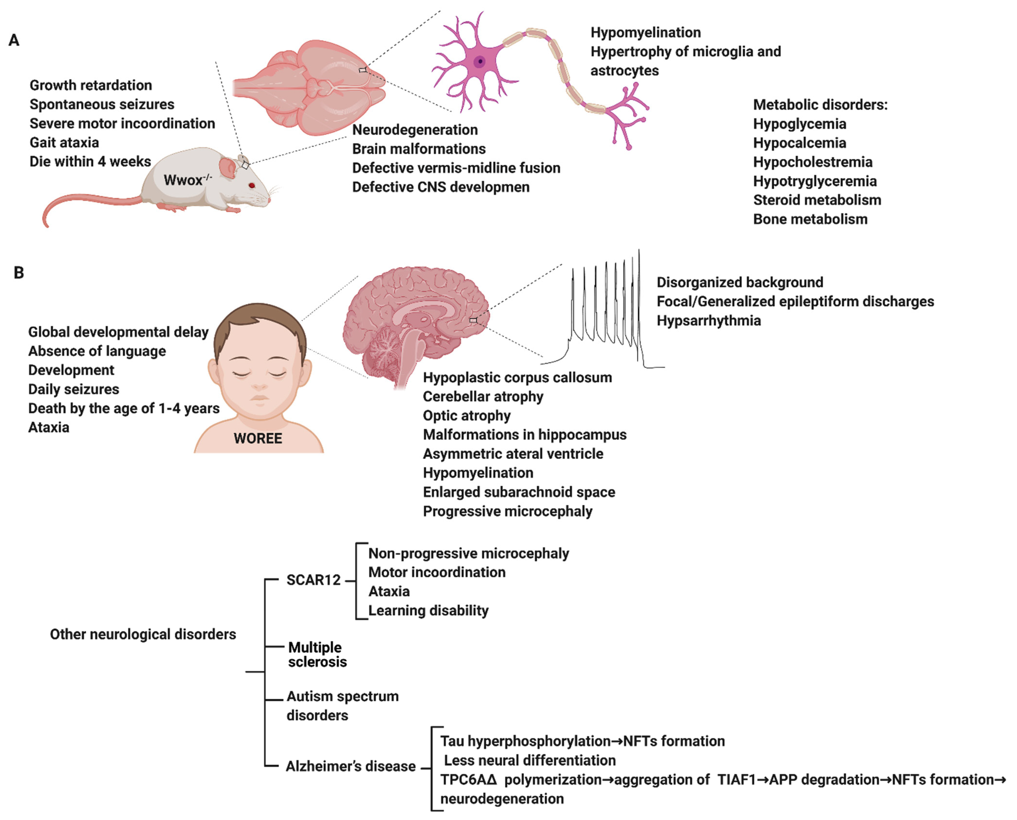
Several animal models of WWOX deficiency have been described in the literature, however, two models in particular report phenocopy epilepsy. Suzuki et al. reported that a spontaneous deletion of 13-bp in exon 9 within the WWOX gene of the lde/lde (lethal dwarfism with epilepsy) rat was associated with dwarfism, ataxic gait, high incidence of epileptic seizures, and postnatal lethality [36] providing direct evidence for the role of WWOX in CNS biology. The WWOX knockout (KO) mouse model exhibits features that phenocopy some symptoms observed in individuals with WWOX variants such as epilepsy, structural brain malformation, and learning impairments [4,37,38,39]. The full spectrum of the model includes growth retardation, severe metabolic disorder (hypoglycemia, hypolipidemia, impaired steroidogenesis, and bone metabolic phenotypes), spontaneous and audiogenic seizures, gait ataxia, severe motor incoordination, imbalance, and premature death by 2 to 3 weeks after birth [4,37,38,39]. The brains of WWOX KO mice show cerebral malformations, microcephaly, incomplete hemisphere separation, neuronal disorganization, heterotopia, defective cerebellar midline fusion, and the degeneration of many neural cell types (Figure 2). Heterotopia (the ectopic displacement of grey matter in the brain) is well known to be associated with seizures [40]. Reduced GABAergic interneurons (PV and NPY) and activation or hypertrophy of microglia and astrocytes in the hippocampus of WWOX KO mice is also consistent with an epileptic phenotype.
Transcriptional analysis of neurospheres derived from WWOX KO neural stem cells reveals alterations in the expression of genes related to neurological disorders, CNS development, and epilepsy [41]. Furthermore, transcriptomic analysis in WWOX-depleted human neural progenitor cells demonstrates that WWOX downregulation significantly alters the expression of genes involved in neuron migration and cytoskeleton organization [20,42]. Despite these studies, it is evident that detailed studies uncovering the molecular and cellular roles of WWOX in pediatric epilepsy are still lacking.

6. WWOX Impairment in Neurological Disorders

There is an emerging role of WWOX in a number of neurological disorders including early-onset epilepsy, autism spectrum disorder (ASD), multiple sclerosis (MS), and Alzheimer’s disease (Figure 2). However, the underlying mechanism of WWOX function is still currently unknown.

6.1. WWOX in Alzheimer’s Disease

Mutations in WWOX have recently been found to be a significant risk factor for disease development in Alzheimer’s disease, an adult-onset neurodegenerative disorder [43]. Furthermore, decreased WWOX protein levels have been observed in Alzheimer’s disease patients compared to age-matched healthy controls [31,44]. In the hippocampus of Alzheimer’s disease patients, an increase in the phosphorylation of the microtubule-associated protein, tau, was reported in a glycogen synthase kinase 3β (GSK-3β)-dependent pathway [44]. WWOX was found to physically interact with and inhibit GSK-3β, preventing tau hyperphosphorylation and transformation into neurofibrillary tangles (NFTs), leading to neuronal loss [45]. WWOX was also shown to promote neural differentiation by suppressing GSK-3β activity, resulting in an increase of the affinity to microtubules of tau, allowing microtubule assembly to prompt neuronal differentiation [45] (Figure 1). It has also been demonstrated that WWOX physically interacts with TPC6AΔ, an aggregated vesicle-trafficking protein isoform that has a critical role in causing caspase activation, tau aggregation, and Aβ generation in patients with Alzheimer’s disease, blocking its self-aggregation [46]. Consequent to WWOX loss, TPC6AΔ undergoes polymerization leading to the aggregation of TGF-β1-induced antiapoptotic factor (TIAF1) caspase activation, which causes amyloid precursor protein (APP) degradation, leading to the generation of amyloid β and the formation of the neurofibrillary tangles (NFTs), causing neurodegeneration (Figure 1) [47,48].

6.2. WWOX in Multiple Sclerosis

Multiple sclerosis (MS) is an immune-mediated disorder that affects the CNS [49]. It is a demyelinating disease that attacks the myelin sheaths covering the axons of nerve cells in the brain and the spinal cord. These attacks cause myelin sheath destruction, which can further lead to axonal damage and loss, inducing many severe neurological disabilities [50]. Lately, a study published by Jäkel et al. using single nucleus RNA-sequencing on post-mortem tissues of 4 MS individuals and 5 healthy controls determined that an alteration in oligodendrocyte population heterogeneity between MS individuals and the controls and a shift in the transcription profile of the different oligodendrocyte sub-clusters in MS tissues. Markedly, in their differential gene expression analysis, WWOX levels were shown to be reduced in the different types of MS lesions, mostly in the chronic active lesions, compared to the controls [51].
Furthermore, several lines of studies have listed WWOX as one of the susceptible genes associated with MS. A genome-wide association study (GWAS) conducted by Beecham et al. on 14,498 individuals with MS and 24,091 healthy controls and joined with previous independent GWAS data from 14,802 MS individuals and 26,703 healthy controls identified 48 susceptibility variants, one of which was the rs12149527 WWOX variant with a joint p = 3.3 × 10⁻¹¹ and an odds ratio (OR) of 1.08 [52]. WWOX rs7201683, another variant, was shown to have a higher frequency in MS individuals than in healthy controls in a study performed on 3 Italian families with MS members, 120 unrelated MS individuals, and healthy controls in order to identify new exonic low-allele frequency variants associated with MS [53]. Furthermore, in a GWAS on 776 MS individuals and 75 healthy controls conducted to study the gray matter pathology indicated by cortical thinning in MS, WWOX was noted to be associated with the thinning of different cortical regions in MS [54]. Additionally, in an analysis of genetic data from 15 different MS genome-associated studies of 47,429 MS individuals and 68,374 healthy controls, the rs12925972 WWOX variant was suggested to be significantly associated with MS susceptibility with a p = 3.07 × 10⁻⁸ and an OR of 1.099 [55]. Altogether, WWOX could play important roles in MS, perhaps due to its emerging function controlling myelination and oligodendrocyte differentiation.

6.3. WWOX in Autism Spectrum Disorders (ASD)

Heterozygous deletions and duplications overlapping the WWOX gene have been observed in individuals with ASD [56,57]. Copy number variants (CNVs) of WWOX have been reported in many ASD individuals characterized with less severe ASD phenotypes and intelligence quotient (IQ) levels approximate to the normal ranges, suggesting that heterozygous WWOX variants were a low-penetrance risk factor for ASD [58,59]. These WWOX variants act as weak risk factors and are generally associated with milder ASD phenotypes.

6.4. WWOX in Early Onset Epilepsy and Ataxia

Consistent with the observations in WWOX animal models, human patients with WWOX bi-allelic loss of function variants (deletions, nonsense, and some missense mutations) were found to be associated with autosomal recessive cerebellar ataxia, epilepsy, optic tract atrophy, retinal degeneration, growth retardation, developmental delay, intellectual disabilities, microcephaly with seizures, and early death [4,5,6,60,61,62,63,64,65,66,67,68,69,70,71,72,73,74,75,76]. The range of phenotypic severity of WWOX-deficient individuals extends from less severe phenotypes with later onset and non-progressive microcephaly, as seen in SCAR12, to more severe phenotypes with progressive microcephaly, seizures, global developmental delay, optic atrophy, and early age lethality, as observed in WOREE. Moreover, brain magnetic resonance images (MRI) of children with WOREE revealed an abnormally thin or hypoplastic corpus callosum, progressive optic atrophy, and brain atrophy as the most common signatures, while delayed myelination and white matter hyperintense signals were reported in some cases [5]. In patients with SCAR12, brain MRIs revealed mild hypoplasia of the cerebellum or cerebellar vermis. There was also a description of a patient with epilepsy of infancy with migrating focal seizures (EIMFS), in which the seizures started at 2.5 months with infantile spasms, and at the age of 6 years, there was profound severe intellectual impairment, microcephaly, spasticity, hypotonia, and scoliosis [73]. Another interesting West syndrome case described a heterozygous 6.8-Mb deletion of WWOX suggesting other genes within this large genomic region might be involved [77]. Although there are limited studies, overall, the phenotypic severity of WWOX deficient patients appears to be variant-specific, suggesting a phenotype–genotype correlation.

7. WWOX Variants and Mutations in Early Onset Epilepsy and Ataxia

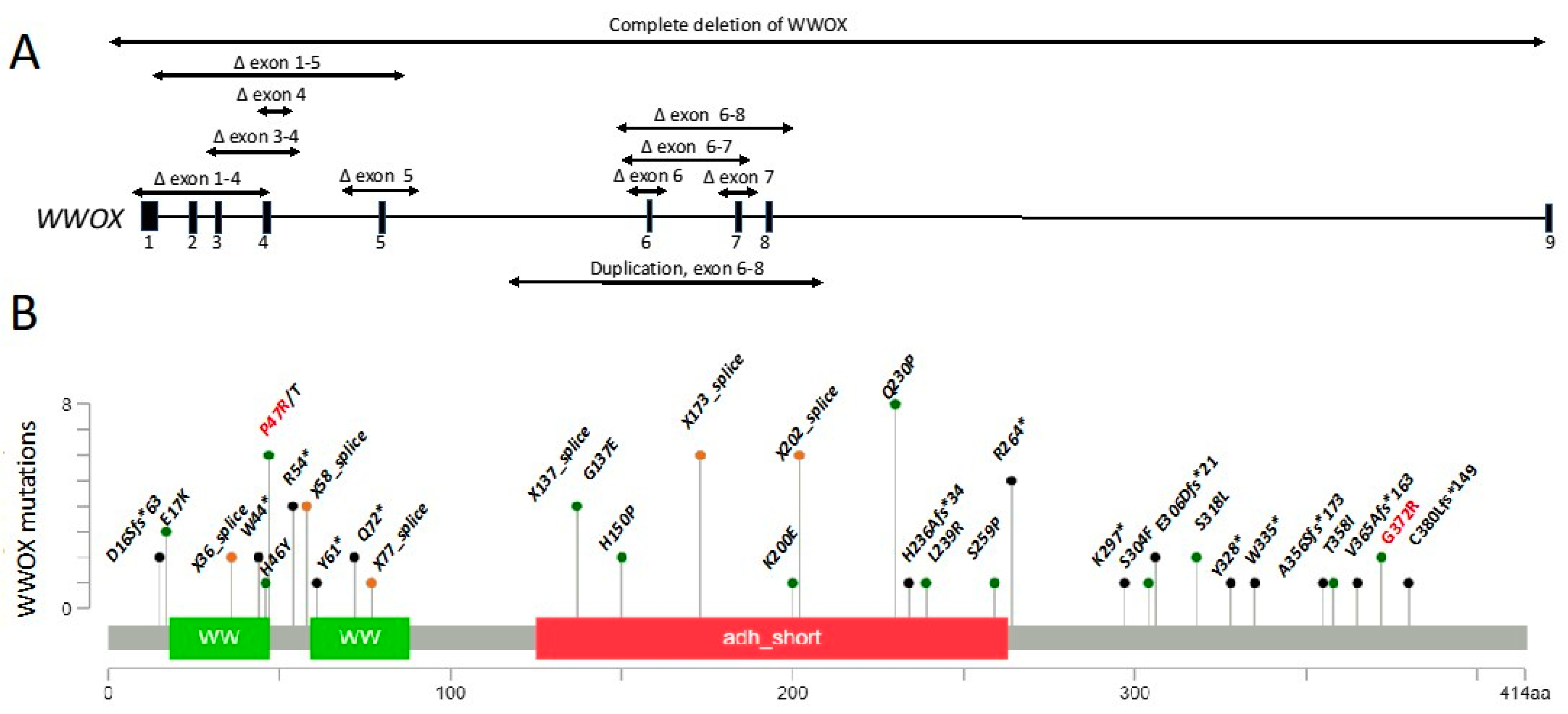
Using Mutationmapper [78], we generated a gene map of all reported pathogenic WWOX variants demonstrating the range of variants spanning the gene (Figure 3). All SCAR12 individuals carry only missense variants (Figure 3 (red), Table 1), however, as only a small number of SCAR12 individuals have been reported to date, it remains unknown if this genotype is common to all SCAR12 patients or if it represents a sampling bias. This may be due to either the reserved protein function in the missense variants or the presence of patient-specific modifiers on the gene. Although the loss of function variants (nonsense, frameshift, splice-site) are more common, the missense variants are also observed in WOREE syndrome individuals. A better understanding of the genotype–phenotype correlations in the SCAR12 and WOREE syndromes will guide research efforts towards a better understanding of the underlying pathophysiology and treatment options of these disorders and assist in clinical management. We encourage clinicians and patients to register their variants and phenotypes on the WWOX foundation website (https://www.wwox.org, 1 April 2021) to facilitate further research on the WWOX gene.
To perform a comprehensive review of all reported WWOX variants, we combined in this review article publicly available data, our own database of patients (Figure S1), peer-reviewed publications in PubMed (https://pubmed.ncbi.nlm.nih.gov/, 1 April 2021), the online mendelian database (OMIM) (https://omim.org/, 1 April 2021), the DECIPHER database (https://decipher.sanger.ac.uk/, 1 April 2021), the ClinVar database (https://www.ncbi.nlm.nih.gov/clinvar/, 1 April 2021), the DGV database (http://dgv.tcag.ca/dgv/app/home, 1 April 2021), and finally data from gnomAD [79,80], to understand variant frequency in healthy controls.

7.1. Published WOREE Cases

In total, 45 variants were reported in 56 WOREE patient cases (Table 1). Of these, 34 were loss of function mutations, and 11 were missense variants. Of the reported WOREE cases described, 19 individuals had either a homozygous or compound heterozygous missense variant. Glutamine to proline substitution on amino acid 230 (Q230P) is an example of one of the missense variants, representing a very conserved amino acid residue change. This can explain the more severe disease phenotype even without a loss of function alleles, or a condition in which a missense mutation was actually causative of a loss of function effect.

7.2. Further Patient Details

Patient data regarding gender, ethnicity, seizure onset, and death are presented where available (Table 1; an extended Table 1 is provided as Table S1). Variants were reported in individuals spanning over 20 countries and various ethnicities. To date, six individuals with SCAR12 variants have been reported in two families with seizures manifesting at less than 2 years of age [4]. WOREE syndrome individuals have many discreet mutations [5,12], with most mutations only described in a single patient (Table 1).

7.3. Data Collected from ClinVar

The dataset was divided into single-gene variations and continuous gene deletions, and subdivided into the five variation calls of benign, likely benign, variant of unknown significance (VUS), likely pathogenic, and pathogenic [81].
We found a total of 544 variants in WWOX deposited in ClinVar (https://www.ncbi.nlm.nih.gov/clinvar/, 1 April 2021), with 224 being benign or likely benign, and 320 being recorded as either variants of unknown significance (VUS), pathogenic, or likely pathogenic variants. These variants included the missense and nonsense point mutations, the partial and complete deletions, and the mutations affecting splicing. Of these variants, 205 were single-gene variants smaller than 500 kb deletions in size, with the remainder representing large deletions of the chromosome, removing multiple genes (including WWOX) at the locus. Because variants in ClinVar can be reported as individual allelic variants without requiring information of the other allele, variants whose second allele was not described were excluded from our study. Out of four variants associated with large deletions between 50 and 500kb, two were already published, and their descriptions appear in Table S1. Two others were not well described, and represent single-allele deletions. A single variant with a deletion between 1 kb and 50 kb was classified as a VUS due to insufficient evidence regarding its pathogenicity, and, furthermore, that variant was excluded from the dataset because no data regarding zygosity was available for that patient. Five additional variants between 51 bp and 1 kb were omitted for the same reasons. Most of the variants reviewed were smaller than 51 bp.
We also compared 103 VUS (Table S2) using the gnomAD database to further characterize these variants. Of these, 93 were missense variants, 6 intronic, 2 nonsense and 2 were indel. We found that 87% of the variants in gnomAD appeared between 1–109 times, with an average of 14 individual alleles per variant. None of these variants were seen in a homozygous state. In total there were 1057 alleles out of approximately 250 thousand alleles in gnomAD, giving an allele frequency of approximately 1:250. We do not know if all the variants are in cis or trans, i.e.: if they are on the same or different haplotypes. If they are indeed on different haplotypes, the expected frequency of disease cases was 1:62,000. If some are actually in cis, the expected disease frequency would be lower. We also assume random mating, but that might not always be the case. For a population of 7.5 billion people, this would result in approximately 121,000 cases of WOREE in the entire population. This number is higher than that of the cases reported in the population, which can be indicated by adding the 56 published patients to the additional 160 cases documented in ClinVar, (ClinVar has allele data and not zygosity data, so we assume all of these variants are combined into pairs). in conclusion, we are aware of a total of 217 reported worldwide cases of WWOX-related patients.
The difference between these two numbers obviously needs addressing. One possibility is that there is an under-diagnosis of WWOX-related neurological syndromes, which may be due to lack of reporting or partial/no genetic workup in cases of unexplained epilepsy. It also could be that many of these variants are benign, which should be addressed in future studies.
The likely-pathogenic and pathogenic alleles were then curated and compared to those of the previously-published individuals and their variants. The most represented type of variant was the loss of function variant, except for the two described families with SCAR12, which were missense variants [4]. Most of the individuals had compound heterozygous or homozygous variants, which is consistent in individuals with a clinical description matching WOREE or SCAR12. We conclude that pathogenic variants in WWOX are most likely recessive (bi-allelic) loss of function mutations, resulting in a non-functional or non-existent protein. This is in concordance with their gnomAD pLI score of 0 (probability of loss of function intolerance) [82].

7.4. Unpublished WWOX Cases

Using the DECIPHER and ClinVar databases, we identified ten previously unpublished individuals with pathogenic WWOX mutations (Table 2 and Table S3). Four patients were found in the DECIPHER database and 6 in ClinVar. Two individuals identified in DECIPHER were described with epileptic encephalopathy, brain ventriculomegaly, cryptorchidism, hydrocele testis, and inguinal hernia, while the other two individuals were described with epileptic encephalopathy and profound global developmental delay.
Of the six cases identified in ClinVar, all individuals had the loss of function mutations and were described as having seizures. Two of the individuals had developmental delay, two had microcephaly, and one had dystonia and an adducted thumb.
It is worth mentioning, as noted previously by Piard et al. [5], that the 46,XY DSD phenotype described by White et al. [7] was not, due to a single heterozygous allelic deletion, noted in any of the published or unpublished cases. This patient harbored a heterozygous deletion of part of the WWOX gene and presented from birth with ambiguous genitalia, undifferentiated gonadal tissue, and immature testis.

8. Future Possibilities for Personalized Medicine Interventions

Antiepileptic drugs (AEDs) are used in the mainstream clinical management of WOREE individuals. However, they are not effective, with most individuals becoming refractory to treatment (Table S1). The poor response of WOREE patients to AEDs challenges the scientific and clinical communities to develop alternative therapeutic approaches. Without exact genotype–phenotype correlations, clinical management is hampered by limited knowledge of the underlying pathophysiology of this disorder, the exact role of WWOX in the brain, and a lack of understanding of whether specific missense variants lead to the gain-of-function or the loss-of-function (LOF) of the WWOX protein.
The development of WWOX-brain organoid cultures and models could aid in this respect. Brain organoids are generated from patient cells (blood leukocytes or skin fibroblasts), through reprogramming into induced pluripotent stem cells (iPSCs). iPSCs have been used in the past to model pediatric neurodevelopmental disorders such as Rett syndrome [83]. The use of iPSC-derived brain organoids has been shown to provide important insights into neurodevelopment and disease mechanisms such as microcephaly and others [84,85], and is intended to serve in WWOX-related disorders as well [86]. This will enable a better understanding of the mechanism of action, and also provide an improved model for future interventions and the testing of candidate drugs and therapies.
Genetic approaches to correct or replace defective genes in monogenetic diseases are continuously developing, but are largely dependent on the nature of the gene and its associated variants. For example, variants occurring in either splice acceptor or splice donor sites that alter the natural splicing machinery of genes are considered severe mutations. Defective splicing due to a splice-site mutation can cause a complete loss of the function phenotype by removing part of the final protein product and/or affecting the overall coding sequence, which could cause a frame shift or premature termination. Both can also lead to the complete elimination of one copy of the gene through nonsense-mediated mRNA decay (NMD). Efforts to reduce or completely ablate the NMD process have recently shown promise [87]. This could be a possible therapy in patients with splice-site mutations such as the case of the Yemenite founder WWOX-splicing mutation [71], and also in patients with any NMD-based allele deletion.
The WWOX loss of function variants involving loss of WWOX protein expression suggest the possibility of introducing the coding region of WWOX by genetic editing or gene therapy. For individuals with complete loss of function mutations, a copy of the WWOX cDNA introduced into target cells could potentially provide a therapeutic option. Recent success in treating spinal muscular atrophy (SMA), a neuromuscular disorder with grave consequences of varying severity, has recently shown success in gene therapy clinical trials where patients with SMA were treated with a partial to complete restoration of the gene through the use of an AAV9 vector [88]. With advances in technology and a better understanding of the underlying pathophysiology of each WWOX variant, it is conceivable that gene therapy will also become available for WWOX-related disease patients with LOF mutations.
Another therapeutic approach is gene editing using the CRISPR-Cas9 system. This system is among other in situ correction gene editing options, holding promise even if there is a need for single-nucleotide correction. The most common mechanism of use for repair is the extraction of patient cells, ex vivo repair using CRISPR-Cas9, and the reimplantation of the corrected cells [89]. This model is not feasible for neurological disorders and hence alternatives to generate systems based on CRISPR-Cas9 that can cross the blood–brain barrier are being developed [90]. CRISPR-Cas9 gene-editing technology is being tested in clinical trials in vitro for Leber’s congenital amaurosis 10 (LCA10), which results in blindness, to correct the CEP290 gene, which is responsible for the disease [91,92,93,94]. Such development is an important step towards the use of gene therapy to tackle diseases with no available treatments. It is thus clear that several approaches for providing precision medicine to patients with WWOX-neurological disorders should be exploited and developed.

9. Concluding Remarks

Here we presented an updated review of the literature describing the WWOX gene in early-onset epilepsy and associated neurological disorders. There are two main medical diagnoses with direct relation to the human germline WWOX pathogenic mutations SCAR12 and WOREE. As the SCAR12 condition was reported only reported in two families (six patients), it qualifies as an ultra-rare condition. The pathogenic mutations described in this gene are missense mutations, which might explain the difference in phenotypes between SCAR12 and WOREE syndrome. This gives rise to the hypothesis that while the complete loss of function variants will cause WOREE syndrome, the missense variants can sometimes maintain some residual protein function, therefore giving rise to the SCAR12 phenotype. The single allele heterozygous loss of function mutations are not expected to lead to severe phenotypes, as was determined from the gnomAD score of pLI = 0. Since there are insufficient medical and genetic diagnostic facilities in undeveloped and developing countries, we assume that there will be more patients diagnosed with WWOX mutations worldwide. It is worthwhile to include the WWOX gene in routine genetic tests in order to provide treatment with precision medicine approaches, as discussed above. To summarize, the “red flags” for WWOX-related disorders should be suspected in children with early-onset refractory epilepsy, profound developmental delay, abnormal EEG patterns, and brain abnormalities, possibly with eye involvement. This combination, alongside an autosomal recessive inheritance pattern, should trigger this diagnosis in patients.
The fact that WWOX-related epilepsy disorders are rare requires a joint effort from the scientific community and patient advocates to achieve a better understanding of WWOX-related diseases and their underlying mechanisms to develop new curative therapeutics. Very recently, two platforms were generated to help reach all the patients and mutations identified globally. A dedicated WWOX Foundation (https://www.wwox.org, 1 April 2021) was established with the aim of increasing awareness, helping develop novel treatment options, and finding a cure for WWOX-related syndromes. A registry database will be established to follow all cases. Another platform exists on the Human Disease Genes website, which provides information on patients with germinal mutations in WWOX, including clinical data, molecular data, management, and research options (https://humandiseasegenes.nl/WWOX/, 1 April 2021).
Further knowledge of the protein structure, domains, and interacting proteins of WWOX will be required to enable a better understanding of the true nature of its mechanism of action. Using animal models to dissect WWOX function in the brain will be instrumental while developing brain organoid models from patient-derived iPSCs will enable a better understanding of the complex interactions between WWOX and other cellular factors in human brain tissue and will serve as an invaluable platform for testing new therapeutic options.

Supplementary Materials

The following are available online at https://www.mdpi.com/article/10.3390/cells10040824/s1, Figure S1: Data collection and analysis, Table S1: Published WWOX-related epilepsy patients-full table related to Table 1. Table S2: ClinVar variants of uncertain significance (VUS) that are linked to WOREE and SCAR12 patients. Table S3: ClinVar pathogenic and likely pathogenic variants that are linked to WOREE and SCAR12.

Author Contributions

Conceptualization, E.B., B.A., W.G., R.I.A.; formal analysis and investigation, E.B., B.A., S.A.-S., S.R.R., D.J.S., D.S., S.A., L.L., P.L.C., W.G., R.I.A.; writing—original draft preparation, E.B., B.A., S.A.-S., S.R.R., D.J.S., S.A., P.L.C., W.G., R.I.A. All authors have read and agreed to the published version of the manuscript.

Funding

This research received no external funding.

Acknowledgments

The Aqeilan’s lab is funded by the European Research Council (ERC) [grant agreement No. 682118], the Proof-of-Concept ERC grant [No. 957543] and the KAMIN grant from the Israel Innovation Authority [No. 69118].

Conflicts of Interest

The authors declare no conflict of interest.

References

  1. Howell, K.B.; Freeman, J.L.; Mackay, M.T.; Fahey, M.C.; Archer, J.; Berkovic, S.F.; Chan, E.; Dabscheck, G.; Eggers, S.; Hayman, M.; et al. The severe epilepsy syndromes of infancy: A population-based study. Epilepsia 2021, 62, 358–370. [Google Scholar] [CrossRef]
  2. Steward, C.A.; Roovers, J.; Suner, M.M.; Gonzalez, J.M.; Uszczynska-Ratajczak, B.; Pervouchine, D.; Fitzgerald, S.; Viola, M.; Stamberger, H.; Hamdan, F.F.; et al. Re-annotation of 191 developmental and epileptic encephalopathy-associated genes unmasks de novo variants in SCN1A. NPJ Genom. Med. 2019, 4, 31. [Google Scholar] [CrossRef] [Green Version]
  3. Shbarou, R.; Mikati, M.A. The Expanding Clinical Spectrum of Genetic Pediatric Epileptic Encephalopathies. Semin. Pediatr. Neurol. 2016, 23, 134–142. [Google Scholar] [CrossRef] [PubMed]
  4. Mallaret, M.; Synofzik, M.; Lee, J.; Sagum, C.A.; Mahajnah, M.; Sharkia, R.; Drouot, N.; Renaud, M.; Klein, F.A.; Anheim, M.; et al. The tumour suppressor gene WWOX is mutated in autosomal recessive cerebellar ataxia with epilepsy and mental retardation. Brain 2014, 137, 411–419. [Google Scholar] [CrossRef] [PubMed] [Green Version]
  5. Piard, J.; Hawkes, L.; Milh, M.; Villard, L.; Borgatti, R.; Romaniello, R.; Fradin, M.; Capri, Y.; Heron, D.; Nougues, M.C.; et al. The phenotypic spectrum of WWOX-related disorders: 20 additional cases of WOREE syndrome and review of the literature. Genet. Med. 2019, 21, 1308–1318. [Google Scholar] [CrossRef] [Green Version]
  6. Shaukat, Q.; Hertecant, J.; El-Hattab, A.W.; Ali, B.R.; Suleiman, J. West syndrome, developmental and epileptic encephalopathy, and severe CNS disorder associated with WWOX mutations. Epileptic Disord. 2018, 20, 401–412. [Google Scholar] [CrossRef] [PubMed]
  7. White, S.; Hewitt, J.; Turbitt, E.; van der Zwan, Y.; Hersmus, R.; Drop, S.; Koopman, P.; Harley, V.; Cools, M.; Looijenga, L.; et al. A multi-exon deletion within WWOX is associated with a 46, XY disorder of sex development. Eur. J. Hum. Genet. 2012, 20, 348–351. [Google Scholar] [CrossRef] [Green Version]
  8. Gribaa, M.; Salih, M.; Anheim, M.; Lagier-Tourenne, C.; H’Mida, D.; Drouot, N.; Mohamed, A.; Elmalik, S.; Kabiraj, M.; Al-Rayess, M.; et al. A new form of childhood onset, autosomal recessive spinocerebellar ataxia and epilepsy is localized at 16q21-q23. Brain 2007, 130, 1921–1928. [Google Scholar] [CrossRef] [Green Version]
  9. Bird, T.D. Hereditary Ataxia Overview. In GeneReviews®; Adam, M.P., Ardinger, H.H., Pagon, R.A., Wallace, S.E., Bean, L.J.H., Mirzaa, G., Amemiya, A., Eds.; University of Washington: Seattle, WA, USA, 1993. [Google Scholar]
  10. Bednarek, A.K.; Laflin, K.J.; Daniel, R.L.; Liao, Q.; Hawkins, K.A.; Aldaz, C.M. WWOX, a novel WW domain-containing protein mapping to human chromosome 16q23.3-24.1, a region frequently affected in breast cancer. Cancer Res. 2000, 60, 2140–2145. [Google Scholar]
  11. Del Mare, S.; Salah, Z.; Aqeilan, R.I. WWOX: Its genomics, partners, and functions. J. Cell. Biochem. 2009, 108, 737–745. [Google Scholar] [CrossRef]
  12. Abu-Remaileh, M.; Dodson, E.J.; Schueler-Furman, O.; Aqeilan, R.I. Pleiotropic Functions of Tumor Suppressor WWOX in Normal and Cancer Cells. J. Biol. Chem. 2015. [Google Scholar] [CrossRef] [PubMed] [Green Version]
  13. Salah, Z.; Aqeilan, R.; Huebner, K. WWOX gene and gene product: Tumor suppression through specific protein interactions. Future Oncol. 2010, 6, 249–259. [Google Scholar] [CrossRef] [PubMed] [Green Version]
  14. Aqeilan, R.I.; Abu-Remaileh, M.; Abu-Odeh, M. The common fragile site FRA16D gene product WWOX: Roles in tumor suppression and genomic stability. Cell. Mol. Life Sci. 2014, 71, 4589–4599. [Google Scholar] [CrossRef] [PubMed]
  15. Aqeilan, R.I.; Croce, C.M. WWOX in biological control and tumorigenesis. J. Cell. Physiol. 2007, 212, 307–310. [Google Scholar] [CrossRef]
  16. Gardenswartz, A.; Aqeilan, R.I. WW domain-containing oxidoreductase’s role in myriad cancers: Clinical significance and future implications. Exp. Biol. Med. 2014, 239, 253–263. [Google Scholar] [CrossRef] [PubMed]
  17. Aldaz, C.M.; Ferguson, B.W.; Abba, M.C. WWOX at the crossroads of cancer, metabolic syndrome related traits and CNS pathologies. Biochim. Biophys. Acta 2014. [Google Scholar] [CrossRef] [PubMed] [Green Version]
  18. Bouteille, N.; Driouch, K.; Hage, P.E.; Sin, S.; Formstecher, E.; Camonis, J.; Lidereau, R.; Lallemand, F. Inhibition of the Wnt/beta-catenin pathway by the WWOX tumor suppressor protein. Oncogene 2009. [Google Scholar] [CrossRef] [PubMed] [Green Version]
  19. Khawaled, S.; Nigita, G.; Distefano, R.; Oster, S.; Suh, S.S.; Smith, Y.; Khalaileh, A.; Peng, Y.; Croce, C.M.; Geiger, T.; et al. Pleiotropic tumor suppressor functions of WWOX antagonize metastasis. Signal Transduct. Target. Ther. 2020, 5, 43. [Google Scholar] [CrossRef] [Green Version]
  20. Kosla, K.; Pluciennik, E.; Styczen-Binkowska, E.; Nowakowska, M.; Orzechowska, M.; Bednarek, A.K. The WWOX Gene Influences Cellular Pathways in the Neuronal Differentiation of Human Neural Progenitor Cells. Front. Cell. Neurosci. 2019, 13, 391. [Google Scholar] [CrossRef] [Green Version]
  21. Ferguson, B.W.; Gao, X.; Zelazowski, M.J.; Lee, J.; Jeter, C.R.; Abba, M.C.; Aldaz, C.M. The cancer gene WWOX behaves as an inhibitor of SMAD3 transcriptional activity via direct binding. BMC Cancer 2013, 13, 593. [Google Scholar] [CrossRef] [Green Version]
  22. Aqeilan, R.I.; Donati, V.; Palamarchuk, A.; Trapasso, F.; Kaou, M.; Pekarsky, Y.; Sudol, M.; Croce, C.M. WW domain-containing proteins, WWOX and YAP, compete for interaction with ErbB-4 and modulate its transcriptional function. Cancer Res. 2005, 65, 6764–6772. [Google Scholar] [CrossRef] [Green Version]
  23. Aqeilan, R.I.; Donati, V.; Gaudio, E.; Nicoloso, M.S.; Sundvall, M.; Korhonen, A.; Lundin, J.; Isola, J.; Sudol, M.; Joensuu, H.; et al. Association of WWOX with ErbB4 in Breast Cancer. Cancer Res. 2007, 67, 9330–9336. [Google Scholar] [CrossRef] [PubMed] [Green Version]
  24. Schuchardt, B.J.; Bhat, V.; Mikles, D.C.; McDonald, C.B.; Sudol, M.; Farooq, A. Molecular Origin of the Binding of WWOX Tumor Suppressor to ErbB4 Receptor Tyrosine Kinase. Biochemistry 2013, 52, 9223–9236. [Google Scholar] [CrossRef] [PubMed] [Green Version]
  25. Fu, J.; Qu, Z.; Yan, P.; Ishikawa, C.; Aqeilan, R.I.; Rabson, A.B.; Xiao, G. The tumor suppressor gene WWOX links the canonical and noncanonical NF-kappaB pathways in HTLV-I Tax-mediated tumorigenesis. Blood 2011, 117, 1652–1661. [Google Scholar] [CrossRef] [Green Version]
  26. Abu-Remaileh, M.; Aqeilan, R.I. Tumor suppressor WWOX regulates glucose metabolism via HIF1alpha modulation. Cell Death Differ. 2014, 21, 1805–1814. [Google Scholar] [CrossRef] [PubMed] [Green Version]
  27. O’Keefe, L.V.; Colella, A.; Dayan, S.; Chen, Q.; Choo, A.; Jacob, R.; Price, G.; Venter, D.; Richards, R.I. Drosophila orthologue of WWOX, the chromosomal fragile site FRA16D tumour suppressor gene, functions in aerobic metabolism and regulates reactive oxygen species. Hum. Mol. Genet. 2011, 20, 497–509. [Google Scholar] [CrossRef] [PubMed] [Green Version]
  28. Abu-Odeh, M.; Salah, Z.; Herbel, C.; Hofmann, T.G.; Aqeilan, R.I. WWOX, the common fragile site FRA16D gene product, regulates ATM activation and the DNA damage response. Proc. Natl. Acad. Sci. USA 2014, 111, E4716–E4725. [Google Scholar] [CrossRef] [Green Version]
  29. Aqeilan, R.I.; Pekarsky, Y.; Herrero, J.J.; Palamarchuk, A.; Letofsky, J.; Druck, T.; Trapasso, F.; Han, S.Y.; Melino, G.; Huebner, K.; et al. Functional association between WWOX tumor suppressor protein and p73, a p53 homolog. Proc. Natl. Acad. Sci. USA 2004, 101, 4401–4406. [Google Scholar] [CrossRef] [Green Version]
  30. Salah, Z.; Bar-mag, T.; Kohn, Y.; Pichiorri, F.; Palumbo, T.; Melino, G.; Aqeilan, R.I. Tumor suppressor WWOX binds to DeltaNp63alpha and sensitizes cancer cells to chemotherapy. Cell Death Dis. 2013, 4, e480. [Google Scholar] [CrossRef] [Green Version]
  31. Liu, C.C.; Ho, P.C.; Lee, I.T.; Chen, Y.A.; Chu, C.H.; Teng, C.C.; Wu, S.N.; Sze, C.I.; Chiang, M.F.; Chang, N.S. WWOX Phosphorylation, Signaling, and Role in Neurodegeneration. Front. Neurosci. 2018, 12, 563. [Google Scholar] [CrossRef]
  32. Nunez, M.I.; Ludes-Meyers, J.; Aldaz, C.M. WWOX protein expression in normal human tissues. J. Mol. Histol. 2006, 37, 115–125. [Google Scholar] [CrossRef] [PubMed] [Green Version]
  33. Chen, S.T.; Chuang, J.I.; Wang, J.P.; Tsai, M.S.; Li, H.; Chang, N.S. Expression of WW domain-containing oxidoreductase WOX1 in the developing murine nervous system. Neuroscience 2004, 124, 831–839. [Google Scholar] [CrossRef]
  34. Aldaz, C.M.; Hussain, T. WWOX Loss of Function in Neurodevelopmental and Neurodegenerative Disorders. Int. J. Mol. Sci. 2020, 21, 8922. [Google Scholar] [CrossRef] [PubMed]
  35. Tochigi, Y.; Takamatsu, Y.; Nakane, J.; Nakai, R.; Katayama, K.; Suzuki, H. Loss of WWOX Causes Defective Development of Cerebral Cortex with Hypomyelination in a Rat Model of Lethal Dwarfism with Epilepsy. Int. J. Mol. Sci. 2019, 20, 3596. [Google Scholar] [CrossRef] [Green Version]
  36. Suzuki, H.; Katayama, K.; Takenaka, M.; Amakasu, K.; Saito, K.; Suzuki, K. A spontaneous mutation of the WWOX gene and audiogenic seizures in rats with lethal dwarfism and epilepsy. Genes Brain Behav. 2009. [Google Scholar] [CrossRef] [PubMed]
  37. Aqeilan, R.I.; Hassan, M.Q.; de Bruin, A.; Hagan, J.P.; Volinia, S.; Palumbo, T.; Hussain, S.; Lee, S.H.; Gaur, T.; Stein, G.S.; et al. The WWOX tumor suppressor is essential for post-natal survival and normal bone metabolism. J. Biol. Chem. 2008, 283, 21629–21639. [Google Scholar] [CrossRef] [PubMed] [Green Version]
  38. Aqeilan, R.I.; Trapasso, F.; Hussain, S.; Costinean, S.; Marshall, D.; Pekarsky, Y.; Hagan, J.P.; Zanesi, N.; Kaou, M.; Stein, G.S.; et al. Targeted deletion of WWOX reveals a tumor suppressor function. Proc. Natl. Acad. Sci. USA 2007, 104, 3949–3954. [Google Scholar] [CrossRef] [Green Version]
  39. Cheng, Y.Y.; Chou, Y.T.; Lai, F.J.; Jan, M.S.; Chang, T.H.; Jou, I.M.; Chen, P.S.; Lo, J.Y.; Huang, S.S.; Chang, N.S.; et al. WWOX deficiency leads to neurodevelopmental and degenerative neuropathies and glycogen synthase kinase 3beta-mediated epileptic seizure activity in mice. Acta Neuropathol. Commun. 2020, 8, 6. [Google Scholar] [CrossRef] [PubMed]
  40. Raymond, A.A.; Fish, D.R.; Stevens, J.M.; Sisodiya, S.M.; Alsanjari, N.; Shorvon, S.D. Subependymal heterotopia: A distinct neuronal migration disorder associated with epilepsy. J. Neurol. Neurosurg. Psychiatry 1994, 57, 1195–1202. [Google Scholar] [CrossRef]
  41. Hussain, T.; Kil, H.; Hattiangady, B.; Lee, J.; Kodali, M.; Shuai, B.; Attaluri, S.; Takata, Y.; Shen, J.; Abba, M.C.; et al. WWOX deletion leads to reduced GABA-ergic inhibitory interneuron numbers and activation of microglia and astrocytes in mouse hippocampus. Neurobiol. Dis. 2019, 121, 163–176. [Google Scholar] [CrossRef]
  42. Iacomino, M.; Baldassari, S.; Tochigi, Y.; Kosla, K.; Buffelli, F.; Torella, A.; Severino, M.; Paladini, D.; Mandara, L.; Riva, A.; et al. Loss of WWOX Perturbs Neuronal Migration and Impairs Early Cortical Development. Front. Neurosci. 2020, 14, 644. [Google Scholar] [CrossRef] [PubMed]
  43. Kunkle, B.W.; Grenier-Boley, B.; Sims, R.; Bis, J.C.; Damotte, V.; Naj, A.C.; Boland, A.; Vronskaya, M.; van der Lee, S.J.; Amlie-Wolf, A.; et al. Genetic meta-analysis of diagnosed Alzheimer’s disease identifies new risk loci and implicates Abeta, tau, immunity and lipid processing. Nat. Genet. 2019, 51, 414–430. [Google Scholar] [CrossRef] [PubMed] [Green Version]
  44. Sze, C.I.; Su, M.; Pugazhenthi, S.; Jambal, P.; Hsu, L.J.; Heath, J.; Schultz, L.; Chang, N.S. Down-regulation of WW domain-containing oxidoreductase induces Tau phosphorylation in vitro. A potential role in Alzheimer’s disease. J. Biol. Chem. 2004, 279, 30498–30506. [Google Scholar] [CrossRef] [PubMed] [Green Version]
  45. Wang, H.Y.; Juo, L.I.; Lin, Y.T.; Hsiao, M.; Lin, J.T.; Tsai, C.H.; Tzeng, Y.H.; Chuang, Y.C.; Chang, N.S.; Yang, C.N.; et al. WW domain-containing oxidoreductase promotes neuronal differentiation via negative regulation of glycogen synthase kinase 3beta. Cell Death Differ. 2012, 19, 1049–1059. [Google Scholar] [CrossRef] [PubMed] [Green Version]
  46. Chang, J.Y.; Lee, M.H.; Lin, S.R.; Yang, L.Y.; Sun, H.S.; Sze, C.I.; Hong, Q.; Lin, Y.S.; Chou, Y.T.; Hsu, L.J.; et al. Trafficking protein particle complex 6A delta (TRAPPC6ADelta) is an extracellular plaque-forming protein in the brain. Oncotarget 2015, 6, 3578–3589. [Google Scholar] [CrossRef] [PubMed]
  47. Chang, J.Y.; Chang, N.S. WWOX dysfunction induces sequential aggregation of TRAPPC6ADelta, TIAF1, tau and amyloid beta, and causes apoptosis. Cell Death Discov. 2015, 1, 15003. [Google Scholar] [CrossRef]
  48. Lee, M.H.; Lin, S.R.; Chang, J.Y.; Schultz, L.; Heath, J.; Hsu, L.J.; Kuo, Y.M.; Hong, Q.; Chiang, M.F.; Gong, C.X.; et al. TGF-beta induces TIAF1 self-aggregation via type II receptor-independent signaling that leads to generation of amyloid beta plaques in Alzheimer’s disease. Cell Death Dis. 2010, 1, e110. [Google Scholar] [CrossRef] [Green Version]
  49. Goldenberg, M.M. Multiple sclerosis review. Pharm. Ther. 2012, 37, 175–184. [Google Scholar]
  50. Podbielska, M.; Banik, N.L.; Kurowska, E.; Hogan, E.L. Myelin recovery in multiple sclerosis: The challenge of remyelination. Brain Sci. 2013, 3, 1282–1324. [Google Scholar] [CrossRef] [PubMed] [Green Version]
  51. Jakel, S.; Agirre, E.; Mendanha Falcao, A.; van Bruggen, D.; Lee, K.W.; Knuesel, I.; Malhotra, D.; Ffrench-Constant, C.; Williams, A.; Castelo-Branco, G. Altered human oligodendrocyte heterogeneity in multiple sclerosis. Nature 2019, 566, 543–547. [Google Scholar] [CrossRef]
  52. International Multiple Sclerosis Genetics, C.; Beecham, A.H.; Patsopoulos, N.A.; Xifara, D.K.; Davis, M.F.; Kemppinen, A.; Cotsapas, C.; Shah, T.S.; Spencer, C.; Booth, D.; et al. Analysis of immune-related loci identifies 48 new susceptibility variants for multiple sclerosis. Nat. Genet. 2013, 45, 1353–1360. [Google Scholar] [CrossRef] [PubMed]
  53. Ziliotto, N.; Marchetti, G.; Scapoli, C.; Bovolenta, M.; Meneghetti, S.; Benazzo, A.; Lunghi, B.; Balestra, D.; Laino, L.A.; Bozzini, N.; et al. C6orf10 Low-Frequency and Rare Variants in Italian Multiple Sclerosis Patients. Front. Genet. 2019, 10, 573. [Google Scholar] [CrossRef] [PubMed] [Green Version]
  54. Matsushita, T.; Madireddy, L.; Sprenger, T.; Khankhanian, P.; Magon, S.; Naegelin, Y.; Caverzasi, E.; Lindberg, R.L.; Kappos, L.; Hauser, S.L.; et al. Genetic associations with brain cortical thickness in multiple sclerosis. Genes Brain Behav. 2015, 14, 217–227. [Google Scholar] [CrossRef] [PubMed] [Green Version]
  55. International Multiple Sclerosis Genetics, C. Multiple sclerosis genomic map implicates peripheral immune cells and microglia in susceptibility. Science 2019, 365. [Google Scholar] [CrossRef] [PubMed] [Green Version]
  56. Xia, K.; Zhang, J.; Ahn, M.; Jha, S.; Crowley, J.J.; Szatkiewicz, J.; Li, T.; Zou, F.; Zhu, H.; Hibar, D.; et al. Genome-wide association analysis identifies common variants influencing infant brain volumes. Transl. Psychiatry 2017, 7, e1188. [Google Scholar] [CrossRef] [PubMed] [Green Version]
  57. Peter, B.; Dinu, V.; Liu, L.; Huentelman, M.; Naymik, M.; Lancaster, H.; Vose, C.; Schrauwen, I. Exome Sequencing of Two Siblings with Sporadic Autism Spectrum Disorder and Severe Speech Sound Disorder Suggests Pleiotropic and Complex Effects. Behav. Genet. 2019, 49, 399–414. [Google Scholar] [CrossRef] [PubMed]
  58. Bacchelli, E.; Cameli, C.; Viggiano, M.; Igliozzi, R.; Mancini, A.; Tancredi, R.; Battaglia, A.; Maestrini, E. An integrated analysis of rare CNV and exome variation in Autism Spectrum Disorder using the Infinium PsychArray. Sci. Rep. 2020, 10, 3198. [Google Scholar] [CrossRef] [Green Version]
  59. Leppa, V.M.; Kravitz, S.N.; Martin, C.L.; Andrieux, J.; Le Caignec, C.; Martin-Coignard, D.; DyBuncio, C.; Sanders, S.J.; Lowe, J.K.; Cantor, R.M.; et al. Rare Inherited and De Novo CNVs Reveal Complex Contributions to ASD Risk in Multiplex Families. Am. J. Hum. Genet. 2016, 99, 540–554. [Google Scholar] [CrossRef] [Green Version]
  60. Abdel-Salam, G.; Thoenes, M.; Afifi, H.H.; Korber, F.; Swan, D.; Bolz, H.J. The supposed tumor suppressor gene WWOX is mutated in an early lethal microcephaly syndrome with epilepsy, growth retardation and retinal degeneration. Orphanet J. Rare Dis. 2014, 9, 12. [Google Scholar] [CrossRef] [Green Version]
  61. Ben-Salem, S.; Al-Shamsi, A.M.; John, A.; Ali, B.R.; Al-Gazali, L. A novel whole exon deletion in WWOX gene causes early epilepsy, intellectual disability and optic atrophy. J. Mol. Neurosci. 2015, 56, 17–23. [Google Scholar] [CrossRef]
  62. Mignot, C.; Lambert, L.; Pasquier, L.; Bienvenu, T.; Delahaye-Duriez, A.; Keren, B.; Lefranc, J.; Saunier, A.; Allou, L.; Roth, V.; et al. WWOX-related encephalopathies: Delineation of the phenotypical spectrum and emerging genotype-phenotype correlation. J. Med. Genet. 2015, 52, 61–70. [Google Scholar] [CrossRef] [PubMed]
  63. Valduga, M.; Philippe, C.; Lambert, L.; Bach-Segura, P.; Schmitt, E.; Masutti, J.P.; Francois, B.; Pinaud, P.; Vibert, M.; Jonveaux, P. WWOX and severe autosomal recessive epileptic encephalopathy: First case in the prenatal period. J. Hum. Genet. 2015, 60, 267–271. [Google Scholar] [CrossRef] [PubMed]
  64. Elsaadany, L.; El-Said, M.; Ali, R.; Kamel, H.; Ben-Omran, T. W44X mutation in the WWOX gene causes intractable seizures and developmental delay: A case report. BMC Med. Genet. 2016, 17, 53. [Google Scholar] [CrossRef] [PubMed] [Green Version]
  65. Tabarki, B.; AlHashem, A.; AlShahwan, S.; Alkuraya, F.S.; Gedela, S.; Zuccoli, G. Severe CNS involvement in WWOX mutations: Description of five new cases. Am. J. Med. Genet. Part A 2015, 167, 3209–3213. [Google Scholar] [CrossRef]
  66. Tarta-Arsene, O.; Barca, D.; Craiu, D.; Iliescu, C. Practical clues for diagnosing WWOX encephalopathy. Epileptic Disord. 2017, 19, 357–361. [Google Scholar] [CrossRef]
  67. Rim, J.H.; Kim, S.H.; Hwang, I.S.; Kwon, S.S.; Kim, J.; Kim, H.W.; Cho, M.J.; Ko, A.; Youn, S.E.; Kim, J.; et al. Efficient strategy for the molecular diagnosis of intractable early-onset epilepsy using targeted gene sequencing. BMC Med. Genom. 2018, 11, 6. [Google Scholar] [CrossRef] [PubMed]
  68. Johannsen, J.; Kortum, F.; Rosenberger, G.; Bokelmann, K.; Schirmer, M.A.; Denecke, J.; Santer, R. A novel missense variant in the SDR domain of the WWOX gene leads to complete loss of WWOX protein with early-onset epileptic encephalopathy and severe developmental delay. Neurogenetics 2018, 19, 151–156. [Google Scholar] [CrossRef] [PubMed]
  69. Kothur, K.; Holman, K.; Farnsworth, E.; Ho, G.; Lorentzos, M.; Troedson, C.; Gupta, S.; Webster, R.; Procopis, P.G.; Menezes, M.P.; et al. Diagnostic yield of targeted massively parallel sequencing in children with epileptic encephalopathy. Seizure 2018, 59, 132–140. [Google Scholar] [CrossRef] [PubMed] [Green Version]
  70. Davids, M.; Markello, T.; Wolfe, L.A.; Chepa-Lotrea, X.; Tifft, C.J.; Gahl, W.A.; Malicdan, M.C.V. Early infantile-onset epileptic encephalopathy 28 due to a homozygous microdeletion involving the WWOX gene in a region of uniparental disomy. Hum. Mutat. 2019, 40, 42–47. [Google Scholar] [CrossRef] [PubMed] [Green Version]
  71. Weisz-Hubshman, M.; Meirson, H.; Michaelson-Cohen, R.; Beeri, R.; Tzur, S.; Bormans, C.; Modai, S.; Shomron, N.; Shilon, Y.; Banne, E.; et al. Novel WWOX deleterious variants cause early infantile epileptic encephalopathy, severe developmental delay and dysmorphism among Yemenite Jews. Eur. J. Paediatr. Neurol. 2019, 23, 418–426. [Google Scholar] [CrossRef] [PubMed]
  72. Yang, C.; Zhang, Y.; Song, Z.; Yi, Z.; Li, F. Novel compound heterozygous mutations in the WWOX gene cause early infantile epileptic encephalopathy. Int. J. Dev. Neurosci. 2019, 79, 45–48. [Google Scholar] [CrossRef] [PubMed]
  73. Burgess, R.; Wang, S.; McTague, A.; Boysen, K.E.; Yang, X.; Zeng, Q.; Myers, K.A.; Rochtus, A.; Trivisano, M.; Gill, D.; et al. The Genetic Landscape of Epilepsy of Infancy with Migrating Focal Seizures. Ann. Neurol. 2019, 86, 821–831. [Google Scholar] [CrossRef] [PubMed]
  74. He, J.; Zhou, W.; Shi, J.; Zhang, B.; Wang, H. A Chinese patient with epilepsy and WWOX compound heterozygous mutations. Epileptic Disord. 2020, 22, 120–124. [Google Scholar] [CrossRef] [PubMed]
  75. Su, T.; Yan, Y.; Xu, S.; Zhang, K.; Xu, S. Early onset epileptic encephalopathy caused by novel compound heterozygous mutation of WWOX gene. Int. J. Dev. Neurosci. 2020, 80, 157–161. [Google Scholar] [CrossRef] [PubMed]
  76. Nashabat, M.; Al Qahtani, X.S.; Almakdob, S.; Altwaijri, W.; Ba-Armah, D.M.; Hundallah, K.; Al Hashem, A.; Al Tala, S.; Maddirevula, S.; Alkuraya, F.S.; et al. The landscape of early infantile epileptic encephalopathy in a consanguineous population. Seizure 2019, 69, 154–172. [Google Scholar] [CrossRef] [PubMed]
  77. Mori, T.; Goji, A.; Toda, Y.; Ito, H.; Mori, K.; Kohmoto, T.; Imoto, I.; Kagami, S. A 16q22.2-q23.1 deletion identified in a male infant with West syndrome. Brain Dev. 2019, 41, 888–893. [Google Scholar] [CrossRef]
  78. Cerami, E.; Gao, J.; Dogrusoz, U.; Gross, B.E.; Sumer, S.O.; Aksoy, B.A.; Jacobsen, A.; Byrne, C.J.; Heuer, M.L.; Larsson, E.; et al. The cBio cancer genomics portal: An open platform for exploring multidimensional cancer genomics data. Cancer Discov. 2012, 2, 401–404. [Google Scholar] [CrossRef] [Green Version]
  79. Wang, Q.; Pierce-Hoffman, E.; Cummings, B.B.; Alfoldi, J.; Francioli, L.C.; Gauthier, L.D.; Hill, A.J.; O’Donnell-Luria, A.H.; Genome Aggregation Database Production, T.; Genome Aggregation Database, C.; et al. Landscape of multi-nucleotide variants in 125,748 human exomes and 15,708 genomes. Nat. Commun. 2020, 11, 2539. [Google Scholar] [CrossRef]
  80. Karczewski, K.J.; Francioli, L.C.; Tiao, G.; Cummings, B.B.; Alfoldi, J.; Wang, Q.; Collins, R.L.; Laricchia, K.M.; Ganna, A.; Birnbaum, D.P.; et al. The mutational constraint spectrum quantified from variation in 141,456 humans. Nature 2020, 581, 434–443. [Google Scholar] [CrossRef]
  81. Richards, S.; Aziz, N.; Bale, S.; Bick, D.; Das, S.; Gastier-Foster, J.; Grody, W.W.; Hegde, M.; Lyon, E.; Spector, E.; et al. Standards and guidelines for the interpretation of sequence variants: A joint consensus recommendation of the American College of Medical Genetics and Genomics and the Association for Molecular Pathology. Genet. Med. 2015, 17, 405–424. [Google Scholar] [CrossRef]
  82. Lek, M.; Karczewski, K.J.; Minikel, E.V.; Samocha, K.E.; Banks, E.; Fennell, T.; O’Donnell-Luria, A.H.; Ware, J.S.; Hill, A.J.; Cummings, B.B.; et al. Analysis of protein-coding genetic variation in 60,706 humans. Nature 2016, 536, 285–291. [Google Scholar] [CrossRef] [Green Version]
  83. Marchetto, M.C.; Carromeu, C.; Acab, A.; Yu, D.; Yeo, G.W.; Mu, Y.; Chen, G.; Gage, F.H.; Muotri, A.R. A model for neural development and treatment of Rett syndrome using human induced pluripotent stem cells. Cell 2010, 143, 527–539. [Google Scholar] [CrossRef] [Green Version]
  84. Lancaster, M.A.; Renner, M.; Martin, C.A.; Wenzel, D.; Bicknell, L.S.; Hurles, M.E.; Homfray, T.; Penninger, J.M.; Jackson, A.P.; Knoblich, J.A. Cerebral organoids model human brain development and microcephaly. Nature 2013, 501, 373–379. [Google Scholar] [CrossRef] [PubMed]
  85. Sidhaye, J.; Knoblich, J.A. Brain organoids: An ensemble of bioassays to investigate human neurodevelopment and disease. Cell Death Differ. 2020. [Google Scholar] [CrossRef] [PubMed]
  86. Steinberg, D.J.; Saleem, A.; Repudi, S.R.; Banne, E.; Mahajnah, M.; Hanna, J.H.; Carlen, P.L.; Aqeilan, R.I. Modeling Genetic Epileptic Encephalopathies using Brain Organoids. bioRxiv 2020. [Google Scholar] [CrossRef]
  87. Denichenko, P.; Mogilevsky, M.; Clery, A.; Welte, T.; Biran, J.; Shimshon, O.; Barnabas, G.D.; Danan-Gotthold, M.; Kumar, S.; Yavin, E.; et al. Specific inhibition of splicing factor activity by decoy RNA oligonucleotides. Nat. Commun. 2019, 10, 1590. [Google Scholar] [CrossRef] [Green Version]
  88. Pattali, R.; Mou, Y.; Li, X.J. AAV9 Vector: A Novel modality in gene therapy for spinal muscular atrophy. Gene Ther. 2019, 26, 287–295. [Google Scholar] [CrossRef]
  89. Li, H.; Yang, Y.; Hong, W.; Huang, M.; Wu, M.; Zhao, X. Applications of genome editing technology in the targeted therapy of human diseases: Mechanisms, advances and prospects. Signal Transduct. Target. Ther. 2020, 5, 1. [Google Scholar] [CrossRef]
  90. Cota-Coronado, A.; Diaz-Martinez, N.F.; Padilla-Camberos, E.; Diaz-Martinez, N.E. Editing the Central Nervous System through CRISPR/Cas9 Systems. Front. Mol. Neurosci. 2019, 12, 110. [Google Scholar] [CrossRef] [Green Version]
  91. Maeder, M.L.; Stefanidakis, M.; Wilson, C.J.; Baral, R.; Barrera, L.A.; Bounoutas, G.S.; Bumcrot, D.; Chao, H.; Ciulla, D.M.; DaSilva, J.A.; et al. Development of a gene-editing approach to restore vision loss in Leber congenital amaurosis type 10. Nat. Med. 2019, 25, 229–233. [Google Scholar] [CrossRef]
  92. Ledford, H. CRISPR treatment inserted directly into the body for first time. Nature 2020, 579, 185. [Google Scholar] [CrossRef] [PubMed] [Green Version]
  93. Ehaideb, S.N.; Al-Bu Ali, M.J.; Al-Obaid, J.J.; Aljassim, K.M.; Alfadhel, M. Novel Homozygous Mutation in the WWOX Gene Causes Seizures and Global Developmental Delay: Report and Review. Transl. Neurosci. 2018, 9, 203–208. [Google Scholar] [CrossRef] [PubMed]
  94. Serin, H.M.; Simsek, E.; Isik, E.; Gokben, S. WWOX-associated encephalopathies: Identification of the phenotypic spectrum and the resulting genotype-phenotype correlation. Neurol. Sci. 2018, 39, 1977–1980. [Google Scholar] [CrossRef] [PubMed]
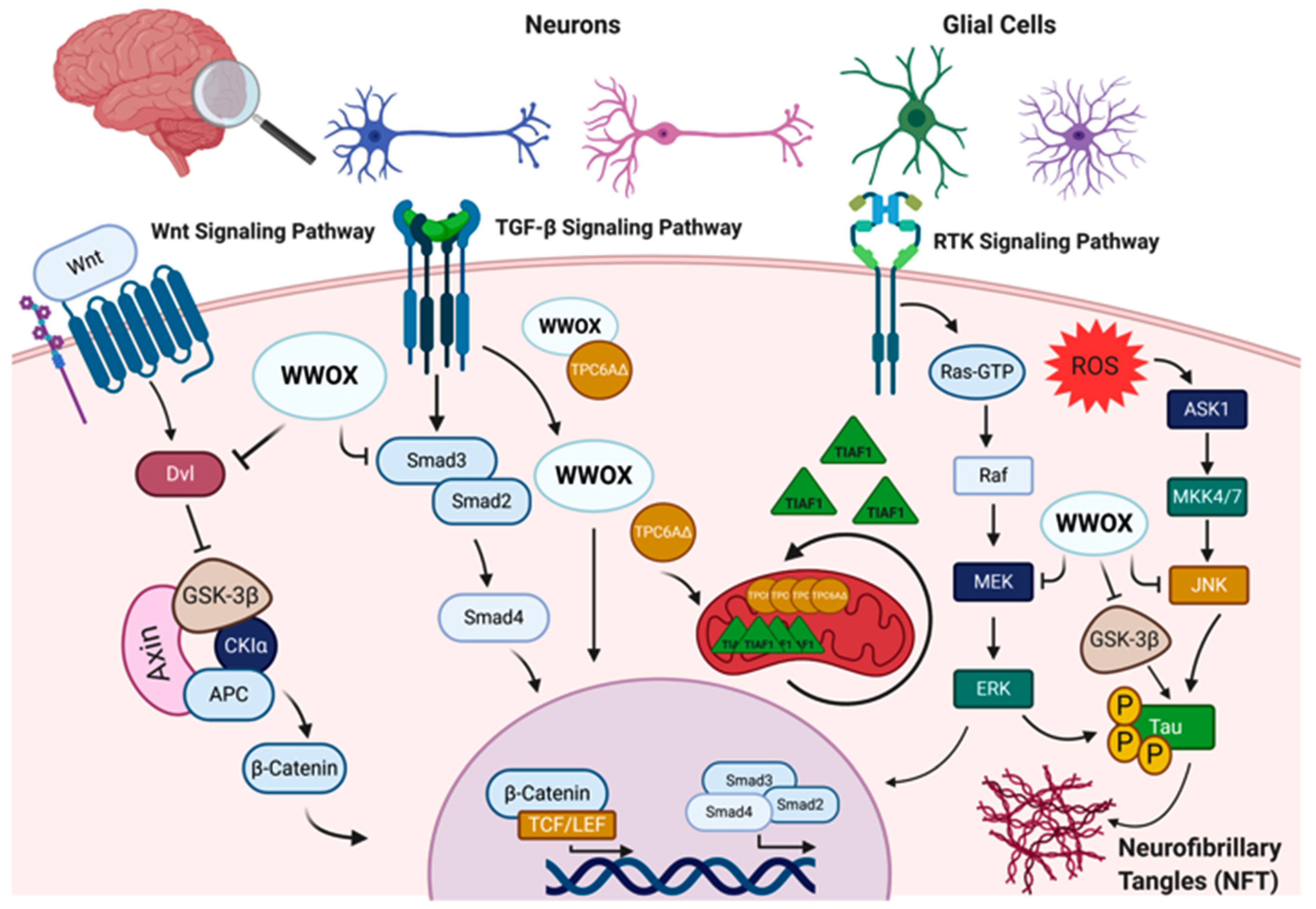
Figure 1. WW domain-containing oxidoreductase (WWOX) interaction partners and signaling pathways in the nervous system. WWOX protein–protein interactions and signaling in the nervous system involves: I. The Wnt/β-catenin signaling pathway, where WWOX negatively regulates this pathway through interaction with Dishevelled (Dvl) proteins and/or glycogen synthase kinase-3 β (GSK-3β). WWOX deficiency has been shown to be associated with increased nuclear β-catenin activity. II. WWOX has been also shown to modulate the transforming growth factor-β (TGF-β) signaling pathway through regulation of the SMAD proteins and the trafficking protein particle complex 6AΔ (TPC6AΔ). In the absence of WWOX, the SMAD/co-SMAD complex is inhibited, and TPC6AΔ and anti-apoptotic factor 1 (TIAF1) aggregate in the mitochondria, contributing to CNS pathologies. III. WWOX could also modulate the receptor tyrosine kinase (RTK) and c-Jun N-terminal kinase (JNK) signaling pathways to regulate tau phosphorylation and the formation of neurofibrillary tangles (NFT). Other abbreviations used: APC, adenomatosis polyposis; CKIα, casein kinase Iα; TCF/LEF, T-cell factor/lymphoid enhancing factor; ROS, reactive oxygen species; ASK1, apoptosis signal-regulating kinase 1; MKK4/7, mitogen-activated kinase kinase 4/7; RAF, rapidly accelerated fibrosarcoma; MEK: mitogen-activated protein Kinase; ERK, extracellular signal-regulated kinase. Figure was created via BioRender.
Cells 10 00824 g001
Figure 2. Summary of the neurological disorders associated with WWOX germline mutations. Upper panel (A) shows phenotypes associated with WWOX deficiency in rodents. Lower panel (B) summarizes neuropathological diseases associated with WWOX gene mutations in humans. NFTs, neurofibrillary tangles; TPC6AΔ, trafficking protein particle complex 6A; TIAF1, TGF-β1 induced antiapoptotic factor; APP, amyloid precursor protein. Figure was generated with BioRender.com.
Cells 10 00824 g002
Figure 3. WWOX variants identified in patients with SCAR12 and WOREE syndrome. (A) Deletions leading to copy number variations in the WWOX gene. (B) Single-nucleotide variations (SNVs) leading to point mutations and protein changes. Mutation diagram circles are colored to indicate type of mutations: green for missense, orange for splice site, and black for truncating mutations including nonsense, frameshift deletion, and frameshift insertion. SCAR12 is mostly associated with missense mutations (red), while WOREE is associated with the deletions, missense, and nonsense mutations (black). Number of patients with increased frequency of a given mutation is indicated (height of the vertical lines).
Cells 10 00824 g003
Table 1. Published WOREE and SCAR12 patients.
NameVariant typeProtein changeCases/referencesGenderEthnicityZygositySeizure onsetDeath
Spinocerebellar ataxia, autosomal recessive 12
NM_016373.4(WWOX):c.139C>A (p.Pro47Thr)MissenseP47T4 siblings [4,8]F/F/M/FSaudi-Arabian Homozygous 12m/9m/9m/9m17-26 y
NM_016373.4(WWOX):c.1114G>C (p.Gly372Arg)MissenseG372R2 siblings [4]M/FIsraeliHomozygous<2 y/<2y5-10 y
Developmental and epileptic encephalopathy 28
NM_016373.4(WWOX):c.160C>T (p.Arg54Ter)NonsenseR54*1 [60]FEgyptianHomozygous2m16m
WWOX-null alleles 2 [63] F/MTurkish 3 m/prenatal22m/Mtp
GRCh37/hg19 16q23.1(chr16:78180603-78208482)x0Deletion (Exon 5) 1 [61]MEmiratiHomozygous2 w5 m
NM_016373.4(WWOX):c.606-1G>ASplicing variant 5 patients from 2 families [65]F/F/F/M/FSaudi ArabianHomozygous2m/3m/2m/2m/3m<3 y/<3 y/<3 y/<3 y/<3 y
NM_016373.4(WWOX):c.46_49del (p.Asp16fs)Deletion (Exon 1)/NonsenseD16fs2 [62]M/FPortugueseCompound heterozygous5 m/5malive at 4y/3 y
NM_016373.4(WWOX):c.140C>G (p.Pro47Arg)MissenseP47R
NM_016373.4(WWOX):c.1005G>A (p.Trp335Ter)NonsenseW335*1 [62]FPortugueseCompound heterozygous 7 w24 m
NM_016373.2(WWOX):c.517-?_605+?delDeletion
NM_016373.3(WWOX):c.-366-?_516+?delDeletion 1 [62]FPortugueseCompound heterozygous2malive at 3y
NM_016373.4:c.517_1056delDeletion (Exon 6 to Exon 8)
NM_016373.4(WWOX):c.889A>T (p.Lys297*)Nonsense 1[62]FPortugueseCompound heterozygous2m38m
deletion encompassing the WWOX locus (2.8 Mb del)Deletion
NM_016373.4(WWOX):c.131G>A (p.Trp44Ter)NonsenseW44*2 [64]F/FQatariHomozygous7 w/7w7y/20 m
NM_016373.4(WWOX):c.918del (p.Glu306fs)Deletion (Exon 8)/NonsenseE306fs1 [66]MRomanianCompound heterozygous4 w<3 y
NM_016373.4(WWOX):c.173-1G>TSplicing variantAsp58Alafs*3
NM_016373.4(WWOX):c.160C>T (p.Arg54Ter)NonsenseR54*2 [93]M/F Saudi ArabianHomozygous2 malive at 3 m/18 m
NM_016373.4(WWOX):c.409+1G>TSplicing variant 1 [93]MSaudi ArabianHomozygous2 malive at 21 m
GRCh37/hg19 16q23.1(chr16:78143675-78149052)Deletion (Exon 3 to Exon 4) 1 [6]MEmiratiHomozygous5 walive at 2 y
NM_016373.4(WWOX):c.606-1G>ASplicing variant 1 [6]MEmiratiHomozygous5 walive at 13 m
NM_016373.4(WWOX):c.689A>C (p.Gln230Pro)Missense Q230P2 [68]F/FAfghanHomozygous2 m/2 malive at 12 y/10 y
NM_016373.4(WWOX):c.716T>G (p.Leu239Arg)MissenseL239R1 [94]FTurkishHomozygous 2 mNA
NM_016373.4:c.1_409delDeletion (Exon 1 to Exon 4) 1 [5]M PakistaniHomozygous7 m2 y
NM_016373.4:c.49G>A (p.Glu17Lys)MissenseE17K 1 [5]FNACompound heterozygous11 wNA
NM_016373.4:c.911C>T (p.Ser304Phe)MissenseS304F
NM_016373.4(WWOX):c.160C>T (p.Arg54Ter)NonsenseR54*1 [5]FFrenchCompound heterozygous2.5 mNA
NM_016373.4:c.1094_1095delTC (p.Val365AlafsTer163)Deletion (Exon 9)/NonsenseV365Afs*163
NM_016373.4:c.231_409del (p.Asp77GlufsTer27)Deletion (Exon 4)D77Efs*271 [5]FItalianCompound heterozygousday 20NA
NM_016373.4:c.607_791+1delDeletion (Exon 7 to Intron 7)
NM_016373.4(WWOX):c.410G>A (p.Gly137Glu)MissenseG137E1 [5]MNACompound heterozygousday 1NA
NM_016373.4(WWOX):c.953C>T (p.Ser318Leu)MissenseS318L
NM_016373.4(WWOX):c.410G>A (p.Gly137Glu)MissenseG137E1 [5]MBritishCompound heterozygousday 12 y6 m
NM_016373.4:c.517_791delDeletion(Exon 6 to Exon 7 )
NM_016373.4:c.517_791delDeletion (Exon 6 to Exon 7) 2 [5]M/MFrenchCompound heterozygous3 m/2.5mNA/NA
NM_016373.4:c.449A>C (p.His150Pro)MissenseH150P
NM_016373.4:c.411_516+1delDeletion(Exon 5 to Intron 5) 1 [5]FFrenchCompound heterozygousday 16 m
NM_016373.4:c.607_791+1delDeletion (Exon 7 to Intron 7)
NM_016373.4(WWOX):c.689A>C (p.Gln230Pro)Missense Q230P1 [5]FMoroccanHomozygous2 m3 y 3 m
NM_016373.4(WWOX):c.689A>C (p.Gln230Pro)Missense Q230P1 [5]FFrenchCompound heterozygous3 wNA
NM_016373.4:c.598A>G (p.Lys200Glu)MissenseK200E
NM_016373.4(WWOX):c.1073C>T (p.Thr358Ile)MissenseT358I
NM_016373.4(WWOX):c.689A>C (p.Gln230Pro)Missense Q230P1 [5]FFrenchCompound heterozygous3 mNA
NM_016373.4:c.1138dupT (p.Cys380LeufsTer149)Duplication/NonsenseC380Lfs*149
NM_016373.4(WWOX):c.689A>C (p.Gln230Pro)Missense Q230P1 [5]FIranianHomozygous2 mNA
NM_016373.4(WWOX):c.790C>T (p.Arg264Ter)NonsenseR264*2 [5]F/MIndianHomozygous7 w/NA8y 11 m/5y 2m
NM_016373.4(WWOX):c.173-1G>T Asp58Alafs*31 [5]FFrenchCompound heterozygousday 20NA
NM_016373.4(WWOX):c.517_1056dupDuplicationHis173_Met352dup
NM_016373.4:c.517_1056delDeletion (Exon 6 to Exon 8) 1 [5]FCaucasianCompound heterozygous stateday 2NA
NM_016373.4(WWOX):c.953C>T (p.Ser318Leu)MissenseS318L
NM_016373.4:c.517_1056delDeletion (Exon 6 to Exon 8 ) 1 [5]MSubSaharanHomozygous1 m27 m
NM_016373.4:c.517_1056delDeletion (Exon 6 to Exon 8) 1 [5]FBritishCompound heterozygousday 52y5m
NM_016373.4(WWOX):c.705dup (p.His236fs)Frameshift
NM_016373.2(WWOX):c.517-?_605+?delDeletion 1 [70]FUSAHomozygous2 w6 y
NM_016373.4:c.517-2A>GSplicing variant 4 [71]F/F/F/MYemenite JewsHomozygous3m/3 w/1m/6w30 m/33 m/9 m/alive at 6 m
NM_016373.4:c.517-2A>GSplicing variant 2 [71]M/FYemenite JewsCompound heterozygous2 w/3 walive at 4y/3 y
NM_016373.4(WWOX):c.689A>C (p.Gln230Pro)Missense Q230P
NM_016373.4:c.173-2A>GSplicing variant 1 [72]MChineseCompound heterozygousday 19NA
NM_016373.4:c.775T>C (p.Ser259Pro)MissenseS259P
NM_016373.3 (and GRCh37/hg19): deletion-chr16:78146639-78151289, chr16:78166192-78184119 Deletion 1 [73]FAustraliancompound heterozygous 2.5malive at 7y
NM_016373.4:c.49G>A (p.Glu17Lys)MissenseE17K
GRCh37/hg19: 16q22.2q23.1(71,689,186–78,530,357)x16.8 Mb Deletion 1 [77]MJapaneseheterozygous16m4 y7m
NM_016373.4:c.229_230+2delGAGTDeletion (Exon 3 to Intron 3 ) 1 [75]FChineseCompound heterozygous8 w5 m
NM_016373.4:c.1065dupA (p.Ala356SerfsTer173)Duplication/NonsenseA356Sfs*173
NM_016373.4(WWOX):c.790C>T (p.Arg264Ter)NonsenseR264*2 [42]M/MSicily ItalyHomozygous45 days/prenatalalive/Mtp
NM_016373.4(WWOX):c.984C>G (p.Tyr328Ter)NonsenseY328*1 [74]MChinesecompound heterozygous 1 m1y
NM_016373.4(WWOX):c.172+1G>CSplicing variant
Table 2. Unpublished patients from DECIPHER and ClinVar databases.
Decipher IDLocationVariant ChangeProtein ChangeSizeCasesZygosityPathogenicityInheritanceSex
40743978098478-78259430Deletion 160.95 kb2HomozygousPathogenicBiparental46XY
38498478133724-78133724c.49G > AE17KSNV2Compound heterozygousLikely pathogenicUnknown46XX
78022830-78216167Deletion 193.34 kbLikely pathogenicMaternally inherited, constitutive in mother
ClinVar
NameVariant changeProtein ChangeCasesZygosityPathogenicityPhenotype(s)GRCh37ChromosomeGRCh37Location
NM_016373.4(WWOX):c.108-2A>TSplice acceptor 1HomozygousLikely pathogenicseizures, Developmental regression, global developmental delay, microcephaly, and Dystonia1678142318
NM_016373.4(WWOX):c.183C>A (p.Tyr61Ter)NonsenseY61*1Compound heterozygousLikely pathogenica phenotype consistent with the WWOX-associated disease.1678143685
NM_016373.4(WWOX):c.918del (p.Glu306fs)Deletion (exon 8)E306fsLikely pathogenic1678466511
NM_016373.4(WWOX):c.214C>T (p.Gln72Ter)NonsenseQ72*1Compound heterozygousPathogenicityseizures1678143716
NC_000016.10:g.(?_78099759)_(78115174_?)delDeletion (exons 1–4) Pathogenicity1678133656–78149071
NM_016373.4(WWOX):c.214C>T (p.Gln72Ter)NonsenseQ72*1Compound heterozygousPathogenicitymicrocephaly, infantile spasms/seizures1678143716
NC_000016.9:g.(?_78198060)_(78198206_?)delDeletion (exon 5) Pathogenicity1678198060–78198206
NM_016373.4(WWOX):c.107+1G>ASplicing donor 1Compound heterozygousPathogenicitydevelopmental delay and early-onset epileptic encephalopathy.1678133783
NM_016373.4(WWOX):c.136C>T (p.His46Tyr)MissinseH46YLikely pathogenic1678142348
NM_016373.4(WWOX):c.409+1G>CSplice donor 1Compound heterozygousLikely pathogenicseizures1678149052
NM_016373.4(WWOX):c.790C>T (p.Arg264Ter)NonsenseR264*Pathogenicity1678458951
Publisher’s Note: MDPI stays neutral with regard to jurisdictional claims in published maps and institutional affiliations.