Article

HPLC-ESI-HRMS/MS-Based Metabolite Profiling and Bioactivity Assessment of Catharanthus roseus

1
Central Department of Chemistry, Tribhuvan University, Kirtipur, Kathmandu 44618, Nepal
2
School of Pharmacy, Sungkyunkwan University, Suwon 16419, Republic of Korea
3
Paraza Pharma, Inc., 2525 Marie-Curie Avenue, Montreal, QC H4S 2E, Canada
*
Authors to whom correspondence should be addressed.
These authors contributed equally to this study.
Current address: Department of Chemical, Biological, and Bioengineering, North Carolina A&T State University, Greensboro, NC 27411, USA.
Plants 2025, 14(15), 2395; https://doi.org/10.3390/plants14152395
Submission received: 19 June 2025 / Revised: 29 July 2025 / Accepted: 30 July 2025 / Published: 2 August 2025

Abstract

A comprehensive metabolic profiling of Catharanthus roseus (L.) G. Don was performed using tandem mass spectrometry, along with an evaluation of the biological activities of its various solvent extracts. Among these, the methanolic leaf extract exhibited mild radical scavenging activity, low to moderate antimicrobial activity, and limited cytotoxicity in both the brine shrimp lethality assay and MTT assay against HeLa and A549 cell lines. High-performance liquid chromatography–electrospray ionization–high-resolution tandem mass spectrometry (HPLC-ESI-HRMS/MS) analysis led to the annotation of 34 metabolites, primarily alkaloids. These included 23 indole alkaloids, two fatty acids, two pentacyclic triterpenoids, one amino acid, four porphyrin derivatives, one glyceride, and one chlorin derivative. Notably, two metabolites—2,3-dihydroxypropyl 9,12,15-octadecatrienoate and (10S)-hydroxypheophorbide A—were identified for the first time in C. roseus. Furthermore, Global Natural Products Social Molecular Networking (GNPS) analysis revealed 18 additional metabolites, including epoxypheophorbide A, 11,12-dehydroursolic acid lactone, and 20-isocatharanthine. These findings highlight the diverse secondary metabolite profile of C. roseus and support its potential as a source of bioactive compounds for therapeutic development.

Graphical Abstract

1. Introduction

Medicinal plants have long served as a vital source of pharmacological lead compounds, with their use dating back over 5000 years [1]. A large portion of the Global South still relies on herbal remedies to treat a range of illnesses, including skin diseases, malaria, diarrhea, dysentery, stomach pain, schistosomiasis, toothache, jaundice, and more [2,3,4]. Bioactive compounds derived from medicinal plants have garnered considerable interest due to their critical role in drug development, particularly in the treatment of infectious diseases, cancer, and cardiovascular disorders—often with minimal side effects [5].
Catharanthus roseus (L.) G. Don, commonly known as Madagascar periwinkle, is a rare undershrub in the Apocynaceae family. Traditionally used for its antidiabetic properties, this plant is now recognized as a rich source of bioactive constituents, producing over 100 alkaloids as well as other nitrogen-containing metabolites such as peptides, purines and their derivatives, amino acids, antibiotics, and amino sugars [6,7,8]. Among its notable compounds, indole alkaloids—found throughout the plant—play a major role in treating cancer, hypertension, diarrhea, diabetes, and Alzheimer’s disease, and have also shown potential in promoting wound healing [9,10,11].
Metabolic profiling is attracting increasing attention due to its potential in drug discovery, as it facilitates the analysis of biosynthetic pathways using the diverse bioactive compounds found in medicinal plants. Metabolomics is a technique that systematically analyzes large numbers of small molecules produced during cellular biochemical processes, revealing the unique chemical fingerprints associated with specific biological functions [12]. Metabolites, which are the end products of enzymatic activity, serve as accurate indicators of metabolic function [13]. There are two main approaches to metabolomic profiling: targeted and untargeted metabolomics. Targeted metabolomics focuses on the precise identification and quantification of specific metabolite groups within a sample, primarily to validate their presence [14]. In contrast, untargeted metabolomics aims to qualitatively and semi-quantitatively profile as many metabolites as possible in a single analysis [15]. Liquid chromatography coupled with mass spectrometry (LC-MS) is the preferred method for global metabolite profiling, as it enables the identification of a wide array of metabolites. Furthermore, Global Natural Product Social Molecular Networking (GNPS) is a web-based platform that clusters metabolites based on the similarity of their MS/MS fragmentation patterns, thereby enhancing metabolite annotation.
More than 130 terpene indole alkaloids (TIAs) have been identified in C. roseus, including vinblastine and vincristine—clinically important anticancer agents synthesized through the dimerization of vindoline and catharanthine [16]. In addition, C. roseus extracts have demonstrated antimicrobial, cytotoxic, and apoptotic activities, as evaluated by minimum inhibitory concentration (MIC), cell viability assays, and flow cytometry analysis, respectively—highlighting their potential in cancer therapy [17]. Moreover, C. roseus exhibits significant antioxidant properties, which can be assessed through assays such as 2,2-diphenyl-1-picrylhydrazyl (DPPH) radical scavenging, ABTS [2,2′-azino-bis(3-ethylbenzothiazoline-6-sulfonic acid)] radical scavenging, and ferric reducing antioxidant power (FRAP) [18]. In one study, Ferreres et al. employed high-performance liquid chromatography coupled with photodiode array detection and electrospray ionization tandem mass spectrometry (HPLC-DAD-ESI-MS/MS) to screen metabolites from various parts of C. roseus. They identified three caffeoylquinic acid derivatives and 15 flavonoids, marking the first comprehensive analysis of non-pigmented phenolic compounds in the species [19]. Similarly, five known indole alkaloids and three novel dimeric indole alkaloids were isolated from the whole plant of C. roseus. The absolute configurations and structures of the novel compounds were elucidated through nuclear magnetic resonance (NMR) spectroscopy and circular dichroism (CD) analysis [20]. Moreover, Yadav et al. evaluated the antibacterial activity of crude extracts obtained from the root, stem, and leaf of C. roseus. These extracts demonstrated inhibitory effects against Bacillus subtilis, Staphylococcus aureus, and Escherichia coli [21].
As part of our ongoing investigations into biologically active metabolites from diverse natural sources [22,23,24,25,26,27], this study aimed to separately evaluate the biochemical and biological activities of the leaf and stem extracts of C. roseus. The leaf extracts exhibited superior biological activity compared to the stem extracts. Based on this observation, we hypothesized that the leaves may contain more potent metabolites than the stems. To explore and identify the secondary metabolites responsible, we conducted HPLC-ESI-MS/MS analysis and GNPS analysis of the leaf extracts. Unlike previous studies that have focused primarily on targeted alkaloid screening or single-platform analyses, our work presents the first combined HPLC-ESI-HRMS/MS and GNPS-based untargeted metabolomic profiling of the methanolic leaf extract of C. roseus. This approach led to the annotation of 34 secondary metabolites, including two compounds, 2,3-dihydroxypropyl 9,12,15-octadecatrienoate and (10S)-hydroxypheophorbide A, reported here for the first time in C. roseus, and uncovered 18 additional metabolites via molecular networking. By integrating comprehensive bioactivity assays with advanced metabolomics, our study expands the chemical inventory of C. roseus and provides new leads for therapeutic development. Here, we present the bioactivity assessment of C. roseus extracts and HPLC-ESI-HRMS/MS-based metabolite profiling.

2. Results

2.1. DPPH Radical Scavenging Assay

The collected stems and leaves of Catharanthus roseus were air-dried, crushed, powdered, and extracted using the maceration technique with four different solvents: methanol, ethyl acetate, dichloromethane (DCM), and hexane. Leaf extracts prepared using methanol, ethyl acetate, DCM, and hexane were labeled as A01, A02, A03, and A04, respectively, while stem extracts prepared with methanol and ethyl acetate were designated A05 and A06, respectively.
The DPPH radical scavenging activity of the C. roseus extracts ranged from 53.12 ± 1.60 to 94.12 ± 1.03 µg/mL. Quercetin, used as the positive control, exhibited an IC50 value of 3.89 ± 0.68 µg/mL. All extracts demonstrated lower scavenging activity compared to quercetin, as shown in Table 1. The IC50 value of the methanolic leaf extract (A01) reported in a previous study was 49.74 ± 0.52 µg/mL, which is comparable to our result, with a difference of approximately 4 µg/mL [28]. In contrast, the stem extracts (A05 and A06) showed a weaker scavenging effect, even at higher concentrations, as illustrated in Figures S1 and S2, suggesting their relatively lower DPPH scavenging potential compared to the leaf extracts. A two-way ANOVA with replication indicated that all comparisons were statistically significant (p < 0.05). A subsequent Tukey’s HSD post hoc test revealed that the scavenging activity of the hexane extract differed significantly from those of the methanol, ethyl acetate, and dichloromethane extracts (p < 0.05). Detailed results are presented in Supplementary Tables S5 and S6.

2.2. Antimicrobial Assays

The antimicrobial activity of the plant extracts (A01–A06) was evaluated against various Gram-positive and Gram-negative bacteria, including Staphylococcus aureus, Shigella sonnei, Klebsiella pneumoniae, and Escherichia coli (Table S1). Among all extracts, the methanolic leaf extract (A01) and the methanolic stem extract (A05) demonstrated the mild levels of inhibition against S. aureus and S. sonnei, weak inhibition against K. pneumoniae, and no detectable inhibition against E. coli. However, they exhibited lower activity than the positive control (neomycin). Accordingly, the minimum inhibitory concentration (MIC) and minimum bactericidal concentration (MBC) of the methanolic and ethyl acetate extracts from both leaves and the stem (A01, A02, A05, and A06) were determined against S. sonnei and S. aureus (Figure 1). The methanolic (A01) and ethyl acetate (A02) extracts showed MIC values of 12.5 mg/mL and MBC values of 25 mg/mL against both bacterial strains. In comparison, A05 and A06 exhibited higher MIC and MBC values (25–50 mg/mL), indicating lower potency. The positive control (neomycin) showed significantly lower MIC and MBC values, with MICs of 0.004 mg/mL and 0.01 mg/mL and MBCs of 0.01 mg/mL and 0.02 mg/mL for S. sonnei and S. aureus, respectively, confirming the validity of the assay (Table S2, Figures S1–S6).

2.3. Brine Shrimp Lethality Assay

The biological activity of methanolic leaf extract (A01) of C. roseus was evaluated using a brine shrimp lethality assay against newly hatched Artemia salina nauplii. The lethality of the methanolic leaf extract (A01) was assessed by calculating mortality percentages based on the number of dead nauplii exposed to different extract concentrations, as summarized in Table S3. The results showed a concentration-dependent increase in mortality, with higher extract concentrations causing greater lethality. All nauplii died in the positive control group, while 100% survival was observed in the negative control. The methanolic leaf extract (A01) exhibited toxicity, with an LC50 value of 914.98 µg/mL, suggesting weak anticancer properties. However, this assay provides only preliminary evidence. Therefore, an MTT assay was subsequently conducted to further evaluate the anticancer potential of the methanolic leaf extract.

2.4. Cytotoxicity Assay

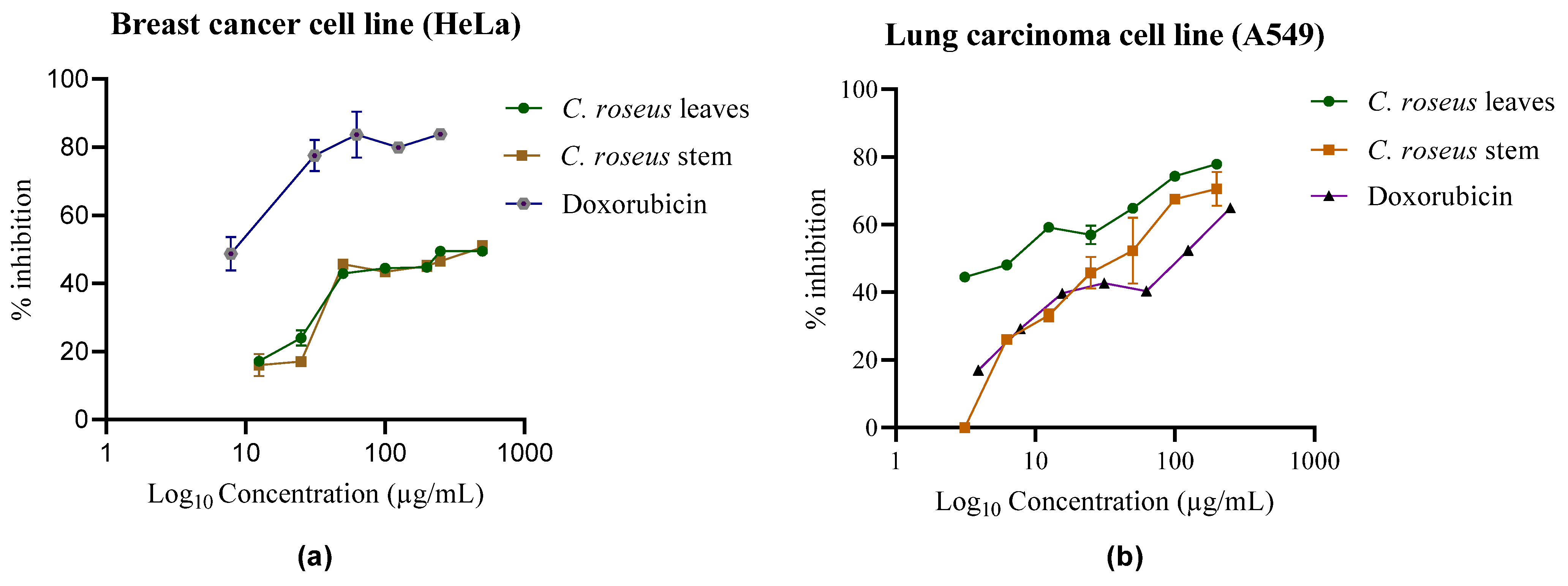
The MTT assay was conducted on lung carcinoma cell lines (A549) and cervical cancer cell lines (HeLa cell lines) to evaluate the cytotoxic effects of methanolic extracts (A01 and A05) from C. roseus. IC50 values were calculated to determine the concentration required to inhibit 50% of cell viability (Figure 2). The methanolic extracts from the stem and leaves exhibited IC50 values of 32.5 ± 0.01 µg/mL and 37.5 ± 0.10 µg/mL, respectively, against HeLa cells. Similarly, against A549 cells, the methanolic leaf and stem extracts showed IC50 values of 35.98 ± 0.89 µg/mL and 31.64 ± 0.12 µg/mL, respectively. As shown in Figure 2, increasing the extract concentration resulted in higher inhibition rates, leading to decreased cell viability. These results suggest that C. roseus extracts possess moderate cytotoxic potential against both cell lines. Additionally, the RealTime-Glo™ (Promega Corporation, Madison, WI, USA) MT Cell Viability Assay performed on the HeLa cell line revealed IC50 values of 45 ± 0.15 µg/mL and 40 ± 0.15 µg/mL for both the leaf and stem methanolic extracts, respectively (Figure S44). Since the assay substrate is reduced only by metabolically active cells, a decrease in luminescence with increasing extract concentration reflects reduced cell viability. Doxorubicin (IC50: ~15.26 ± 0.91 µg/mL) was included as a positive control to evaluate the assay’s robustness and dynamic range.

2.5. Metabolite Profiling Using HPLC-ESI-HRMS/MS

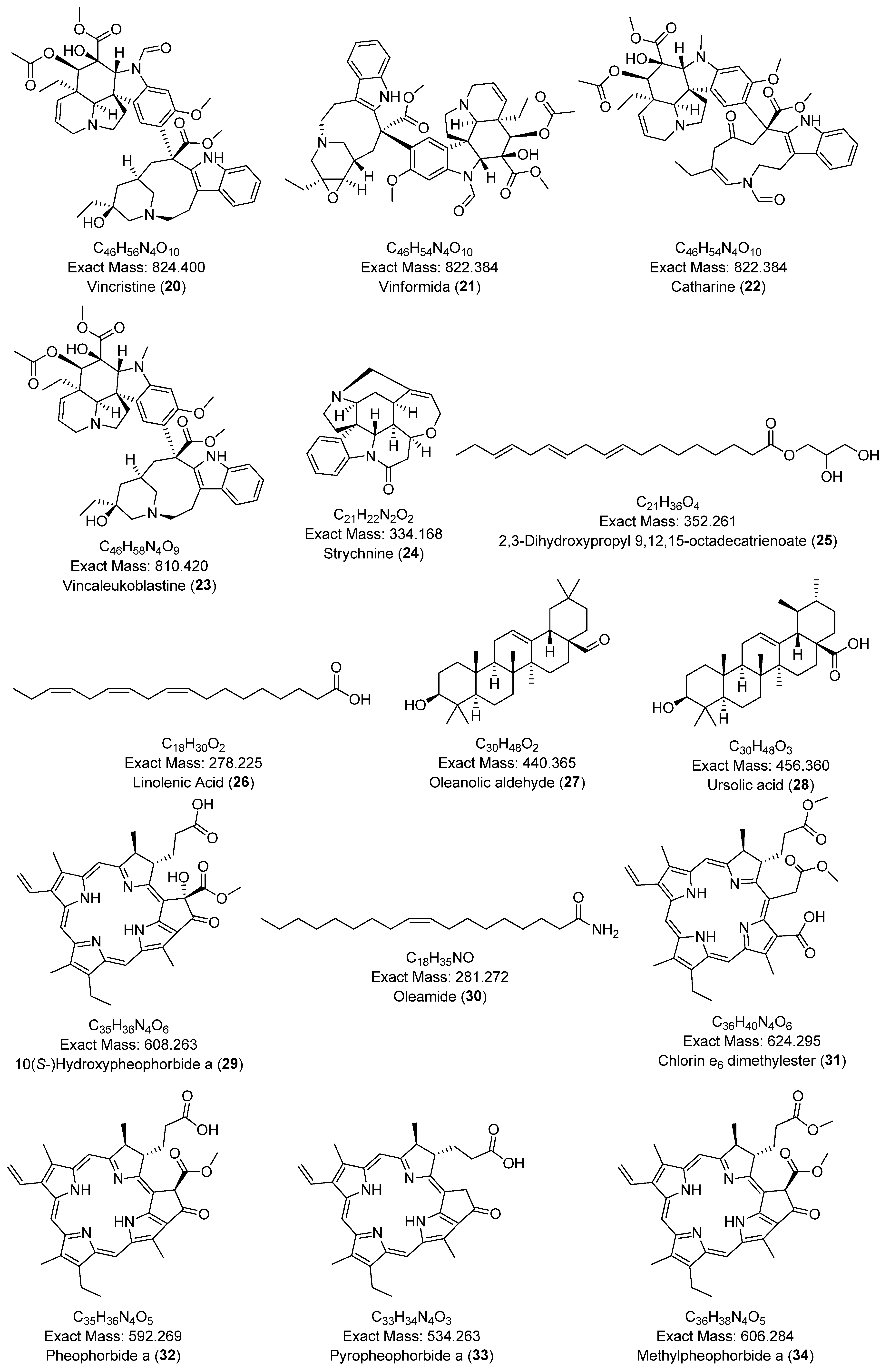
A comprehensive phytochemical profiling of the methanolic leaf extract of C. roseus (A01) was conducted using HPLC-ESI-HRMS/MS, based on its antibacterial and free radical scavenging activities. Figure 3 presents the total ion chromatogram (TIC) with annotated metabolites. A total of 34 secondary metabolites—including 22 indole alkaloids, 2 fatty acids, 2 pentacyclic triterpenoids, 1 amino acid, 4 porphyrin derivatives, 1 glyceride, and 1 chlorin derivative—were identified in positive ionization mode, as listed in Table 2. To the best of our knowledge, two metabolites—2,3-dihydroxypropyl 9,12,15-octadecatrienoate and (10S)-hydroxypheophorbide A—were identified for the first time in C. roseus. A few peaks could not be annotated and are marked with an asterisk (*) due to low signal intensity and insufficient fragmentation peaks. The chemical structures of the annotated metabolites are illustrated in Figure 4. The BPCs and MS profiles of all detected metabolites are presented in Figures S7–S40.

2.6. GNPS Analysis

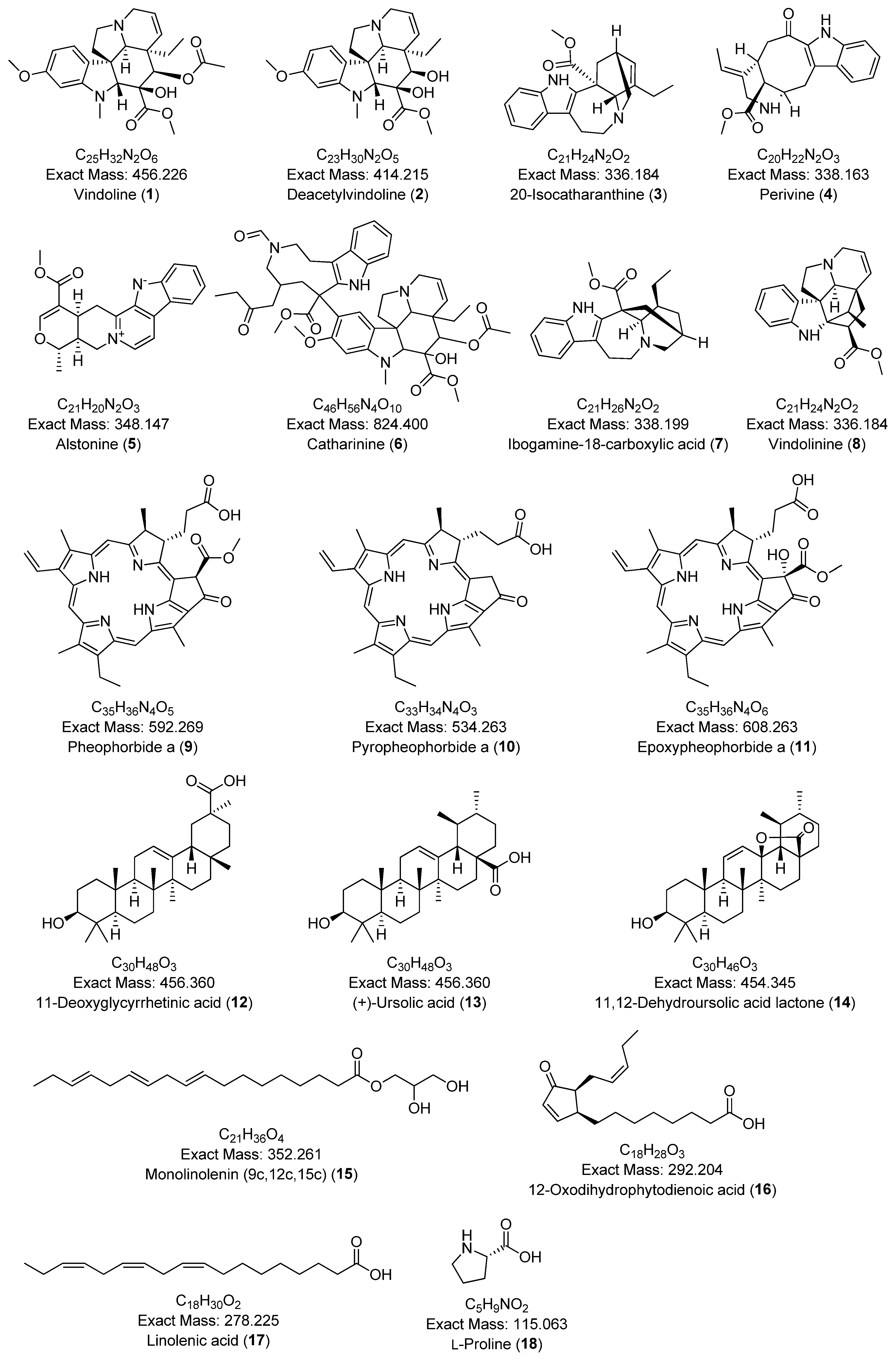
Given the radical scavenging activity and notable antimicrobial efficacy of the methanolic leaf extract of C. roseus (A01) against Staphylococcus aureus and Shigella sonnei, a comprehensive phytochemical profile was generated using MS/MS analysis and a GNPS-based metabolomics platform. A total of 18 compounds were tentatively identified from the leaf extract (A01), including 11 tryptophan-derived alkaloids, 3 triterpenoids, 3 fatty acids, and 1 amino acid, as listed in Table 3. The chemical structures of these compounds are shown in Figure 5.
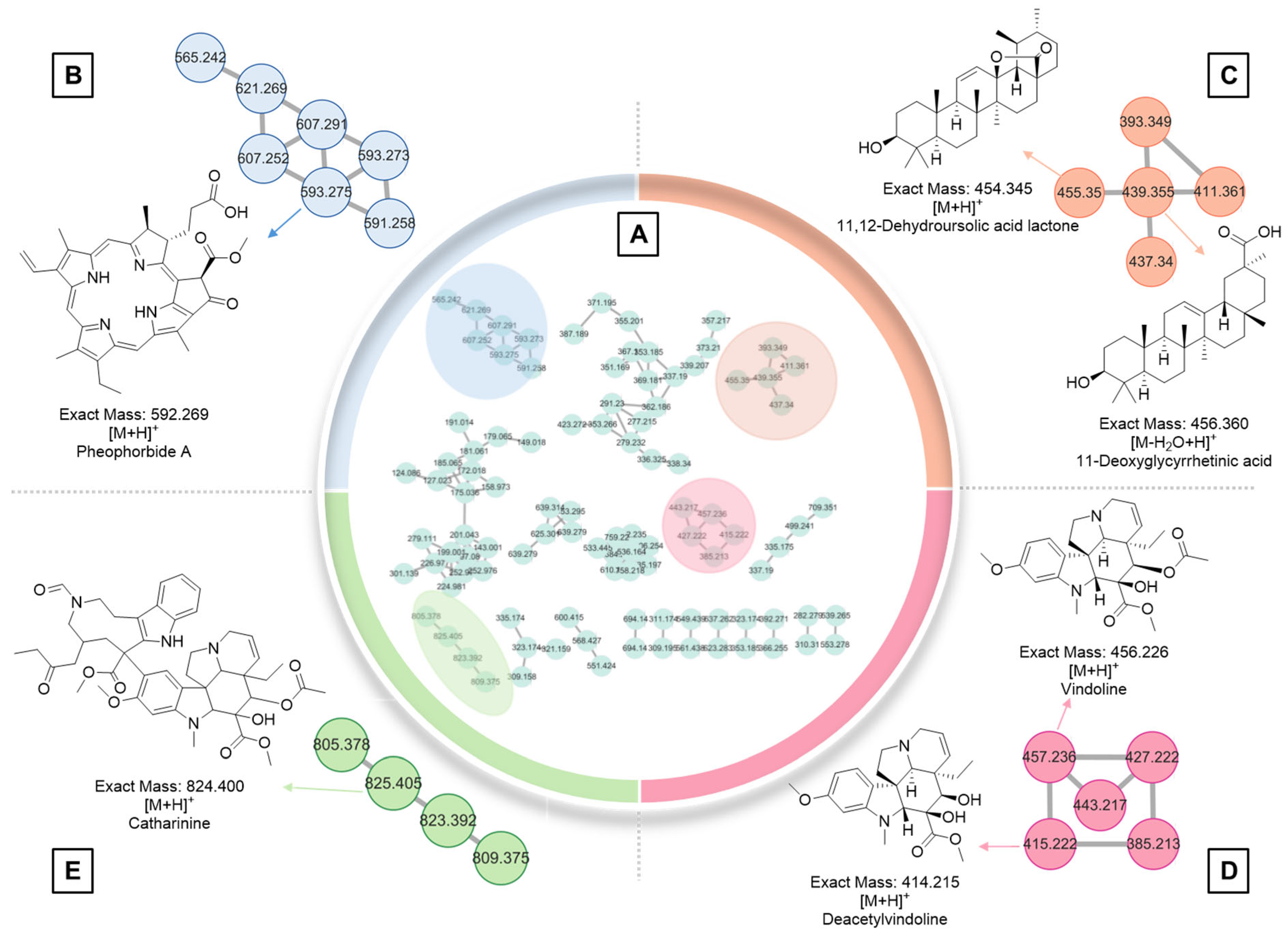
Molecular networking is a computational tool that visually represents the chemical space of complex samples. It operates on the principle that structurally similar molecules tend to produce comparable fragmentation patterns in tandem mass spectrometry (MS/MS). Each metabolite is represented as a node, typically labeled with its m/z (mass-to-charge) value, and nodes are grouped into clusters based on similarities in their MS/MS fragmentation profiles. In the methanolic leaf extract of C. roseus (A01), 176 molecular ions were detected and connected by 219 edges, forming a network. These ions were distributed into 10 multi-node clusters, one cluster with three nodes, eight clusters with two nodes, and 75 single, unconnected nodes (Figure 6A). Among these, four major clusters were identified (Figure 6). One node at m/z 593.275 was annotated as pheophorbide a (Figure 6B). Two ions at m/z 455.350 and 456.360 corresponded to the triterpenoids, 11,12-dehydroursolic acid lactone and 11-deoxoglycyrrhetinic acid, respectively (Figure 6C). Previous work by Huang et al. demonstrated the triterpene biosynthetic capacity of C. roseus through the identification of two key enzymes, CrAS and CrAO, which are essential for the formation of pentacyclic triterpenes such as ursolic acid and oleanolic acid [52]. In addition, vindoline—one of the most abundant metabolites in C. roseus—was detected at m/z 457.236 (Figure 6D). Deacetylvindoline (m/z 415.222) was also found in the same cluster, suggesting a potential biochemical relationship or precursor-product linkage. Another metabolite, catharanthine, an indole alkaloid with a molecular ion at m/z 825.405, was identified as one of more than 130 known terpenoid indole alkaloids in C. roseus (Figure 6E) [16].
Table 3. Annotated secondary metabolites from the methanolic leaf extract (A01) of C. roseus identified through GNPS analysis.
NoAnnotated CompoundAccurate
Mass (Da)
Precursor
Ion
Adduct TypeMS2 Fragmentation
Pattern
Molecular FormulaRetention Time
(mins)
Error (ppm)Reference
Tryptophan alkaloids
1Vindoline456.226457.230[M+H]+188.106C25H32N2O69.2413.1[53]
2Deacetylvindoline414.215415.220[M+H]+188.101, 173.078C23H30N2O58.074.8[54]
320-Isocatharanthine336.184337.191[M+H]+144.081, 93.070C21H24N2O28.543.0[55]
4Perivine338.163339.17[M+H]+234.127, 144.080,
130.065, 93.069
C20H22N2O36.183.0[56]
5Alstonine348.147349.155[M+H]+235.087, 207.092C21H20N2O38.652.9[43]
6Catharinine824.400825.407[M+H]+765.385, 556.280,
341.186, 144.081
C46H56N4O1010.872.4[57]
7Ibogamine-18-carboxylic acid338.199339.207[M+H]+339.208, 144.081C21H26N2O28.660.0[58]
8Vindolinine336.184337.19[M+H]+320.163, 177.090,
144.080, 117.069
C21H24N2O26.840.0[37]
9Pheophorbide a592.269593.269[M+H]+593.276, 533.255C35H36N4O520.8110.2[50]
10Pyropheophorbide a534.263535.270[M+H]+535.270, 507.275,
435.254
C33H34N4O321.071.9[50]
11Epoxypheophorbide a608.263609.272[M+H]+609.272, 591.261,
559.235, 531.240
C35H36N4O619.991.6[50]
Triterpenoids
1211-Deoxyglycyrrhetinic acid456.360439.357[M-H2O+H]+439.357, 189.164,
121.101, 95.086
C30H48O37.324.5[59]
13(+)-Ursolic acid456.360457.368[M+H]+189.163, 95.085C30H48O319.314.4[60]
1411,12-Dehydroursolic acid lactone454.345455.352[M+H]+437.344, 247.170,
133.101, 119.086
C30H46O318.314.3[61]
Fatty acids
15Monolinolenin (9c,12c,15c)352.261353.270[M+H]+261.221, 95.086,
81.070, 67.055
C21H36O417.7611.1[62]
1612-Oxodihydrophytodienoic acid292.204277.216[M-H2O+H]+235.170, 107.087,
93.071, 79.056
C18H28O314.843.6[63]
17Linolenic acid278.225279.232[M+H]+95.085, 81.069
67.054
C18H30O217.240.0[64]
Amino acid
18L-Proline115.063116.071[M+H]+116.071, 70.065C5H9NO20.9917.2[65]

3. Discussion

Catharanthus roseus is rich in organic bioactive compounds, primarily alkaloids, flavonoids, and polyphenolic compounds, which feature diverse functional groups such as hydroxyl, carbonyl, and heterocycles [65]. Given its significant ethnobotanical value and wide-ranging applications, further untargeted metabolomic profiling is essential to uncover additional secondary metabolites and to validate its broader therapeutic potential. This study primarily aimed to assess the antioxidant, antimicrobial, and cytotoxic activities of C. roseus extracts, followed by the annotation of secondary metabolites to confirm and expand upon previous experimental findings.
The radical scavenging activity of the methanolic leaf extract of C. roseus (A01) was found to be 53.12 ± 1.60 µg/mL, closely aligning with a previously reported value of 49.74 ± 0.52 µg/mL [28]. This activity may be attributed to the presence of alkaloids and flavonoids, which are known for their free radical-scavenging properties and ability to reduce oxidative stress. Additionally, metabolites such as quercetin, ursolic acid, vindoline, coronaridine, alstonine, ajmalicine, and coronaridine have been reported to contribute to the moderate radical scavenging potential of C. roseus [66,67,68,69]. The extract also demonstrated weak antibacterial activity against Shigella sonnei, Staphylococcus aureus, and Klebsiella pneumoniae, but showed no efficacy against Escherichia coli. Furthermore, the brine shrimp lethality assay of A01 revealed cytotoxic potential, with an LC50 value of less than 1000 µg/mL. Although a correlation was observed between radical scavenging activity and lethality, this does not imply a causal relationship. The methanolic extract (A01) also exhibited significant cytotoxicity against HeLa and A549 cell lines, with IC50 values of 37.5 ± 0.10 µg/mL and 35.98 ± 0.89 µg/mL, respectively. Further studies are required to isolate the specific bioactive constituents responsible for these effects and to elucidate their underlying mechanisms of action. Alkaloids such as coronaridine, vindorosine, vincristine, vinformide, catharanthine, alstonine, and porphyrin derivatives have previously been reported to possess cytotoxic activity, supporting the findings from the MTT and brine shrimp assays [16,65,69].
Based on the effective biological activities observed in the methanolic leaf extract of C. roseus, metabolic profiling was performed using HPLC-ESI-HRMS/MS analysis, resulting in the identification of 34 secondary metabolites. Most of these were indole alkaloids, including terpene indole alkaloids, corynanthean-type, tryptophan-derived, oxindole, and β-carboline alkaloids. L-proline, an amino acid known to accumulate under saline stress conditions, was also detected [70]. Preakuammicine, a biosynthetic intermediate exclusively detected in C. roseus, has not yet been isolated but is considered crucial in the formation of type II and type III monoterpene indole alkaloids, such as strychnine, ajmalicine, and serpentine [28,71]. Quercetin, a well-known flavonoid with antioxidant and anti-inflammatory properties, is associated with cardiovascular and neuroprotective effects [67]. Perivine, previously reported in both Lanceus pich and C. roseus, exhibits potent extracellular virucidal activity against the Vaccinia virus, the causative agent of smallpox [29]. Mitraphylline, identified in Uncaria tomentosa and C. roseus, is known to inhibit cytokine production and suppress tumor necrosis factor-α (TNF-α) expression, contributing to its anti-inflammatory potential [30,72]. Several alkaloids—including catharanthine, tabersonine, and vindolinine—share the same molecular mass and are closely linked within the indole alkaloid biosynthetic pathway. Catharanthine serves as a key precursor to tabersonine [73], while vindoline can be derived from either catharanthine or tabersonine through enzymatic transformations such as oxidation, methylation, and reduction [74]. Vindoline and catharanthine are further coupled through peroxidase-mediated reactions to form vinblastine [75]. Pleiocarpamine, derived from geissoschizine, has been reported to exhibit neuroprotective effects and is used in the treatment of neurological disorders [76]. Deacetylvindoline, an essential intermediate in vindoline biosynthesis, is produced via hydroxylation of desacetoxyvindoline [77]. Vindoline, in turn, is a crucial precursor for both vinblastine and vincristine [38], and exhibits various pharmacological activities including antioxidant, antihyperlipidemic, antiproliferative, and antidiabetic effects [68]. Understanding the biosynthetic pathways leading to vinblastine and vincristine is vital for pharmaceutical development. Additionally, tubotaiwine, first identified in Pleiocarpa tubicina, has shown strong antiplasmodial activity, with an IC50 value of 8.5 Μm [38,78]. Coronaridine, found in Tabernaemontana catharinensis and related species, has demonstrated cytotoxic activity by inducing cell death in hepatocellular carcinoma cells, though it caused minimal DNA damage and had limited effects on the plasma membrane [79]. Similarly, alstonine, a tryptophan-derived alkaloid, has been shown to possess anxiolytic, antipsychotic, antiproliferative, and antimalarial activities [69]. Moreover, previous studies have shown that ajmalicine, found in Rauvolfia, Mitragyna species, and C. roseus, exhibits antimicrobial, antihypertensive, and vasodilatory properties [80]. Similarly, vindorosine, vincristine, vinformide, and catharanthine have demonstrated anticancer activity by inducing apoptosis and disrupting microtubule dynamics, thereby inhibiting cell division [16]. In the present study, strychnine—previously reported in Strychnos species—was also identified. This compound is a potent convulsant that enhances neuronal activity in the central nervous system by inhibiting glycine receptors [81]. Strychnine is biosynthesized through a sequence of oxidation and reduction reactions from geissoschizine to Wieland–Gumlich aldehyde [82]. Additionally, 2,3-dihydroxypropyl 9,12,15-octadecatrienoate—previously isolated from Citrullus colocynthis and Chenopodium album—has been reported to exhibit central nervous system depressant, anti-inflammatory, and antifungal activities [83,84]. To the best of our knowledge, this is the first report of its detection in C. roseus. Two fatty acids were also annotated in this study. Linolenic acid, commonly found in Glycine max, is known for its anti-inflammatory and antioxidant effects and has therapeutic potential for cardiovascular and neurological diseases [85]. Oleamide, another fatty acid identified, is known to induce sleep, stimulate vanilloid receptors, and mimic neurotransmitter activity at cannabinoid receptors, suggesting a potential role in cardiac muscle development [86]. Furthermore, two pentacyclic triterpenoids—ursolic acid and oleanoic aldehyde—were identified. Ursolic acid is known for its antimicrobial, antiviral, antioxidant, and hepatoprotective properties, while oleanoic aldehyde, typically found in grapes, has been reported to promote insulin synthesis and reduce blood glucose levels, functioning as an antihyperglycemic agent [87]. In addition, four porphyrin derivatives were annotated, all of which are known as photodynamic sensitizers with demonstrated anticancer potential [88].
The GNPS technique enabled the simultaneous analysis of multiple mass spectra, allowing for enhanced visualization and interpretation of complex spectral datasets [89]. This approach also revealed structural relationships among molecules, resulting in the identification of 18 metabolites. However, many polyphenols and flavonoids were not detected, likely due to matrix effects or ion suppression. Ion suppression occurs when co-eluting compounds in a sample matrix interfere with the ionization of target analytes, thereby reducing signal intensity or preventing detection [90]. Several metabolites identified in this study—such as perivine, vindolinine, catharanthine, vindoline, ajmalicine, ursolic acid, and vincristine—have been previously reported to exhibit antimicrobial activity, which supports the antimicrobial effects observed in the methanolic leaf extract [91,92]. Metabolic profiling not only enhances our understanding of biosynthetic pathways but also provides opportunities to identify and manipulate key metabolic steps for the generation of novel bioactive compounds with therapeutic potential. Future studies should focus on isolating individual metabolites, followed by detailed structural elucidation using techniques such as NMR spectroscopy and comprehensive bioactivity evaluations to assess their pharmaceutical relevance. Furthermore, optimization of extraction protocols may enhance metabolite recovery and facilitate the discovery of previously undetectable compounds, thereby contributing to the advancement of pharmacological research.

4. Materials and Methods

4.1. Chemicals

Solvents, including methanol (≥99.0%), ethanol (≥99.9%), hexane (≥99.0%), di-chloromethane (DCM, ≥99.0%), and ethyl acetate (≥99.5%), were of analytical grade from Merck (Darmstadt, Germany) and Thermo Fischer Scientific (Mumbai, India). Gallic acid (≥98.0%), potassium acetate (≥99.0%), and Folin–Ciocalteu’s (FC) phenol reagent were purchased from Loba Chemie (Mumbai, India). In addition, anhydrous sodium carbonate (≥98.5%), tannic acid (≥99.5%), iron(III) nitrate nonahydrate (≥98.0%), Mueller–Hinton broth, Mueller–Hinton agar, and Mueller agar growth nutrients were purchased from HiMedia Laboratories (Mumbai, India). Similarly, dimethyl sulfoxide (DMSO, ≥99.0%) was purchased from the Silico research laboratory (Madhya Pradesh, India). Acetic acid (≥99.7%) was obtained from Control Drug House (Gujarat, India). Quercetin (≥95.0%) and 2,2-diphenyl-1-picrylhydrazyl (DPPH, ≥95.0%) were procured from Srikem Laboratories (Mumbai, India). Positive controls, neomycin and doxorubicin (≥98.0%), were purchased from Sigma-Aldrich (St. Louis, MO, USA).

4.2. Plant Collection and Extract Preparation

Catharanthus roseus (L.) G. Don (stems and leaves) was collected in July 2023 from the Kathmandu Valley, Nepal (27°42′2.7684″ N, 85°18′0.5040″ E), based on its traditional medicinal use. The species was identified at the National Herbarium and Plant Laboratories, Godawari, Nepal, and registered under voucher number RB01. The collected plant materials were air-dried, crushed, powdered, and stored in airtight plastic bags for further processing. Plant extracts were prepared using the maceration technique in four different solvents: methanol, ethyl acetate, dichloromethane (DCM), and hexane, as detailed in the Supplementary Materials. Leaf extracts were labeled as A01 (methanol), A02 (ethyl acetate), A03 (DCM), and A04 (hexane), while stem extracts were designated A05 (methanol) and A06 (ethyl acetate).

4.3. DPPH Radical Scavenging Assay

DPPH is a stable free radical that exhibits a strong absorbance at 517 nm, which decreases upon reduction by an antioxidant. The radical scavenging assay was performed according to the protocol described by Blois (1958) [93]. Briefly, 100 µL of each extract at various concentrations was dispensed in triplicate into 96-well plates. For the negative control, 100 µL of methanol (for samples A01 and A05) or 50% DMSO (for A02, A03, A04, and A06) was used, depending on the solvent used for extract dissolution. Initial absorbance was measured at 517 nm using a microplate spectrophotometer, followed by the addition of 100 µL of 0.1 mM DPPH solution to each well. The plates were incubated in the dark for 30 min, after which the final absorbance was recorded. Quercetin was used as the positive control. The degree of color change from purple to yellow indicated scavenging activity, and the percentage of radical scavenging was calculated using the following equation [94]:
% I n h i b i t i o n = A c o n t r o l A e x t r a c t A c o n t r o l × 100
Acontrol = Absorbance of DPPH solution (control, without sample);
Aextract = Absorbance of the test sample or reference sample and DPPH.
GraphPad Prism 8.0.2 (accessed on 4 January 2025) was used to calculate the IC50 value of different extracts of C. roseus for DPPH radical scavenging activity. All data are presented as mean ± standard deviation (n = 3). A two-way ANOVA with replication was performed, followed by Tukey’s HSD post hoc test to assess the effects of extract type and concentration on the DPPH assay. A p-value < 0.05 was considered statistically significant.

4.4. Antimicrobial Assay

The antimicrobial activity of the extracts was evaluated using the agar well diffusion method on Mueller–Hinton Agar (MHA) plates [95]. Minimum inhibitory concentration (MIC) and minimum bactericidal concentration (MBC) were determined using the broth microdilution method, following the guidelines of the Clinical and Laboratory Standards Institute (CLSI) [96], as detailed in the Supplementary Materials. The antimicrobial activity of various extracts of C. roseus was determined against ATCC strains, including gram-positive bacteria, Staphylococcus aureus ATCC 43300, and Gram-negative bacteria, Escherichia coli ATCC 25912, Klebsiella pneumoniae ATCC 700603, and Shigella sonnei ATCC 25931.

4.5. Brine Shrimp Assay

The brine shrimp lethality assay is widely used to evaluate the toxicity and bioactivity of physiologically active compounds, including those from plant extracts. In this study, the assay was conducted using methanol-based leaf (A01) and stem (A05) extracts of Catharanthus roseus, following the standard protocol described by Olowa and Nuñeza (2013) [97]. To prepare the stock solution, 20 mg of the methanolic leaf extract (A01) was dissolved in 2 mL of 50% DMSO to yield a concentration of 10,000 ppm. Serial dilutions were performed to obtain seven concentrations: 1000, 800, 500, 250, 125, 100, and 50 µg/mL. For each concentration, 500 µL of the solution was added to separate test tubes containing 4 mL of artificial seawater and 10 matured brine shrimp nauplii. Each concentration was tested in triplicate (21 test tubes in total). After 24 h of incubation, the number of surviving nauplii was recorded using disposable pipettes. Potassium dichromate served as the positive control, while 50% DMSO was used as the negative control. For the assay, the brine shrimp (Artemia salina) cysts (100 mg; Oji Art Industries, Soka city, Japan) were hatched in 1 L of artificial seawater prepared by dissolving the required salts in distilled water (see Table S4 for composition). The hatching chamber was maintained at 30 °C under continuous illumination (100 W white lamp, ~2000 lux) with gentle aeration. After 48 h, the resulting 48-h-old nauplii, which aggregated on the illuminated side, were collected for the lethality assay.

4.6. Cytotoxicity Assay

Cytotoxicity was assessed using both the MTT assay and the RealTime-Glo™ MT Cell Viability Assay on HeLa (cervical cancer) and A549 (lung carcinoma) cell lines. For subculturing, Dulbecco’s Modified Eagle Medium (DMEM; Sigma-Aldrich) supplemented with penicillin–streptomycin and fetal bovine serum (FBS) was used to prepare the complete growth medium. Cells were seeded in 96-well plates at a density of approximately 1 × 10⁵ to 1 × 10⁶ cells per well in 100 µL of growth medium. Cell numbers were determined using a hemocytometer. The plates were incubated overnight in a CO2 incubator at 37 °C with 5% CO2 and 95% humidity. After incubation, 100 µL of C. roseus methanolic extract (from both leaves and stems), prepared at various concentrations in 1% DMSO, was added to each well. After 48 h of treatment, the medium was replaced with fresh medium, and 20 µL of MTT stock solution (5 mg/mL) was added to each well. The plate was incubated for an additional 4 h in the CO2 incubator. Following incubation, the supernatant was removed, and 100 µL of DMSO was added to each well to dissolve the formazan crystals. After 30 min of gentle shaking at room temperature, absorbance was measured at 570 nm using a microplate reader (Synergy LX Multimode Reader, BioTek, Winooski, VT, USA) [98]. GraphPad Prism 8.0.2 (accessed from 8 February 2025) was used to calculate the IC50 value of different extracts of C. roseus for the cytotoxicity assay.

4.7. Real-Time-Glo™ MT Cell Viability Assay

The RealTime-Glo™ MT Cell Viability Assay was used to determine the number of viable cells based on luminescence, which reflects the metabolic activity of live cells. The assay was performed according to the standard protocol provided in the Promega Technical Bulletin. HeLa cells were seeded at appropriate densities into 96-well plates. Serially diluted methanolic extracts from both the stem and leaf of C. roseus were added to the wells and incubated for 24 h. Doxorubicin was used as the positive control, and 1% DMSO served as the negative control. To prepare the 2× RealTime-Glo reagent, 996 µL of culture medium, 1 mL of RealTime-Glo buffer, and 2 µL each of NanoLuc® enzyme and MT cell viability substrate (both at 1000× concentration) were combined and mixed thoroughly using a vortex mixer. Equal volumes of the prepared reagent were then added to each well. The plate was incubated for 1 h, after which luminescence was measured using a microplate luminometer (Synergy LX Multimode Reader, BioTek, Winooski, VT, USA) with an integration time of 0.25 s per well.

4.8. Untargeted Metabolomics Using HPLC-ESI-HRMS/MS Analysis

The methanolic leaf extract of C. roseus (A01) was chemically profiled using high-performance liquid chromatography–electrospray ionization–high-resolution tandem mass spectrometry (HPLC-ESI-HRMS/MS) on an Agilent G6545B quadrupole time-of-flight (Q-TOF) mass spectrometer (Agilent Technologies, Santa Clara, CA, USA) at Sungkyunkwan University. The sample was prepared at a concentration of 2 mg/mL in HPLC-grade solvent and injected into the autosampler in 150 µL aliquots. Chromatographic separation was achieved using an Acquity® UPLC BEH reverse-phase C18 column (150 mm × 2.1 mm, 1.7 µm particle size; Waters Corporation, Milford, MA, USA). The mobile phases consisted of water (A) and acetonitrile (B), both acidified with 0.1% formic acid. The gradient program was as follows: 5% B (0–2 min), 20% B (5 min), 100% B (20 min), and returning to 5% B (23–25 min). The flow rate was maintained at 0.4 mL/min with an injection volume of 3 µL. Mass spectrometric data were acquired in positive ion mode over an m/z range of 50–1700 Da, with a full width at half maximum (FWHM) of 3000. Collision energies were set at 15 V and 40 V for MS/MS fragmentation. Mass spectral data were acquired and processed using Agilent MassHunter Workstation Software LC/MS Data Acquisition for 6200 series TOF/6500 series Q-TOF, version B.09.00, build 9.0.9044.1 SP1 (Agilent Technologies, Santa Clara, CA, USA).

4.9. Metabolic Profiling and Identification of Secondary Metabolites

Secondary metabolites were identified and annotated using MestReNova software (version 12.0.0; https://mestrelab.com/; accessed 6–30 March 2024) [99]. Molecular ions were determined based on the base peak chromatogram (BPC). Precursor ions were primarily detected in the protonated form [M+H]+, with some observed as sodium adducts [M+Na]+, [2M+H]+, and [2M+Na]+ in positive ion mode. Annotation of molecular ion peaks with strong and distinct fragment ions was performed using public databases, including PubChem (https://pubchem.ncbi.nlm.nih.gov/; accessed 25–30 March 2024) [100], ChemSpider (http://www.chemspider.com/; accessed 25–30 March 2024) [101], and LOTUS (https://lotus.naturalproducts.net/; accessed 25–30 March 2024) [102]. The literature references were also consulted for further confirmation. Additionally, the raw data were converted into Mascot Generic Format (.mgf) files using the ProteoWizard tool [103]. The resulting .mgf files were analyzed using SIRIUS (version 5.8.0; accessed 7–30 March 2024) for molecular formula prediction, compound validation, and dereplication of unknown metabolites based on MS2 fragmentation patterns.

4.10. Molecular Networking with GNPS

Raw spectral files in “.d” format were first converted to “.mzXML” format and uploaded to the GNPS platform via the recommended FTP client. Molecular networking was performed using established GNPS workflows_ for MS/MS data analysis and visualization. The resulting molecular network was exported in “.graphml” format and imported into Cytoscape (version 3.9.1) for further analysis and customized visualization.

5. Conclusions

This study comprehensively evaluated the antioxidant, antimicrobial, and cytotoxic properties of C. roseus leaf and stem extracts, with a particular focus on the methanolic leaf extract (A01). The leaf extract demonstrated mild DPPH radical scavenging activity and weak antibacterial efficacy, particularly against Staphylococcus aureus and Shigella sonnei. Cytotoxic assays revealed moderate activity against HeLa and A549 cancer cell lines, with IC50 values supporting moderate anticancer potential. Untargeted metabolomic profiling using HPLC-ESI-HRMS/MS led to the annotation of 34 secondary metabolites, including indole alkaloids, triterpenoids, fatty acids, and porphyrin derivatives. Notably, several bioactive compounds such as vindoline, catharanthine, vincristine, and ursolic acid—previously associated with antioxidant, antimicrobial, and antiproliferative effects—were identified. Two metabolites, 2,3-dihydroxypropyl 9,12,15-octadecatrienoate and (10S)-hydroxypheophorbide A, were reported in C. roseus for the first time. GNPS analysis further corroborated the metabolite diversity and revealed structural relationships within key compound classes such as tryptophan-derived alkaloids and triterpenes. Taken together, these findings highlight the metabolic richness and therapeutic potential of C. roseus, particularly its leaves. The integration of biological assays with advanced metabolomics offers valuable insight into the plant’s bioactive constituents. Future investigations should focus on the isolation of individual metabolites, detailed structural characterization, and mechanistic studies to further explore their pharmacological applications.

Supplementary Materials

The following supporting information can be downloaded at: https://www.mdpi.com/article/10.3390/plants14152395/s1, Table S1: Zone of inhibition of different extracts of Catharanthus roseus; Table S2: MIC and MBC of different extracts against two bacterial strains; Table S3: Calculation of LC50 of methanolic (leaves) (A01) extract of C. roseus; Table S4: Composition of artificial seawater; Table S5: Analysis of variance of DPPH radical scavenging activity; Table S6: post hoc Tukey HSD test of DPPH radical scavenging activity; Figure S1: Plot of % inhibition of A05 (methanolic extract of the stem) vs. concentration (µg/mL); Figure S2: Plot of % inhibition of A06 (ethyl acetate extract of the stem) vs. concentration (µg/mL); Figure S3: Antimicrobial activity of various extracts of C. roseus against Shigella sonnei and Klebsiella pneumonia; Figure S4: MIC of different extracts of C. roseus against S. sonnei; Figure S5: MIC of different extracts of C. roseu against S. aureus (For PC, dilution was started from 0.25 mg); Figure S6: MBC of different extract of leaves and stem of C. roseus along with positive control against S. aureus in the right and S. sonnei in the left; Figure S7: BPC and MS profile of L-proline (1); Figure S8: BPC and MS profile of preakuammicine (2); Figure S9: BPC and MS profile of quercetin (3); Figure S10: BPC and MS profile of perivine (4); Figure S11: BPC and MS profile of mitraphylline (ajmalicine oxindole B) (5); Figure S12: BPC and MS profile of catharanthin (6); Figure S13: BPC and MS profile of tabersonine (7); Figure S14: BPC and MS profile of yohimbine (8); Figure S15: BPC and MS profile of geissoschizine (9); Figure S16: BPC and MS profile of quebrachidine (vincarine) (10); Figure S17: BPC and MS profile of pleiocarpamine (11); Figure S18: BPC and MS profile of deacetylvindoline (12); Figure S19: BPC and MS profile of vindolinine (13); Figure S20: BPC and MS profile of tubotaiwine (14); Figure S21: BPC and MS profile of alstonine (15); Figure S22: BPC and MS profile of coronaridine (16); Figure S23: BPC and MS profile of ajmalicine (17); Figure S24: BPC and MS profile of vindoline (18); Figure S25: BPC and MS profile of vindorosine (19); Figure S26: BPC and MS profile of vincristine (20); Figure S27: BPC and MS profile of vinformida (formyl leurosine) (21); Figure S28: BPC and MS profile of catharine (22); Figure S29: BPC and MS profile of vincaleukoblastine (23); Figure S30: BPC and MS profile of strychnine (24); Figure S31: BPC and MS profile of 2,3-dihydroxypropyl 9,12,15-octadecatrienoate (25); Figure S32: BPC and MS profile of linolenic acid (26); Figure S33: BPC and MS profile of oleanolic aldehyde (27); Figure S34: BPC and MS profile of ursolic acid (28); Figure S35: BPC and MS profile of 10(S)-hydroxypheophorbide a (29); Figure S36: BPC and MS profile of oleamide (30); Figure S37: BPC and MS profile of chlorin e6 dimethylester (31); Figure S38: BPC and MS profile of pheophorbide a (32); Figure S39: BPC and MS profile of pyropheophorbide a (33); Figure S40: BPC and MS profile of methylpheophorbide a (34); Figure S41: Observed MS/MS profiles of the protonated molecular ions at m/z 353.268 (a) and m/z 609.270 (b); Figure S42. Observed fragmentation pattern of 2,3-dihydroxypropyl 9,12,15-octadecatrienoate in (+)-ESI mode; Figure S43. Observed fragmentation pattern of (10S)-hydroxypheophorbide a in (+)-ESI mode; Figure S44. A graphical representation of luminescence vs. concentration of C. roseus (stem and leaves)

Author Contributions

Conceptualization, N.P. and K.H.K.; methodology, S.J., C.H. and R.B.; formal analysis, S.J., C.H., R.B.; A.G., S.B. and K.R.S.; writing—original draft preparation, S.J., C.H. and K.H.K.; writing—review and editing, N.P. and K.H.K.; supervision, N.P. and K.H.K.; funding acquisition, N.P. and K.H.K. All authors have read and agreed to the published version of the manuscript.

Funding

This work was supported by KCRE Excellent Student Thesis Grant 08032023 and the University Grants Commission, Nepal (Award No. CRIG-78/79-S&T-01). This work was supported by a National Research Foundation of Korea (NRF) grant funded by the Korean government (MSIT; grant numbers RS-2019-NR040057 and RS-2021-NR059240) and the Korea Environment Industry & Technology Institute (KEITI) through a project to make multi-ministerial national biological research resources more advanced funded by the Korea Ministry of Environment (MOE; 2021003420003).

Institutional Review Board Statement

Not applicable.

Informed Consent Statement

Not applicable.

Data Availability Statement

Data available on request from the authors.

Conflicts of Interest

Author Salyan Bhattarai was employed by the company Paraza Pharma, Inc. The remaining authors declare that the research was conducted in the absence of any commercial or financial relationships that could be construed as a potential conflict of interest.

References

  1. Castiglioni, A. A History of Medicine; Routledge: London, UK, 2019. [Google Scholar]
  2. Parsaeimehr, A.; Martinez-Chapa, S.; Parra-Saldívar, R. Medicinal plants versus skin disorders: A survey from ancient to modern herbalism. In The Microbiology of Skin, Soft Tissue, Bone and Joint Infections; Elsevier: Amsterdam, The Netherlands, 2017; pp. 205–221. [Google Scholar]
  3. Rakotoarivelo, N.H.; Rakotoarivony, F.; Ramarosandratana, A.V.; Jeannoda, V.H.; Kuhlman, A.R.; Randrianasolo, A.; Bussmann, R.W. Medicinal plants used to treat the most frequent diseases encountered in Ambalabe rural community, Eastern Madagascar. J. Ethnobiol. Ethnomed. 2015, 11, 68. [Google Scholar] [CrossRef]
  4. Tiwari, B.; Raut, B. Screening of selected medicinal plants of Pokhara valley for their antimicrobial activities. J. Nat. Hist. Mus. 2009, 24, 16–20. [Google Scholar] [CrossRef]
  5. Newman, D.J.; Cragg, G.M. Natural products as sources of new drugs from 1981 to 2014. J. Nat. Prod. 2016, 79, 629–661. [Google Scholar] [CrossRef] [PubMed]
  6. Jaleel, C.A.; Gopi, R.; Manivannan, P.; Gomathinayagam, M.; Sridharan, R.; Panneerselvam, R. Antioxidant potential and indole alkaloid profile variations with water deficits along different parts of two varieties of Catharanthus roseus. Colloids Surf. B Biointerfaces 2008, 62, 312–318. [Google Scholar] [CrossRef]
  7. Gajalakshmi, S.; Vijayalakshmi, S.; Devi, R.V. Pharmacological activities of Catharanthus roseus: A perspective review. Int. J. Pharma Bio Sci. 2013, 4, 431–439. [Google Scholar]
  8. Mishra, J.N.; Verma, N.K. A brief study on Catharanthus roseus: A review. Int. J. Res. Pharm. Pharmaceut. Sci. 2017, 2, 20–23. [Google Scholar]
  9. Kumar, S.; Singh, B.; Singh, R. Catharanthus roseus (L.) G. Don: A review of its ethnobotany, phytochemistry, ethnopharmacology and toxicities. J. Ethnopharmacol. 2022, 284, 114647. [Google Scholar] [CrossRef] [PubMed]
  10. Nayak, B.; Pinto Pereira, L.M. Catharanthus roseus flower extract has wound-healing activity in Sprague Dawley rats. BMC Complement. Altern. Med. 2006, 6, 41. [Google Scholar] [CrossRef] [PubMed]
  11. Pereira, D.M.; Ferreres, F.; Oliveira, J.M.; Gaspar, L.; Faria, J.; Valentão, P.; Sottomayor, M.; Andrade, P.B. Pharmacological effects of Catharanthus roseus root alkaloids in acetylcholinesterase inhibition and cholinergic neurotransmission. Phytomedicine 2010, 17, 646–652. [Google Scholar] [CrossRef]
  12. Rivera, R.; Garrido, N. Metabolomics. In Oxidants, Antioxidants and Impact of the Oxidative Status in Male Reproduction; Elsevier: Amsterdam, The Netherlands, 2019; pp. 277–285. [Google Scholar]
  13. Patti, G.J.; Yanes, O.; Siuzdak, G. Metabolomics: The apogee of the omics trilogy. Nat. Rev. Mol. Cell Biol. 2012, 13, 263–269. [Google Scholar] [CrossRef]
  14. Putri, S.P.; Yamamoto, S.; Tsugawa, H.; Fukusaki, E. Current metabolomics: Technological advances. J. Biosci. Bioeng. 2013, 116, 9–16. [Google Scholar] [CrossRef] [PubMed]
  15. Ganzera, M.; Sturm, S. Recent advances on HPLC/MS in medicinal plant analysis—An update covering 2011–2016. J. Pharm. Biomed. Anal. 2018, 147, 211–233. [Google Scholar] [CrossRef]
  16. Almagro, L.; Fernández-Pérez, F.; Pedreño, M.A. Indole alkaloids from Catharanthus roseus: Bioproduction and their effect on human health. Molecules 2015, 20, 2973–3000. [Google Scholar] [CrossRef] [PubMed]
  17. Rajashekara, S.; Reena, D.; Mainavi, M.V.; Sandhya, L.S.; Baro, U. Biological isolation and characterization of Catharanthus roseus (L.) G. Don methanolic leaves extracts and their assessment for antimicrobial, cytotoxic, and apoptotic activities. BMC Complement. Med. Ther. 2022, 22, 328. [Google Scholar] [CrossRef]
  18. Pham, H.N.T.; Sakoff, J.A.; Van Vuong, Q.; Bowyer, M.C.; Scarlett, C.J. Screening phytochemical content, antioxidant, antimicrobial and cytotoxic activities of Catharanthus roseus (L.) G. Don stem extract and its fractions. Biocatal. Agric. Biotechnol. 2018, 16, 405–411. [Google Scholar] [CrossRef]
  19. Ferreres, F.; Pereira, D.M.; Valentão, P.; Andrade, P.B.; Seabra, R.M.; Sottomayor, M. New phenolic compounds and antioxidant potential of Catharanthus roseus. J. Agric. Food Chem. 2008, 56, 9967–9974. [Google Scholar] [CrossRef]
  20. Wang, C.-H.; Wang, G.-C.; Wang, Y.; Zhang, X.-Q.; Huang, X.-J.; Zhang, D.-M.; Chen, M.-F.; Ye, W.-C. Cytotoxic dimeric indole alkaloids from Catharanthus roseus. Fitoterapia 2012, 83, 765–769. [Google Scholar] [CrossRef]
  21. Yadav, A.K.; Ambasta, S.K.; Prasad, S.K.; Trvedi, M. In-vitro evaluation of antibacterial property of Catharanthus roseus (Linn.) G. Don. Var.“rosea” and “alba”. Int. J. Pharm. Sci. 2018, 10, 55–58. [Google Scholar] [CrossRef]
  22. Lee, S.; Jang, M.; Ryoo, R.; Roh, J.; Ko, S.-K.; Kim, K.H. New autophagy-modulating lanostane-type triterpenoids from a hallucinogenic poisonous mushroom Gymnopilus orientispectabilis. Arch. Pharmacal. Res. 2024, 47, 272–287. [Google Scholar] [CrossRef]
  23. Lee, D.E.; Park, K.H.; Hong, J.-H.; Kim, S.H.; Park, K.-M.; Kim, K.H. Anti-osteoporosis effects of triterpenoids from the fruit of sea buckthorn (Hippophae rhamnoides) through the promotion of osteoblast differentiation in mesenchymal stem cells, C3H10T1/2. Arch. Pharmacal. Res. 2023, 46, 771–781. [Google Scholar] [CrossRef] [PubMed]
  24. Gil, T.-Y.; Kim, H.-J.; Kim, H.-M.; Sim, H.-Y.; Choi, W.; Lee, B.S.; Kim, K.H.; An, H.-J. Aster glehni ethanol extract inhibits inflammatory responses regulating skin barrier molecules in human keratinocytes. Nat. Prod. Sci. 2024, 30, 262–267. [Google Scholar] [CrossRef]
  25. Lee, J.-E.; Jeong, S.Y.; Li, Z.; Kim, H.-Y.; Kim, H.-W.; Yoo, M.J.; Jang, H.J.; Kim, D.-K.; Cho, N.; Yoo, H.M. Development of a screening platform to discover natural products active against SARS-CoV-2 infection using lung organoid models. Biomater. Res. 2023, 27, 18. [Google Scholar] [CrossRef] [PubMed]
  26. You, C.-L.; Lee, S.-J.; Lee, J.; Vuong, T.A.; Lee, H.-Y.; Jeong, S.Y.; Alishir, A.; Walker, A.S.; Bae, G.-U.; Kim, K.H. Inonotus obliquus upregulates muscle regeneration and augments function through muscle oxidative metabolism. Int. J. Biol. Sci. 2023, 19, 4898. [Google Scholar] [CrossRef]
  27. Cho, C.H.; Chae, S.H.; Kim, S.H.; Kim, K.H. Phenolic compounds isolated from Juncus decipiens and their effects on osteoblast differentiation in the mouse mesenchymal stem cell line C3H10T1/2. Nat. Prod. Sci. 2024, 30, 135–142. [Google Scholar] [CrossRef]
  28. Yashoda, K.; Deegendra, K.; Bimala, S. Antioxidant, ptp 1b inhibition and α-amylase inhibition property and gc-ms analysis of methanolic leaves extract of Achyanthes aspear and Catharanthus roseus of Nepal. Int. J. Pharm. Sci. 2021, 13, 49–55. [Google Scholar]
  29. Aniszewski, T. Alkaloid chemistry. In Alkaloids; Elsevier: Amsterdam, The Netherlands, 2015; pp. 99–193. [Google Scholar]
  30. Liu, J.; Liu, Y.; Pan, Y.-j.; Zu, Y.-G.; Tang, Z.-H. Determination of alkaloids in Catharanthus roseus and Vinca minor by high-performance liquid chromatography–tandem mass spectrometry. Anal. Lett. 2016, 49, 1143–1153. [Google Scholar] [CrossRef]
  31. Ali, I.; Bashir, M. Isolation and structural studies on the alkaloids in flowers of Catharanthus roseus. J. Nat. Prod. 1984, 47, 554–555. [Google Scholar] [CrossRef]
  32. Hisiger, S.; Jolicoeur, M. Analysis of Catharanthus roseus alkaloids by HPLC. Phytochem. Rev. 2007, 6, 207–234. [Google Scholar] [CrossRef]
  33. Kutney, J.P.; Choi, L.S.; Kolodziejczyk, P.; Sleigh, S.K.; Stuart, K.L.; Worth, B.R.; Kurz, W.; Chatson, K.; Constabel, F. Alkaloid production in Catharanthus roseus cell cultures: Isolation and characterization of alkaloids from one cell line. Phytochemistry 1980, 19, 2589–2595. [Google Scholar] [CrossRef]
  34. Zenk, M.H. Enzymatic synthesis of ajmalicine and related indole alkaloids. J. Nat. Prod. 1980, 43, 438–451. [Google Scholar] [CrossRef]
  35. Yu, J.; Wearing, X.Z.; Cook, J.M. A General strategy for the synthesis of vincamajine-related indole alkaloids: Stereocontrolled total synthesis of (+)-dehydrovoachalotine,(−)-vincamajinine, and (−)-11-Methoxy-17-epivincamajine as well as the related quebrachidine diol, vincamajine diol, and vincarinol1. J. Org. Chem. 2005, 70, 3963–3979. [Google Scholar]
  36. Bashir, M. Isolation of new alkaloids from Catharanthus roseus. Planta Med. 1983, 49, 124–125. [Google Scholar] [PubMed]
  37. Eng, J.G.M.; Shahsavarani, M.; Smith, D.P.; Hájíček, J.; De Luca, V.; Qu, Y. A Catharanthus roseus Fe (II)/α-ketoglutarate-dependent dioxygenase catalyzes a redox-neutral reaction responsible for vindolinine biosynthesis. Nat. Commun. 2022, 13, 3335. [Google Scholar] [CrossRef] [PubMed]
  38. Shams, K.; Nazif, N.; Azim, N.; Shafeek, K.; Missiry, M.; Ismail, S.; Nasr, M. Isolation and characterization of antineoplastic alkaloids from Catharanthus roseus L. Don. cultivated in Egypt. Afr. J. Tradit. Complement. Altern. Med. 2009, 6, 118–122. [Google Scholar] [CrossRef]
  39. Adizov, S.M.; Ziyavitdinov, J.; Tashkhodjaev, B.; Aripova, S.; Eshboev, F.; Azimova, S.S. Alkaloids of a cultivar of Catharanthus roseus, their biological activity, and crystal structures of ajmalicine and vindolinine. Chem. Nat. Compd. 2024, 60, 1203–1206. [Google Scholar] [CrossRef]
  40. Kisaka, H.; Karakawa, S.; Miwa, T.; Hirano, H.; Onuki, T.; Iyo, M. Analysis and characteristics of coronaridine, an alkaloid found in Catharanthus roseus. Plant Biotechnol. 2024, 41, 387–392. [Google Scholar] [CrossRef]
  41. Goboza, M.; Meyer, M.; Aboua, Y.G.; Oguntibeju, O.O. In vitro antidiabetic and antioxidant effects of different extracts of Catharanthus roseus and its indole alkaloid, vindoline. Molecules 2020, 25, 5546. [Google Scholar] [CrossRef]
  42. Al-Amin, M.; Eltayeb, N.M.; Rahiman, S.S.F.; Khairuddean, M.; Salhimi, S.M. UPLC-ESI-QTOF-MS/MS and 1H-NMR identification of alkaloids in potent fraction of Catharanthus roseus leaves inhibits migration and invasion of MDA-MB-231 cells. Biologia 2022, 77, 3291–3303. [Google Scholar] [CrossRef]
  43. Wang, B.; Liu, L.; Chen, Y.-Y.; Li, Q.; Li, D.; Liu, Y.-P.; Luo, X.-D. Monoterpenoid indole alkaloids from Catharanthus roseus cultivated in Yunnan. Nat. Prod. Commun. 2015, 10, 1934578X1501001217. [Google Scholar] [CrossRef]
  44. Zhou, H.; Tai, Y.; Sun, C.; Pan, Y. Rapid identification of vinca alkaloids by direct-injection electrospray ionisation tandem mass spectrometry and confirmation by high-performance liquid chromatography–mass spectrometry. Phytochem. Anal. 2005, 16, 328–333. [Google Scholar] [CrossRef]
  45. Stavrinides, A.; Tatsis, E.C.; Foureau, E.; Caputi, L.; Kellner, F.; Courdavault, V.; O’Connor, S.E. Unlocking the diversity of alkaloids in Catharanthus roseus: Nuclear localization suggests metabolic channeling in secondary metabolism. Chem. Biol. 2015, 22, 336–341. [Google Scholar] [CrossRef]
  46. Rani, S.; Singh, V.; Sharma, M.K.; Sisodia, R. GC–MS based metabolite profiling of medicinal plant-Catharanthus roseus under cadmium stress. Plant Physiol. Rep. 2021, 26, 491–502. [Google Scholar] [CrossRef]
  47. Huang, L.; Li, J.; Ye, H.; Li, C.; Wang, H.; Liu, B.; Zhang, Y. Molecular characterization of the pentacyclic triterpenoid biosynthetic pathway in Catharanthus roseus. Planta 2012, 236, 1571–1581. [Google Scholar] [CrossRef]
  48. Cieckiewicz, E.; Angenot, L.; Gras, T.; Kiss, R.; Frédérich, M. Potential anticancer activity of young Carpinus betulus leaves. Phytomedicine 2012, 19, 278–283. [Google Scholar] [CrossRef]
  49. Vicente, M.d.G.H.; Smith, K.M. Amino acid derivatives of chlorin-e6—A review. Molecules 2023, 28, 3479. [Google Scholar] [CrossRef]
  50. Lee, H.; Park, H.-Y.; Jeong, T.-S. Pheophorbide a derivatives exert antiwrinkle effects on UVB-induced skin aging in human fibroblasts. Life 2021, 11, 147. [Google Scholar] [CrossRef] [PubMed]
  51. Bueno, H.R.; Pinzón, J.R.; Daza, M.C.; Doerr, M. A computational study on the photophysics of methylpheophorbide a. Phys. Chem. Chem. Phys. 2025, 27, 10376. [Google Scholar] [CrossRef]
  52. Kamatchi, P.A.C.; Maheswaran, R.; Sivanandhan, S.; Ignacimuthu, S.; Balakrishna, K.; Reegan, A.D.; Arivoli, S. Bioefficacy of ursolic acid and its derivatives isolated from Catharanthus roseus (L.) G. Don leaf against Aedes aegypti, Culex quinquefasciatus, and Anopheles stephensi larvae. Environ. Sci. Pollut. Res. Int. 2023, 30, 69321–69329. [Google Scholar] [CrossRef] [PubMed]
  53. Nataraj, R.; Jagadish, M.; Varadaraju, K.R.; Bindu, J. Isolation, purification and characterization of vindoline from Catharanthus roseus. Int. J. Sci. Res. Arch. 2023, 8, 310–319. [Google Scholar] [CrossRef]
  54. Magnotta, M.; Murata, J.; Chen, J.; De Luca, V. Expression of deacetylvindoline-4-O-acetyltransferase in Catharanthus roseus hairy roots. Phytochemistry 2007, 68, 1922–1931. [Google Scholar] [CrossRef]
  55. Bourbon, G.; Leroy, R.; Machado-Rodrigues, C.; Massiot, G. Catharanthine borane–An unexpected reaction product. Phytochem. Lett. 2022, 48, 40–42. [Google Scholar] [CrossRef]
  56. Balsevich, J.; Hogge, L.; Berry, A.; Games, D.; Mylchreest, I. Analysis of indole alkaloids from leaves of Catharanthus roseus by means of supercritical fluid chromatography/mass spectrometry. J. Nat. Prod. 1988, 51, 1173–1177. [Google Scholar] [CrossRef]
  57. Kutney, J.; Boulet, C.; SIU LEUNG CHOI, L.; Gustowski, W.; McHugh, M.; Nakano, J. Alkaloid production in Catharanthus roseus (L.) G. Don cell cultures. XIV: The role of unstable dihydropyridinium intermediates in the biosynthesis of bisindole alkaloids. Heterocycles (Sendai) 1988, 27, 613–620. [Google Scholar] [CrossRef]
  58. Ali Khan, M.S.; Mat Jais, A.M.; Afreen, A. Prostaglandin analogous and antioxidant activity mediated gastroprotective action of Tabernaemontana divaricata (L.) R. Br. flower methanolic extract against chemically induced gastric ulcers in rats. BioMed Res. Int. 2013, 2013, 185476. [Google Scholar] [CrossRef]
  59. Inte, V.; Ragasa, C.Y.; Rideout, J. Triterpenes, hydrocarbons and an antimutagenic alkaloid from Catharanthus roseus (Apocynaceae). Asia Life Sci. 1998, 7, 11–21. [Google Scholar]
  60. Yu, F.; Thamm, A.M.; Reed, D.; Villa-Ruano, N.; Quesada, A.L.; Gloria, E.L.; Covello, P.; De Luca, V. Functional characterization of amyrin synthase involved in ursolic acid biosynthesis in Catharanthus roseus leaf epidermis. Phytochemistry 2013, 91, 122–127. [Google Scholar] [CrossRef] [PubMed]
  61. Sultana, N. Clinically useful anticancer, antitumor, and antiwrinkle agent, ursolic acid and related derivatives as medicinally important natural product. J. Enzyme Inhib. Med. Chem. 2011, 26, 616–642. [Google Scholar] [CrossRef] [PubMed]
  62. Witzel, K.; Djalali Farahani-Kofoet, R.; Döll, S.; Lindemann, V.; Cramer, B.; Humpf, H.-U.; Zrenner, R. Metabolomics and dual proteomics identify contrasting patterns of major pathways affected in asparagus shoot upon Fusarium infection. Plant Soil 2024, 2024, 1–20. [Google Scholar] [CrossRef]
  63. Hisamatsu, Y.; Goto, N.; Sekiguchi, M.; Hasegawa, K.; Shigemori, H. Oxylipins arabidopsides C and D from Arabidopsis thaliana. J. Nat. Prod. 2005, 68, 600–603. [Google Scholar] [CrossRef] [PubMed]
  64. Pushpalatha, G.G.L.; Devaraj, A.; GC, A. Artificial intelligence and machine learning approach based in-silico ADMETox and pharmacokinetic profile of α-linolenic acid from Catharanthus roseus (L.) G. Don. J. Drug Deliv. Ther. 2022, 12, 96–109. [Google Scholar]
  65. Sharma, P.; Singla, N.; Kaur, R.; Bhardwaj, U. A review on phytochemical constituents and pharmacological properties of Catharanthus roseus (L.) G. Don. J. Med. Plants Stud. 2024, 12, 131–156. [Google Scholar] [CrossRef]
  66. Xiang, W.; Xia, Z.; Xu, L. UPLC-MS/MS profiling, antioxidant, α-glucosidase inhibitory, cholinesterase inhibitory, and cardiovascular protection potentials of Jialing 20 (Morus multicaulis Perr.) mulberry branch extract. Foods 2021, 10, 2659. [Google Scholar] [CrossRef]
  67. Shebeko, S.; Zupanets, I.; Popov, O.; Tarasenko, O.; Shalamay, A. Effects of quercetin and its combinations on health. In Polyphenols: Mechanisms of Action in Human Health and Disease; Elsevier: Amsterdam, The Netherlands, 2018; pp. 373–394. [Google Scholar]
  68. Oguntibeju, O.O.; Aboua, Y.; Goboza, M. Vindoline—A natural product from Catharanthus roseus reduces hyperlipidemia and renal pathophysiology in experimental type 2 diabetes. Biomedicines 2019, 7, 59. [Google Scholar] [CrossRef]
  69. Olawale, F.; Adetunji, T.L.; Adetunji, A.E.; Iwaloye, O.; Folorunso, I.M. The therapeutic value of alstonine: An updated review. S. Afr. J. Bot. 2023, 152, 288–295. [Google Scholar] [CrossRef]
  70. Jaleel, C.A.; Gopi, R.; Sankar, B.; Manivannan, P.; Kishorekumar, A.; Sridharan, R.; Panneerselvam, R. Studies on germination, seedling vigour, lipid peroxidation and proline metabolism in Catharanthus roseus seedlings under salt stress. S. Afr. J. Bot. 2007, 73, 190–195. [Google Scholar] [CrossRef]
  71. Benayad, S.; Ahamada, K.; Lewin, G.; Evanno, L.; Poupon, E. Preakuammicine: A long-awaited missing link in the biosynthesis of monoterpene indole alkaloids. Eur. J. Org. Chem. 2016, 2016, 1494–1499. [Google Scholar] [CrossRef]
  72. Rojas-Duran, R.; González-Aspajo, G.; Ruiz-Martel, C.; Bourdy, G.; Doroteo-Ortega, V.; Alban-Castillo, J.; Robert, G.; Auberger, P.; Deharo, E. Anti-inflammatory activity of mitraphylline isolated from Uncaria tomentosa bark. J. Ethnopharmacol. 2012, 143, 801–804. [Google Scholar] [CrossRef] [PubMed]
  73. Qu, Y.; Easson, M.E.; Simionescu, R.; Hajicek, J.; Thamm, A.M.; Salim, V.; De Luca, V. Solution of the multistep pathway for assembly of corynanthean, strychnos, iboga, and aspidosperma monoterpenoid indole alkaloids from 19E-geissoschizine. Proc. Natl. Acad. Sci. USA 2018, 115, 3180–3185. [Google Scholar] [CrossRef]
  74. Liu, T.; Huang, Y.; Jiang, L.; Dong, C.; Gou, Y.; Lian, J. Efficient production of vindoline from tabersonine by metabolically engineered Saccharomyces cerevisiae. Commun. Biol. 2021, 4, 1089. [Google Scholar] [CrossRef] [PubMed]
  75. Costa, M.M.R.; Hilliou, F.; Duarte, P.; Pereira, L.G.; Almeida, I.; Leech, M.; Memelink, J.; Barceló, A.R.; Sottomayor, M. Molecular cloning and characterization of a vacuolar class III peroxidase involved in the metabolism of anticancer alkaloids in Catharanthus roseus. Plant Physiol. 2008, 146, 403–417. [Google Scholar] [CrossRef] [PubMed]
  76. Takayama, H. Total syntheses of lycopodium and monoterpenoid indole alkaloids based on biosynthesis-inspired strategies. Chem. Pharm. Bull. 2020, 68, 103–116. [Google Scholar] [CrossRef]
  77. Balsevich, J. Altered alkaloid pattern in dark grown seedlings of Catharanthus roseus. The isolation and characterization of 4-desacetoxy-vindoline: A novel indole alkaloid and proposed precursor of vindoline. Heterocycles 1986, 24, 2415–2421. [Google Scholar] [CrossRef]
  78. Sevik Kilicaslan, O.; Cretton, S.; Quirós-Guerrero, L.; Bella, M.A.; Kaiser, M.; Mäser, P.; Ndongo, J.T.; Cuendet, M. Isolation and structural elucidation of compounds from Pleiocarpa bicarpellata and their in vitro antiprotozoal activity. Molecules 2022, 27, 2200. [Google Scholar] [CrossRef]
  79. Rizo, W.F.; Ferreira, L.E.; Colnaghi, V.; Martins, J.S.; Franchi, L.P.; Takahashi, C.S.; Beleboni, R.O.; Marins, M.; Pereira, P.S.; Fachin, A.L. Cytotoxicity and genotoxicity of coronaridine from Tabernaemontana catharinensis A. DC in a human laryngeal epithelial carcinoma cell line (Hep-2). Genet. Mol. Biol. 2013, 36, 105–110. [Google Scholar] [CrossRef] [PubMed]
  80. Sudha, C.; Reddy, B.O.; Ravishankar, G.; Seeni, S. Production of ajmalicine and ajmaline in hairy root cultures of Rauvolfia micrantha Hook f. a rare and endemic medicinal plant. Biotechnol. Lett. 2003, 25, 631–636. [Google Scholar] [CrossRef] [PubMed]
  81. Young, A.B.; Snyder, S.H. Strychnine binding associated with glycine receptors of the central nervous system. Proc. Natl. Acad. Sci. USA 1973, 70, 2832–2836. [Google Scholar] [CrossRef] [PubMed]
  82. Hong, B.; Grzech, D.; Caputi, L.; Sonawane, P.; López, C.E.R.; Kamileen, M.O.; Hernández Lozada, N.J.; Grabe, V.; O’Connor, S.E. Biosynthesis of strychnine. Nature 2022, 607, 617–622. [Google Scholar] [CrossRef]
  83. Idan, S.A.; Al-Marzoqi, A.H.; Hameed, I.H. Spectral analysis and anti-bacterial activity of methanolic fruit extract of Citrullus colocynthis using gas chromatography-mass spectrometry. Afr. J. Biotechnol. 2015, 14, 3131–3158. [Google Scholar]
  84. Javaid, A.; Ali, A.; Khan, I.H.; Ferdosi, M.F. Leaves of Chenopodium album as source of natural fungicides against Sclertium rolfsii. Arab. J. Chem. 2023, 16, 104677. [Google Scholar] [CrossRef]
  85. Barve, K.H.; Kulkarni, Y.A.; Gaikwad, A.B. Nutraceuticals as therapeutic agents for inflammation. In Fruits, Vegetables, and Herbs; Elsevier: Amsterdam, The Netherlands, 2016; pp. 121–147. [Google Scholar]
  86. Hiley, C.R.; Hoi, P.M. Oleamide: A fatty acid amide signaling molecule in the cardiovascular system? Cardiovasc. Drug Rev. 2007, 25, 46–60. [Google Scholar] [CrossRef]
  87. Eddouks, M. Phytotherapy in the Management of Diabetes and Hypertension; Bentham Science Publishers: Singapore, 2016; Volume 2. [Google Scholar]
  88. Kramer-Marek, G.; Serpa, C.; Szurko, A.; Widel, M.; Sochanik, A.; Snietura, M.; Kus, P.; Nunes, R.M.; Arnaut, L.G.; Ratuszna, A. Spectroscopic properties and photodynamic effects of new lipophilic porphyrin derivatives: Efficacy, localisation and cell death pathways. J. Photochem. Photobiol. B Biol. 2006, 84, 228. [Google Scholar] [CrossRef] [PubMed]
  89. Nephali, L.; Steenkamp, P.; Burgess, K.; Huyser, J.; Brand, M.; van der Hooft, J.J.; Tugizimana, F. Mass spectral molecular networking to profile the metabolome of Biostimulant bacillus strains. Front. Plant Sci. 2022, 13, 920963. [Google Scholar] [CrossRef]
  90. Yang, Z.; Fan, J.; Wang, J.; Fan, X.; Ouyang, Z.; Wang, H.-W.; Zhou, X. Electrospray-assisted cryo-EM sample preparation to mitigate interfacial effects. Nat. Methods 2024, 21, 1023–1032. [Google Scholar] [CrossRef]
  91. Muhammad, M.; Basit, A.; Wahab, A.; Li, W.-J.; Shah, S.T.; Mohamed, H.I. Response mechanism of plant stresses to secondary metabolites production. In Fungal Secondary Metabolites; Elsevier: Amsterdam, The Netherlands, 2024; pp. 469–492. [Google Scholar]
  92. Zhu, J.; Wang, M.; Wen, W.; Yu, R. Biosynthesis and regulation of terpenoid indole alkaloids in Catharanthus roseus. Phcog. Rev. 2015, 9, 24–28. [Google Scholar]
  93. Blois, M.S. Antioxidant determinations by the use of a stable free radical. Nature 1958, 181, 1199–1200. [Google Scholar] [CrossRef]
  94. Alabri, T.H.A.; Al Musalami, A.H.S.; Hossain, M.A.; Weli, A.M.; Al-Riyami, Q. Comparative study of phytochemical screening, antioxidant and antimicrobial capacities of fresh and dry leaves crude plant extracts of Datura metel L. J. King Saud. Univ. Sci. 2014, 26, 237–243. [Google Scholar] [CrossRef]
  95. Valgas, C.; de Souza, S.M.; Smânia, E.F.; Smânia, A., Jr. II Screening methods to determine antibacterial activity of natural products. Braz. J. Microbiol. 2007, 38, 369–380. [Google Scholar] [CrossRef]
  96. Elshikh, M.; Ahmed, S.; Funston, S.; Dunlop, P.; McGaw, M.; Marchant, R.; Banat, I.M. Resazurin-based 96-well plate microdilution method for the determination of minimum inhibitory concentration of biosurfactants. Biotechnol. Lett. 2016, 38, 1015–1019. [Google Scholar] [CrossRef] [PubMed]
  97. Olowa, L.F.; Nuñeza, O.M. Brine shrimp lethality assay of the ethanolic extracts of three selected species of medicinal plants from Iligan City, Philippines. Int. Res. J. Biol. Sci. 2013, 2, 74–77. [Google Scholar]
  98. Van Meerloo, J.; Kaspers, G.J.; Cloos, J. Cell sensitivity assays: The MTT assay. Methods Mol. Biol. 2011, 731, 237–245. [Google Scholar]
  99. Willcott, M.R. MestRe nova. J. Am. Chem. Soc. 2009, 131, 13180. [Google Scholar] [CrossRef]
  100. Kim, S.; Chen, J.; Cheng, T.; Gindulyte, A.; He, J.; He, S.; Li, Q.; Shoemaker, B.A.; Thiessen, P.A.; Yu, B. PubChem 2025 update. Nucleic Acids Res. 2025, 53, D1516–D1525. [Google Scholar] [CrossRef]
  101. Pence, H.E.; Williams, A. ChemSpider: An online chemical information resource. J. Chem. Educ. 2010, 87, 1123–1124. [Google Scholar] [CrossRef]
  102. Mun, T.; Bachmann, A.; Gupta, V.; Stougaard, J.; Andersen, S.U. Lotus base: An integrated information portal for the model legume Lotus japonicus. Sci. Rep. 2016, 6, 39447. [Google Scholar] [CrossRef] [PubMed]
  103. Holman, J.D.; Tabb, D.L.; Mallick, P. Employing ProteoWizard to convert raw mass spectrometry data. Curr. Protoc. Bioinform. 2014, 46, 13.24.1–13.24.9. [Google Scholar] [CrossRef] [PubMed]
Figure 1. (a) MIC values and (b) MBC values of different C. roseus extracts against Staphylococcus aureus and Shigella sonnei. A01 and A02 represent the methanolic and ethyl acetate extracts of the leaves, respectively, while A05 and A06 represent the methanolic and ethyl acetate extracts of the stem, respectively. Neomycin was used as the positive control. All data are presented as mean ± standard deviation (n = 3).
Plants 14 02395 g001
Figure 2. Cytotoxic activity of methanolic extracts of C. roseus and doxorubicin against (a) HeLa and (b) A549 cell lines. All data are presented as mean ± standard deviation (n = 3).
Plants 14 02395 g002
Figure 3. Total ion chromatogram (TIC) of the methanolic leaf extract (A01) of C. roseus obtained via HPLC-ESI-HRMS/MS in positive ion mode, showing annotated metabolites. (* Unidentified peaks).
Plants 14 02395 g003
Figure 4. Chemical structures of annotated compounds from the methanolic leaf extract (A01) of C. roseus, identified by HPLC-ESI-MS/MS analysis.
Plants 14 02395 g004aPlants 14 02395 g004b
Figure 5. Chemical structures of annotated specialized metabolites from the methanolic leaf extract (A01) of C. roseus identified via GNPS analysis.
Plants 14 02395 g005
Figure 6. GNPS analysis and annotated specialized metabolites from the methanolic leaf extract (A01) of C. roseus: (A) Overview of the GNPS and classification of annotated compounds. (B) Enlarged view of the cluster containing pheophorbide A. (C) Enlarged view of the triterpenoid cluster. (D) Enlarged view of the tryptophan alkaloid cluster. (E) Enlarged view of the catharanthine cluster.
Plants 14 02395 g006
Table 1. DPPH radical scavenging potential of the leaf extracts of C. roseus and quercetin as a positive control.
Extracts 1IC50 (µg/mL)
A0153.12 ± 1.60
A0294.12 ± 1.03
A0392.69 ± 0.90
A0454.27 ± 1.48
Quercetin 23.894 ± 0.68
1 A01, A02, A03, and A04 represent the methanolic, ethyl acetate, DCM, and hexane extracts of C. roseus leaves, respectively. 2 positive control. All data are presented as mean ± standard deviation (n = 3).
Table 2. Annotated secondary metabolites identified in positive ion mode by HPLC-ESI-MS/MS analysis of the methanolic leaf extract (A01) of C. roseus.
NoAnnotated CompoundExact MassObserved MassDetected IonMolecular FormulaRDBE 1Absolute Error (ppm)Retention Time (min)CSI Finger ID Score (%)References
1L-Proline115.062116.071[M+H]+C5H9NO22.00.821.00-[6]
2Preakuammicine352.178353.186[M+H]+C21H24N2O311.00.115.8070.44[28]
3Quercetin302.042303.049[M+H]+C15H10O711.02.186.03-[19]
4Perivine338.162339.17[M+H]+C20H22N2O311.02.296.2771.65[29]
5Mitraphylline (Ajmalicine oxindole B)368.173369.181[M+H]+C21H24N2O411.01.056.4468.86[30]
6Catharanthin336.400337.192[M+H]+C21H24N2O211.03.326.7192.76[31]
7Tabersonine336.180337.192[M+H]+C21H24N2O211.00.197.0763.10[30]
8Yohimbine354.193355.200[M+H]+C21H26N2O310.01.037.2593.60[32]
9Geissoschizine352.178353.186[M+H]+C21H24N2O311.00.777.3981.20[33]
10Quebrachidine (Vincarine)352.178353.186[M+H]+C21H24N2O311.01.117.5277.30[34]
11Pleiocarpamine322.167323.176[M+H]+C20H22N2O211.00.607.80-[35]
12Deacetylvindoline414.214415.223[M+H]+C23H30N2O510.00.228.07-[36]
13Vindolinine336.180337.192[M+H]+C21H24N2O211.01.768.1659.86[37]
14Tubotaiwine324.183325.191[M+H]+C20H24N2O210.00.088.3497.62[38]
15Alstonine348.147349.155[M+H]+C21H20N2O313.01.728.4378.59[39]
16Coronaridine338.200339.207[M+H]+C21H26N2O210.01.578.6185.46[40]
17Ajmalicine352.178353.186[M+H]+C21H24N2O311.00.468.7586.01[33]
18Vindoline456.225457.235[M+H]+C25H32N2O611.02.949.2097.78[41]
19Vindorosine426.214427.222[M+H]+C24H30N2O511.01.329.25-[42]
20Vincristine824.400825.407[M+H]+C46H56N4O1021.00.1010.8894.99[43]
21Vinformida822.383823.392[M+H]+C46H54N4O1022.02.5111.0692.29[44]
22Catharine822.383823.392[M+H]+C46H54N4O1022.00.8711.4785.52[44]
23Vincaleukoblastine822.383823.392[M+H]+C46H54N4O1022.00.8711.8388.57[44]
24Strychnine334.167335.176[M+H]+C21H22N2O212.00.9614.55-[45]
252,3-Dihydroxypropyl 9,12,15-octadecatrienoate352.260353.268[M+H]+C21H36O44.00.6017.7795.83[46]
26Linolenic acid278.224279.232[M+H]+C18H30O24.01.5618.7299.27[46]
27Oleanolic aldehyde438.349439.356[M+H]+C30H46O28.01.3518.9468.87[47]
28Ursolic acid456.359457.367[M+H]+C30H48O37.01.7618.99-[47]
29(10S)-Hydroxypheophorbide a608.262609.270[M+H]+C35H36N4O620.01.0519.58-[48]
30Oleamide281.271282.280[M+H]+C18H35NO2.01.9519.67100[46]
31Chlorin e6 dimethylester624.294625.302[M+H]+C36H40N4O619.00.4419.7686.30[49]
32Pheophorbide a592.268593.276[M+H]+C35H36N4O520.00.4420.3589.80[50]
33Pyropheophorbide a534.260535.270[M+H]+C33H34N4O319.00.3721.0780.36[50]
34Methylpheophorbide a606.283607.294[M+H]+C36H38N4O520.00.2821.6283.07[51]
1 Ring Double Bond Equivalents (RDBE) value represents the number of rings and/or double bonds present in a molecule.
Disclaimer/Publisher’s Note: The statements, opinions and data contained in all publications are solely those of the individual author(s) and contributor(s) and not of MDPI and/or the editor(s). MDPI and/or the editor(s) disclaim responsibility for any injury to people or property resulting from any ideas, methods, instructions or products referred to in the content.