Article

Impact of Sulfurization Temperature on the Formation and Properties of Chalcogenide Perovskites

1
Key Laboratory of Magnetic Molecules and Magnetic Information Materials of Ministry of Education, School of Chemistry and Materials Science, Shanxi Normal University, Taiyuan 030006, China
2
School of Physics, Xi’an Jiaotong University, Xi’an 710049, China
3
Department of Physics, University at Buffalo, The State University of New York, Buffalo, NY 14260, USA
4
Advanced Materials Research Central, Northwest Institute for Nonferrous Metal Research, Xi’an 710016, China
*
Author to whom correspondence should be addressed.
These authors contributed equally to this work.
Molecules 2025, 30(6), 1198; https://doi.org/10.3390/molecules30061198
Submission received: 22 February 2025 / Revised: 4 March 2025 / Accepted: 5 March 2025 / Published: 7 March 2025

Abstract

Chalcogenide perovskites have gained attention as alternative semiconductor materials, yet their experimental investigation remains limited. This study investigates the synthesis and characterization of a series of chalcogenide perovskite powder samples via the sulfurization of oxide precursors at different temperatures. Zr- and Hf-based chalcogenide perovskites adopted a perovskite structure with a Pnma space group, while Ti-based chalcogenides formed hexagonal phases. The minimum synthesis temperature varied among materials and was correlated with the strength of the A cation–oxygen bonds. The synthesized chalcogenide perovskites exhibit bandgaps suitable for solar cell absorption layers, and the photoluminescence (PL) results indicate that SrZrS3, SrHfS3, CaZrS3, and CaHfS3 are promising candidates for light-emitting semiconductors.

1. Introduction

Over the past two decades, significant progress has been made in photovoltaic and photoluminescent applications using organic–inorganic hybrid perovskite materials [1,2]. However, organic–inorganic halide perovskites face critical challenges, including susceptibility to water-induced decomposition, instability under light exposure [3], and the environmental and health risks posed by the inherent toxicity of lead-containing elements [4]. These issues urgently require solutions. To address these challenges, extensive efforts have been made, such as replacing organic groups at the A-site and lead at the B-site with alternatives like CsSnI3, which has shown some promising results [5]. However, the instability of divalent Sn makes it an unfavorable replacement option. To solve the problems mentioned above, some outstanding researchers have put forward several solutions. For example, the creative approach of encapsulating perovskite crystals in transparent glasses is a highly efficient way to address the issues of the poor stability and lead toxicity of perovskites in practical devices. This not only improves stability and reduces toxicity but also allows for the customization of optoelectronic properties, opening up new possibilities for perovskite-based optoelectronic and photonic applications [6]. Based on first-principles calculations, chalcogenide perovskites have been proposed as potential alternatives; it should be noted that while all group 16 elements of the periodic table are chalcogens, the term chalcogenide is more commonly used to refer to sulfides, selenides, and tellurides, rather than oxides [7]. Previous studies have explored various materials as precursors, and numerous advantages of this material system as a precursor have been presented [8,9,10]. Their bandgap and light absorption properties, calculated and verified experimentally, demonstrate potential for photovoltaic and light-emitting applications [11,12,13,14].
Currently, a wide range of synthesis techniques exists for chalcogenide perovskites, particularly sulfide perovskites, including the sulfurization of oxide perovskites, the sintering of binary sulfide powders, and solution-based methods [15,16,17,18]. Low-temperature synthesis is critical in advancing the practical applications of these materials [19]. Synthesizing high-quality BaZrS3 thin films from oxide targets using pulsed laser deposition (PLD) necessitates temperatures above 900 °C [20]. To enable low-temperature thin film deposition, using chalcogenide perovskite targets is a viable option, as shown in our previous studies [19,21]. To prepare these targets, sulfurizing oxide perovskite powder samples with CS2 remains a convenient method for synthesizing high-purity chalcogenide perovskites, as other solid-state reaction methods often introduce unwanted impurities and secondary phases. While several studies have investigated the synthesis and properties of specific chalcogenide perovskite powders at particular temperatures [22], a comprehensive comparison of the synthesis conditions, structures, and properties across different chalcogenide perovskite materials has yet to be conducted. In this study, we synthesized nine types of sulfide samples by sulfurizing oxide perovskites with CS2 at different temperatures to investigate the impact of temperature on the structure and properties of chalcogenide perovskites. Our results show that different materials require different minimum temperatures to form the chalcogenide perovskite structure with the Pnma space group. We categorized the products into three series: Ti-based, Zr-based, and Hf-based sulfides. While Zr-based and Hf-based compounds can form chalcogenide perovskites at specific temperatures, Ti-based compounds form hexagonal phases, which are consistent with earlier studies.
Our work expands on experimentally produced chalcogenide perovskite material systems exhibiting optoelectronic properties. In particular, CaZrS3 and CaHfS3 chalcogenide perovskite powders were reported for the first time. These materials were suggested to be promising thermoelectric materials based on first-principles calculations. Additionally, the optical properties of CaZr1−xHfxS3 indicate its potential for photovoltaic applications [23,24].

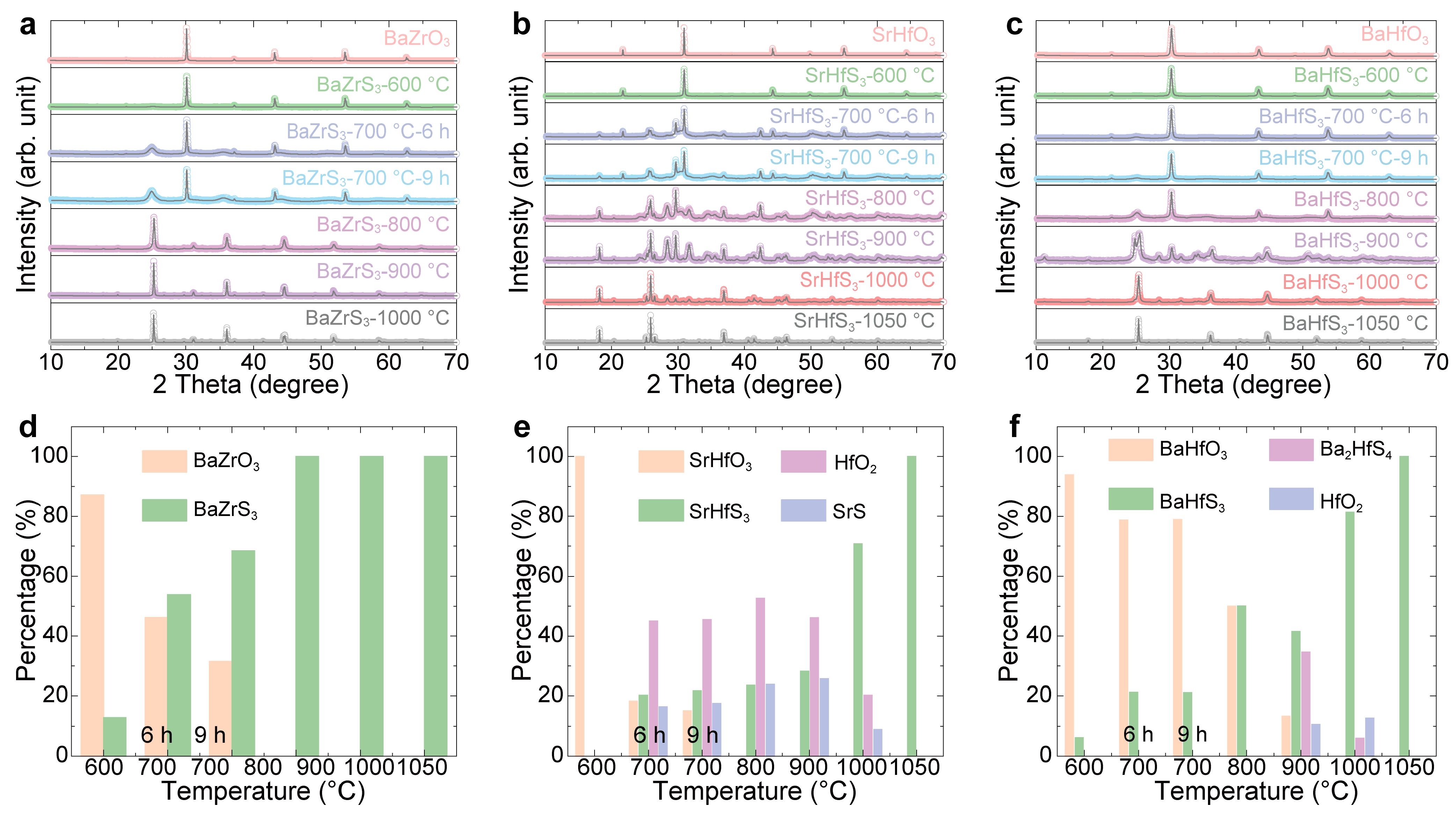
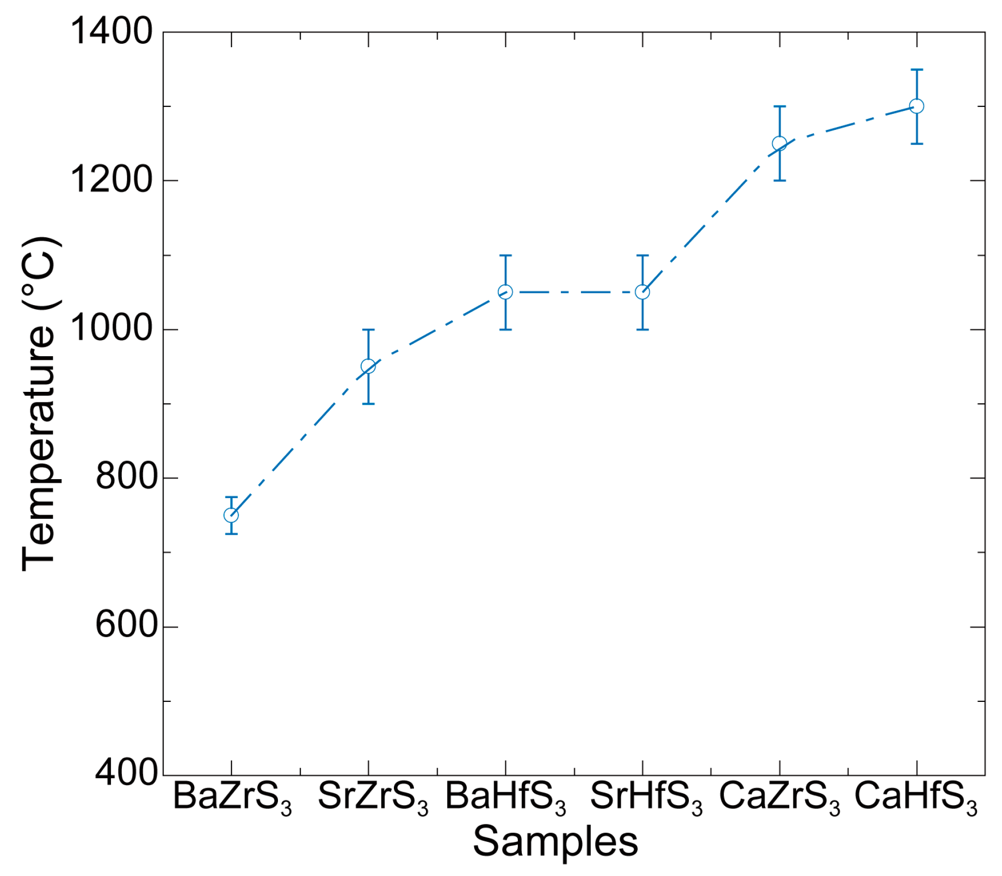
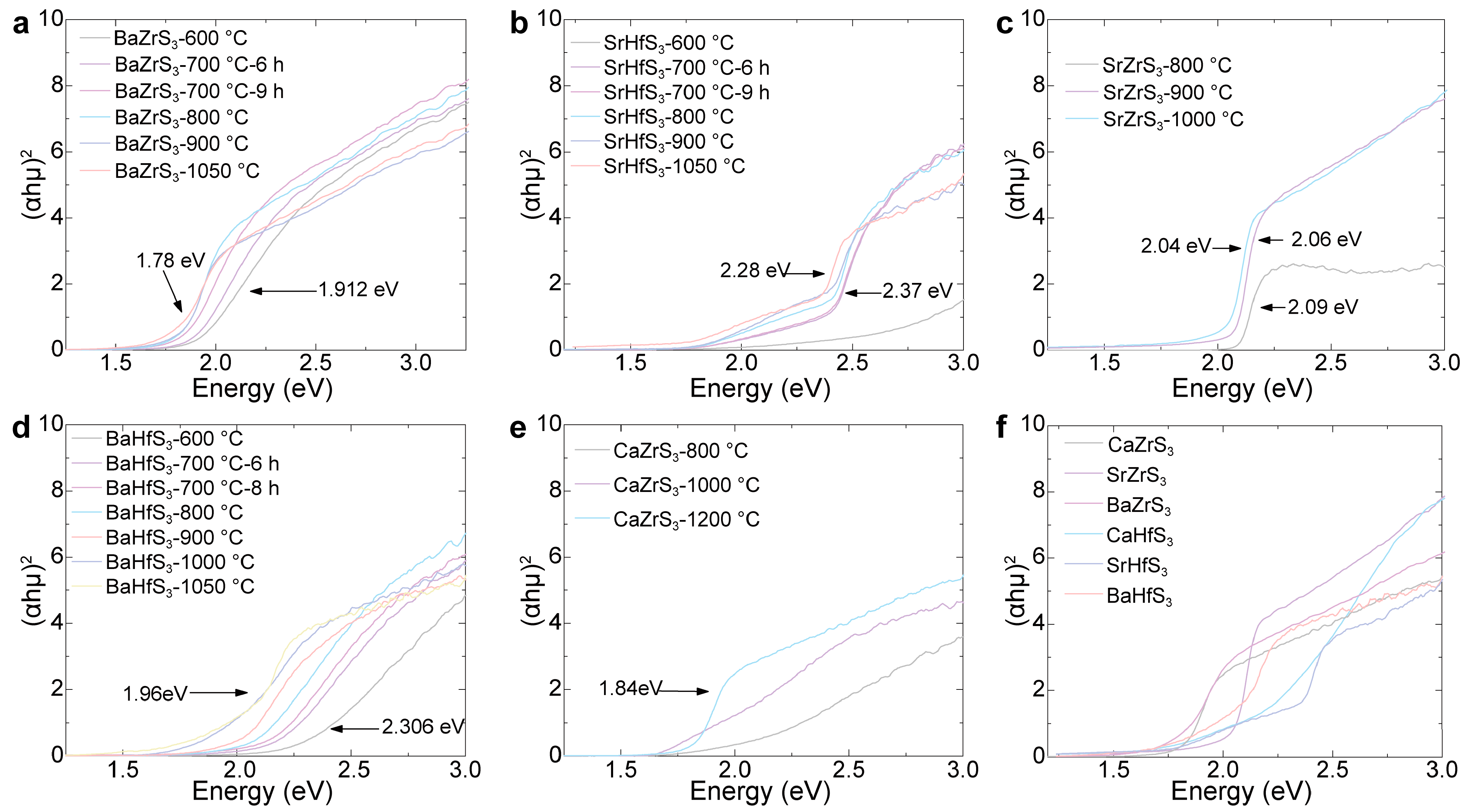
2. Results and Discussion

In this work, we used CS2 to sulfurize oxide perovskites into chalcogenide perovskites with argon as the carrier gas. The sulfurization temperatures were adjusted for different oxide perovskites to obtain sulfide samples. The structure and stability of the perovskites were estimated using the Goldschmidt tolerance coefficient (t) and octahedral factor (μ) [25]. A stable distorted perovskite structure is expected when 0.71≤ t ≤0.91 and μ ≥ 0.41. Otherwise, non-perovskite structures, such as hexagonal or orthorhombic phases with a needle-like morphology, are likely to form. The previous literature shows that Zr- and Hf-based chalcogenide perovskites (ABS3) can form stable perovskite structures with a Pnma space group. However, the t for Ti-based chalcogenides is 0.33, which is below the threshold of 0.41 to form a stable perovskite structure [7,26]. Thus, attempts to fabricate Ti-based chalcogenide materials result in non-perovskite structures (see Figures S5 and S6). In the main text, we focus on the results of Zr- and Hf-based chalcogenide perovskites.
As shown in Figure 1, the sulfurized samples exhibit a clear trend of darkening color with increasing sulfurization temperature. Initially, all oxide perovskite powders are white, indicating minimal visible light absorption. However, as sulfurization temperature increases, the color of the powder samples progressively darkens. For example, the color transitions from white for BaZrO3 to light red when sulfurized at 600 °C, deepens to dark red at 700 °C, and turns to gray-black above 800 °C, suggesting strong absorption in the visible range. A similar darkening trend is observed in the other materials. BaHfO3 shifts from white to light yellow when sulfurized at 600 °C and becomes dark red at 1000 °C. SrHfO3, though showing a more gradual change, transitions from light yellow at 600 °C to dark yellow-green at 1000 °C. CaZrS3 sulfurized at 1200 °C appears dark red, while SrZrS3 sulfurized at 1000 °C shows a bright red hue. CaHfS3 samples processed at 1200 °C develop a dark yellow color, reinforcing the trend of increasing absorption with increasing sulfurization temperature. Even the non-perovskite-phase Ti-based samples sulfurized at 1000 °C exhibit a pronounced black appearance, as shown in Figure S5.
To investigate the structural evolution of the products sulfurized at different temperatures, XRD was measured on these samples. The XRD patterns of BaZrO3, BaHfO3, and SrHfO3 sulfurized at various temperatures are shown in Figure 2a–c, respectively. The Rietveld refinement results for BaZrO3, BaHfO3, and SrHfO3 sulfurized at different temperatures are presented in Figures S1–S3, from which we extracted phase content and analyzed the relationship between phase composition and temperature. The proportions of oxide perovskites, chalcogenide perovskites, and intermediate phases at different temperature are shown in Figure 2d–f, respectively. From these results, the amount of oxide perovskite decreases with increasing sulfurization temperature for all samples, indicating the substitution of oxygen with sulfur. However, the minimum temperature required for complete sulfurization varies with the specific material. As seen in Figure 2d–f, BaZrS3 requires around 800 °C for a complete reaction, while SrHfS3 and BaHfS3 require temperatures exceeding 1000 °C. It is interesting to observe that BaZrO3 is converted into BaZrS3 directly with different fractions at different temperatures without any intermediate step. Earlier studies also suggest the presence of oxysulfides, as seen from the redshift in the absorption spectrum with increasing sulfurization temperature [15]. The oxysulfides are likely amorphous as they are not detected by XRD. In contrast, the sulfurization of SrHfO3 and BaHfO3 tended to form binary compounds at intermediate temperatures. (The refined XRD of SrZrS3, CaZrS3, and CaHfS3 can be found in Figure S4).
For the Zr series, BaZrS3 requires 800 °C for complete sulfurization, SrZrS3 requires approximately 1000 °C, and CaZrS3 needs temperatures exceeding 1200 °C. Thus, there is a trend of requiring increased sulfurization temperature with decreasing size of the A cation. This trend also applies to the Hf series: both BaHfS3 and SrHfS3 require 1100 °C for complete sulfurization, while the yield of CaHfS3 reaches only about 74.15% even at 1200 °C. These results are summarized in Figure 3, which shows the approximate lowest sulfurization temperatures for the Zr- and Hf-based chalcogenide perovskites. The increasing sulfurization temperature with decreasing size of the A cation reflects the increasing strength of the A-O bonds in the oxide perovskite precursors as one moves down the alkaline earth metal group (Ba, Sr, Ca). This trend is directly related to the electronegativity of the alkaline earth metal. As electronegativity increases from Ba to Sr to Ca, the electronegativity difference between the A cation and O increases, strengthening the bonding (i.e., the bond becomes more ionic). Since the sulfurization of these oxides requires the breaking of the A-O bonds, more energy is required to break the Ca-O bond than the Ba-O bond, which is provided by the higher sulfurization temperature. Therefore, the formation of CaZrS3 requires the highest sulfurization temperature, while BaZrS3 requires the lowest.
The bandgap of samples sulfurized at different temperatures was determined using UV-Vis spectroscopy. The variation in bandgap with sulfurization temperature is shown in Figure 4. For BaZrS3, SrHfS3, SrZrS3, and BaHfS3, the measured bandgap decreases as the sulfurization temperature increases, as illustrated in Figure 4a–d. This systematic redshift in bandgap, of the order of hundreds of meV, can be attributed to the presence of oxysulfide structural motifs in the partially sulfurized samples. If oxides and sulfides coexisted as separate phases, the UV-Vis measurements would reflect a superposition of two distinct band edge transitions instead of a smooth bandgap shift. The fact that XRD does not show an oxysulfide phase suggests that these oxysulfide structural motifs are not crystalline.
The measured bandgap values also correlate with the visual color of the samples at different temperatures. For samples sulfurized at high temperatures, the bandgap values for BaZrS3, SrZrS3, BaHfS3, and SrHfS3 match closely with theoretical calculations. However, the bandgap values for CaZrS3 and CaHfS3, shown in Figure 4e,f, are measured to be 1.84 eV and 2.13 eV, respectively, which are lower than the calculated values of 1.96 and 2.31 eV. This discrepancy is likely due to a high concentration of sulfur vacancies introduced at elevated temperatures, which alters the electronic structure and reduces the bandgap. Additional experiments are needed to determine the bandgap of CaZrS3 and CaHfS3 more accurately.
We further characterized the PL properties of the six Zr- and Hf-based perovskites. In Figure 5, we present normalized PL plots for easy comparison. Notably, all six materials exhibit PL in the visible range, with emission peaks ranging from 1.7 eV to 2.38 eV, confirming that they are all direct gap semiconductors. As we all know, emission peak values are closely related to bandgaps because they result from the radiative recombination of electrons and holes. When an electron in the conduction band recombines with a hole in the valence band, a photon is emitted, and its energy corresponds to the emission peak. However, defects and impurities in the materials are likely to have a certain impact on the bandgaps and emission characteristics. The emission peak values and bandgaps for the four Ba- and Sr-based perovskites are similar and closely align with theoretical predictions [7]. However, there are significant differences in the values of emission peaks and bandgaps for the two Ca-based perovskites, as shown in Table 1, which is derived from Figure 4f and Figure 5a. The full-width at half-maximum (FWMH) values can be seen in Figure 5b. SrZrS3 has the narrowest FWMH of 155.9 meV. This value is close to the 122 meV obtained from characterization [22], while CaHfS3 exhibits the highest FWHM of 491.1 meV. The measured PL peak of CaHfS3 is 1.93 eV. However, it has a relatively large FWHM. We speculate that this is because the final product is composed of 25.85% HfS2 and 74.15% CaHfS3. Bulk HfS2 exhibits an indirect bandgap, with reported values of 2.64 eV [27] or 2.58 eV [28]. As a result, the FWHM broadens towards the high-energy side, leading to its relatively large value.

3. Experimental Section

3.1. Synthesis of Chalcogenide Perovskite Powder Samples

We fabricated nine kinds of oxide perovskites for sulfurization following the ABO3 structure (A: Ca, Sr, and Ba; B: Ti, Zr, and Hf). All oxide perovskite ceramic samples were fabricated with a solid-state reaction method. First, ACO3 and BO2 were mixed with a stoichiometric ratio of 1:1 and ball milled for 8 h to ensure homogeneity. The mixture was then dried and placed into a muffle furnace for 8 h at 1200 °C. Next, the oxide perovskite ABO3 underwent sulfidation treatment at different temperatures in a tube furnace filled with carbon disulfide (CS2) and argon (Ar), following the procedure outlined in our previous work [29]. The different treatment conditions are as follows: sulfurization at 600 °C for 9 h, at 700 °C for 6 h, and at 800 °C for 4 h, or 2 h sulfidation at temperatures exceeding 900 °C.

3.2. Powder Characterization Techniques

The crystal diffraction patterns of powders were recorded by X-ray diffractometer (XRD, Bruker D8-Advance, Bruker AXS, Karlsruhe, Germany, Diffrac. Suite) equipped with a Cu Kα radiation source (λ =1.5418 Å). Measurements were performed under the θ–2θ scanning mode and continuous scanning with a step size of 0.02°. Raman spectra (Laser Raman Spectrometer, HORIBA, Tokyo, Japan, LabSpec6 version: 6.4.4.16) were obtained with a HORIBA Raman spectrometer using 532 nm laser excitation. The scanning electron microscopy (SEM) images and energy-dispersive X-ray spectrosco-py (EDX) analysis were acquired with a Focused Ion Beam-Scanning Electron Microscope (FIB-SEM, JSM-7000F, JEOL Japan Electronics Co., Ltd., Tokyo, Japan)-Carl Zeiss AURIGA CrossBeam with an Oxford EDS system. The absorption spectra were collected from a Cary series UV-Vis-NIR (Hitachi U-4100, Hitachi Limited, Tokyo, Japan) spectrophotometer measuring from 400 nm to 800 nm. Photoluminescence (PL, Laser Raman Spectrometer, HORIBA, Tokyo, Japan, LabSpec6 version: 6.4.4.16) was measured by a HORIBA Raman spectrometer with 325 nm and 532 nm lasers.

4. Conclusions

In conclusion, this study systematically investigated the impact of sulfurization temperature on the structural and optical properties of chalcogenide perovskites, among which CaHfS3 and CaZrS3 were reported for the first time. The minimum temperatures required for the formation of chalcogenide perovskites from different oxide precursors vary due to the different energy barriers that must be overcome during sulfur substitution. Breaking the Ca-O bond requires the highest energy, while Ba-O requires the lowest, and therefore CaZrS3 and CaHfS3 require the highest sulfurization temperatures. The six Zr- and Hf-based chalcogenide perovskite materials were found to have emission peaks ranging from 1.7 eV to 2.38 eV. The systematic studies of the series of chalcogenide perovskite materials offer valuable insights for their potential applications in optoelectronics.

Supplementary Materials

The following supporting information can be downloaded at https://www.mdpi.com/article/10.3390/molecules30061198/s1: Figure S1: Plots of powder XRD patterns with Rietveld analysis. (a) BaZrO3 and BaZrO3 sulfurized at different temperatures: (b) 600 °C, (c) 700 °C for 6 h, (d) 700 °C for 9 h, (e) 800 °C, (f) 900 °C, and (g) 1000 °C; Figure S2: Plots of powder XRD patterns with Rietveld analysis. (a) SrHfO3 and SrHfO3 sulfurized at different temperatures: (b) 600 °C, (c) 700 °C for 6 h, (d) 700 °C for 9 h, (e) 800 °C, (f) 900 °C, (g) 1000 °C, and (h) 1050 °C; Figure S3: Plots of powder XRD patterns with Rietveld analysis. (a) BaHfO3 and BaHfO3 sulfurized at different temperatures: (b) 600 °C, (c) 700 °C for 6 h, (d) 700 °C for 9 h, (e) 800 °C, (f) 900 °C, (g) 1000 °C, and (h) 1050 °C; Figure S4: Plots of powder XRD patterns with Rietveld analysis. (a) SrZrO3 sulfurized at different temperatures: (b) 800 °C, (c) 900 °C, and (d) 1000 °C. (e) CaZrO3 and (f) CaZrO3 sulfurized at 1200 °C. (g) CaHfO3 and (h) CaHfO3 sulfurized at 1200 °C; Figure S5: Photos of CaTiO3, SrTiO3, and BaTiO3 powder as well as CaTiO3, SrTiO3, and BaTiO3 powder sulfurized at 1000 °C; Figure S6: Plots of powder XRD patterns with Rietveld analysis. (a) SrTiO3, (b) BaTiO3, (c) SrTiO3, and (d) BaTiO3 sulfurized at 1000 °C and (e) CaTiO3 and CaTiO3 sulfurized at 1000 °C; Figure S7: The room-temperature Raman spectra of (a) BaZrO3, (b) BaHfO3, and (c) SrHfO3 sulfurized at different temperatures. (d) The room-temperature Raman spectra of CaZrS3 sulfurized at 1200 °C, SrZrS3 sulfurized at 1000 °C, and CaHfS3 sulfurized at 1200 °C; Figure S8: The room-temperature Raman spectra of (a) SrTiO3 and (b) BaTiO3 sulfurized at 1000 °C; Figure S9: Typical SEM images of BaHfO3, BaZrO3, and CaZrO3 after sulfurization at different temperatures are presented as follows: For BaHfO3, images (a–c) correspond to sulfurization temperatures of 600 °C, 800 °C, and 1200 °C, respectively. For BaZrO3, images (d–f) correspond to sulfurization temperatures of 600 °C, 800 °C, and 1000 °C. And for CaZrO3, images (g–i) show the samples sulfurized at 800 °C, 1000 °C, and 1200 °C; Figure S10: The FWHMs of the (121) peak as a function of temperature. (a) BaZrO3, (b) SrHfO3, and (c) BaHfO3 sulfurized at different temperatures; Figure S11: (a) The ratios of S to S + O concentrations in various samples at different temperatures, along with the EDX spectra of (b) BaZrS3, (c) BaHfS3, (d) SrHfS3, and (e) CaZrS3 sulfurized at high temperatures.

Author Contributions

Fabricated the chalcogenide perovskites samples, P.Z., L.Y. and B.B.; structural and optical characterizations, P.Z., L.Y. and B.B.; XRD data analysis, S.K., K.C. and H.H.; data analysis, P.Z., L.Y., L.S. and K.G.; writing—original draft, P.Z., L.Y. and Z.Y.; writing—review and editing, P.Z., L.Y. and Z.Y.; funding acquisition, Z.Y. All authors have read and agreed to the published version of the manuscript.

Funding

This work is supported by the Higher Education Science and Technology Innovation Plan Project of Shanxi (2024L146) and the Fundamental Research Program of Shanxi Province (202403021222252).

Institutional Review Board Statement

Not applicable.

Informed Consent Statement

Not applicable.

Data Availability Statement

Data are contained within the article and Supplementary Materials.

Acknowledgments

The authors are grateful to the State Key Lab of Advanced Technology for Materials Synthesis and Processing (Wuhan University of Technology) for their financial support.

Conflicts of Interest

The authors declare no conflicts of interest.

References

  1. Kojima, A.; Teshima, K.; Shirai, Y.; Miyasaka, T. Organometal Halide Perovskites as Visible-Light Sensitizers for Photovoltaic Cells. J. Am. Chem. Soc. 2009, 131, 6050–6051. [Google Scholar] [CrossRef] [PubMed]
  2. Metcalf, I.; Sidhik, S.; Zhang, H.; Agrawal, A.; Persaud, J.; Hou, J.; Even, J.; Mohite, A.D. Synergy of 3D and 2D Perovskites for Durable, Efficient Solar Cells and Beyond. Chem. Rev. 2023, 123, 9565–9652. [Google Scholar] [CrossRef]
  3. Xie, H.B.; Lira-Cantu, M. Multi-Component Engineering to Enable Long-Term Operational Stability of Perovskite Solar Cells. J. Phys. Energy 2020, 2, 024008. [Google Scholar] [CrossRef]
  4. Ren, M.; Qian, X.; Chen, Y.; Wang, T.; Zhao, Y. Potential Lead Toxicity and Leakage Issues on Lead Halide Perovskite Photovoltaics. J. Hazard. Mater. 2022, 426, 127848. [Google Scholar] [CrossRef]
  5. Hossain, M.K.; Toki, G.F.I.; Samajdar, D.P.; Mushtaq, M.; Rubel, M.H.K.; Pandey, R.; Madan, J.; Mohammed, M.K.A.; Islam, M.R.; Rahman, M.F.; et al. Deep Insights into the Coupled Optoelectronic and Photovoltaic Analysis of Lead-Free CsSnI3 Perovskite-Based Solar Cell Using DFT Calculations and SCAPS-1D Simulations. ACS Omega 2023, 8, 22466–22485. [Google Scholar] [CrossRef] [PubMed]
  6. Konidakis, I.; Karagiannakia, A.; Stratakis, E. Advanced Composite Glasses with Metallic, Perovskite, and Two-Dimensional Nanocrystals for Optoelectronic and Photonic Applications. Nanoscale 2022, 14, 2966–2989. [Google Scholar] [CrossRef]
  7. Sun, Y.Y.; Agiorgousis, M.L.; Zhang, P.; Zhang, S. Chalcogenide Perovskites for Photovoltaics. Nano Lett. 2015, 15, 581–585. [Google Scholar] [CrossRef]
  8. Mercy, E.N.V.; Srinivasan, D.; Marasamy, L. Emerging BaZrS3 and Ba(Zr,Ti)S3 Chalcogenide Perovskite Solar Cells: A Numerical Approach Toward Device Engineering and Unlocking Efficiency. ACS Omega 2024, 9, 4359–4376. [Google Scholar] [CrossRef] [PubMed]
  9. Han, Y.; Fang, X.; Shi, Z. Advances in Chalcogenide Perovskites: Fundamentals and Applications. Appl. Phys. Rev. 2024, 11, 021338. [Google Scholar] [CrossRef]
  10. Ivaniuk, K.; Cherpak, V.; Stakhira, P.; Baryshnikov, G.; Minaev, B.; Hotra, Z.; Turyk, P.; Zhydachevskii, Y.; Volyniuk, D.; Aksimentyeva, O.; et al. BaZrO3 Perovskite Nanoparticles as Emissive Material for Organic/Inorganic Hybrid Light-Emitting Diodes. Dyes Pigm. 2017, 145, 399–403. [Google Scholar] [CrossRef]
  11. Nishigaki, Y.; Nagai, T.; Nishiwaki, M.; Aizawa, T.; Kozawa, M.; Hanzawa, K.; Kato, Y.; Sai, H.; Hiramatsu, H.; Hosono, H.; et al. Extraordinary Strong Band-Edge Absorption in Distorted Chalcogenide Perovskites. Sol. RRL 2020, 4, 1900555. [Google Scholar] [CrossRef]
  12. Suhail, M.; Abbas, H.; Khan, M.B.; Khan, Z.H. Chalcogenide Perovskites for Photovoltaic Applications: A Review. J. Nanopart. Res. 2022, 24, 142. [Google Scholar] [CrossRef]
  13. Surendran, M.; Chen, H.; Zhao, B.; Thind, A.S.; Singh, S.; Orvis, T.; Zhao, H.; Han, J.-K.; Htoon, H.; Kawasaki, M.; et al. Epitaxial Thin Films of a Chalcogenide Perovskite. Chem. Mater. 2021, 33, 7457–7464. [Google Scholar] [CrossRef]
  14. Hanzawa, K.; Iimura, S.; Hiramatsu, H.; Hosono, H. Material Design of Green-Light-Emitting Semiconductors: Perovskite-Type Sulfide SrHfS3. J. Am. Chem. Soc. 2019, 141, 5343–5349. [Google Scholar] [CrossRef]
  15. Perera, S.; Hui, H.; Zhao, C.; Xue, H.; Sun, F.; Deng, C.; Gross, N.; Milleville, C.; Xu, X.; Watson, D.F.; et al. Chalcogenide Perovskites-an Emerging Class of Ionic Semiconductors. Nano Energy 2016, 22, 129–135. [Google Scholar] [CrossRef]
  16. Meng, W.W.; Saparov, B.; Hong, F.; Wang, J.B.; Mitzi, D.B.; Yan, Y.F. Alloying and Defect Control within Chalcogenide Perovskites for Optimized Photovoltaic Application. Chem. Mater. 2016, 28, 821–829. [Google Scholar] [CrossRef]
  17. Yang, R.Q.; Jess, A.D.; Fai, C.; Hages, C.J. Low-Temperature, Solution-Based Synthesis of Luminescent Chalcogenide Perovskite BaZrS3 Nanoparticles. J. Am. Chem. Soc. 2022, 144, 15928–15931. [Google Scholar] [CrossRef]
  18. Yasmin, N.; Safdar, M.; Ali, G.; Khan, H.M.; Mirza, M. Solar Light Harvesting Nanomaterial (BaZrS3) for Photocatalytic Activity and OER Reaction. J. Phys. Chem. Solids 2023, 172, 111056. [Google Scholar] [CrossRef]
  19. Yu, Z.; Wei, X.; Zheng, Y.; Hui, H.; Bian, M.; Dhole, S.; Seo, J.-H.; Sun, Y.-Y.; Jia, Q.; Zhang, S.; et al. Chalcogenide Perovskite BaZrS3 Thin-Film Electronic and Optoelectronic Devices by Low Temperature Processing. Nano Energy 2021, 85, 105959. [Google Scholar] [CrossRef]
  20. Wei, X.; Hui, H.; Zhao, C.; Deng, C.; Han, M.; Yu, Z.; Sheng, A.; Roy, P.; Chen, A.; Lin, J.; et al. Realization of BaZrS3 Chalcogenide Perovskite Thin Films for Optoelectronics. Nano Energy 2020, 68, 104317. [Google Scholar] [CrossRef]
  21. Liang, Y.; Zhang, Y.; Xu, J.; Ma, J.; Jiang, H.; Li, X.; Zhang, B.; Chen, X.; Tian, Y.; Han, Y.; et al. Parametric Study on Controllable Growth of SrZrS3 Thin Films with Good Conductivity for Photodetectors. Nano Res. 2023, 16, 7867–7873. [Google Scholar] [CrossRef]
  22. Niu, S.; Huyan, H.; Liu, Y.; Yeung, M.; Ye, K.; Blankemeier, L.; Orvis, T.; Sarkar, D.; Singh, D.J.; Kapadia, R.; et al. Bandgap Control via Structural and Chemical Tuning of Transition Metal Perovskite Chalcogenides. Adv. Mater. 2017, 29, 1604733. [Google Scholar] [CrossRef] [PubMed]
  23. Kassa, M.D.; Woldemariam, M.M.; Debelo, N.G.; Gebremeskel, D.K.; Habura, K.H. A Computational Exploration of Structural, Electronic, and Optical Properties of Hf- Substituted CaZr1−x HfxS3 (x = 0.25, 0.50, and 0.75) for Photovoltaic Applications. Phys. Scripta 2025, 100, 015974. [Google Scholar] [CrossRef]
  24. Song, X.; Shai, X.; Deng, S.; Wang, J.; Li, J.; Ma, X.; Li, X.; Wei, T.; Ren, W.; Gao, L.; et al. Anisotropic Chalcogenide Perovskite CaZrS3: A Promising Thermoelectric Material. J. Phys. Chem. C 2022, 126, 11751–11760. [Google Scholar] [CrossRef]
  25. Sopiha, K.V.; Comparotto, C.; Márquez, J.A.; Scragg, J.J.S. Chalcogenide Perovskites: Tantalizing Prospects, Challenging Materials. Adv. Opt. Mater. 2021, 10, 2101704. [Google Scholar] [CrossRef]
  26. Niu, S.; Joe, G.; Zhao, H.; Zhou, Y.; Orvis, T.; Huyan, H.; Salman, J.; Mahalingam, K.; Urwin, B.; Wu, J.; et al. Giant Optical Anisotropy in a Quasi-One-Dimensional Crystal. Nat. Photonics 2018, 12, 392–396. [Google Scholar] [CrossRef]
  27. Wang, D.; Zhang, X.; Guo, G.; Gao, S.; Li, X.; Meng, J.; Yin, Z.; Liu, H.; Gao, M.; Cheng, L.; et al. Large-Area Synthesis of Layered HfS2(1−x)Se2x Alloys with Fully Tunable Chemical Compositions and Bandgaps. Adv. Mater. 2018, 30, 1803285. [Google Scholar] [CrossRef]
  28. Wang, D.; Meng, J.; Zhang, X.; Guo, G.; Yin, Z.; Liu, H.; Cheng, L.; Gao, M.; You, J.; Wang, R. Selective Direct Growth of Atomic Layered HfS2 on Hexagonal Boron Nitride for High Performance Photodetectors. Chem. Mater. 2018, 30, 3819–3826. [Google Scholar] [CrossRef]
  29. Kong, S.; Dong, H.; Yu, Z.; Guo, J.; Cao, K.; Chen, K.; Ke, X.; Zhou, C.; Deng, J.; Yang, S.; et al. Ti-S Antibonding Coupling Enables Enhanced Bandgap Tuning in Ti-Substituted BaHfS3 Perovskite. Ceram. Int. 2024, 50, 10889–10896. [Google Scholar] [CrossRef]
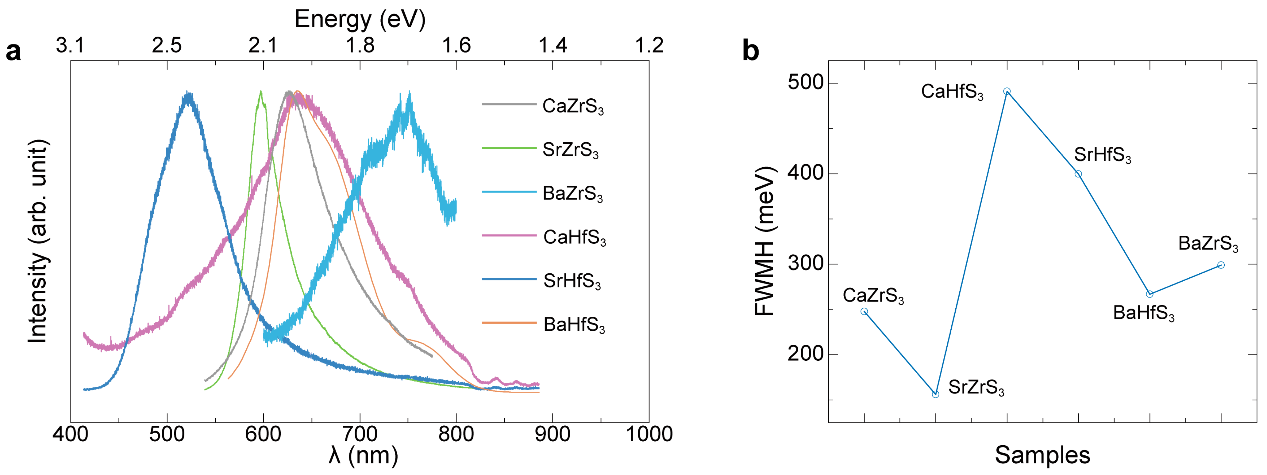
Figure 1. Photos of BaZrO3, BaHfO3, SrHfO3, CaZrO3, CaHfO3, and SrZrO3 powders, as well as these same powders (BaZrO3, BaHfO3, SrHfO3, CaZrO3, CaHfO3, and SrZrO3) sulfurized at different temperatures.
Molecules 30 01198 g001
Figure 2. XRD patterns of (a) BaZrO3, (b) SrHfO3, and (c) BaHfO3 sulfurized at different temperatures and the molar percentage of the final products for (d) BaZrO3, (e) SrHfO3, and (f) BaHfO3 sulfurized at different temperatures.
Molecules 30 01198 g002
Figure 3. Minimum sulfurization temperatures for different oxide perovskites converted to chalcogenide perovskites.
Molecules 30 01198 g003
Figure 4. A Tauc plot of different samples sulfurized at different temperatures: (a) BaZrS3, (b) SrHfS3, (c) SrZrS3, (d) BaHfS3, and (e) CaZrS3. (f) A comparison of six sulfur-based chalcogenide perovskites’ bandgaps.
Molecules 30 01198 g004
Figure 5. (a) The normalized PL spectra and (b) FWMH of the PL spectra of SrZrS3, CaZrS3, CaHfS3, SrHfS3, BaZrS3, and BaHfS3.
Molecules 30 01198 g005
Table 1. Comparison of Zr- and Hf-based perovskite bandgap and PL peak.
SamplesBandgap (eV)PL Peak (eV)
BaZrS31.781.79
CaZrS31.841.98
BaHfS31.961.95
SrZrS32.042.08
CaHfS32.131.93
SrHfS32.282.28
Disclaimer/Publisher’s Note: The statements, opinions and data contained in all publications are solely those of the individual author(s) and contributor(s) and not of MDPI and/or the editor(s). MDPI and/or the editor(s) disclaim responsibility for any injury to people or property resulting from any ideas, methods, instructions or products referred to in the content.

