
Research Progress on 18F-Labeled Agents for Imaging of Myocardial Perfusion with Positron Emission Tomography


1
Department of Nuclear Medicine, Beijing Anzhen Hospital, Capital Medical University, Beijing 100029, China
2
State Key Laboratory of Molecular Vaccinology and Molecular Diagnostics & Center for Molecular Imaging and Translational Medicine, School of Public Health, Xiamen University, Xiamen 361102, China
*
Author to whom correspondence should be addressed.
Molecules 2017, 22(4), 562; https://doi.org/10.3390/molecules22040562
Submission received: 13 January 2017
/
Revised: 14 February 2017
/
Accepted: 20 February 2017
/
Published: 30 March 2017
(This article belongs to the Section Medicinal Chemistry)
Abstract
:Coronary artery disease (CAD) is the leading cause of death in the world. Myocardial perfusion imaging (MPI) plays a significant role in non-invasive diagnosis and prognosis of CAD. However, neither single-photon emission computed tomography nor positron emission tomography clinical MPI agents can absolutely satisfy the demands of clinical practice. In the past decades, tremendous developments happened in the field of 18F-labeled MPI tracers. This review summarizes the current state of 18F-labeled MPI tracers, basic research data of those tracers, and the future direction of MPI tracer research.
1. Introduction
Though the treatments of coronary artery disease (CAD) have seen prominent improvements over the past decades, CAD is still the leading cause of death in the world. Single-photon emission computed tomography (SPECT) myocardial perfusion imaging (MPI), using radiotracers such as 99mTc-sestamibi, 99mTc-tetrofosmin and 201Tl, is the commonly used, standard, and non-invasive clinical screening tool for detecting CAD, risk stratification, and guidance of therapeutic interventions. Its sensitivity and specificity for detecting significant coronary stenosis was 87% and 73%, respectively, in a meta-analysis involving 4480 subjects [1]. However, the application of SPECT MPI is limited for the following reasons: inferior spatial and temporal resolution, incapability of absolute quantification, non-uniform attenuation correction and high uptake in the organs adjacent to the heart [2].
PET isotopes emit positrons. When a positron meets a nearby electron, they annihilate each other and emit two 511-keV photons in opposite direction (discharged at 180° to each other). Only the coincidental detection of two 511-keV photons can be recorded in PET scanners and reconstructed into PET images [3]. Compared with SPECT, PET technology offers a better resolution and effective correction of photo-attenuation and scatter [4], leading to absolute quantification of regional myocardial blood flow and coronary flow reserve [5]. Besides that, patients are exposed to less radiation owing to the short half-lives of positron isotopes [6,7]. Hence, the need for and use of PET applications in healthcare facilities is increasing tremendously [3,8,9,10]. In 2016, the American Society of Nuclear Cardiology and the Society of Nuclear Medicine and Molecular Imaging published a joint position statement on the clinical indications for the significant underutilization of myocardial perfusion PET in America [11,12]. 13N-NH3, 82Rb, and 15O-H2O are representative clinical PET MPI tracers [13]. As the half-lives of 15O-H2O and 13N-NH3 are very short (2 and 10 min, respectively) [14], on-site cyclotrons are required. 82Rb has a very short half-life (75 s) but can be conveniently supplied from a bedside generator. However, it has a low myocardial ejection fraction at high flow rates. Hence, it is imperative to develop novel and preferable PET MPI agents.
Compared with the aforementioned isotopes, 18F has much shorter average positron range (1.03 nm), which results in better spatial resolution and contrast. It also has a longer half-life (110 min), so 18F-labeled radiotracers can be supplied at regional cyclotrons and allow treadmill exercises [15]. Thus, the development of 18F-labeled MPI agents becomes a hot topic of interest for many researchers. Numerous novel agents have been synthesized and studied in the past decade. Especially a series of reports about 18F-flurpiridaz evidently enhanced researchers’ confidence on the future of PET MPI agents. Several published reviews have summarized the characteristics of PET MPI agents and compared them with 13N-NH3, 82Rb, and 15O-H2O [14,16,17,18,19]. However, most of them focused on the MPI agents in clinic status. In this study, we summarized 18F-labeled radiotracers in both clinical and preclinical status.
According to the chemical structures and mechanism, 18F-labeled MPI agents under investigation can be divided into two types: lipophilic cations and analogues of mitochondrial complex-1 (MC-1) inhibitors. Herein, we compared the characteristics, mechanism, and research status of those different tracers.
2. Lipophilic Cations
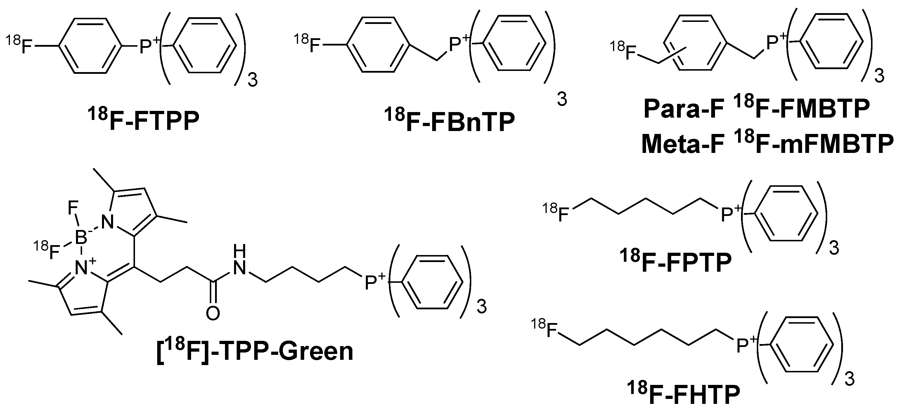
Mitochondria take up 20–30% of the myocardial intracellular volume, making it an ideal target for MPI. The activation energy of lipophilic cations for moving through hydrophobic barrier of a biological membrane is far lower than that of other cations. Hence, lipophilic cations can pass through phospholipid bilayers of mitochondria without requiring a specific uptake mechanism [20]. Because of delocalized positive charge of lipophilic cations, they accumulated substantially in the mitochondria in a membrane potential-dependent manner [21]. Myocardial ischemia can cause cell death. Loss of mitochondrial membrane potential is an early event in cell death [22]. Lipophilic cations can be used to detect myocardial abnormalities because the uptake of which is sensitive with mitochondrial voltage. Lipophilic cations for MPI include two types such as ammonium cations (Figure 1) and phosphonium cations (Figure 2).
2.1. Ammonium Cations
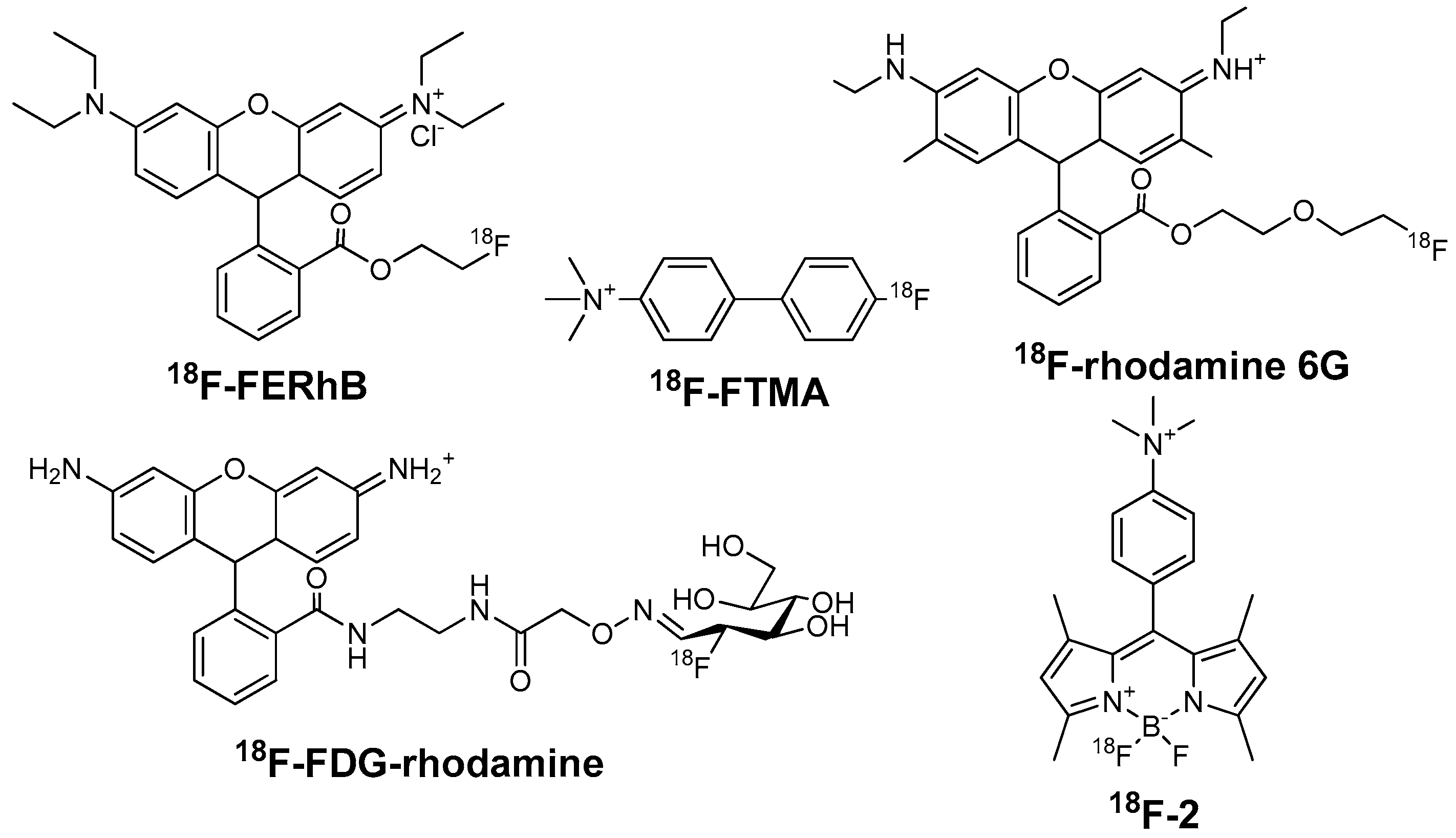
2.1.1. 4-18F-Fluorotri-N-Methylanilinium Iodide (18F-FTMA)
Studenov et al. synthesized four 18F-labeled ammonium salts, represented by 18F-FTMA [23]. The studies of acetylcholinesterase (AcChE) inhibition suggested that the myocardial accumulation of 18F-FTMA was probably due to the binding with myocardial AcChE (Ki: 46–49 μM). The biodistribution study in mice revealed that 18F-FTMA had low myocardial uptake and heart/liver ratio (<0.5 during 5–60 min post injection (p.i.)). Hence, it had a limited potential for MPI.
2.1.2. 18F-Labeled Rhodamines
Rhodamine can accumulate in the mitochondria in proportion to mitochondrial membrane potential. 18F-FERhB was developed for MPI because the lead unlabeled compound rhodamine-123 could accumulate well in the heart of mouse [24]. The imaging capability of 18F-FERhB was related to its stability. For instance, 71% of 18F-FERhB got hydrolyzed in mouse serum at 2 h p.i., leading to poor myocardial uptake in the microPET image of a mouse. Whereas 86% of 18F-FERhB was still intact in rat serum at the same condition, resulting in considerable increase of myocardial uptake in rats [25]. In the biodistribution study of rats, the myocardial uptake (2.06 ± 0.61% ID/g at 60 min p.i.) was over twice the liver uptake and over 25 times the blood uptake. However, the myocardial image of the rat indicated that the uptake in heart and liver was approximately equal so it might not be a competitive MPI agent. Researchers supposed that 18F-FERhB might have better performance in human, since it had a better stability in human serum.
Maddahi et al. used 18F-FDG as the radiointermediate, and reported the preclinical evaluation of 18F-FDG-rhodamine [26]. 18F-FDG-rhodamine had a good stability in human plasma in vitro. The heart uptake of 18F-FDG-rhodamine was 11.24 ± 1.97% ID/g in rats, which was nearly 4 times higher than other radiofluorinated rhodamine analogues. The low lipophilic characteristic (log P = −1.64 ± 0.03) leads to the low liver uptake. The heart/liver ratio was 21.20 at 60 min p.i. The myocardial extraction of 18F-FDG-rhodamine was 27.63 ± 5.12% during the first 15 min of perfusion period, which was higher than 99mTc-MIBI (15 ± 1%) and lower than 201Tl (30 ± 5%). Besides that, Maddahi et al. mentioned that 18F-FDG-rhodamine hydrolysed in vivo in mice as 18F-FERhB. They suggested that mice might not be the suitable animal models for the tests of rhodamine-related compounds [26].
On the other hand, Bartholomä et al. developed a range of different rhodamine cores (rhodamine 6G, rhodamine 101, and tetramethylrhodamine) labeled with 18F. They used various rhodamine lactones as the precursors and used 18F-fluorodiethylene glycol ester as the prosthetic group [27]. Rhodamine 6G could locate in the mitochondria of isolated rat cardiomyocytes and had superior pharmacologic properties than others. Further first-in-human clinical studies with 18F-rhodamine 6G are on the way. So far, there is no follow-up report published in literature concerning its stability and clinical application in human.
2.1.3. 18F-Labeled BODIPY Derivatives
Boron-dipyrromethene (BODIPY) is a class of fluorescent dyes. It contains dipyrromethene and a disubstituted boron atom. BODIPY derivatives accumulate in mitochondria in a mitochondrial membrane potential-dependent manner [28]. They can be labeled with 18F by SnCl4-promoted 18F-19F isotopic exchange in aqueous solutions. Li et al. reported a series of 18F-labeled BODIPY derivatives [29,30,31]. Most of them had low heart/liver ratio, which limited their potential as MPI agents. 10-(4-(trimethylammonio)phenyl)-5-fluoro-5-18F-fluoro-1,3,7,9-tetramethyl-5H-dipyrrolo[1,2-c:2′,1′f][1,3,2] diazaborinin-4-ium-5-uide (18F-2) was the representative compound [31]. The heart uptake and heart/liver ratio of 18F-2 was 8.75% ± 1.04% ID/g and 2.19 ± 0.42 at 60 min p.i. in mice. In addition, all seven reported agents showed certain bone uptake (from 1.03 ± 0.21 to 2.74 ± 0.15% ID/g at 60 min p.i. in mice), indicating the decomposition of agents in vivo. So far, 18F-labeled BODIPY derivatives haven’t showed remarkable biological properties. However, since BODIPY can be developed as PET/optical dual-modality agents, and the labeling method is mild and simple, novel BODIPY derivatives may be competitive in the future.
2.2. Phosphonium Cations
Phosphonium cations were studied much widely than ammonium cations. Most of them showed superior properties in both MPI and detection of apoptosis. The modification of phosphonium cations was focused on the labeling methods and biological properties. The representative agents were 18F-FBnTP, 18FTPP, 18F-FPTP, 18F-FHTP, and 18F-mFMBTP.
2.2.1. 18F-Fluorobenzyl Triphenyl Phosphonium (18F-FBnTP) Cation
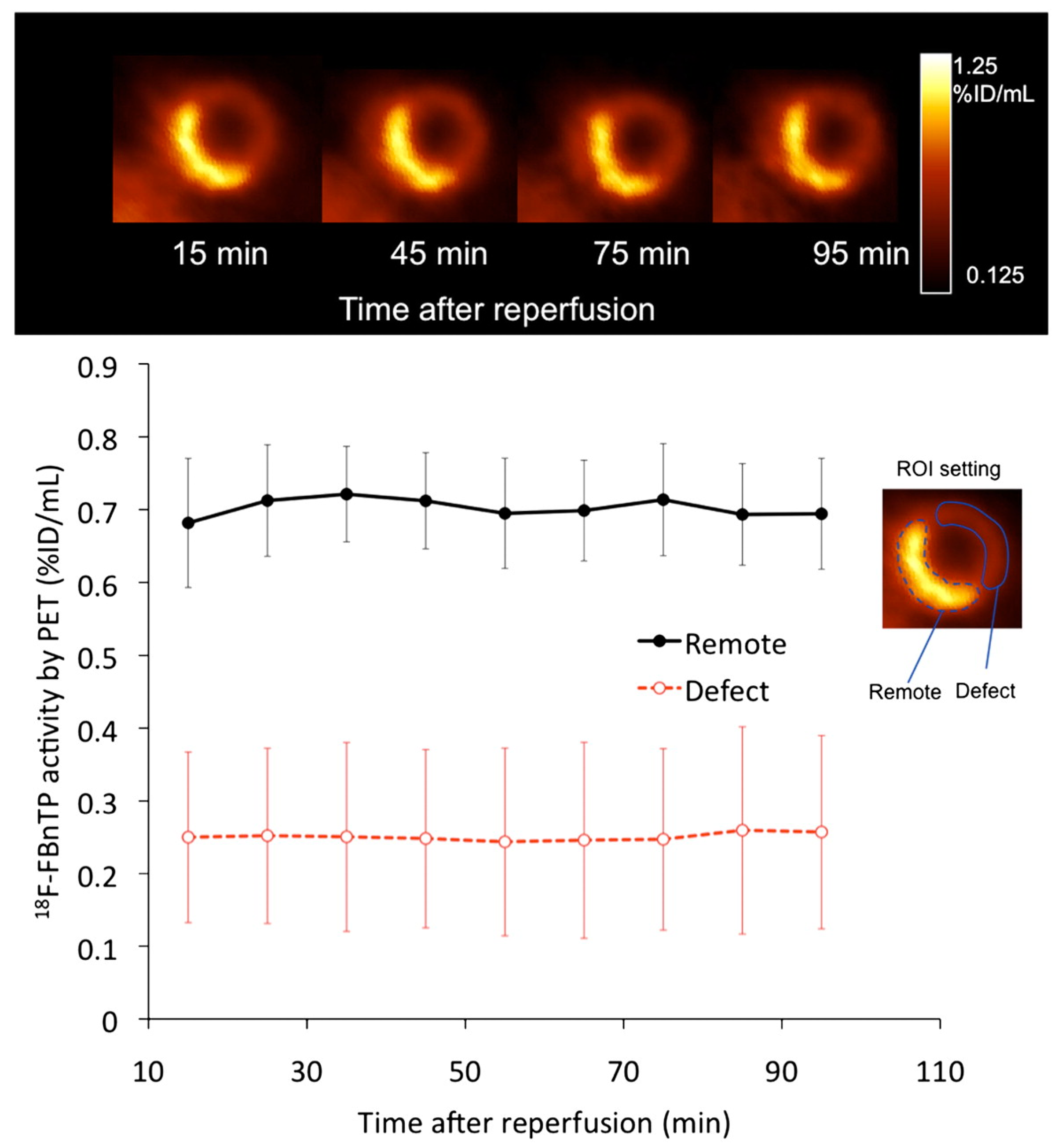
18F-FBnTP is the incipient 18F-labeled phosphonium cation [32]. In the imaging study of dogs, it showed notable initial uptake and prolonged retention in the myocardium [21]. The clearance from blood pool was rapid (half-life: 19.5 ± 4.4 s), reaching 26.2 ± 7.8% and 13.4 ± 6.3% of activity in the left ventricular wall at 5 and 10 min, respectively. At 60 min p.i., the heart/blood, heart/lung, and heart/liver ratios were 16.6:1, 12.2:1, and 1.2:1, respectively. The detailed anatomy of the heart including the papillary muscle and the left and right atria could be easily recognized because of low background activity in combination with extensive uptake and prolonged retention in the myocardium. 18F-FBnTP was eliminated mainly via kidneys than hepatobiliary tract. It is sensitive in detecting small flow defects with similar accuracy all over the myocardium, including the inferior aspect adjacent to the liver.
Compared with ex vivo tissue staining, the ischemic area after coronary occlusion assessed by PET was 16% smaller [33]. Compared with 99mTc-tetrofosmin, the accuracy of 18F-FBnTP was far better in the determination of mild and severe stenosis. In addition, 18F-FBnTP showed stable delineation of the ischemic area with no appreciable washout or redistribution (Figure 3) compared with 201Tl [34]. At the beginning, the main limitation of 18F-FBnTP was its radiosynthesis. The first report of radiosynthesis took four steps (82 min) with poor radiochemical yield (6%), making it inconvenient for clinical application [21]. Since then, researchers have devoted their efforts to simplifying the synthesis [35,36]. In 2016, Zhang et al. reported a one-step synthesis of 18F-FBnTP by a copper-mediated 18F-fluorination reaction with a pinacolyl arylboronate precursor [37]. The total radiochemical yield was 60 ± 18% without correction. This was a huge step for the promotion of 18F-FBnTP.
2.2.2. (4-18F-Fluorophenyl)triphenylphosphonium (18F-FTPP) Cation
Since 18F-FBnTP exhibited remarkable biological properties for MPI, researchers tried to develop further novel phosphonium cations with higher radiochemical yield and better biological properties. Zhen et al. reported 18F-FTPP (also named 18F-TPP) as a potential MPI agent [38]. 18F-FTPP was originally developed for tumor imaging. However, besides tumor accumulation, it also showed significant myocardial uptake.
The radiochemical yield of 18F-FTPP was 10–15% at end of synthesis (EOS). The biodistribution and imaging studies in rats indicated a rapid accumulation of 18F-FTPP in the heart (1–2 min) with stable retention for at least 1 h [39]. The heart uptake of 18F-FTPP (1.51 ± 0.04% ID/g in rats at 30 min p.i.) was similar with 99mTc-MIBI. The clearance of 18F-FTPP from non-target tissues was fast, resulting in high heart/blood ratios (75:1) and favorable heart/lung (4:1) and heart/liver ratios (8:1). In the coronary occlusion model of rabbits, 18F-FTPP showed diminished activity in the area of left anterior descending occlusion. The heart uptake of 18F-FTPP in the occluded myocardial regions of interest was comparable to that of 13N-NH3. Compared with 18F-FBnTP, 18F-FTPP distributes its positive charge over all four aryl groups attached to the phosphorus atom and generates a more uniform lipophilic cationic sphere. However, its potential still needs to be extensively evaluated in further animal studies.
2.2.3. 18F-Labeled Fluoroalkylphosphonium Derivatives
Kim et al. prepared a series of tracers, such as (5-18F-fluoropentyl)triphenylphosphonium cation (18F-FPTP), (6-18F-fluorohexyl)triphenylphosphonium cation (18F-FHTP), (2-(2-18F-fluoro-ethoxy)ethyl)triphenylphosphonium cation (18F-FETP), and (2-(2-18F-fluoroethoxy)ethyl) tris(4-methoxyphenyl)phosphonium cation (18F-FETMP). 18F-FPTP, 18F-FHTP, and 18F-FETMP used 18F-fluoroalkyl-4-methylbenzenesulfonate as radiointermediates [32,40,41]. The radiochemical yields of those tracers were 10–20%, which was similar with that of 18F-FTPP.
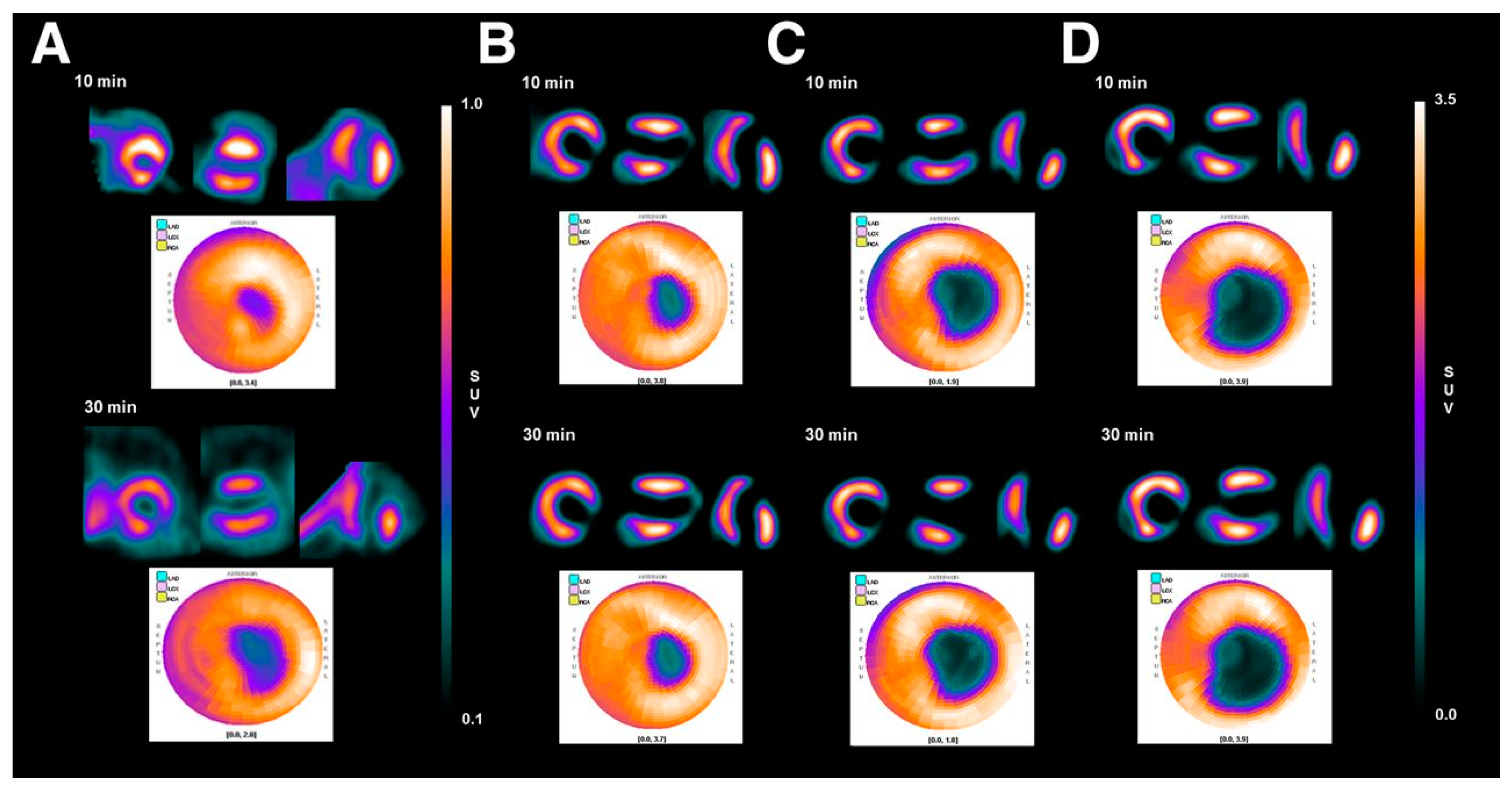
18F-FPTP and 18F-FHTP have similar structures and physicochemical properties. The biodistribution of these two tracers are similar in most of the organs except liver. The liver clearance rate of 18F-FHTP was much faster than that of 18F-FPTP in mice, resulting in over twice heart/liver ratios of 18F-FHTP (25.53 ± 5.88 at 2 h p.i.) than that of 18F-FPTP (10.72 ± 2.17 at 2 h p.i.). The heart/blood ratios of 18F-FHTP were also admirable (138.61 ± 8.10 at 2 h p.i.). In the imaging studies of rats, the myocardial uptakes of both 18F-FHTP and 18F-FPTP were stable at a constant level for up to 1 h p.i. [40]. Kim et al. compared 18F-FPTP, 18F-FHTP, and 18F-FETP with 13N-NH3 in rat models (Figure 4) [42]. They found that the first-pass extraction fraction values of these four radio-agents are comparable at low flow velocity (0.5 mL/min), but 18F-FPTP, 18F-FHTP, and 18F-FETP had significantly higher extraction fractions than 13N-NH3 at higher flow velocity (4.0, 8.0, and 16.0 mL/min, p < 0.05). Small animal PET images with 18F-FPTP demonstrated an excellent image quality with a clear delineation of the borders of defects, which was consistent with the size validated by 2,3,5-triphenyltetrazolium chloride (TTC) staining (r2 = 0.92, p < 0.001) [40].
The structure of 18F-FETMP contains methoxy and ethoxy groups to attenuate the lipophilicity. However, the liver uptake of 18F-FETMP was much higher than that of 18F-FPTP and 18F-FHTP, leading to much slower liver clearance [41]. Researchers supposed that the clearance of these radiotracers from liver was dependent on both lipophilicity and the functional groups of the compounds [43]. It is worth mentioning that the studies of these radiotracers in larger animals haven’t been reported yet. Hence, their perspectives for MPI need further investigations with big animal models.
2.2.4. 4-(18F-Fluoromethyl)benzyltriphenyl Phosphonium (18F-FMBTP) Cation and (3-(18F-Fluoro-methyl)benzyl)trisphenylphosphonium (18F-mFMBTP) Cation
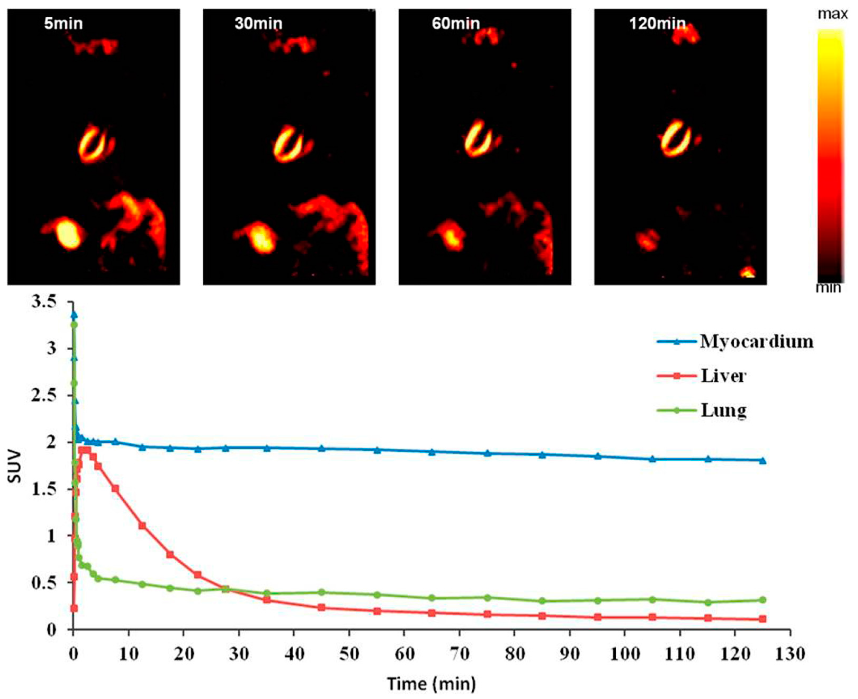
Zhao et al. reported the preparation of 18F-FMBTP and 18F-mFMBTP using another kind of radiointermediates, p- or m-substituted 1-halomethyl-18F-fluoromethylbenzenes [44]. They improved the reaction condition and increased the radiochemical yields of p- or m-radio-intermediates from 12% and 26% to 85% and 92%, respectively. The radiochemical yields of final radiotracers 18F-FMBTP and 18F-mFMBTP were ~50%, which were far higher than 18F-FHTP (10%–20%). 18F-mFMBTP had good retention in the myocardium (26.82 ± 3.46% ID/g at 120 min p.i.) and faster liver clearance (1.02 ± 0.2% ID/g at 120 min p.i.). The heart/liver, heart/lung, and heart/blood ratios of 18F-mFMBTP were 26.25, 9.97, and 83.98 at 120 min p.i., respectively, which was comparable with 18F-FPTP and 18F-FHTP. There was certain uptake in the bone of mice. However, fortunately no obvious bone uptake was observed in the PET images of rats (Figure 5) and dogs (the heart/bone ratios of 18F-mFMBTP in dogs were >10 in 120 min p.i.). MicroPET studies of 18F-mFMBTP resulted in high contrast images with sustained prominent myocardium uptake and markedly low liver and lung uptake up to 120 min p.i. Furthermore, the heart/liver and heart/lung standardized uptake value (SUV) ratios of 18F-mFMBTP in dogs were calculated as 2.83 and 15.19 at 30 min p.i., and 7.76 and 35.28 at 120 min p.i., respectively. On the other hand, other organs and tissues had low background uptake because of excellent metabolic properties of the compound.
In brief, p- or m-substituted 1-halomethyl-18F-fluoromethylbenzenes can evidently raise the radiochemical yields of phosphonium cations [45]. Since there was certain uptake of 18F-FMBTP and 18F-mFMBTP in the bone of mice, the stability and uptake of these radiotracers in bones and other organs of big animals still need to be studied carefully.
Besides the agents above, Yuan et al. used the triphenylphosphonium group as a mitochondrial delivery vehicle. They connected the triphenylphosphonium group with BODIPY Green, and developed 18F-TPP-Green [46]. In summary, there is an enormous progress in the development of 18F-labeled phosphonium cations, especially in the radiosynthesis. Varieties of labeling methods have been used for the preparation of 18F-labeled phosphonium cations. The radiochemical yield has been increased from 6% to 60%. In the meantime, some novel 18F-labeled phosphonium cations exhibit favorable metabolic properties in the preliminary research. However, most of them have not been studied thoroughly. Additional researches are required to understand the implications of 18F-labeled phosphonium cations in MPI of humans.
3. Analogues of MC-1 Inhibitors
MC-1 is the first enzyme of the electron transport complexes. It locates in the inner mitochondrial membrane [47]. MC-1 has an extremely complex structure with over 40 subunits and a molecular mass of approximately 1000 kD [45]. The inhibitors of MC-1 such as rotenone, quinazoline, and pyridazinone can specifically bind to MC-1 and accumulate in the mitochondria. The heart uptake of these compounds is correlated with the myocardial blood flow. Hence, MC-1 inhibitor analogues are developed for MPI (Figure 6).
3.1. 18F-Fluorodihydrorotenone (18F-FDHR)
Rotenone is a neutral lipophilic compound that can inhibit the activity of MC-1. It is widely used as an insecticide. Marshall et al. found that 125I-iodorotenone was superior to 99mTc-sestamibi as a blood flow tracer in the isolated rabbit heart [48]. Later, they prepared 18F-fluorodihydrorotenone (18F-FDHR) [49]. In the study of an isolated rabbit heart, 18F-FDHR was more closely related to coronary flow than 201Tl. Researchers considered that 18F-FDHR was a better blood flow tracer than 201Tl. Unfortunately, there is no follow-up study yet to confirm the tracer properties of 18F-FDHR in animal models.
3.2. (2-Tert-butyl-4-chloro-5-[4-(2-18F-fluoroethoxymethyl)-benzyloxy]-2H-pyridazin-3-one (18F-Flurpiridaz)
Yu et al. developed a series of 18F-RP1003, 18F-RP1004, 18F-RP1005, and 18F-flurpiridaz (previously named as BMS-747158-02) radiotracers based on different kinds of MC-1 inhibitors [50]. Among them, pyridaben is considered as the best lead compound. 18F-flurpiridaz, a pyridaben analogue specific binding with the PSST subunit of MC-1, is the most promising MPI agent for clinical implementation [51].
18F-flurpiridaz has now been in Phase III clinical trials [52]. The recent reviews considered it as the most promising tracer for MPI [16,17,53]. The biodistribution studies of 18F-flurpiridaz in mice showed a significant myocardial uptake and good retention properties (9.5 ± 0.5% ID/g at 60 min p.i.). The heart/liver and heart/lung ratios were 8.3 and 14.1, respectively [54]. In the imaging studies of mouse, rat, rabbit, pig, and non-human primate models, 18F-flurpiridaz demonstrated excellent properties with distinct visualization of the right and left ventricular myocardium and contrast between the heart and surrounding organs [51,54,55,56,57]. In coronary occlusion and ischemia/reperfusion models of rats, the images of 18F-flurpiridaz displayed clear and stable delineation in the non-perfused segments of myocardium. Sherif et al. demonstrated that the uptake of 18F-flurpiridaz in the defect area of myocardium determined by PET was closely correlated with TTC staining (r = 0.89, p < 0.01) [57]. Furthermore, the uptake of 18F-flurpiridaz did not change at different time points of acquisition in the infarct area of rats produced by ligating the left anterior descending artery.
In Phase I clinical trial in human subjects (n = 13), no significant adverse events related with 18F-flurpiridaz administration were reported [6]. The largest mean dose was absorbed by the kidneys (0.066 mSv/MBq), followed by the heart wall (0.048 mSv/MBq). The radiation dose of 18F-flurpiridaz is comparable to or less than that of 18F-FDG [6]. In a Phase II, multicenter clinical trial comprising 143 patients, 18F-flurpiridaz had more favorable diagnostic accuracy for evaluating multi-coronary artery stenosis, compared with SPECT MPI agents 99mTc-sestamibi, 99mTc-tetrofosmin, and 201Tl (Figure 7) [58]. In the Phase III clinical trial comprising 72 sites and 795 subjects [52], 18F-flurpiridaz showed a significant reduction in radiation exposure (6.1 ± 0.4 mSv) compared with SPECT (13.2 ± 3.3 mSv). In obese subjects, 18F-flurpiridaz showed statistically superior sensitivity, specificity, accuracy, diagnostic confidence, and image quality.
3.3. 2-Tert-butyl-4-chloro-5-(4-(2-18F-fluoroethoxy))benzyloxy-2H-pyridazin-3-one (18F-FP1OP) and 4-Chloro-2-tert-butyl-5-[2-[[1-[2-[2-[18F]fluroethoxy]ethoxymethyl]-1H-1,2,3-triazol-4-yl]methyl]phenyl-methoxy]-3(2H)-pyridazinone ([18F]Fmpp2)
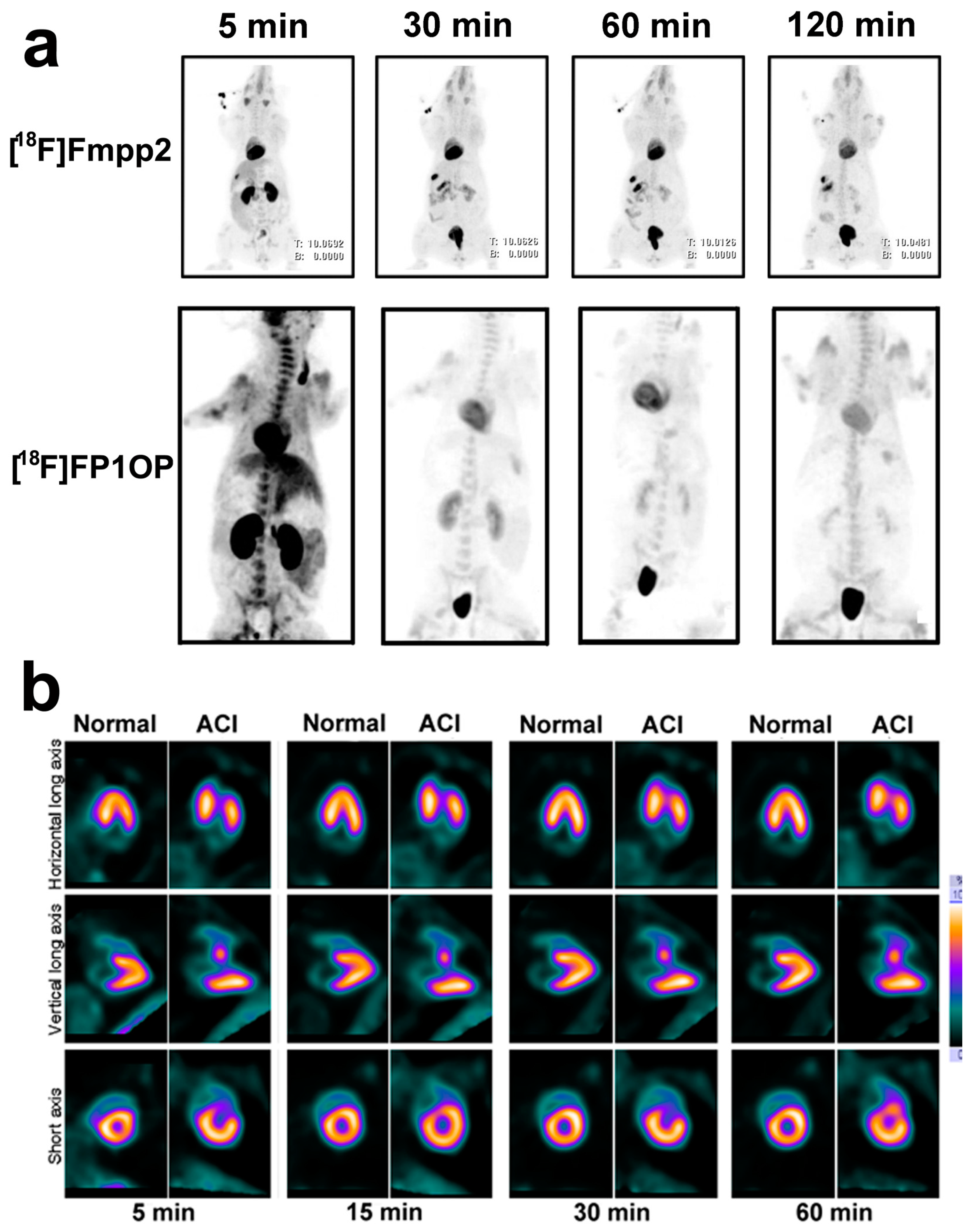
To improve the liver clearance of pyridaben analogues such as 18F-flurpiridaz, Mou et al. introduced the polyethylene glycol (PEG) group into the structure and prepared 18F-FP1OP, 18F-FP2OP, and 18F-FP3OP [59,60]. All three tracers had low initial liver uptake (2.72 ± 0.33% ID/g, 6.14 ± 0.48% ID/g, and 2.71 ± 0.93% ID/g, respectively, at 2 min p.i.), indicating that PEG group could be an available functional group to decrease the liver uptake of those tracers. Follow-up study findings also confirmed this point of view [61,62].
18F-FP1OP had shown the prominent potential properties among those three tracers. In the imaging study in pigs, the heart/liver and heart/lung SUV ratios were 1.83 and 4.53 at 2 min p.i., 2.73 and 7.39 at 30 min p.i., and 3.03 and 8.77 at 60 min p.i., respectively. 18F-FP1OP can distinguish the normal myocardium, ischemic myocardium, and infarct myocardium after acute infarction (Figure 8). However, the stability of 18F-FP1OP in water solution is not good, which limits its further application.
Mou et al. hypothesized that the instability of 18F-FP1OP might due to its phenolic group [61]. Subsequently, they replaced p-substituted phenolic group with 6-methylene-2-pyridyl and 6-methylene-2-phenyl, prepared 18F-FPTP2 [61] and 18F-Fmp2 [61], respectively. Both 18F-FPTP2 and 18F-Fmp2 exhibited excellent stability in water and murine plasma, indicating the replacement of phenolic group was an effective strategy. 18F-FPTP2 showed a significant initial heart uptake (39.70 ± 2.81% ID/g at 2 min p.i.) and moderate retention (20.09 ± 1.93% ID/g at 60 min p.i.), indicating that a variety of aromatic rings could be used to form pyridaben analogues. This result may expand the design of pyridaben analogues for MPI.
However, the biological properties of 18F-FPTP2 and 18F-Fmp2 are not as good as 18F-FP1OP. Recently, they developed [18F]Fmpp1, [18F]Fmpp2 and [18F]Fmpp3 [62]. Among these three tracers, [18F]Fmpp2 exhibited the best properties. It was stable in water for at least 3 h. In the whole-body PET/CT images of mini-swine (Figure 8a), it showed excellent initial heart SUV (7.12 at 5 min p.i.) and good retention (5.75 at 120 min p.i.). The heart/liver SUV ratios were 4.12, 5.42 and 5.99 at 30, 60 and 120 min after injection, respectively. Compared with 18F-flurpiridaz, [18F]Fmpp2 has much faster liver clearance, so it may provide better quality images earlier (15–30 min p.i.). Moreover, unlike other MPI agents, the kidney uptake of [18F]Fmpp2 was low from 30 to 120 min p.i., which might decrease the radiation dose. The metabolic stability of [18F]Fmpp2 in mice, rats and Chinese mini-swine was different. In the heart of mice, 45% activities of [18F]Fmpp2 were metabolized at 60 min p.i. while only 13% activities of [18F]Fmpp2 were metabolized in both the hearts of rat and Chinese mini-swine at 60 min p.i.. Nevertheless, more studies need to be done to evaluate the potential of [18F]Fmpp2 in ischemic or acute myocardial infarction animal models.
Unlike 18F-labeled phosphonium cations, the labeling methods of 18F-labeled MC-1 inhibitors are similar, by substituting tosyl group with 18F. Radiochemical yields of 18F-labeled MC-1 inhibitors are, on the whole, favorable. The performance evaluation of 18F-labeled analogues of MC-1 inhibitors, especially 18F-flurpiridaz, is studied much extensively than 18F-labeled phosphonium cations, and thus they might be the target tracers for MPI in the coming future.
4. Conclusions and Perspectives
SPECT is still the first choice of MPI, especially in the developing countries. In 2012, the Chinese Society of Nuclear Medicine performed a general survey of 30 provinces regarding the status of nuclear medicine in China [63]. According to the survey data, the total SPECT examinations were more than 1.44 million cases per year. MPI studies constituted 7% of them. The total PET examinations were 0.31 million cases per year. The cardiac PET examinations constituted only 0.62% of them. In 2016, the survey data indicated that the SPECT and PET examinations in China increase to 2.1 million and 0.47 million cases per year [64]. The cardiac PET examinations constituted only 0.8% of PET examinations. We believe that the almost no PET MPI examinations in China must be due to the lack of commercialized PET agents for MPI.
However, the disadvantages of SPECT mentioned above make it difficult to satisfy the clinical application in the coming future. An ideal myocardial perfusion tracer should include the following characteristics: high myocardial extraction fraction, excellent image quality, absolute quantification of myocardial blood flow, one-day protocol for rest and stress MPI, and possibility for long-distance transportation. Due to the excellent features, 18F radiotracers become the most prominent isotopes for MPI.
18F-flurpiridaz is the most promising MPI agent. The clinical trials with 18F-flurpiridaz have shown exciting results. Meanwhile, several 18F-labeled radiotracers such as 18F-FBnTP, 18F-FTTP, 18F-FHTP, and 18F-Fmpp2 have shown remarkable properties in preclinical studies as well. The characters of those MPI agents were brief summarized in Table 1. Many of them exhibited high heart uptake and heart/liver ratios. But most of them still need further studies to meet the criteria of clinic, such as defect delineation, polar maps reversibility, etc.
In general, lipophilic cations exhibit superior heart/liver and heart/blood ratios at early time points in small animals. For example, the heart/liver and heart/blood ratios of 18F-FHTP are 11.90 ± 3.37 and 71.87 ± 21.63 at 30 min p.i., respectively, in mice. Hence, clear images can be obtained early after injection of 18F-FHTP. However, the performances of lipophilic cations in larger animals have not yet been studied extensively. Further great efforts are needed to prove the properties of lipophilic cations in preclinical studies with larger animals and in human clinical trials. On the other hand, studies on 18F-labeled MC-1 inhibitors, especially pyridaben analogues, have been extended from preclinical studies involving mice to human clinical trials. Although their target/non-target ratios are not as high as lipophilic cations in mice, they are still more likely to be used in clinical practice because of prolonged retention in myocardium and low background uptake. There was certain concern about the safety of MC-1 inhibitors as the inhibition of MC-1 activity might lead to the death of animals. Fortunately, numerous studies have demonstrated that the use of MC-1 inhibitors is safe for MPI due to their extremely low chemical dose.
In addition, the studies of structure-activity relationship based on lipophilic cations or analogues of MC-1 inhibitors reveal that there is extensive scope for the modification of structures. For instance, the benzene ring of phosphonium cations can be connected with various groups and different kinds of aromatic rings can be used as the “side chain” of pyridaben. Thus, more novel 18F-labeled MPI tracers may be developed. The superior properties of 18F-labeled MPI tracers may likely increase the acceptance of cardiac PET as a routine diagnostic tool in future.
Acknowledgments
This work was sponsored by the National Natural Science Foundation of China (No. 81301251 and 81471707).
Author Contributions
T.M. contributed to writing the article. X.Z. contributed literature review and critical revisions to the article.
Conflicts of Interest
The authors declare no conflict of interest.
Abbreviations
| CAD | coronary artery disease |
| SPECT | single-photon emission computed tomography |
| MPI | myocardial perfusion imaging |
| PET | positron emission tomography |
| MC-1 | mitochondrial complex-1 |
| SUV | standardized uptake value |
| AcChE | acetylcholinesterase |
| p.i. | post injection |
| PEG | polyethylene glycol |
| NDC | non-decay-corrected |
| DC | decay-corrected |
| 18F-FTMA | 4-18F-fluorotri-N-methylanilinium |
| 18F-FERhB | 2-18F-fluoroethylrhodamine B |
| 18F-FDG-rhodamine | 18F-fluoro-2-deoxy-d-glucose-rhodamine |
| BODIPY | Boron-dipyrromethene |
| 18F-2 | 10-(4-(trimethylammonio)phenyl)-5-fluoro-5-18F-fluoro-1,3,7,9-tetramethyl-5H-dipyrrolo-[1,2-c:2′,1′f][1,3,2] diazaborinin-4-ium-5-ide |
| 18F-FBnTP | 18F-fluorobenzyltriphenylphosphonium |
| 18F-FTPP | (4-18F-fluorophenyl)triphenylphosphonium cation |
| 18F-FPTP | (18F-fluoropentyl)triphenylphosphonium cation |
| 18F-FHTP | (6-18F-Fluorohexyl) triphenylphosphonium cation |
| 18F-FETMP | (2-(2-18F-fluoroethoxy)ethyl) tris(4-methoxyphenyl)phosphonium cation |
| 18F-FMBTP | 4-(18F-fluoromethyl)benzyltriphenylphosphonium |
| 18F-mFMBTP | (3-(18F-fluoromethyl) benzyl)trisphenylphosphonium |
| 18F-FDHR | 18F-fluorodihydrorotenone |
| 18F-flurpiridaz (BMS-747158-02) | 2-tert-butyl-4-chloro-5-[4-(2-18F-fluoroethoxymethyl)benzyloxy]-2H-pyridazin-3-one |
| 18F-FP1OP | 2-tert-butyl-4-chloro-5-(4-(2-18F-fluoroethoxy))benzyloxy-2H-pyridazin-3-one |
| 18F-FPTP2 | 2-tert-butyl-4-chloro-5-((6-((4-(2-18F-fluoroethyl)-1H-1,2,3-triazol-1-yl)methyl)-2-pyridinyl)methoxy)-3(2H)-pyridazinone |
| 18F-Fmp2 | 4-chloro-2-tert-butyl-5-[2-[[1-[2-[18F]fluoroethyl]-1H-1,2,3-triazol-4-yl]methyl]phenyl-methoxy]-3(2H)-pyridazinone |
| [18F]Fmpp2 | 4-chloro-2-tert-butyl-5-[2-[[1-[2-[2-[18F]fluroethoxy]ethoxymethyl]-1H-1,2,3-triazol-4-yl]-methyl]phenylmethoxy]-3(2H)-pyridazinone |
References
- Klocke, F.J.; Baird, M.G.; Lorell, B.H.; Bateman, T.M.; Messer, J.V.; Berman, D.S.; O’Gara, P.T.; Carabello, B.A.; Russell, R.O.J.; Cerqueira, M.D.; et al. ACC/AHA/ASNC Guidelines for the Clinical Use of Cardiac Radionuclide Imaging-Executive Summary: A Report of the American College of Cardiology/American Heart Association Task Force on Practice Guidelines (ACC/AHA/ASNC Committee to Revise the 1995 Guidelines for the Clinical Use of Cardiac Radionuclide Imaging). Circulation 2003, 108, 1404–1418. [Google Scholar] [PubMed]
- Guludec, D.; Lautamäki, R.; Knuuti, J.; Bax, J.J.; Bengel, F.M. Present and future of clinical cardiovascular PET imaging in Europe—A position statement by the European Council of Nuclear Cardiology (ECNC). Eur. J. Nucl. Med. Mol. Imaging 2008, 35, 1709–1724. [Google Scholar] [CrossRef] [PubMed]
- Dilsizian, V.; Bacharach, S.L.; Beanlands, R.S.; Bergmann, S.R.; Delbeke, D.; Dorbala, S.; Gropler, R.J.; Knuuti, J.; Schelbert, H.R.; Travin, M.I. ASNC imaging guidelines/SNMMI procedure standard for positron emission tomography (PET) nuclear cardiology procedures. J. Nucl. Cardiol. 2016, 23, 1187–1226. [Google Scholar] [CrossRef] [PubMed]
- Dilsizian, V. Transition from SPECT to PET myocardial perfusion imaging: A desirable change in nuclear cardiology to approach perfection. J. Nucl. Cardiol. 2016, 23, 337–338. [Google Scholar] [CrossRef] [PubMed]
- Sciagrà, R.; Passeri, A.; Bucerius, J.; Verberne, H.J.; Slart, R.H.J.A.; Lindner, O.; Gimelli, A.; Hyafil, F.; Agostini, D.; Übleis, C.; et al. Clinical use of quantitative cardiac perfusion PET: Rationale, modalities and possible indications. Position paper of the Cardiovascular Committee of the European Association of Nuclear Medicine (EANM). Eur. J. Nucl. Med. Mol. Imaging 2016, 43, 1530–1545. [Google Scholar] [CrossRef] [PubMed]
- Maddahi, J.; Czernin, J.; Lazewatsky, J.; Huang, S.; Dahlbom, M.; Schelbert, H.; Sparks, R.; Ehlgen, A.; Crane, P.; Zhu, Q.; et al. Phase I, First-in-Human Study of BMS747158, a Novel 18F-Labeled Tracer for Myocardial Perfusion PET: Dosimetry, Biodistribution, Safety, and Imaging Characteristics After a Single Injection at Rest. J. Nucl. Med. 2011, 52, 1490–1498. [Google Scholar] [CrossRef] [PubMed]
- Senthamizhchelvan, S.; Bravo, P.E.; Esaias, C.; Lodge, M.A.; Merrill, J.; Hobbs, R.F.; Sgouros, G.; Bengel, F.M. Human Biodistribution and Radiation Dosimetry of 82Rb. J. Nucl. Med. 2010, 51, 1592–1599. [Google Scholar] [CrossRef] [PubMed]
- Bober, R.M.; Thompson, C.D.; Morin, D.P. The effect of coronary revascularization on regional myocardial blood flow as assessed by stress positron emission tomography. J. Nucl. Cardiol. 2016, 1–14. [Google Scholar] [CrossRef] [PubMed]
- Srivastava, A.V.; Ananthasubramaniam, K. Guiding coronary revascularization using PET stress myocardial perfusion imaging: The proof is in the pudding. J. Nucl. Cardiol. 2016, 1–5. [Google Scholar] [CrossRef] [PubMed]
- Taqueti, V.R.; Dorbala, S. The role of positron emission tomography in the evaluation of myocardial ischemia in women. J. Nucl. Cardiol. 2016, 23, 1008–1015. [Google Scholar] [CrossRef] [PubMed]
- Bateman, T.M.; Dilsizian, V.; Beanlands, R.S.; DePuey, E.G.; Heller, G.V.; Wolinsky, D.A. American Society of Nuclear Cardiology and Society of Nuclear Medicine and Molecular Imaging Joint Position Statement on the Clinical Indications for Myocardial Perfusion PET. J. Nucl. Med. 2016, 57, 1654–1656. [Google Scholar] [CrossRef] [PubMed]
- Dilsizian, V. Highlights from the Updated Joint ASNC/SNMMI PET Myocardial Perfusion and Metabolism Clinical Imaging Guidelines. J. Nucl. Med. 2016, 57, 1327–1328. [Google Scholar] [CrossRef] [PubMed]
- Heller, G.V.; Calnon, D.; Dorbala, S. Recent advances in cardiac PET and PET/CT myocardial perfusion imaging. J. Nucl. Cardiol. 2009, 16, 962–969. [Google Scholar] [CrossRef] [PubMed]
- Sarikaya, I. Cardiac applications of PET. Nucl. Med. Commun. 2015, 36, 971–985. [Google Scholar] [CrossRef] [PubMed]
- Hung, G.; Wang, Y.; Su, H.; Hsieh, T.; Ko, C.; Yen, R. New Trends in Radionuclide Myocardial Perfusion Imaging. Acta Cardiol. Sin. 2016, 32, 156–166. [Google Scholar] [PubMed]
- Maddahi, J.; Packard, R.R.S. Cardiac PET Perfusion Tracers: Current Status and Future Directions. Semin. Nucl. Med. 2014, 44, 333–343. [Google Scholar] [CrossRef] [PubMed]
- Schindler, T.H. Positron-Emitting Myocardial Blood Flow Tracers and Clinical Potential. Prog. Cardiovasc. Dis. 2015, 57, 588–606. [Google Scholar] [CrossRef] [PubMed]
- Lin, X.; Zhang, J.; Wang, X.; Tang, Z.; Zhang, X.; Lu, J. Development of radiolabeled compounds for myocardial perfusion imaging. Curr. Pharm. Des. 2012, 18, 1041–1057. [Google Scholar] [CrossRef] [PubMed]
- Rischpler, C.; Park, M.J.; Fung, G.S.K.; Javadi, M.; Tsui, B.M.W.; Higuchi, T. Advances in PET myocardial perfusion imaging: F-18 labeled tracers. Ann. Nucl. Med. 2012, 26, 1–6. [Google Scholar] [CrossRef] [PubMed]
- Murphy, M.P.; Smith, R.A.J. Targeting Antioxidants to Mitochondria by Conjugation to Lipophilic Cations. Annu. Rev. Pharmacol. Toxicol. 2007, 47, 629–656. [Google Scholar] [CrossRef] [PubMed]
- Madar, I.; Ravert, H.T.; Du, Y.; Hilton, J.; Volokh, L.; Dannals, R.F.; Frost, J.J.; Hare, J.M. Characterization of Uptake of the New PET Imaging Compound 18F-Fluorobenzyl Triphenyl Phosphonium in Dog Myocardium. J. Nucl. Med. 2006, 47, 1359–1366. [Google Scholar] [PubMed]
- Kroemer, G. Mitochondrial control of apoptosis: An introduction. Biochem. Biophys. Res. Commun. 2003, 304, 433–435. [Google Scholar] [CrossRef]
- Studenov, A.R.; Berridge, M.S. Synthesis and properties of 18F-labeled potential myocardial blood flow tracers. Nucl. Med. Biol. 2001, 28, 683–693. [Google Scholar] [CrossRef]
- Vora, M.M.; Dhalla, M. In vivo studies of unlabeled and radioiodinated rhodamine-123. Nucl. Med. Biol. 1992, 19, 405–410. [Google Scholar] [CrossRef]
- Gottumukkala, V.; Heinrich, T.K.; Baker, A.; Dunning, P.; Fahey, F.H.; Treves, S.T.; Packard, A.B. Biodistribution and stability studies of [18F]Fluoroethylrhodamine B, a potential PET myocardial perfusion agent. Nucl. Med. Biol. 2010, 37, 365–370. [Google Scholar] [CrossRef] [PubMed]
- AlJammaz, I.; Al-Otaibi, B.; AlHindas, H.; Okarvi, S.M. Novel synthesis and initial preclinical evaluation of 18F-[FDG] labeled rhodamine: A potential PET myocardial perfusion imaging agent. Nucl. Med. Biol. 2015, 42, 804–808. [Google Scholar] [CrossRef] [PubMed]
- Bartholomä, M.D.; Zhang, S.; Akurathi, V.; Pacak, C.A.; Dunning, P.; Fahey, F.H.; Cowan, D.B.; Ted Treves, S.; Packard, A.B. 18F-labeled rhodamines as potential myocardial perfusion agents: Comparison of pharmacokinetic properties of several rhodamines. Nucl. Med. Biol. 2015, 42, 796–803. [Google Scholar] [CrossRef] [PubMed]
- Nigam, S.; Burke, B.P.; Davies, L.H.; Domarkas, J.; Wallis, J.F.; Waddell, P.G.; Waby, J.S.; Benoit, D.M.; Seymour, A.M.; Cawthorne, C.; et al. Structurally optimised BODIPY derivatives for imaging of mitochondrial dysfunction in cancer and heart cells. Chem. Commun. 2016, 52, 7114–7117. [Google Scholar] [CrossRef] [PubMed]
- Li, Z.; Lin, T.; Liu, S.; Huang, C.; Hudnall, T.W.; Gabbaï, F.P.; Conti, P.S. Rapid aqueous [18F]-labeling of a bodipy dye for positron emission tomography/fluorescence dual modality imaging. Chem. Commun. 2011, 47, 9324–9326. [Google Scholar] [CrossRef] [PubMed]
- Liu, S.; Li, D.; Shan, H.; Gabbaï, F.P.; Li, Z.; Conti, P.S. Evaluation of 18F-labeled BODIPY dye as potential PET agents for myocardial perfusion imaging. Nucl. Med. Biol. 2014, 41, 120–126. [Google Scholar] [CrossRef] [PubMed]
- Chansaenpak, K.; Wang, H.; Wang, M.; Giglio, B.; Ma, X.; Yuan, H.; Hu, S.; Wu, Z.; Li, Z. Synthesis and Evaluation of [18F]-Ammonium BODIPY Dyes as Potential Positron Emission Tomography Agents for Myocardial Perfusion Imaging. Chemistry 2016, 22, 12122–12129. [Google Scholar] [CrossRef] [PubMed]
- Ravert, H.T.; Madar, I.; Dannals, R.F. Radiosynthesis of 3-[18F]fluoropropyl and 4-[18F]fluorobenzyl triarylphosphonium ions. J. Label. Compd. Radiopharm. 2004, 47, 469–476. [Google Scholar] [CrossRef]
- Madar, I.; Ravert, H.; DiPaula, A.; Du, Y.; Dannals, R.F.; Becker, L. Assessment of Severity of Coronary Artery Stenosis in a Canine Model Using the PET Agent 18F-Fluorobenzyl Triphenyl Phosphonium: Comparison with 99mTc-Tetrofosmin. J. Nucl. Med. 2007, 48, 1021–1030. [Google Scholar] [CrossRef] [PubMed]
- Higuchi, T.; Fukushima, K.; Rischpler, C.; Isoda, T.; Javadi, M.S.; Ravert, H.; Holt, D.P.; Dannals, R.F.; Madar, I.; Bengel, F.M. Stable Delineation of the Ischemic Area by the PET Perfusion Tracer 18F-Fluorobenzyl Triphenyl Phosphonium After Transient Coronary Occlusion. J. Nucl. Med. 2011, 52, 965–969. [Google Scholar] [CrossRef] [PubMed]
- Ravert, H.T.; Holt, D.P.; Dannals, R.F. A microwave radiosynthesis of the 4-[18F]-fluorobenzyltriphenylphosphonium ion. J. Label. Compd. Radiopharm. 2015, 57, 695–698. [Google Scholar] [CrossRef] [PubMed]
- Tominaga, T.; Ito, H.; Ishikawa, Y.; Ren, I.; Ishiwata, K.; Furumoto, S. Radiosynthesis and preliminary biological evaluation of a new 18F-labeled triethylene glycol derivative of triphenylphosphonium. J. Label. Compd. Radiopharm. 2016, 59, 117–123. [Google Scholar] [CrossRef] [PubMed]
- Zhang, Z.; Zhang, C.; Lau, J.; Colpo, N.; Bénard, F.; Lin, K.S. One-step synthesis of 4-[18F]fluorobenzyltriphenylphosphonium cation for imaging with positron emission tomography. J. Label. Compd. Radiopharm. 2016, 59, 467–471. [Google Scholar] [CrossRef] [PubMed]
- Cheng, Z.; Subbarayan, M.; Chen, X.; Gambhir, S.S. Synthesis of (4-[18F]fluorophenyl)triphenylphosphonium as a potential imaging agent for mitochondrial dysfunction. J. Label. Compd. Radiopharm. 2005, 48, 131–137. [Google Scholar] [CrossRef]
- Shoup, T.M.; Elmaleh, D.R.; Brownell, A.; Zhu, A.; Guerrero, J.L.; Fischman, A.J. Evaluation of (4-[18F]Fluorophenyl)triphenylphosphonium Ion. A Potential Myocardial Blood Flow Agent for PET. Mol. Imaging Biol. 2010, 13, 511–517. [Google Scholar] [CrossRef] [PubMed]
- Kim, D.; Kim, H.; Yu, K.; Min, J. Synthesis of [18F]-labeled (2-(2-fluoroethoxy)ethyl)tris(4-methoxyphenyl)phosphonium cation as a potential agent for positron emission tomography myocardial imaging. Nucl. Med. Biol. 2012, 39, 1093–1098. [Google Scholar] [CrossRef] [PubMed]
- Kim, D.; Yu, K.; Bom, H.; Min, J. Synthesis of (4-[18F]Fluorophenyl)triphenylphosphonium as a mitochondrial voltage sensor for PET. Nucl. Med. Mol. Imaging 2007, 41, 561–565. [Google Scholar]
- Kim, D.; Kim, H.S.; Reder, S.; Zheng, J.H.; Herz, M.; Higuchi, T.; Pyo, A.; Bom, H.; Schwaiger, M.; Min, J. Comparison of 18F-Labeled Fluoroalkylphosphonium Cations with 13N-NH3 for PET Myocardial Perfusion Imaging. J. Nucl. Med. 2015, 56, 1581–1586. [Google Scholar] [CrossRef] [PubMed]
- Kim, D.; Min, J. Radiolabeled Phosphonium Salts as Mitochondrial Voltage Sensors for Positron Emission Tomography Myocardial Imaging Agents. Nucl. Med. Mol. Imaging 2016, 50, 185–195. [Google Scholar] [CrossRef] [PubMed]
- Zhao, Z.; Yu, Q.; Mou, T.; Liu, C.; Yang, W.; Fang, W.; Peng, C.; Lu, J.; Liu, Y.; Zhang, X. Highly efficient one-pot labeling of new phosphonium cations with fluorine-18 as potential PET agents for myocardial perfusion imaging. Mol. Pharm. 2014, 11, 3823–3831. [Google Scholar] [CrossRef] [PubMed]
- Degli Esposti, M. Inhibitors of NADH-ubiquinone reductase: An overview. Biochim. Biophys. Acta-Bioenerg. 1998, 1364, 222–235. [Google Scholar] [CrossRef]
- Yuan, H.; Cho, H.; Chen, H.H.; Panagia, M.; Sosnovik, D.E.; Josephson, L. Fluorescent and radiolabeled triphenylphosphonium probes for imaging mitochondria. Chem. Commun. 2013, 49, 10361–10363. [Google Scholar] [CrossRef] [PubMed]
- Hunte, C.; Zickermann, V.; Brandt, U. Functional Modules and Structural Basis of Conformational Coupling in Mitochondrial Complex I. Science 2010, 329, 448–451. [Google Scholar] [CrossRef] [PubMed]
- Marshall, R.C.; Powers-Risius, P.; Reutter, B.W.; Taylor, S.E.; VanBrocklin, H.F.; Huesman, R.H.; Budinger, T.F. Kinetic Analysis of 125I-Iodorotenone as a Deposited Myocardial Flow Tracer: Comparison with 99mTc-Sestamibi. J. Nucl. Med. 2001, 42, 272–281. [Google Scholar] [PubMed]
- Marshall, R.C.; Powers-Risius, P.; Reutter, B.W.; O’Neil, J.P.; La Belle, M.; Huesman, R.H.; VanBrocklin, H.F. Kinetic Analysis of 18F-Fluorodihydrorotenone as a Deposited Myocardial Flow Tracer: Comparison to 201Tl. J. Nucl. Med. 2004, 45, 1950–1959. [Google Scholar] [PubMed]
- Yu, M.; Guaraldi, M.; Kagan, M.; Mistry, M.; McDonald, J.; Bozek, J.; Yalamanchili, P.; Hayes, M.; Azure, M.; Purohit, A.; et al. Assessment of 18F-labeled mitochondrial complex I inhibitors as PET myocardial perfusion imaging agents in rats, rabbits, and primates. Eur. J. Nucl. Med. Mol. Imaging 2008, 36, 63–72. [Google Scholar] [CrossRef] [PubMed]
- Nekolla, S.G.; Reder, S.; Saraste, A.; Higuchi, T.; Dzewas, G.; Preissel, A.; Huisman, M.; Poethko, T.; Schuster, T.; Yu, M.; et al. Evaluation of the Novel Myocardial Perfusion Positron-Emission Tomography Tracer 18F-BMS-747158-02: Comparison to 13N-Ammonia and Validation With Microspheres in a Pig Model. Circulation 2009, 119, 2333–2342. [Google Scholar] [CrossRef] [PubMed]
- Bateman, T.M.; Maddahi, J.; Udelson, J.; Beanlands, R.; Knuuti, J.; Heller, G.; Berman, D.; Lazewatsky, J.; Orlandi, C. Improved assessment of CAD in obese subjects with flurpiridaz F18 PET myocardial perfusion imaging: A subset analysis of the flurpiridaz F18 301 phase 3 study. J. Am. Coll. Cardiol. 2016, 67, 1578. [Google Scholar] [CrossRef]
- Brunken, R.C. Promising New 18F-Labeled Tracers for PET Myocardial Perfusion Imaging. J. Nucl. Med. 2015, 56, 1478–1479. [Google Scholar] [CrossRef] [PubMed]
- Yalamanchili, P.; Wexler, E.; Hayes, M.; Yu, M.; Bozek, J.; Kagan, M.; Radeke, H.S.; Azure, M.; Purohit, A.; Casebier, D.S.; et al. Mechanism of uptake and retention of F-18 BMS-747158-02 in cardiomyocytes: A novel PET myocardial imaging agent. J. Nucl. Cardiol. 2007, 14, 782–788. [Google Scholar] [CrossRef] [PubMed]
- Higuchi, T.; Nekolla, S.G.; Huisman, M.M.; Reder, S.; Poethko, T.; Yu, M.; Wester, H.; Casebier, D.S.; Robinson, S.P.; Botnar, R.M.; et al. A New 18F-Labeled Myocardial PET Tracer: Myocardial Uptake After Permanent and Transient Coronary Occlusion in Rats. J. Nucl. Med. 2008, 49, 1715–1722. [Google Scholar] [CrossRef] [PubMed]
- Huisman, M.C.; Higuchi, T.; Reder, S.; Nekolla, S.G.; Poethko, T.; Wester, H.; Ziegler, S.I.; Casebier, D.S.; Robinson, S.P.; Schwaiger, M. Initial Characterization of an 18F-Labeled Myocardial Perfusion Tracer. J. Nucl. Med. 2008, 49, 630–636. [Google Scholar] [CrossRef] [PubMed]
- Sherif, H.M.; Saraste, A.; Weidl, E.; Weber, A.W.; Higuchi, T.; Reder, S.; Poethko, T.; Henriksen, G.; Casebier, D.; Robinson, S.; et al. Evaluation of a Novel 18F-Labeled Positron-Emission Tomography Perfusion Tracer for the Assessment of Myocardial Infarct Size in Rats. Circ. Cardiovasc. Imaging 2009, 2, 77–84. [Google Scholar] [CrossRef] [PubMed]
- Berman, D.S.; Maddahi, J.; Tamarappoo, B.K.; Czernin, J.; Taillefer, R.; Udelson, J.E.; Gibson, C.M.; Devine, M.; Lazewatsky, J.; Bhat, G. Phase II Safety and Clinical Comparison With Single-Photon Emission Computed Tomography Myocardial Perfusion Imaging for Detection of Coronary Artery Disease. J. Am. Coll. Cardiol. 2013, 61, 469–477. [Google Scholar] [CrossRef] [PubMed]
- Mou, T.; Jing, H.; Yang, W.; Fang, W.; Peng, C.; Guo, F.; Zhang, X.; Pang, Y.; Ma, Y. Preparation and biodistribution of [18F]FP2OP as myocardial perfusion imaging agent for positron emission tomography. Bioorg. Med. Chem. 2010, 18, 1312–1320. [Google Scholar] [CrossRef] [PubMed]
- Mou, T.; Zhao, Z.; Fang, W.; Peng, C.; Guo, F.; Liu, B.; Ma, Y.; Zhang, X. Synthesis and Preliminary Evaluation of 18F-Labeled Pyridaben Analogues for Myocardial Perfusion Imaging with PET. J. Nucl. Med. 2012, 53, 472–479. [Google Scholar] [CrossRef] [PubMed]
- Mou, T.; Zhao, Z.; Zhang, P.; Fang, W.; Peng, C.; Lu, J.; Wang, Q.; Ma, Y.; Zhang, X. Synthesis and bio-evaluation of New 18F-Labeled Pyridaben Analogs with Improved Stability for Myocardial Perfusion Imaging in Mice. Chem. Biol. Drug Des. 2015, 86, 351–361. [Google Scholar] [CrossRef] [PubMed]
- Mou, T.; Zhao, Z.; You, L.; Li, Y.; Qian, W.; Wei, F.; Jie, L.; Cheng, P.; Zhang, X. Synthesis and Evaluation of 18F-labeled Pyridaben Analogues for Myocardial Perfusion Imaging in Mice, Rats and Chinese mini-swine. Sci. Rep. 2016, 6, 33450. [Google Scholar] [CrossRef] [PubMed]
- Chinese Society of Nuclear Medicine. A brief report on the results of the national survey of nuclear medicine in 2012. Chin. J. Nucl. Med. Mol. Imaging 2012, 32, 357. [Google Scholar]
- Chinese Society of Nuclear Medicine. A brief report on the results of the national survey of nuclear medicine in 2016. Chin. J. Nucl. Med. Mol. Imaging 2016, 36, 479–480. [Google Scholar]
Sample Availability: Not available. |
Figure 1.
The structures of several 18F-labeled ammonium lipophilic cations.

Figure 2.
The structures of several 18F-labeled phosphonium lipophilic cations.

Figure 3.
Representative short-axis small-animal PET images of 18F-FBnTP at different time points after tracer administration during short-term occlusion, followed by reperfusion. Graph shows mean uptake in defect and remote area. %ID = percentage injected dose; RO = region of interest [34].
Figure 3.
Representative short-axis small-animal PET images of 18F-FBnTP at different time points after tracer administration during short-term occlusion, followed by reperfusion. Graph shows mean uptake in defect and remote area. %ID = percentage injected dose; RO = region of interest [34].

Figure 4.
Short-, vertical long-, and horizontal long-axis and polar map images of 13N-NH3 (A); 18F-FPTP (B); 18F-FHTP (C); or 18F-FETP (D) in each representative animal. Data were collected between 0–10 and 20–30 min after radiotracer injection (37 MBq). SUV = standardized uptake value [42].
Figure 4.
Short-, vertical long-, and horizontal long-axis and polar map images of 13N-NH3 (A); 18F-FPTP (B); 18F-FHTP (C); or 18F-FETP (D) in each representative animal. Data were collected between 0–10 and 20–30 min after radiotracer injection (37 MBq). SUV = standardized uptake value [42].

Figure 5.
Coronal microPET images in a normal rat. The heart was visible with excellent ratios of heart/liver and heart/lung, and fast clearance from small intestine at 5, 30, 60, and 120 min after iv injection of [18F]mFMBTP, respectively. Time–activity curves generated from dynamic PET images. [18F]mFMBTP accumulated specifically in the heart. The [18F]mFMBTP had excellent heart/liver and heart/lung ratios and in liver and lung was washed out rapidly but was retained in the myocardium for the whole time [44].

Figure 6.
The structures of several 18F-labeled mitochondrial complex-1 inhibitors.
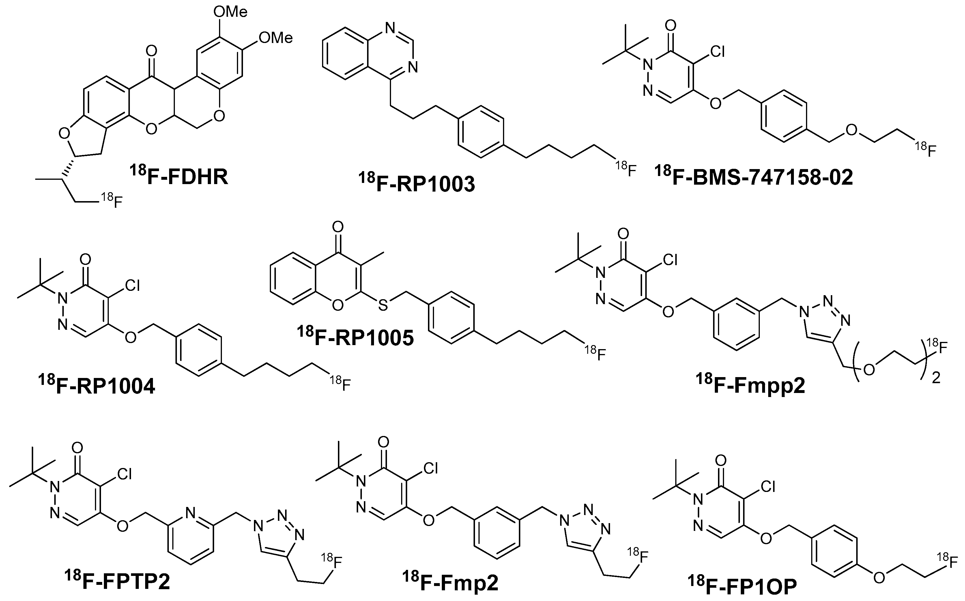
Figure 7.
The 18F-flurpiridaz PET (top) and MIBI SPECT (bottom) images from an 82-year-old man with shortness of breath and an occluded native proximal left anterior descending (LAD) coronary artery and an occluded left internal mammary graft to the LAD and no other significant native CAD. The 18F-flurpiridaz images show a severe reversible perfusion defect throughout the territory of the occluded proximal LAD, whereas the MIBI images show only a moderate perfusion defect in the distal LAD territory (apical slices) [58].
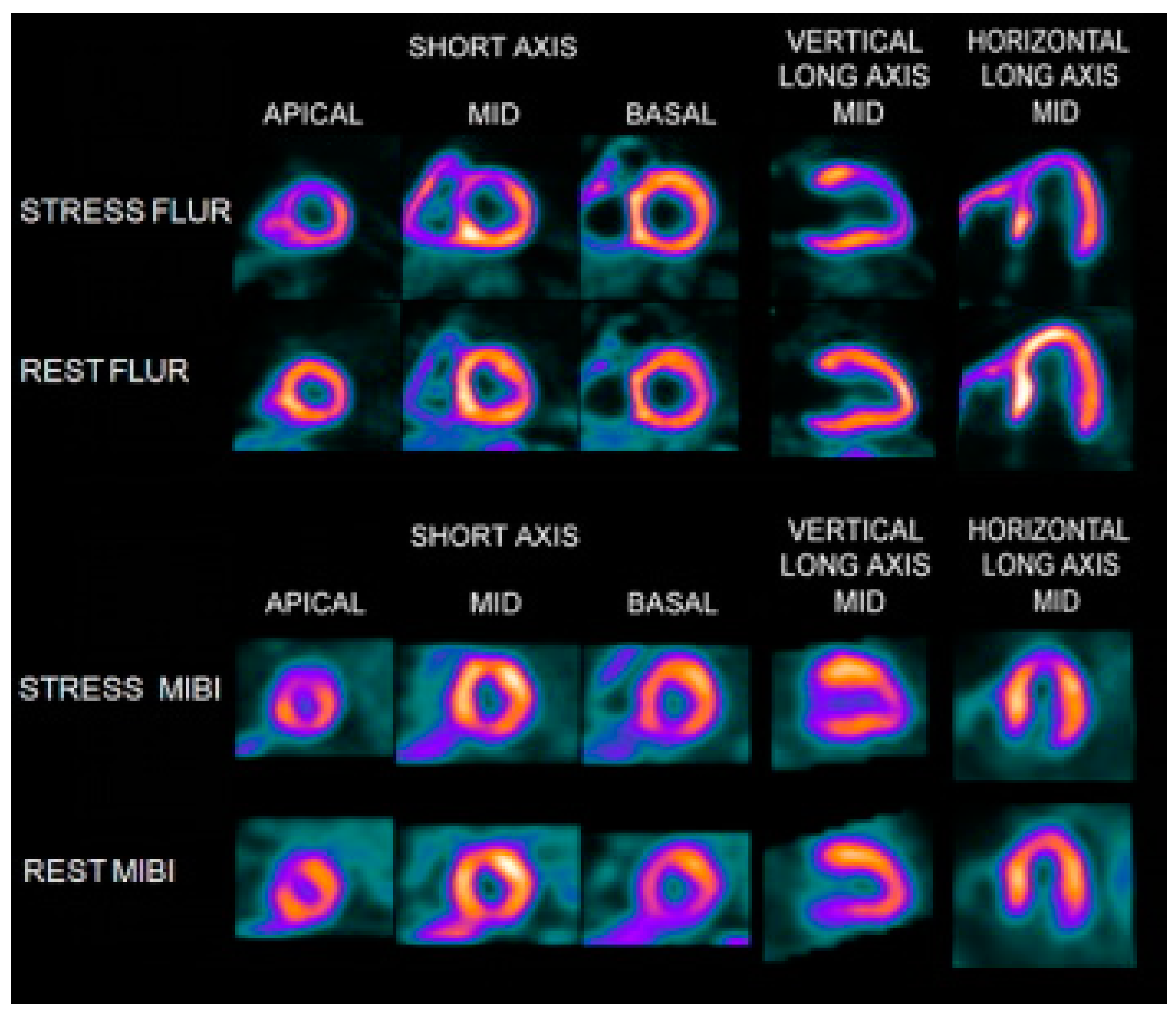
Figure 8.
(a) Whole-body planar images of healthy Chinese mini swine. Images were obtained with 55 MBq of [18F]FP1OP [60] or 37 MBq of [18F]Fmpp2 [62] in 5% ethanol solution at 2, 30, 60, and 120 min after injection. B = urinary bladder; H = heart; K = kidney; (b) Cardiac PET images of healthy Chinese mini swine (normal) and Chinese mini swine with acute myocardial infarction (AMI). Images were obtained with 55 MBq of [18F]FP1OP in 5% ethanol solution at 2, 15, 30, and 60 min after injection. Arrows indicate perfusion defect sites of infarction areas (apical and anterior walls) [60].

Table 1.
Comparison of representative 18F-labeled compounds as potential MPI agents.
| Probes | 18F-FDG-Rhodamine | 18F-FBnTP | 18F-FTTP | 18F-mFMBTP | 18F-Flurpiridaz | 18F-Fmpp2 |
|---|---|---|---|---|---|---|
| Class | ammonium cation | phosphonium cation | phosphonium cation | phosphonium cation | MC-1 inhibitors | MC-1 inhibitors |
| Charge | cationic | cationic | cationic | cationic | neutral | neutral |
| Log P | −1.64 ± 0.03 | -- | 1.78 ± 0.05 | 1.05 ± 0.01 | -- | 1.73 ± 0.05 |
| RCY (%) | 97.0 ± 1.9 (based on 18F-FDG) | 62 ± 1.4 (NDC) | 10–15 (EOS) | 50 (DC) | 25 (DC) | 58 ± 7.1 (DC) |
| Heart Uptake (%ID/g) | 11.24 ± 1.97 (rat) | -- | 1.51 ± 0.04 (rat) | 27.39 ± 1.46 (mice) | 9.5 ± 0.5 (mice) | 27.15 ± 3.58 (mice) |
| Heart/Liver Ratio | 21.2 (rat) | 1.2 (dog) | 8 (rat) | 4.84 (mice) | 8.3 (mice) | 3.96 (mice) |
| Heart/Blood Ratio | 28.10 (rat) | 16.6 (dog) | 75.5 (rat) | 23.82 (mice) | -- | 10.29 (mice) |
| Time point (min) * | -- | 60 | 30 | 30 | 60 | 30 |
| Current status | rats | dogs | rabbits | dogs | Clinic trial (Phase III) | pigs |
| References | 26 | 21, 32, 37 | 39 | 44 | 53 | 62 |
* Time point means the time point of heart uptake and heart/liver ratios in Table 1.
© 2017 by the authors. Licensee MDPI, Basel, Switzerland. This article is an open access article distributed under the terms and conditions of the Creative Commons Attribution (CC BY) license (http://creativecommons.org/licenses/by/4.0/).
Share and Cite
MDPI and ACS Style
Mou, T.; Zhang, X. Research Progress on 18F-Labeled Agents for Imaging of Myocardial Perfusion with Positron Emission Tomography. Molecules 2017, 22, 562. https://doi.org/10.3390/molecules22040562
AMA Style
Mou T, Zhang X. Research Progress on 18F-Labeled Agents for Imaging of Myocardial Perfusion with Positron Emission Tomography. Molecules. 2017; 22(4):562. https://doi.org/10.3390/molecules22040562
Chicago/Turabian StyleMou, Tiantian, and Xianzhong Zhang. 2017. "Research Progress on 18F-Labeled Agents for Imaging of Myocardial Perfusion with Positron Emission Tomography" Molecules 22, no. 4: 562. https://doi.org/10.3390/molecules22040562