Article

Hypoglycemic, Antioxidant Activities, and Probiotic Characteristics of Lacticaseibacillus rhamnosus LBUX2302 Isolated from Stool Samples of Neonates

by
Pedro A. Reyes-Castillo
1,†,
Ana Laura Esquivel-Campos
2,†,
Edgar Torres-Maravilla
3,
Eduardo Zúñiga-León
2,
Felipe Mendoza-Pérez
2,
Rosa González-Vázquez
4,5,
María Guadalupe Córdova-Espinoza
4,5,6,
María Angélica Gutiérrez-Nava
7,
Raquel González-Vázquez
8,* and
Lino Mayorga-Reyes
2,*
1
Doctorado en Ciencias Biologicas y de la Salud, Universidad Autonoma Metropolitana, Mexico City 04960, Mexico
2
Laboratorio de Biotecnologia, Departamento de Sistemas Biologicos, Universidad Autonoma Metropolitana Unidad Xochimilco, Mexico City 04960, Mexico
3
Facultad de Medicina Mexicali, Universidad Autonoma de Baja California, Mexicali 21000, Mexico
4
Laboratorio de Bacteriologia Medica, Escuela Nacional de Ciencias Biologicas, Instituto Politecnico Nacional (IPN), Mexico City 11350, Mexico
5
Unidad Medica de Alta Especialidad, Hospital de Especialidades, “Dr. Antonio Fraga Mouret”, Centro Medico Nacional La Raza, Instituto Mexicano del Seguro Social (IMSS), Mexico City 02990, Mexico
6
Laboratorio de Inmunologia, Escuela Militar de Graduados de Sanidad, Mexico City 11200, Mexico
7
Laboratorio de Ecologia Microbiana, Departamento de Sistemas Biologicos, Universidad Autonoma Metropolitana Unidad Xochimilco, Ciudad de Mexico 04960, Mexico
8
Laboratorio de Biotecnologia, Departamento de Sistemas Biologicos, Secihti-Universidad Autonoma Metropolitana Unidad Xochimilco, Mexico City 04960, Mexico
*
Authors to whom correspondence should be addressed.
These authors contributed equally to this work.
Life 2025, 15(5), 804; https://doi.org/10.3390/life15050804
Submission received: 22 March 2025 / Revised: 2 May 2025 / Accepted: 13 May 2025 / Published: 18 May 2025

Abstract

:
Lacticaseibacillus species have shown potential in managing hyperglycemia, hypercholesterolemia, and oxidative stress, depending on the strain and species. This study aimed to isolate a novel Lacticaseibacillus rhamnosus strain from healthy newborns and assess its hypoglycemic and antioxidative activity, along with other probiotic properties. A non-hemolytic L. rhamnosus LBUX2302 was isolated, and it exhibited survival rates of 2.7%, 22%, and 27.5% at pH 2, 3, and 5 for 120 min. It metabolized various carbon sources and showed resistance to gentamicin, dicloxacillin, and penicillin; coaggregated with Salmonella typhi ATCC14028, Staphylococcus aureus STCC6538, and Escherichia coli O157:H7. L. rhamnosus LBUX2302 showed hydrophobicity, autoaggregation, and adhesion to HaCat, HeLa, MCF-7, SK-LU-1, and SW620 cell lines. It also exhibited extracellular activity of bile salt hydrolase. Enzymatic inhibition assays revealed 66% and 24% inhibitions of α-amylase and α-glucosidase, respectively. Its cell-free supernatant inhibited DPPH (89%), hydroxyl (81%), and superoxide anion radicals (61%). Also, antioxidant activity was observed in whole cells and cell fragments. Finally, the presence of ferulic acid activity was detected. The results highlight L. rhamnosus LBUX2302 as a promising probiotic with hypoglycemic and antioxidant effects, warranting further in vivo evaluation for its possible inclusion in functional food and health formulations.

1. Introduction

Lactic acid bacteria (LAB) comprise the Lacticaseibacillus genus, in which the rhamnosus species is included [1]. LAB have been isolated from various sources, such as fermented dairy and non-dairy products, the gastrointestinal tract of humans, animals, and insects, as well as human breast milk [2,3]. FAO/WHO suggested that potential probiotics should be capable of surviving passage through the digestive tract. This means they must be resistant to gastric juices and be able to grow in the presence of bile under conditions found in the intestines. As with any bacteria, antibiotic resistance exists among some LAB, and this resistance may be related to chromosomal- or plasmid-located genes [4].
Subsequently, their functional properties should be assessed, including adhesion to the epithelial surface, antimicrobial and antioxidant activity, hydrophobicity, and self-aggregation [5,6]. LAB can attach to the intestinal epithelium via various mechanisms, including passive forces, electrostatic and hydrophobic interactions, steric forces, lipoteichoic acids, and distinct surface structures. This ability to adhere may inhibit pathogenic bacteria from binding to intestinal cells [7].
LAB can reduce cholesterol through the enzymatic activity of bile salt hydrolase (BSH) [8]. Certain Lacticaseibacillus strains have shown hypoglycemic potential, attributed to their ability to inhibit the enzymatic activity of α-amylase and α-glucosidase. This inhibition slows glucose uptake, promoting a more stable glycemic profile, and suggests a potential therapeutic application for the management of type 2 diabetes mellitus (T2DM) [9,10]. T2DM and oxidative stress are closely linked due to hyperglycemia-induced overproduction of reactive oxygen species (ROS), such as superoxide anion (O−2), hydrogen peroxide (H2O2), and hydroxyl radicals (OH). Some Lacticaseibacillus strains have shown antioxidant activity by increasing different free radical scavenging activities and superoxide dismutase enzyme activity, which could help to reduce oxidative stress, thus impacting glucose homeostasis [11].
L. rhamnosus isolated from cheese showed antibacterial effects against Escherichia coli, Staphylococcus aureus, and Pseudomonas aeruginosa [12]. Lacticaseibacillus rhamnosus LR22 has been reported as beneficial against constipation [13]. Due to the above, this work focused on isolating a new strain of L. rhamnosus from healthy newborns and evaluating its hypoglycemic and antioxidative activity, along with other probiotic properties.

2. Materials and Methods

2.1. Strain Isolation

LAB isolation was performed using stool samples from healthy newborns from an obstetric clinic of the public health service in Mexico. The research protocol was approved by the Ethics Committee of the clinic under registration number 36,068. Details about isolation are found in Reyes-Castillo et al. (2023) [10]. We did not handle the stool samples directly as we received the purified strains, which were subsequently examined for their morphology and Gram staining characteristics.

2.2. Molecular Identification

A Wizard® Genomic DNA Purification Kit (Promega, Tokyo, Japan) was used to perform gDNA extraction, following the manufacturer’s protocol. The primers used to amplify a segment of the genomic DNA of our isolated strain were 27F (AGAGTTTGATCMGGCTCAG) and 1491R (TACGGYTACCTTGTTAGGATT). The conditions for the PCR reaction were according to Galkiewicz and Kellogg, 2008 [14]. The amplified product was sequenced by the Integrated Microbiome Resource (Halifax, NS, Canada). Sequence alignment and comparisons were conducted using MEGA5 and NCBI’s Basic Local Alignment Search Tool (http://blast.ncbi.nlm.nih.gov/Blast.cgi) (accessed on 18 May 2024).

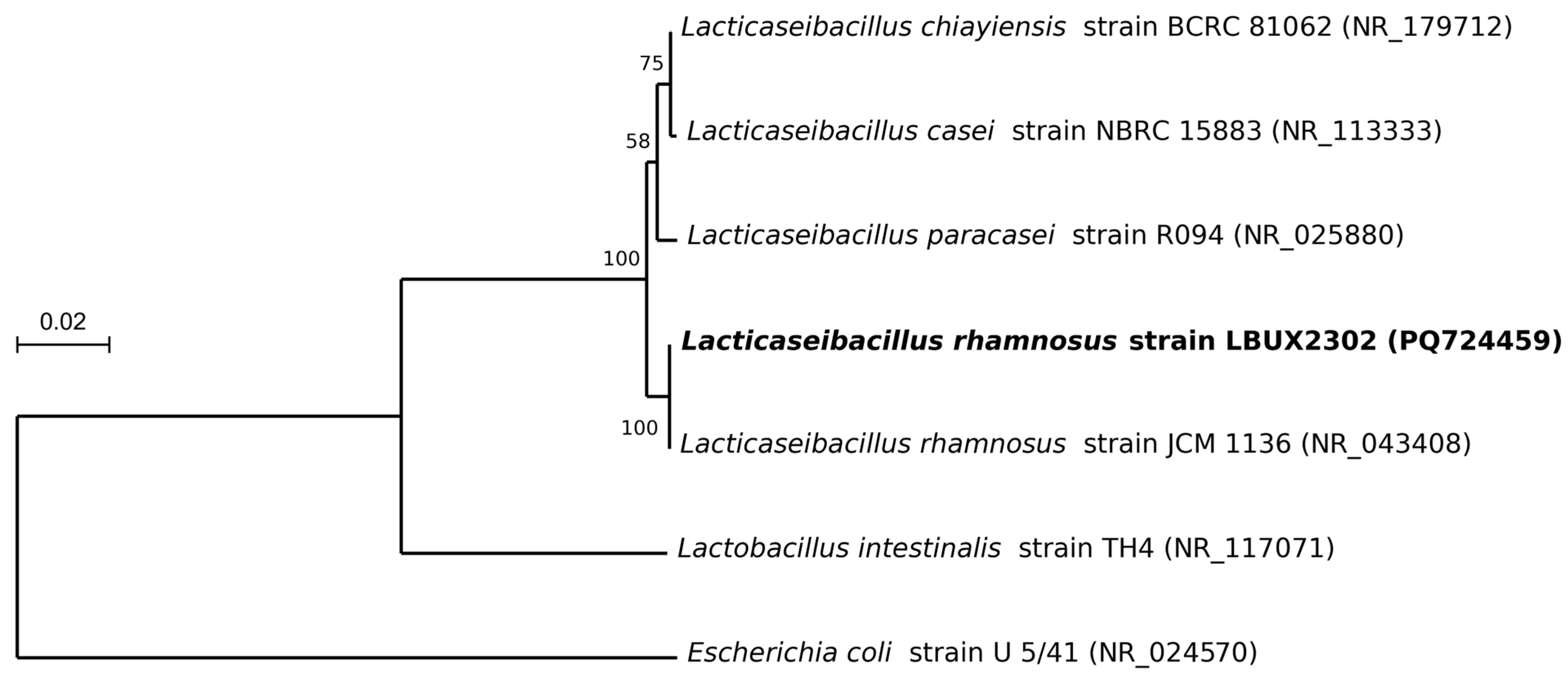
2.3. Phylogeny Analysis

The phylogenetic tree was constructed using MEGA software (version 11.0.13) [15] by the Maximum Likelihood method with the Kimura 2-parameter model [16]. The 16S rRNA gene sequences were obtained from seven Lactobacillus species deposited in the NCBI database. Bootstrap values of the tree were computed by resampling 1000 replications.

2.4. Catalase Test

A 12 h culture plate of pure bacterial cells was used to evaluate catalase activity by placing a drop of hydrogen peroxide on the center of a clean slide. Then, using a sterile bacteriological loop or stick, a small amount of the bacterial colony was transferred and mixed with the H2O2 drop. The reaction was observed immediately. The positive control was Staphylococcus aureus ATCC6538 [17], while Lacticaseibacillus casei isolated from a commercial productwas the negative control. This strain was used as the control in the next experiments. Catalase-positive bacteria produced bubbles due to the release of oxygen, whereas catalase-negative bacteria did not generate bubbles. Triplicates of the test were performed [18].

2.5. Hemolysis Test

A bacterial suspension containing 1 × 10⁹ CFU/mL was seeded onto blood agar and incubated at 37 °C for 48 h. S. aureus ATCC6538 [17] and L. casei [18] were used as controls. Hemolysis was considered positive when complete red blood cell lysis was observed, whereas the absence of hemolysis was classified as a negative result. Triplicates of the test were performed [10].

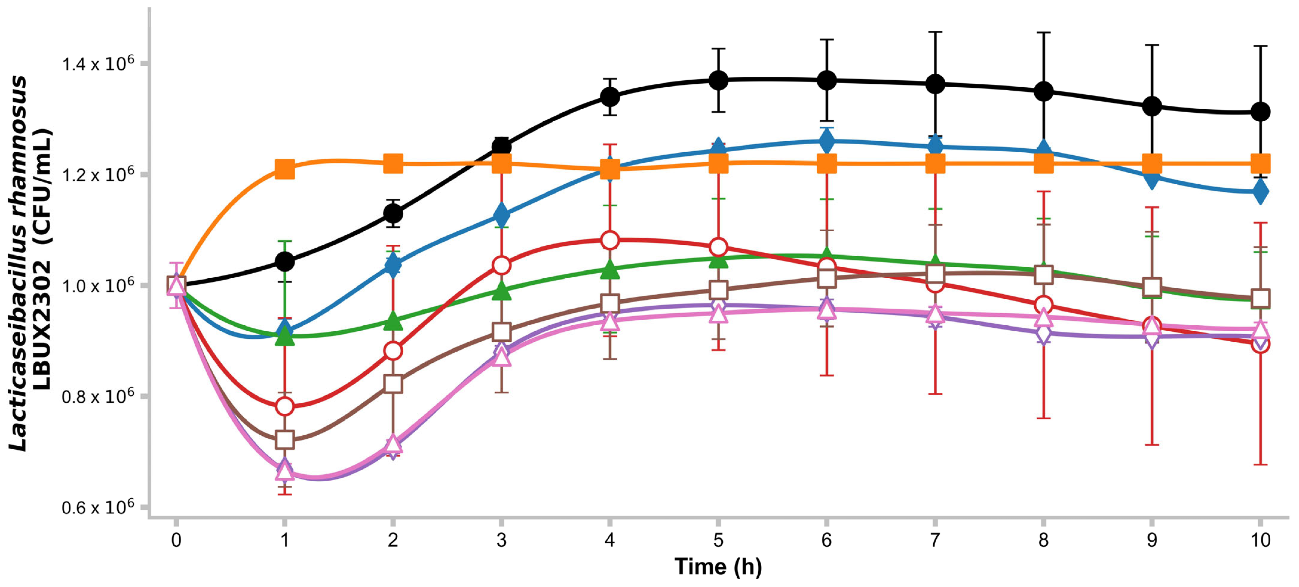
2.6. Growth Kinetics on Different Carbon Sources

Bacterial growth was monitored over 10 h at 2 h intervals using glucose, sucrose, fructooligosaccharides (FOS), lactose, lactulose, and raffinose at 0.01% (w/v) concentrations in a minimal medium (pH 7) containing 0.02 g/L NaCl, 1 g/L (NH4)2 SO4, 0.02 g/L, CaCl2 ۔ H2O, 0.4 g/L MgSO4 7-H2O, 0.72 g/L, K2H PO4, 0.72 g/L, KH2 PO4, and 0.01% (w/v) yeast extract. Bacterial growth was measured using a microplate reader and analyzed with Gen5® software (version 2x). Triplicates of the test were performed [10].

2.7. Antimicrobial Activity

This activity was evaluated against E. coli ATCC25922 [19], E. coli O157:H7 [20], Salmonella typhi ATCC14028 [21], and S. aureus ATCC6538 [17] by diffusion plate assay, as reported by Reyes-Castillo et al. (2023) [10]. Absence of inhibition was considered as negative (−), weak inhibition as (+), and strong inhibition as (++). Antimicrobial resistance testing was conducted in triplicate. L. casei was used as a control [18]. Triplicates of the test were performed [9].

2.8. Antibiotic Resistance
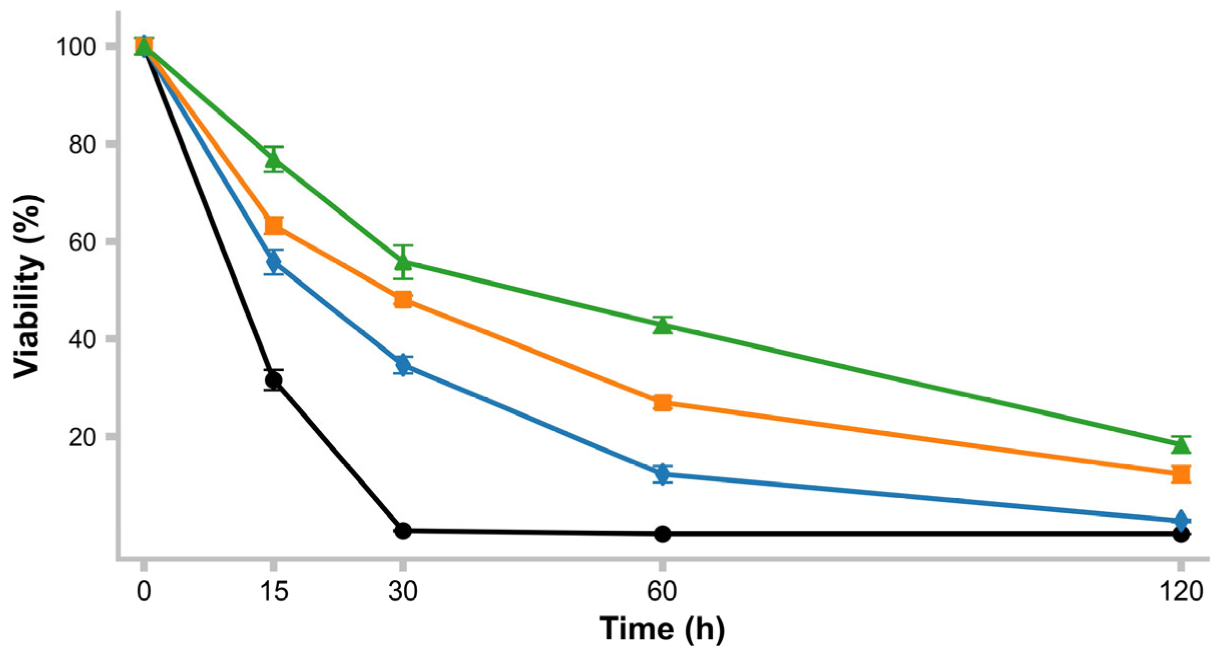
This ability was assessed by the diffusion test, using Multibac discs for Gram-positive bacteria of Investigación Diagnostica, Ciudad de Mexico, Mexico, according to Reyes-Castillo et al. (2023) [10]. The discs contained 1 µg of dicloxacillin, 5 µg of ciprofloxacin, 10 µg of ampicillin and gentamicin, 15 µg of erythromycin, 25 µg of trimethoprim-sulfamethoxazole, 30 µg of cefotaxime, cephalothin, clindamycin, vancomycin, and tetracycline, and 10 U of penicillin. Triplicates of the test were performed. L. casei was used as a control.

2.9. pH Resistance

Prior to the experiment, a standard growth curve of L. rhamnosus and L. casei (control) was determined to establish the amount of CFU needed to inoculate 10 mL of MRS broth to obtain, after 8 h of incubation at 37 °C, 1 × 10⁹ CFU/mL. To prepare the inoculum of the desired concentration, 10 mL of MRS was inoculated and incubated at 37 °C for 24 h. Then, each culture was centrifuged for 5 min at 10,000× g, the pellet was washed twice with PBS (0.1 M, pH 7) and suspended in PBS adjusted to different pH values (1.5, 2, 3, and 5) using 1N HCl and incubated at 37 °C for 120 min. The viability was determined by the plate count method [18] at 0, 15, 30, 60, and 120 min. Using the following expression, the percentage of viability was determined under the test conditions. Triplicates of the test were performed [22].
%   V i a b i l i t y = U F C / m L t i m e   m i n U F C / m L t i m e 0   m i n 100

2.10. Hydrophobicity

Hydrophobicity was evaluated according to Vinderola et al. 2003 [23] to assess non-specific adhesion (the ability to adhere to hydrocarbons). Previously, a suspension of L. rhamnosus with an optical density (OD) of 1.0 at 600 nm (A0) was prepared. This suspension was vigorously mixed for 1 min with xylene in a 1:3 ratio and incubated at 37 °C for one hour. The aqueous component was separated, and its OD600 nm was measured (A1). L. casei was the control strain. Triplicates of the test were performed. Hydrophobicity was defined as follows:
%   H i d r o p h o b i c i t y = 1 A 1 A 0 100

2.11. Autoaggregation

Before the experiment, a bacterial culture was prepared as in the hydrophobicity test and was considered A0. The suspension was incubated at 37 °C for 2 h. Then, 0.1 mL was transferred and mixed with 1.9 mL of PBS. OD600 nm was determined (A1). L. casei was the control strain. Triplicates of the test were performed [24]. Autoaggregation was defined as follows:
%   A u t o a g g r e g a t i o n = 1 A 1 A 0 100

2.12. Coaggregation

This assay involved preparing a cell suspension as described in the hydrophobicity tests and mixing 1 mL of the suspension with an equal volume of each pathogen (E. coli, S. typhi, and S. aureus). The OD600 nm was measured and designated as A0. The mix was then incubated for 2 h at 37 °C, after which the OD600 nm was measured and considered as At. L. casei was used as the control strain. Triplicates of the test were performed. The coaggregation percentage was defined as in Zuo et al. (2023) [24].
%   C o a g g r e g a t i o n = A 0 A T / A 0 100

2.13. Qualitative Assay of Bile Salt Tolerance

For bile salt tolerance assessment, the method of Gao et al. (2021) [9] was used. In brief, 10 μL of a fresh culture of the isolated strain was inoculated onto plates containing MRS agar containing 0.1%, 0.3%, and 0.5% w/v of glycocholic acid (GcCA), glycodeoxycholic acid (GDxCA), taurocholic acid (TcCA), taurodeoxycholic acid (TDxCA), and oxgall (OXGL) (Sigma Aldrich, St. Louis, MO, USA). Each assay was incubated aerobically for 48 h at 37 °C. The appearance of a halo surrounding the colony after incubation was considered an indicator of tolerance to bile salt (BS). Triplicates of the test were performed.

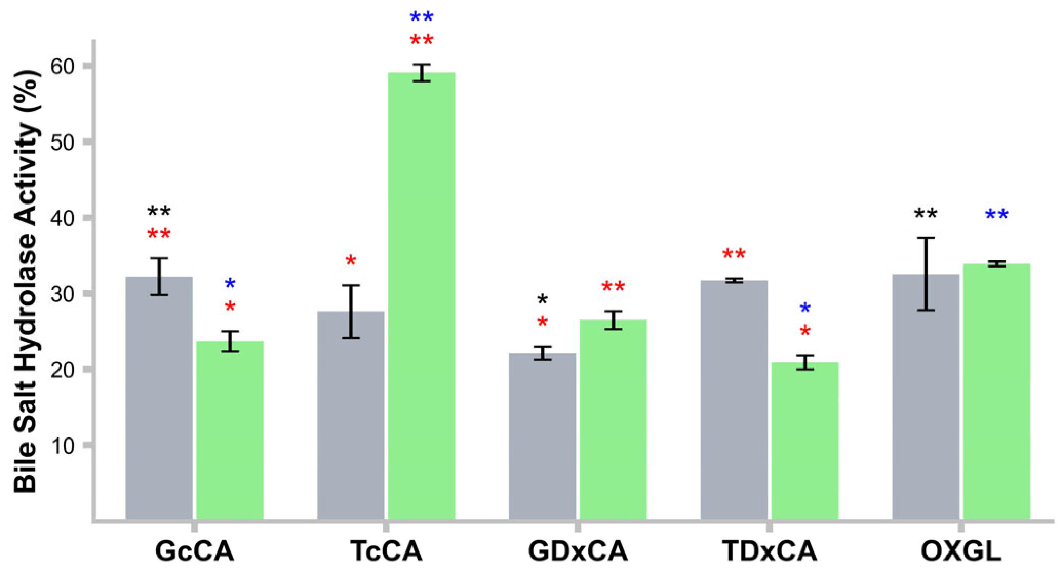
2.14. Bile Salt Hydrolase (BSH) Activity

This activity was assessed using whole cells (WC) according to González–Vázquez et al. (2015) [18], using an inoculum of 1 × 10⁹ CFU/mL in PBS containing 0.5% w/v of GcCA, GDxCA, TcCA, TDxCA, and OXGL and incubated for 48 h. L. casei was used as a control. A standard curve of glycine was established. Triplicates of the test were performed. BSH activity was calculated as follows:
%   B S H   a c t i v i t y = G l y c i n   c o n c e n t r a t i o n s a m p l e G l y c i n   c o n c e n t r a t i o n c o n t r o l 100

2.15. 2,2-Diphenyl-1 Picrylhydrazyl (DPPH) Radical Inhibitory Activity

To analyze the inhibition of DPPH radical activity by L. rhamnosus LBUX02, a solution containing 1 mL of 0.2 mmol/L of DPPH solubilized in CH3OH was mixed with 1 mL of WC or cell-free supernatant (CFs), or cell fragments Cfg, and maintained in darkness for 30 min. OD517 nm was measured. L. casei was the control strain. Triplicates of the test were performed. DPPH inhibitory activity was calculated as described by Gao et al. (2021) [9]:
%   D P P H   i n h i b i t o r y   a c t i v i t y = 1 A s a m p l e A b l a n k A c o n t r o l 100

2.16. Hydroxyl Radical Inhibitory Activity

In this assay, 1 mL of WC, CFs, or Cfg was mixed with 1.0 mL of phenanthroline (2.5 mM), 1 mL of FeSO4 (2.5 mM), and 1 mL of PBS (pH 7.4), then the mixture was incubated at 37 °C for 90 min. To inhibit the reaction, 1 mL of 20 mM H2O2 was added, and the OD517 nm was measured. L. casei was the control strain. Triplicates of the test were performed. Inhibitory ability was determined as described by Yan et al. (2019) [25]:
%   H y d r o x y l   r a d i c a l   i n h i b i t o r y   a c t i v i t y = 1 A s a m p l e A b l a n k A c o n t r o l A b l a n k 100

2.17. Superoxide Anion Radical Inhibitory Activity

The inhibitory activity was evaluated using 1 mL per sample of WC, CFs, or Cfg mixed with 3 mL of Tris–HCl solution (pH 8.2) and incubated at 25 °C for 20 min. Afterwards, 0.4 mL of pyrogallol (25 mM) was added, and the reaction was maintained at room temperature for 4 min. Then, 0.5 mL of HCl was added to inhibit the reaction, and OD325 nm was measured. L. casei was the control strain. Triplicates of the test were performed. Inhibitory activity was determined as follows [25]:
%   S u p e r o x i d e   a n i o n   r a d i c a l   i n h i b i t o r y   a c t i v i t y = ( 1 A s a m p l e A b l a n k ) 100

2.18. Qualitative Ferulic Acid Activity (EFA)

EFA activity on the plate was tested in agreement with the methodology reported by Tomaro-Duchesneau et al. 2012 [26]. Before the assay, LAB strains were inoculated in MRS broth supplemented with 1% ethyl 4-hydroxy-3-methoxycinnamate (EFA), 1.33 mM, and then they were incubated anaerobically under standard conditions. Meanwhile, MRS EFA agar plates were prepared using MRS agar (pH = 6.5 and 1.5% w/v). Once tempered, 0.3 mL of EFA (prepared at 10% w/v in N, N-dimethylformamide) was added per 20 mL of agar. Sterile Whatman #3 paper discs, impregnated with LAB strains (MRS-EFA culture), were placed onto the dried MRS EFA plates, and incubated at 37 °C for 48 h. No activity was considered as a negative (−) test, a moderate test was considered as positive (+), and total activity was considered as double positive (++). L. casei was the control strain. Triplicates of the test were performed.

2.19. Inhibition of α-Amylase

This experiment was carried out according to Won et al. (2021) [27]. In brief, a previous culture of L. rhamnosus was prepared. The experiment was carried out by plating the bacteria at a concentration of 1 × 10⁹ CFU/mL onto MRS broth and incubating at 37 °C for 24 h. Afterwards, the culture was centrifuged for 15 min at 8000× g at 4 °C. Then, 250 μL of α-amylase (pre-incubated at 25 °C for 10 min, 0.5 mg/mL) was mixed with 250 μL of CFs. The mix was then incubated at 25 °C for 10 min with 250 μL of 1% (w/v) starch solution in 0.02 M PBS. To finish the reaction, 500 μL of DNS dye reagent was added, and the test was boiled for 5 min, then cooled to room temperature and diluted fourfold with distilled water. OD540 nm was determined using 1 mL of the diluted solution. L. casei was the control strain. Triplicates of the test were performed. The percentage of α-amylase inhibition was defined as follows:
%   α a m y l a s e   i n h i b i t i o n = A c o n t r o l A s a m p l e A c o n t r o l 100

2.20. Inhibition of α-Glucosidase

The inhibition of this enzyme was determined according to Won et al. (2021) [27]. L. rhamnosus strain (1 × 10⁹ CFU/mL) was grown on MRS broth under standard conditions. Then the culture was centrifuged at 8000× g for 15 min at 4 °C. A volume of 25 μL of supernatant was used to mix it with 150 μL of 0.01 M PBS (pH 7.0) and 75 μL of p-nitrophenyl glucopyranoside (PNPG) solution (0.2 M). This mixture was incubated at 37 °C for 10 min. To start the reaction, 50 μL of α-glucosidase (0.17 U/mL) was added. The reaction was incubated at 37 °C for 10 min, and it was finished by adding 1 mL of 0.1 M Na2CO3. OD405 nm was quantified to determine the p-nitrophenol released. The experiment was performed in triplicate, with L. casei as the control strain. The percentage of α-glucosidase inhibition was defined as follows:
%   α g l u c o s i d a s e   i n h i b i t i o n = 1 C D A B 100

2.21. Adhesion to Cancer Cell Lines

Cell adhesion assays were performed using different cancer cell lines, including SW-620 (colon cancer), Hella (cervical cancer), MCF-7 (breast cancer), HaCaT (aneuploid immortal keratinocyte cell), and SK-LU-1 (lung adenocarcinoma). Dulbecco’s Modified Eagle’s medium (Lonza, Basel, Suiza), with 5% fetal bovine serum (Gibco), 1% L-glutamine (Lonza, Basel, Suiza), and 1% antibiotics (penicillin/streptomycin, Gibco), was used for all cell line proliferations. Then, 4 × 10⁵ cells per well were seeded and incubated under standard conditions with 5% CO2. Triplicates of the test were performed [28].
Before the adhesion assay, a fresh culture of L. rhamnosus at 1 × 10⁸ CFU/mL was prepared. The bacteria were then incubated with the cancer cell lines at 37 °C with 5% CO2 for 1 h. The culture was washed with PBS and 300 µL of trypsin-EDTA (Gibco, Waltham, MA, USA). Adherent Lactobacillus cells were quantified by plate counting and expressed as CFU/mL. Adhesion was reported as the percentage of adhered bacterial cells with respect to the Lactobacillus initially added.

2.22. Statistical Analysis

Significant differences in all experiments were determined using the Kruskal–Wallis test, followed by Dunn’s post hoc test with a 95% confidence interval, performed using GraphPad Prism 5.01 software.

3. Results

3.1. Identification and Phylogeny

The isolate obtained from stool samples exhibited a rod-like morphology and was Gram-positive. The DNA amplified via PCR was sequenced, and BLASTN (version 2.16.1+) analysis revealed 99.6% identity with Lacticaseibacillus rhamnosus (Figure 1). The strain was designated as L. rhamnosus LBUX2302 (GenBank accession: PQ724459.1).

3.2. Growth Kinetics on Different Carbon Sources

L. rhamnosus LBUX2302 exhibited optimum growth at pH 7.0. and was able to metabolize all tested carbohydrates (Figure 2). The highest growth rate was observed in glucose (control). The exponential phase for glucose and saccharose lasted between 3 h, whereas for FOS, it was 1 h; however, no significant differences were detected. Differences (p ≤ 0.05) between glucose and lactose, raffinose, lactulose, xylan, and xylose were found. Sucrose showed significant differences compared to lactulose and xylose (Figure 1). Additionally, FOS exhibited significant differences compared to lactulose, xylan, and xylose. For raffinose, lactulose, xylan, and xylose, an initial phase was observed during the first hour, followed by an exponential phase between 1 and 3 h. After 4 h, the strain maintained stable metabolic activity over time (Figure 2).
Significant differences (p ≤ 0.05) in the growth of L. rhamnosus LBUX2302 when utilizing glucose vs. lactose, raffinose, lactulose, xylan, and xylose; sucrose vs. lactulose, and xylose; and FOS vs. lactulose, xylan, and xylose were found.

3.3. Catalase and Hemolysis

Neither L. rhamnosus LBUX2302 nor L. casei exhibited catalase or hemolytic activity. Conversely, S. aureus (negative control) displayed both activities, suggesting that the isolates are safe for use, which is an essential criterion for probiotic microorganisms (Table 1A).

3.4. Antimicrobial Activity

In the case of antimicrobial properties, L. rhamnosus LBUX2302 and L. casei strains inhibited the growth of pathogenic or non-pathogenic bacteria, either completely or moderately. The L. rhamnosus LBUX2302 strain totally inhibited S. typhi ATCC14028, a bacterium in which virulent genes such as fmA = fimbria production factor, invA = invasion factor, SpvR and SpvC = systemic infection (inhibition activation of macrophage), and Stn = enterotoxigenic substances production have been reported [21]. Also, it inhibited the E. coli ATTC25922 strain, a non-pathogenic strain commonly used as a control strain for antibiotic susceptibility testing [19]. In the case of E. coli 0157:H7, it was partially inhibited; this bacterium has shown pathogenicity that primarily affects children, the elderly, and immunosuppressed individuals, causing mild cases of diarrhea to hemolytic uremic syndrome [20]. L. rhamnosus LBUX2302 inhibited the S. aureus ATCC6538 strain, which has been used as a control for antibiotic susceptibility testing [17] (Table 1B).

3.5. Antibiotic Resistance

Probiotics must be evaluated for their antibiotic resistance and pathogenicity [17]. L. rhamnosus LBUX2302 and L. casei strains were sensitive to several antibiotics, exhibiting resistance to β-lactam and aminoglycoside antibiotics (Table 1C). L. rhamnosus LBUX2302 showed resistance to gentamicin (aminoglycoside), dicloxacillin, and penicillin (β-lactam antibiotics). L. casei specifically showed resistance to β-lactam antibiotics, such as vancomycin, dicloxacillin, and penicillin, and to protein synthesis inhibitors such as gentamicin.

3.6. Bile Salt Tolerance

Bile salt tolerance is an essential characteristic for evaluating the viability of LAB in the intestine. L. rhamnosus LBUX2302 and L. casei showed tolerance to primary and secondary bile salts at concentrations of 0.1%, 0.3%, and 0.5% (Table 1D).

3.7. Hydrophobicity and Autoaggregation

The cell surface hydrophobicity of L. rhamnosus LBUX2302 was 61%. The control strain did not exhibit hydrophobicity. A significant difference was observed between the strains. Autoaggregation in probiotics plays a fundamental role in their adhesion to the intestinal epithelium. L. rhamnosus LBUX2302 exhibited an autoaggregation rate of (53.5%), which was lower than that of the control strain (Table 1E).

3.8. Coaggregation

L. rhamnosus LBUX2302 showed a high average percentage of 70% of coaggregation with other microorganisms, specifically E. coli ATCC25922, S. aureus ATCC6538, and S. typhi ATCC14028, compared to the coaggregation ability of L. casei (Table 1F).

3.9. Ferulic Acid Activity

Both L. rhamnosus LBUX2302 and the control strain exhibited ferulic acid activity (Table 1G).

3.10. BSH Activity in CFs

Both L. rhamnosus LBUX2302 (32.2%) and L. casei (24%) exhibited BSH activity in GcCA, with no significant differences. Regarding TcCA, the control strain (58%) showed greater BSH activity than the L. rhamnosus LBUX2302 strain (30%). However, L. rhamnosus LBUX2302 (33%) displayed higher BSH activity in the presence of TDxCA versus the control strain (20%) (Figure 3). Significant differences in BSH activity were found in L. rhamnosus LBUX2302 between GcCA vs. GDxCA and GDxCA vs. OXGL. For L. casei, significant differences were found in GcCA vs. TcCA, TcCA vs. TDxCA, and GcCA and TDxCA with OXGL. When comparing both strains, significant differences were present in all bile salts except OXGL.

3.11. pH Survival

The survival rate at different pH levels is shown in Figure 4. L. rhamnosus LBUX2302 was unable to survive at pH 1.5 for 120 min. However, at pH 2, 3, and 5, survival rates were 2.7%, 22%, and 27.5%, respectively, after 120 min, with no significant differences detected.

3.12. Antioxidant Activity

The CFs of L. rhamnosus LBUX2302 and L. casei strains exhibited high antioxidant inhibition of DPPH. In contrast, the WC of L. casei demonstrated an antioxidant capacity 15 times greater than that of the L. rhamnosus LBUX2302 strain. For L. rhamnosus LBUX2302, a significant difference (p ≤ 0.05) was observed among the three treatments. Regarding L. casei, significant differences (p ≤ 0.05) were found between CFs and Cfg. (Figure 5a). The CFs of L. rhamnosus LBUX2302 were able to eliminate 80% of hydroxyl radicals, whereas the CFs of L. casei exhibited a 43% inhibition rate (p ≤ 0.05). No significant differences were observed among the different cell fractions of L. rhamnosus LBUX2302. However, for L. casei, significant differences (p ≤ 0.05) were found between CFs and Cfg. Additionally, significant differences (p ≤ 0.05) were observed between both strains in the CFs fractions (Figure 5b).
Regarding superoxide anion radical inhibition, L. rhamnosus LBUX2302 and the control stain exhibited significant differences (p ≤ 05) among their respective cell fractions. When comparing superoxide inhibition across the different cell fractions between the two strains, significant differences (p ≤ 05) were observed in both the CFs and the Cfg (Figure 5c).

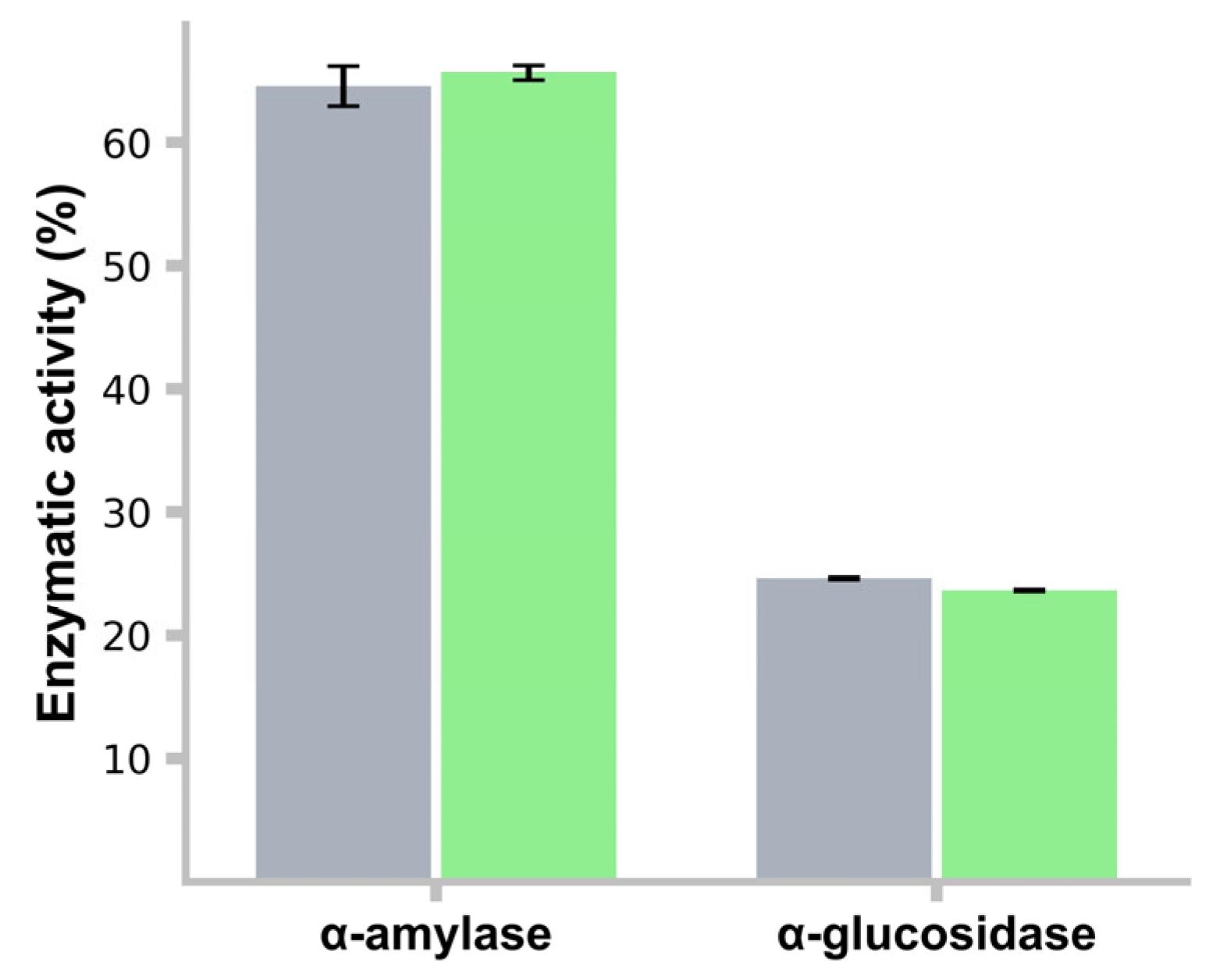
3.13. α-Amylase and α-Glucosidase Activity Inhibition

The L. rhamnosus LBUX2302 strain exhibited 64.5% inhibition of α-amylase activity, showing no significant differences compared to the control strain (64%). Regarding α-glucosidase inhibition, L. rhamnosus LBUX2302 (23%) and the control strain (22.5%) demonstrated lower inhibition compared to α-amylase activity, with no significant differences (Figure 6). Between the two strains for either enzymatic activity, no significant differences were found.

3.14. Adhesion to Cancer Cell Lines

L. rhamnosus LBUX2302 exhibited a higher adhesion capacity to MCF-7, SK-LU-1, and SW-620 cell lines (71%, 75%, and 74%) with respect to HaCaT (38%). These results suggest that adhesion depends on the origin of the cell line and its interaction with the strain species (Figure 7).

4. Discussion

The phenotypic characterization of probiotic microorganisms provides valuable information about their potential applications in the food, animal, and human health industries. The isolation of novel microorganisms with hypoglycemic and antioxidant properties requires systematic investigation. T2DM is a chronic disease characterized by hyperglycemia and insulin resistance. Genetic factors, dietary habits, obesity, sedentary lifestyle, and lack of exercise contribute to its development [29,30]. Postprandial glucose is considered the most important parameter in controlling the risk of diabetes. One of the strategies to keep postprandial glucose at adequate levels is the inhibition of the α-amylase and α-glucosidase enzymes, which catalyze the digestive process of carbohydrates. Conventional treatment involves pharmacological intervention, which may have adverse effects. An alternative approach is the use of probiotic bacteria to manage the disease [27,31,32]. The inhibitory activity shown by L. rhamnosus LBUX2302 (60% to 25%) over α-amylase and α-glucosidase is consistent with previously reported findings [31,33]. This inhibition may contribute to reducing blood glucose levels and slowing glucose absorption. Our results serve as an indicator for scaling up to in vivo evaluations to confirm the potential of L. rhamnosus LBUX2302 as a therapeutic agent for hyperglycemia. Lactiplantibacillus plantarum CCFM0236has demonstrated the ability to inhibit α-glucosidase and α-amylase, thereby reducing postprandial hyperglycemia. Huligere et al. (2023) [34] have demonstrated that different Lacticaseibacillus strains present in the human gut possess α-amylase and α-glucosidase inhibitory activity, reducing blood glucose responses in vitro.
L. rhamnosus LBUX2302 also exhibited strong antioxidant activity, particularly in the CFs, with high DPPH, hydroxyl, and superoxide anion inhibitory capacities. These antioxidative properties of L. rhamnosus LBUX2302 can be applied in fermented beverages and animal feed consumption since it is possible to reduce oxidative damage caused by stress and limit the use of synthetic antioxidants. This reduction in oxidative damage is probably due to the action of enzymes such as superoxide dismutase, glutathione peroxidase, glutathione reductase, and vitamins C and E, or to the action of bioactive peptides, which are polysaccharides such as the ones produced by the intestinal microbiota, as suggested by Lepecka et al. (2023) [35] Our results agree with those reported by Won et al. (2021) [27] and Wanget al. (2022) [36], who described L. plantarum and L. paracasei strains that showed high hypoglycemic, antioxidant, and probiotic properties. Oxidative stress, which implies a disequilibrium between the production of ROS and the defense system against oxidation, contributes to DM complications, the damage of genetic material, oxidation of proteins, and peroxidation of lipids [37]. Kaprasob et al. (2019) [38] have suggested that bioprocessing of cashews and apple juice through fermentation with L. plantarum and L. casei was effective for obtaining antioxidant nutraceuticals, which were relevant to DM2. Antioxidant activity has been reported for various LAB, including L. rhamnosus, L. lactis, and L. plantarum [39]. This activity has been associated with probiotic-induced glycemic improvement, suggesting that probiotics may modulate oxidative stress and contribute to glucose regulation [34,40].
Bile salt tolerance is essential for some probiotics to maintain viability in the intestinal environment. L. rhamnosus LBUX2302 and L. casei exhibited tolerance to various bile salt concentrations, which was mediated by the activation of surface proteins [41]. BSH activity plays a crucial role in the host metabolism, affecting energy regulation, lipid absorption, and cholesterol metabolism [42]. In this study, L. rhamnosus LBUX2302 and L. casei exhibited BSH activity in primary and secondary bile salts, as well as OXGL, which may contribute to cholesterol and triglycerides reduction [43]. Dong and Lee (2018) [44] have reported that BSH activity in L. rhamnosus E9 showed a preference for glycocholic bile salts. This activity could be attributed to the genes encoding this enzyme in L. rhamnosus [45].
On the other hand, L. casei exhibited a higher affinity for TcCA compared to other bile salts. Notably, in human bile salts, the proportion of glycine to taurine conjugates is 3:1 [46]. Similar BSH activity results have been reported for L. casei by various authors. González-Vázquez (2015) [18] identified BSH activity in L. casei Shirota using TcCA and TDxCA as substrates, with higher activity observed in TcCA. Lee et al. (2023) [47] have reported that supplementation with L. reuteri NCIMB 30242 could alleviate serum cholesterol levels by inhibiting intracellular cholesterol synthesis and promoting intestinal cholesterol excretion.
Autoaggregation, coaggregation, and hydrophobicity are key factors influencing bacterial adhesion [48]. L. rhamnosus LBUX2302 exhibited 54% of autoaggregation (Table 1E), indicating possible potential biofilm formation and prolonged colonization in the digestive tract [49,50]. A hydrophobicity assay revealed a 61% affinity to hydrocarbons (Table 1E), aligning with previously reported values for L. rhamnosus strains [51].
The autoaggregation of probiotic bacteria is an essential process for adhesion and is considered the first step in bacterial colonization [52]. This process leads to the formation of structured bacterial communities that facilitate cell-to-cell interaction and communication, the exchange of genetic material, adherence, and colonization in different environments [53]. Our results were superior to those reported by Grigoryan et al. (2018) [54] and Zawistowska-Rojek et al. (2022) [55], who have found autoaggregation percentages of 44.3% to L. rhamnosus INA-5.1%, 39.4% to L. rhamnosus R-2002, 10.1% to L. rhamnosus LrA, 14.6% to L. rhamnosus LrB, 15% to L. rhamnosus LrC, and 12.5% to L. rhamnosus LrD. In contrast, Sophatha et al. (2020) [56] have reported high autoaggregation levels in L. rhamnosus SD4 and SD11 (55–60%), which are similar to the results obtained in our study. The literature indicates that autoaggregation percentages can vary within the same species, likely due to differences in the probiotic strain’s origin and incubation time [57]. L. rhamnosus LBUX2302 showed a higher adhesion capacity in several cell lines, such as human colon cancer, cervical cancer, human breast cancer, and lung adenocarcinoma, which indicates that this microorganism could have new functional properties not yet explored. Thananimit, et al. (2022) [58] demonstrated that L. rhamnosus SD1, SD4, and SD11 exhibit high adhesion capacity in Caco-2 and HIEC-6 cells, with adhesion percentages ranging from 39% to 80%. However, no previous studies have reported the adhesion of Lacticaseibacillus in cell lines such as Hela, MCF-7, HaCaT, SK-LU-1, and SW-260. Evaluating adhesion in these cell lines could reveal new probiotic functions.
The coaggregation assay demonstrated strong adhesion of L. rhamnosus LBUX2302 to non-pathogenic and pathogenic bacteria, particularly E. coli ATCC25922, S. aureus ATCC6538, and S. typhi ATCC14028 (Table 1F), suggesting its potential to prevent pathogen colonization [59]. Our results were consistent with those reported by Cozzolino et al. (2020) [51], who found that L. rhamnosus GG exhibited high coaggregation with pathogenic bacteria, including P. mirabilis and E. coli. In contrast, Zawistowska-Rojek et al. (2022) [55] have reported lower coaggregation values for L. rhamnosus GG with E. coli, S. typhimurium, and E. faecalis. High coaggregation values indicate a strong capacity for barrier formation, which can prevent pathogenic colonization [59].
L. rhamnosus LBUX2302 showed weak antimicrobial inhibition against E. coli O157:H and S. aureus ATCC6538. Inturri et al. (2019) [60] have demonstrated that L. rhamnosus HN001 inhibited E. coli ATCC25922, E. coli ATCC35218, and S. typhi STN12. Davoodabadi et al. (2015) [61] have found that L. rhamnosus S19 inhibited various E. coli strains, including those with enteroaggregative, antihemorrhagic, antiinvasive, enteropathogenic, and enterotoxigenic activities. Furthermore, Johnson-Henry et al. (2008) [62] have reported that L. rhamnosus GG protects epithelial monolayers against the influence of E. coli O157:H7 on tight junctions claudin-1 and ZO-1.
The potential mechanism by which L. rhamnosus SCB0119 induces S. aures cell death may involve genomic instability due to interference with DNA repair pathways through the expression of the genes that encode ATP synthase, responsible for ATP hydrolysis [63]. Finally, probiotic antimicrobial activity can be mediated through multiple mechanisms, including organic acid production leading to acidification, bacteriocin production, competition for nutrients and adhesion sites in the host mucosa, and the induction of proinflammatory responses that facilitate pathogen elimination, both in vitro and in vivo [64,65,66]. Nonetheless, additional studies are required to elucidate the specific mechanism underlying the antimicrobial activity of L. rhamnosus LBUX2302.
The isolation of microorganisms with probiotic characteristics must include an evaluation of their abilities to tolerate relevant conditions, such as acidity and bile acids. The acidity that probiotics face in the gastrointestinal tract is a factor that influences their viability [67]. In this study, L. rhamnosus LBUX2302 exhibited a survival range between 22 and 27.5% at pH 2 and 3 over 120 min. Probiotics are intrinsically acid-resistant; they must withstand the harsh conditions of the stomach to reach the intestine alive, where they can colonize and exert beneficial effects [68]. The resistance of microorganisms to acidic pH may be attributed to a univariant gradient between cytoplasmic and extracellular pH, as well as proton extrusion via the F0F1-ATPase mechanism, which facilitates the survival in the gastrointestinal tract [69].
Safety is a crucial criterion for bacterial strains used for the food industry [70]. According to the European Food Safety Authority (EFSA), bacterial identification and hemolytic activity are among the primary criteria for assessing probiotic safety. In this study, we found no hemolytic activity in L. rhamnosus LBUX2302 or the control strain (Table 1A). Wang et al. (2021) [71] similarly reported an absence of hemolytic activity in LAB isolated from infant feces. Other LAB, such as L. rhamnosus CA15, L. rhamnosus CWKu-12, and L. rhamnosus SS73, have also been found to lack hemolytic activity [72,73] to produce hemolysin proteins, unlike the S. aureus species.
Antibiotics are one of the alternatives for treating infectious diseases caused by Gram-positive and Gram-negative bacteria in humans and animals. Their overuse has led to the emergence of antibiotic-resistant bacteria, which has caused an international public health problem [74]. Additionally, we assessed the antibiotic profile of L. rhamnosus LBUX2302, which exhibited resistance to gentamicin, dicloxacillin, and penicillin. LAB have been previously reported to exhibit intrinsic resistance to aminoglycosides (gentamicin) [75], which indicates that there are no genes that encode transferable resistance determinants and that it may be due to chromosomal zones of the bacteria [10]. Therefore, according to the qualified presumption of safety criteria, they should be considered safe. Furthermore, another author has suggested that the antibiotic resistance detected in L. rhamnosus GG is natural [76]. It is important to note that antibiotic-resistant LAB do not necessarily represent a health risk; nevertheless, additional research is required to confirm whether antibiotic-resistant genes in LAB are capable of being transferred [36]. In 2023, Shahali et al. [77] reported a high percentage of resistance to gentamicin, streptomycin, and ciprofloxacin in many LAB strains. The potential mechanism of gentamicin resistance in LAB strains is the absence of an antibiotic transporter, as gentamicin susceptibility is associated with the ability of the antibiotic to cross the bacterial membrane [78]. Previous studies have suggested that Lactobacillus spp. is susceptible to penicillin and β-lactams. Our findings indicate that L. rhamnosus LBUX2302 exhibited resistance to dicloxacillin and penicillin. These results are consistent with those reported by Hasan et al. (2020) [79], who found that L. rhamnosus MT539286 was resistant to the β-lactam antibiotics amoxicillin and oxacillin. Although L. rhamnosus is recognized as safe (GRAS) and has been granted a Qualified Presumption of Safety (QPS) status [80], it can still present antibiotic resistance and potentially transfer resistance genes to other bacteria via mobile genetic elements such as plasmids. Due to the need to perform a genomic characterization of L. rhamnosus LBUX2302, we are currently analyzing its genome to determine whether resistance to β-lactams and aminoglycosides is transferable or encoded at the chromosomal level. Specifically, we aim to identify intrinsic resistance genes related to the antibiotics gentamicin, penicillin, and dicloxacillin.
The Lactobacillus genus can efficiently grow on mono-, di-, tri-, and oligosaccharides [81]. Carbohydrates serve as an essential energy source for both the host and gut microbiota. In general, Lactobacillus species can metabolize these carbohydrates, producing compounds of interest for health and food industries [82,83]. In this study, we observed that L. rhamnosus LBUX2302 was able to metabolize monosaccharides and oligosaccharides (Figure 1), likely due to the expression of genes encoding glycosyl hydrolase enzymes. Our results are consistent with those reported by Ceapa et al. (2015) [84], who found that L. rhamnosus GG could utilize a wide range of mono-, di-, and polysaccharides, and polyols. In addition, Li et al. (2024) [85] have identified several genes encoding enzymes associated with carbohydrate metabolism in L. rhamnosus LR-ZB1107-01, including glycoside hydrolases, glycosyltransferases, esterases, and carbohydrate-binding modules. Furthermore, L. rhamnosus LR-ZB1107-01 possesses a complex phosphoenolpyruvate (PEP)–phosphotransferase system (PTS), which is responsible for the phosphorylation and transportation of sugars such as saccharose, lactose, maltose/glucose, mannitol, cellobiose, mannose, and fructose into the cell [85,86]. These sugar metabolism and transport systems are likely involved in the environmental adaptation capacity of L. rhamnosus. Moreover, we found that L. rhamnosus LBUX2302 can utilize FOS for growth, a result similar to the one reported by Kaewarsar et al. (2023) [87]. Additionally, Niu et al. (2023) [88] suggested that L. rhamnosus AS1.2466T can grow in the presence of FOS and, when administered simultaneously in mice, extends its colonization time in the intestine and ileum. The proposed mechanism for FOS degradation involves the fructanhydrolase enzyme, which has been identified in L. paracasei 1195 and functions in conjunction with the mannose PTS transporter complex [89]. However, this mechanism has not yet been identified in L rhamnosus species. Therefore, a key perspective for future research that we suggest is to sequence the complete genome of L. rhamnosus LBUX2302 and annotate the genes involved in all the activities determined in this study, and to test the effects in vivo models to demonstrate safety and corroborate the activities found.

5. Conclusions

This study indicates that L. rhamnosus LBUX2302 is a safe strain with promising probiotic properties. It demonstrates tolerance to bile salts, high hydrophobicity, and the ability to autoaggregate and congregate, along with strong adhesion capacity to MCF-7, SK-LU-1, and SW-620 cell lines. Furthermore, it exhibits significant hypoglycemic and antioxidant potential. However, further genomic characterization is necessary to confirm its safety and functionality at the genetic level. Once this characterization is completed, L. rhamnosus LBUX2302 could be considered for in vivo studies and potential applications in the pharmaceutical and food industries.

Author Contributions

Conceptualization, L.M.-R., R.G.-V. (Raquel González-Vázquez), M.A.G.-N., E.T.-M.; Methodology, P.A.R.-C., E.Z.-L., R.G.-V. (Raquel González-Vázquez), A.L.E.-C.; Investigación, L.M.-R., R.G.-V. (Raquel González-Vázquez), M.A.G.-N., E.T.-M., P.A.R.-C., E.Z.-L., F.M.-P., R.G.-V. (Rosa González-Vázquez), M.G.C.-E.; Resources, L.M.-R., R.G.-V. (Raquel González-Vázquez), M.A.G.-N.; Data curation, P.A.R.-C., E.Z.-L., R.G.-V. (Raquel González-Vázquez), L.M.-R.; Writing-original draft, review and editing, L.M.-R., R.G.-V. (Raquel González-Vázquez), M.A.G.-N., E.T.-M., P.A.R.-C., E.Z.-L., A.L.E.-C., F.M.-P., R.G.-V. (Rosa González-Vázquez), M.G.C.-E.; Supervision, L.M.-R., R.G.-V. (Raquel González-Vázquez), M.A.G.-N., E.T.-M. All authors have read and agreed to the published version of the manuscript.

Funding

This research was funded by (i) General Rectory of Universidad Autonoma Metropolitana, through 2024 call for research project proposals addressing current challenges; and (ii) Rectory of the Universidad Autonoma Metropolitana, campus Xochimilco, through the support program for final projects 2024.

Institutional Review Board Statement

The study was conducted in accordance with the Declaration of Helsinki and approved by the Institutional Review Board of Instituto Mexicano del Seguro Social (protocol code 36068 accepted on 23 August 2023). Nevertheless, we did not handle the stools samples directly as we received the purified strains.

Informed Consent Statement

Informed consent was obtained from all subjects involved in the study.

Data Availability Statement

The genomic sequencing data of L. rhamnosus LBUX2302 have been registered in Gen Bank under the accession code PQ724459.1.

Acknowledgments

Pedro Reyes-Castillo acknowledges Secihti for the scholarship number 2020-000026-02NACF-13604.

Conflicts of Interest

The authors declare no conflicts of interest. The funders had no role in the design of the study; in the collection, analyses, or interpretation of data; in the writing of the manuscript; or in the decision to publish the results.

Abbreviations

Am: ampicillin; BS: bile salt; BSH: Bile salt hydrolase; Cf: cephalothin; Cfg: cell fragments; CFs: cell-free supernatant; CFU: colony forming unit; Cfx: cefotaxime; Clm: clindamycin; Cpf: ciprofloxacin; Dc: dicloxacillin; DM2: type 2 diabetes; DNS: 3,5-Dinitrosalicylic acid; DM: Diabetes mellitus; DPPH: 2,2-diphenyl-1 picrylhydrazyl; E: erythromycin; EFA: ferulic acid activity; EFSA: European Food Safety Authority; FAO/WHO: Food and Agriculture Organization/codex alimentarius; FOS: fructooligosaccharides; GcCA: glycocholic acid; GDxCA: glycodeoxycholic acid; Ge: gentamicin; GRAS: generally recognized as safe; HaCat: spontaneously immortalized human keratinocyte cell line; HeLa: Henrietta Lacks cell line; H2O2: hydrogen peroxide; LAB: Lactic acid bacteria; NCBI: National Center of Biotechnology Information; MCF-7: breast cancer cell line; MRS: de Man Rogosa medium; OD: optical density; OH-: hydroxyl radicals; OXGL: oxgall; O−2: superoxide anion; PBS: Phosphate buffer solution; Pe: penicillin; PEP: phosphoenolpyruvate; PNPG: p-nitrophenyl glucopyranoside; QPS: Qualified Presumption of Safety; ROS: reactive oxygen species; SK-LU-1: human lung adenocarcinoma cell line; Stx: trimethoprim-sulfamethoxazole; SW620: adherent cell line isolated from the large intestine of a patient with Dukes-C colorectal cancer; TcCA: taurocholic acid; TDxCA: taurodeoxycholic acid; Te: tetracycline; Va: vancomycin; WC: whole cells.

References

  1. Zheng, J.; Wittouck, S.; Salvetti, E.; Franz, C.; Harris, H.M.B.; Mattarelli, P.; O’Toole, P.W.; Pot, B.; Vandamme, P.; Walter, J.; et al. A taxonomic note on the genus Lactobacillus: Description of 23 novel genera, emended description of the genus Lactobacillus Beijerinck 1901, and union of Lactobacillaceae and Leuconostocaceae. Int. J. Syst. Evol. Microbiol. 2020, 70, 2782–2858. [Google Scholar] [CrossRef] [PubMed]
  2. Mora-Villalobos, J.A.; Montero-Zamora, J.; Barboza, N.; Rojas-Garbanzo, C.; Usaga, J.; Redondo-Solano, M.; Schroedter, L.; Olszewska-Widdrat, A.; López-Gómez, J.P. Multi-product lactic acid bacteria fermentations: A review. Fermentation 2020, 6, 23. [Google Scholar] [CrossRef]
  3. Rossi, F. Special Issue “Functional Characterization of Lactic Acid Bacteria”: Editorial. Microorganisms 2023, 11, 1190. [Google Scholar] [CrossRef]
  4. Hotel, A.C.P.; Cordoba, A. Health and nutritional properties of probiotics in food including powder milk with live lactic acid bacteria. Prevention 2001, 5, 1–10. [Google Scholar]
  5. Latif, A.; Shehzad, A.; Niazi, S.; Zahid, A.; Ashraf, W.; Iqbal, M.W.; Rehman, A.; Riaz, T.; Aadil, R.M.; Khan, I.M.; et al. Probiotics: Mechanism of action, health benefits and their application in food industries. Front. Microbiol. 2023, 14, 1216674. [Google Scholar] [CrossRef]
  6. Saarela, M.; Mogensen, G.; Fonden, R.; Matto, J.; Mattila-Sandholm, T. Probiotic bacteria: Safety, functional and technological properties. J. Biotechnol. 2000, 84, 197–215. [Google Scholar] [CrossRef]
  7. Poimenidou, S.V.; Skarveli, A.; Saxami, G.; Mitsou, E.K.; Kotsou, M.; Kyriacou, A. Inhibition of Listeria monocytogenes Growth, Adherence and Invasion in Caco-2 Cells by Potential Probiotic Lactic Acid Bacteria Isolated from Fecal Samples of Healthy Neonates. Microorganisms 2023, 11, 363. [Google Scholar] [CrossRef]
  8. de Vos, W.M.; Tilg, H.; Van Hul, M.; Cani, P.D. Gut microbiome and health: Mechanistic insights. Gut 2022, 71, 1020–1032. [Google Scholar] [CrossRef]
  9. Gao, J.; Li, X.; Zhang, G.; Sadiq, F.A.; Simal-Gandara, J.; Xiao, J.; Sang, Y. Probiotics in the dairy industry—Advances and opportunities. Compr. Rev. Food Sci. Food Saf. 2021, 20, 3937–3982. [Google Scholar] [CrossRef]
  10. Reyes-Castillo, P.A.; González-Vázquez, R.; Torres-Maravilla, E.; Bautista-Hernández, J.I.; Zúñiga-León, E.; Leyte-Lugo, M.; Mateos-Sánchez, L.; Mendoza-Pérez, F.; Gutiérrez-Nava, M.A.; Reyes-Pavón, D.; et al. Bifidobacterium longum LBUX23 Isolated from Feces of a Newborn; Potential Probiotic Properties and Genomic Characterization. Microorganisms 2023, 11, 1648. [Google Scholar] [CrossRef]
  11. Wang, H.; Li, L. Comprehensive Evaluation of Probiotic Property, Hypoglycemic Ability and Antioxidant Activity of Lactic Acid Bacteria. Foods 2022, 11, 1363. [Google Scholar] [CrossRef] [PubMed]
  12. Afshari, A.; Hashemi, M.; Tavassoli, M.; Eraghi, V.; Noori, S.M.A. Probiotic bacteria from 10 different traditional Iranian cheeses: Isolation, characterization, and investigation of probiotic potential. Food Sci. Nutr. 2022, 10, 2009–2020. [Google Scholar] [CrossRef] [PubMed]
  13. He, Y.; Na, R.; Niu, X.; Xiao, B.; Yang, H. Lactobacillus rhamnosus and Lactobacillus casei affect various stages of Gardnerella species biofilm formation. Front. Cell. Infect. Microbiol. 2021, 11, 568178. [Google Scholar] [CrossRef]
  14. Galkiewicz, J.P.; Kellogg, C.A. Cross-kingdom amplification using bacteria-specific primers: Complications for studies of coral microbial ecology. Appl. Environ. Microbiol. 2008, 74, 7828–7831. [Google Scholar] [CrossRef]
  15. Thompson, J.D.; Higgins, D.G.; Gibson, T.J. CLUSTAL W: Improving the sensitivity of progressive multiple sequence alignment through sequence weighting, position-specific gap penalties and weight matrix choice. Nucleic Acids Res. 1994, 22, 4673–4680. [Google Scholar] [CrossRef]
  16. Kimura, M. A simple method for estimating evolutionary rates of base substitutions through comparative studies of nucleotide sequences. J. Mol. Evol. 1980, 16, 111–120. [Google Scholar] [CrossRef]
  17. Makarova, O.; Johnston, P.; Walther, B.; Rolff, J.; Roesler, U. Complete Genome Sequence of the Disinfectant Susceptibility Testing Reference Strain Staphylococcus aureus subsp. aureus ATCC 6538. Genome Announc. 2017, 5, e00293-17. [Google Scholar] [CrossRef]
  18. Gonzalez-Vazquez, R.; Azaola-Espinosa, A.; Mayorga-Reyes, L.; Reyes-Nava, L.A.; Shah, N.P.; Rivera-Espinoza, Y. Isolation, Identification and Partial Characterization of a Lactobacillus casei Strain with Bile Salt Hydrolase Activity from Pulque. Probiotics Antimicrob. Proteins 2015, 7, 242–248. [Google Scholar] [CrossRef]
  19. Minogue, T.D.; Daligault, H.A.; Davenport, K.W.; Bishop-Lilly, K.A.; Broomall, S.M.; Bruce, D.C.; Chain, P.S.; Chertkov, O.; Coyne, S.R.; Freitas, T. Complete genome assembly of Escherichia coli ATCC 25922, a serotype O6 reference strain. Genome Announc. 2014, 2, 10-1128. [Google Scholar] [CrossRef]
  20. Hilborn, E.D.; Mshar, P.A.; Fiorentino, T.R.; Dembek, Z.F.; Barrett, T.J.; Howard, R.T.; Cartter, M.L. An outbreak of Escherichia coli O157 [ratio] H7 infections and haemolytic uraemic syndrome associated with consumption of unpasteurized apple cider. Epidemiol. Infect. 2000, 124, 31–36. [Google Scholar] [CrossRef]
  21. Agbankpe, A.J.; Dougnon, T.V.; Balarabe, R.; Deguenon, E.; Baba-Moussa, L. In vitro assessment of antibacterial activity from Lactobacillus spp. strains against virulent Salmonella species isolated from slaughter animals in Benin. Vet. World 2019, 12, 1951. [Google Scholar] [CrossRef] [PubMed]
  22. Su, J.; Wang, T.; Li, Y.-Y.; Li, J.; Zhang, Y.; Wang, Y.; Wang, H.; Li, H. Antioxidant properties of wine lactic acid bacteria: Oenococcus oeni. Appl. Microbiol. Biotechnol. 2015, 99, 5189–5202. [Google Scholar] [CrossRef] [PubMed]
  23. Vinderola, C.G.; Reinheimer, J.A. Lactic acid starter and probiotic bacteria: A comparative “in vitro” study of probiotic characteristics and biological barrier resistance. Food Res. Int. 2003, 36, 895–904. [Google Scholar] [CrossRef]
  24. Zuo, F.; Yu, R.; Feng, X.; Chen, L.; Zeng, Z.; Khaskheli, G.B.; Ma, H.; Chen, S. Characterization and in vitro properties of potential probiotic Bifidobacterium strains isolated from breast-fed infant feces. Ann. Microbiol. 2016, 66, 1027–1037. [Google Scholar] [CrossRef]
  25. Yan, F.; Li, N.; Yue, Y.; Wang, C.; Zhao, L.; Evivie, S.E.; Li, B.; Huo, G. Screening for Potential Novel Probiotics With Dipeptidyl Peptidase IV-Inhibiting Activity for Type 2 Diabetes Attenuation in vitro and in vivo. Front. Microbiol. 2020, 10, 2855. [Google Scholar] [CrossRef]
  26. Tomaro-Duchesneau, C.; Saha, S.; Malhotra, M.; Coussa-Charley, M.; Al-Salami, H.; Jones, M.; Labbé, A.; Prakash, S. Lactobacillus fermentum NCIMB 5221 has a greater ferulic acid production compared to other ferulic acid esterase producing Lactobacilli. Int. J. Probiotics Prebiotics 2012, 7, 23–32. [Google Scholar]
  27. Won, G.; Choi, S.I.; Park, N.; Kim, J.E.; Kang, C.H.; Kim, G.H. In Vitro Antidiabetic, Antioxidant Activity, and Probiotic Activities of Lactiplantibacillus plantarum and Lacticaseibacillus paracasei Strains. Curr. Microbiol. 2021, 78, 3181–3191. [Google Scholar] [CrossRef]
  28. González-Vázquez, R.; Zúñiga-León, E.; Torres-Maravilla, E.; Leyte-Lugo, M.; Mendoza-Pérez, F.; Hernández-Delgado, N.C.; Pérez-Pastén-Borja, R.; Azaola-Espinosa, A.; Mayorga-Reyes, L. Genomic and biochemical characterization of bifidobacterium pseudocatenulatum JCLA3 isolated from human intestine. Microorganisms 2022, 10, 2100. [Google Scholar] [CrossRef]
  29. Punthakee, Z.; Goldenberg, R.; Katz, P. Definition, Classification and Diagnosis of Diabetes, Prediabetes and Metabolic Syndrome. Can. J. Diabetes 2018, 42 (Suppl. S1), S10–S15. [Google Scholar] [CrossRef]
  30. Salis, S.; Virmani, A.; Priyambada, L.; Mohan, M.; Hansda, K.; Beaufort, C. ‘Old Is Gold’: How Traditional Indian Dietary Practices Can Support Pediatric Diabetes Management. Nutrients 2021, 13, 4427. [Google Scholar] [CrossRef]
  31. Kumari, V.B.C.; Huligere, S.S.; Alotaibi, G.; Al Mouslem, A.K.; Bahauddin, A.A.; Shivanandappa, T.B.; Ramu, R. Antidiabetic Activity of Potential Probiotics Limosilactobacillus spp., Levilactobacillus spp., and Lacticaseibacillus spp. Isolated from Fermented Sugarcane Juice: A Comprehensive In Vitro and In Silico Study. Nutrients 2023, 15, 1882. [Google Scholar] [CrossRef] [PubMed]
  32. Li, X.; Wang, N.; Yin, B.; Fang, D.; Jiang, T.; Fang, S.; Zhao, J.; Zhang, H.; Wang, G.; Chen, W. Effects of Lactobacillus plantarum CCFM0236 on hyperglycaemia and insulin resistance in high-fat and streptozotocin-induced type 2 diabetic mice. J. Appl. Microbiol. 2016, 121, 1727–1736. [Google Scholar] [CrossRef] [PubMed]
  33. Muganga, L.; Liu, X.; Tian, F.; Zhao, J.; Zhang, H.; Chen, W. Screening for lactic acid bacteria based on antihyperglycaemic and probiotic potential and application in synbiotic set yoghurt. J. Funct. Foods 2015, 16, 125–136. [Google Scholar] [CrossRef]
  34. Huligere, S.S.; Chandana Kumari, V.B.; Alqadi, T.; Kumar, S.; Cull, C.A.; Amachawadi, R.G.; Ramu, R. Isolation and characterization of lactic acid bacteria with potential probiotic activity and further investigation of their activity by α-amylase and α-glucosidase inhibitions of fermented batters. Front. Microbiol. 2023, 13, 1042263. [Google Scholar] [CrossRef]
  35. Łepecka, A.; Szymański, P.; Okoń, A.; Zielińska, D. Antioxidant activity of environmental lactic acid bacteria strains isolated from organic raw fermented meat products. Lwt 2023, 174, 114440. [Google Scholar] [CrossRef]
  36. Wang, G.; Chen, Y.; Xia, Y.; Song, X.; Ai, L. Characteristics of probiotic preparations and their applications. Foods 2022, 11, 2472. [Google Scholar] [CrossRef]
  37. Ardeshirlarijani, E.; Tabatabaei-Malazy, O.; Mohseni, S.; Qorbani, M.; Larijani, B.; Baradar Jalili, R. Effect of probiotics supplementation on glucose and oxidative stress in type 2 diabetes mellitus: A meta-analysis of randomized trials. DARU J. Pharm. Sci. 2019, 27, 827–837. [Google Scholar] [CrossRef]
  38. Kaprasob, R.; Sarkar, D.; Kerdchoechuen, O.; Laohakunjit, N.; Khanongnuch, C.; Shetty, K. Beneficial lactic acid bacteria based bioprocessing of cashew apple juice for targeting antioxidant nutraceutical inhibitors as relevant antidotes to type 2 diabetes. Process Biochem. 2019, 82, 40–50. [Google Scholar] [CrossRef]
  39. Kim, S.; Lee, J.Y.; Jeong, Y.; Kang, C.-H. Antioxidant Activity and Probiotic Properties of Lactic Acid Bacteria. Fermentation 2022, 8, 29. [Google Scholar] [CrossRef]
  40. Sathiyaseelan, A.; Saravanakumar, K.; Han, K.; Naveen, K.V.; Wang, M.-H. Antioxidant and Antibacterial Effects of Potential Probiotics Isolated from Korean Fermented Foods. Int. J. Mol. Sci. 2022, 23, 10062. [Google Scholar] [CrossRef]
  41. Kebouchi, M.; Galia, W.; Genay, M.; Soligot, C.; Lecomte, X.; Awussi, A.A.; Perrin, C.; Roux, E.; Dary-Mourot, A.; Le Roux, Y. Implication of sortase-dependent proteins of Streptococcus thermophilus in adhesion to human intestinal epithelial cell lines and bile salt tolerance. Appl. Microbiol. Biotechnol. 2016, 100, 3667–3679. [Google Scholar] [CrossRef] [PubMed]
  42. Bustos, A.Y.; Font de Valdez, G.; Fadda, S.; Taranto, M.P. New insights into bacterial bile resistance mechanisms: The role of bile salt hydrolase and its impact on human health. Food Res. Int. 2018, 112, 250–262. [Google Scholar] [CrossRef]
  43. Hernández-Gómez, J.G.; López-Bonilla, A.; Trejo-Tapia, G.; Ávila-Reyes, S.V.; Jiménez-Aparicio, A.R.; Hernández-Sánchez, H. In vitro bile salt hydrolase (BSH) activity screening of different probiotic microorganisms. Foods 2021, 10, 674. [Google Scholar] [CrossRef]
  44. Dong, Z.; Lee, B.H. Bile salt hydrolases: Structure and function, substrate preference, and inhibitor development. Protein Sci. 2018, 27, 1742–1754. [Google Scholar] [CrossRef]
  45. Kaya, Y.; Kök, M.Ş.; Öztürk, M. Molecular cloning, expression and characterization of bile salt hydrolase from Lactobacillus rhamnosus E9 strain. Food Biotechnol. 2017, 31, 128–140. [Google Scholar] [CrossRef]
  46. Miyazaki, T.; Ueda, H.; Ikegami, T.; Honda, A. Upregulation of Taurine Biosynthesis and Bile Acid Conjugation with Taurine through FXR in a Mouse Model with Human-like Bile Acid Composition. Metabolites 2023, 13, 824. [Google Scholar] [CrossRef]
  47. Lee, M.; Park, J.; Kim, O.K.; Kim, D.; Han, M.J.; Kim, S.H.; Kim, T.H.; Lee, J. Lactobacillus reuteri NCIMB 30242 (LRC) Inhibits Cholesterol Synthesis and Stimulates Cholesterol Excretion in Animal and Cell Models. J. Med. Food 2023, 26, 529–539. [Google Scholar] [CrossRef]
  48. Guan, C.; Chen, X.; Jiang, X.; Zhao, R.; Yuan, Y.; Chen, D.; Zhang, C.; Lu, M.; Lu, Z.; Gu, R. In vitro studies of adhesion properties of six lactic acid bacteria isolated from the longevous population of China. RSC Adv. 2020, 10, 24234–24240. [Google Scholar] [CrossRef]
  49. Rajab, S.; Tabandeh, F.; Shahraky, M.K.; Alahyaribeik, S. The effect of lactobacillus cell size on its probiotic characteristics. Anaerobe 2020, 62, 102103. [Google Scholar] [CrossRef]
  50. Wang, S.; Li, L.; Yu, L.; Tian, F.; Zhao, J.; Zhai, Q.; Chen, W. Natural aggregation of Lactobacillus: Mechanisms and influencing factors. Food Biosci. 2024, 62, 105007. [Google Scholar] [CrossRef]
  51. Cozzolino, A.; Vergalito, F.; Tremonte, P.; Iorizzo, M.; Lombardi, S.J.; Sorrentino, E.; Luongo, D.; Coppola, R.; Di Marco, R.; Succi, M. Preliminary Evaluation of the Safety and Probiotic Potential of Akkermansia muciniphila DSM 22959 in Comparison with Lactobacillus rhamnosus GG. Microorganisms 2020, 8, 189. [Google Scholar] [CrossRef] [PubMed]
  52. Monteagudo-Mera, A.; Rastall, R.A.; Gibson, G.R.; Charalampopoulos, D.; Chatzifragkou, A. Adhesion mechanisms mediated by probiotics and prebiotics and their potential impact on human health. Appl. Microbiol. Biotechnol. 2019, 103, 6463–6472. [Google Scholar] [CrossRef] [PubMed]
  53. Isenring, J.; Geirnaert, A.; Lacroix, C.; Stevens, M.J.A. Bistable auto-aggregation phenotype in Lactiplantibacillus plantarum emerges after cultivation in in vitro colonic microbiota. BMC Microbiol. 2021, 21, 268. [Google Scholar] [CrossRef]
  54. Grigoryan, S.; Bazukyan, I.; Trchounian, A. Aggregation and Adhesion Activity of Lactobacilli Isolated from Fermented Products In Vitro and In Vivo: A Potential Probiotic Strain. Probiotics Antimicrob. Proteins 2018, 10, 269–276. [Google Scholar] [CrossRef]
  55. Zawistowska-Rojek, A.; Kośmider, A.; Stępień, K.; Tyski, S. Adhesion and aggregation properties of Lactobacillaceae strains as protection ways against enteropathogenic bacteria. Arch. Microbiol. 2022, 204, 285. [Google Scholar] [CrossRef]
  56. Sophatha, B.; Piwat, S.; Teanpaisan, R. Adhesion, anti-adhesion and aggregation properties relating to surface charges of selected Lactobacillus strains: Study in Caco-2 and H357 cells. Arch. Microbiol. 2020, 202, 1349–1357. [Google Scholar] [CrossRef]
  57. Krausova, G.; Hyrslova, I.; Hynstova, I. In Vitro Evaluation of Adhesion Capacity, Hydrophobicity, and Auto-Aggregation of Newly Isolated Potential Probiotic Strains. Fermentation 2019, 5, 100. [Google Scholar] [CrossRef]
  58. Thananimit, S.; Pahumunto, N.; Teanpaisan, R. Characterization of short chain fatty acids produced by selected potential probiotic lactobacillus strains. Biomolecules 2022, 12, 1829. [Google Scholar] [CrossRef]
  59. Tuo, Y.; Yu, H.; Ai, L.; Wu, Z.; Guo, B.; Chen, W. Aggregation and adhesion properties of 22 Lactobacillus strains. J. Dairy. Sci. 2013, 96, 4252–4257. [Google Scholar] [CrossRef]
  60. Inturri, R.; Trovato, L.; Volti, G.L.; Oliveri, S.; Blandino, G. In vitro inhibitory activity of Bifidobacterium longum BB536 and Lactobacillus rhamnosus HN001 alone or in combination against bacterial and Candida reference strains and clinical isolates. Heliyon 2019, 5, e02891. [Google Scholar] [CrossRef]
  61. Davoodabadi, A.; Soltan Dallal, M.M.; Lashani, E.; Tajabadi Ebrahimi, M. Antimicrobial Activity of Lactobacillus spp. Isolated From Fecal Flora of Healthy Breast-Fed Infants Against Diarrheagenic Escherichia coli. Jundishapur J. Microbiol. 2015, 8, e27852. [Google Scholar] [CrossRef] [PubMed]
  62. Johnson-Henry, K.C.; Donato, K.A.; Shen-Tu, G.; Gordanpour, M.; Sherman, P.M. Lactobacillus rhamnosus strain GG prevents enterohemorrhagic Escherichia coli O157:H7-induced changes in epithelial barrier function. Infect. Immun. 2008, 76, 1340–1348. [Google Scholar] [CrossRef]
  63. Peng, H.; Zhou, G.; Yang, X.M.; Chen, G.J.; Chen, H.B.; Liao, Z.L.; Zhong, Q.P.; Wang, L.; Fang, X.; Wang, J. Transcriptomic Analysis Revealed Antimicrobial Mechanisms of Lactobacillus rhamnosus SCB0119 against Escherichia coli and Staphylococcus aureus. Int. J. Mol. Sci. 2022, 23, 15159. [Google Scholar] [CrossRef]
  64. Yasmin, I.; Saeed, M.; Khan, W.A.; Khaliq, A.; Chughtai, M.F.J.; Iqbal, R.; Tehseen, S.; Naz, S.; Liaqat, A.; Mehmood, T.; et al. In vitro Probiotic Potential and Safety Evaluation (Hemolytic, Cytotoxic Activity) of Bifidobacterium Strains Isolated from Raw Camel Milk. Microorganisms 2020, 8, 354. [Google Scholar] [CrossRef]
  65. Kim, M.J.; Ku, S.; Kim, S.Y.; Lee, H.H.; Jin, H.; Kang, S.; Li, R.; Johnston, T.V.; Park, M.S.; Ji, G.E. Safety Evaluations of Bifidobacterium bifidum BGN4 and Bifidobacterium longum BORI. Int. J. Mol. Sci. 2018, 19, 1422. [Google Scholar] [CrossRef]
  66. Žuntar, I.; Petric, Z.; Bursać Kovačević, D.; Putnik, P. Safety of Probiotics: Functional Fruit Beverages and Nutraceuticals. Foods 2020, 9, 947. [Google Scholar] [CrossRef]
  67. Gunzburg, W.H.; Aung, M.M.; Toa, P.; Ng, S.; Read, E.; Tan, W.J.; Brandtner, E.M.; Dangerfield, J.; Salmons, B. Efficient protection of microorganisms for delivery to the intestinal tract by cellulose sulphate encapsulation. Microb. Cell Fact. 2020, 19, 216. [Google Scholar] [CrossRef]
  68. Ayyash, M.M.; Abdalla, A.K.; AlKalbani, N.S.; Baig, M.A.; Turner, M.S.; Liu, S.-Q.; Shah, N.P. Invited review: Characterization of new probiotics from dairy and nondairy products—Insights into acid tolerance, bile metabolism and tolerance, and adhesion capability. J. Dairy. Sci. 2021, 104, 8363–8379. [Google Scholar] [CrossRef]
  69. Lund, P.A.; De Biase, D.; Liran, O.; Scheler, O.; Mira, N.P.; Cetecioglu, Z.; Fernández, E.N.; Bover-Cid, S.; Hall, R.; Sauer, M.; et al. Understanding How Microorganisms Respond to Acid pH Is Central to Their Control and Successful Exploitation. Front. Microbiol. 2020, 11, 556140. [Google Scholar] [CrossRef]
  70. Laulund, S.; Wind, A.; Derkx, P.M.F.; Zuliani, V. Regulatory and Safety Requirements for Food Cultures. Microorganisms 2017, 5, 28. [Google Scholar] [CrossRef]
  71. Wang, X.; Wang, W.; Lv, H.; Zhang, H.; Liu, Y.; Zhang, M.; Wang, Y.; Tan, Z. Probiotic Potential and Wide-spectrum Antimicrobial Activity of Lactic Acid Bacteria Isolated from Infant Feces. Probiotics Antimicrob. Proteins 2021, 13, 90–101. [Google Scholar] [CrossRef] [PubMed]
  72. Pino, A.; Vaccalluzzo, A.; Caggia, C.; Balzaretti, S.; Vanella, L.; Sorrenti, V.; Ronkainen, A.; Satokari, R.; Randazzo, C.L. Lacticaseibacillus rhamnosus CA15 (DSM 33960) as a Candidate Probiotic Strain for Human Health. Nutrients 2022, 14, 4902. [Google Scholar] [CrossRef] [PubMed]
  73. Rodríguez Díaz, J.A.; Hernández García, J.E.; Sebastián Frizzo, L.; Fernández León, K.J.; Sánchez, L.; Solenzal Valdivia, Y. Caracterización in vitro de propiedades probióticas de Lactobacillus ssp. aislados del tracto digestivo de abejas. Rev. Salud Anim. 2021, 43. [Google Scholar]
  74. Serwecińska, L. Antimicrobials and antibiotic-resistant bacteria: A risk to the environment and to public health. Water 2020, 12, 3313. [Google Scholar] [CrossRef]
  75. Li, Y.; Li, L.; Kromann, S.; Chen, M.; Shi, L.; Meng, H. Antibiotic resistance of Lactobacillus spp. and Streptococcus thermophilus isolated from Chinese fermented milk products. Foodborne Pathog. Dis. 2019, 16, 221–228. [Google Scholar] [CrossRef]
  76. Capurso, L. Thirty Years of Lactobacillus rhamnosus GG: A Review. J. Clin. Gastroenterol. 2019, 53, S1–S41. [Google Scholar] [CrossRef]
  77. Shahali, A.; Soltani, R.; Akbari, V. Probiotic Lactobacillus and the potential risk of spreading antibiotic resistance: A systematic review. Res. Pharm. Sci. 2023, 18, 468–477. [Google Scholar] [CrossRef]
  78. Campedelli, I.; Mathur, H.; Salvetti, E.; Clarke, S.; Rea, M.C.; Torriani, S.; Ross, R.P.; Hill, C.; O’Toole, P.W. Genus-Wide Assessment of Antibiotic Resistance in Lactobacillus spp. Appl. Environ. Microbiol. 2019, 85, e01738-18. [Google Scholar] [CrossRef]
  79. Hasan, M.; Arif, A.; Hasnain, A.; Abbas, T. Antibiotic susceptibility and antibacterial activity of neutralized cell-free supernatant of Lactobacillus rhamnosus MT539286 against Foodborne and Clinical pathogens. Int. J. Endorsing Health Sci. Res. (IJEHSR) 2020, 9, 4–9. [Google Scholar] [CrossRef]
  80. Anisimova, E.; Gorokhova, I.; Karimullina, G.; Yarullina, D. Alarming Antibiotic Resistance of Lactobacilli Isolated from Probiotic Preparations and Dietary Supplements. Antibiotics 2022, 11, 1557. [Google Scholar] [CrossRef]
  81. Chamberlain, M.; O’Flaherty, S.; Cobián, N.; Barrangou, R. Metabolomic Analysis of Lactobacillus acidophilus, L. gasseri, L. crispatus, and Lacticaseibacillus rhamnosus Strains in the Presence of Pomegranate Extract. Front. Microbiol. 2022, 13, 863228. [Google Scholar] [CrossRef] [PubMed]
  82. Indira, M.; Venkateswarulu, T.C.; Abraham Peele, K.; Nazneen Bobby, M.; Krupanidhi, S. Bioactive molecules of probiotic bacteria and their mechanism of action: A review. 3 Biotech. 2019, 9, 306. [Google Scholar] [CrossRef] [PubMed]
  83. Mora-Flores, L.P.; Moreno-Terrazas Casildo, R.; Fuentes-Cabrera, J.; Pérez-Vicente, H.A.; de Anda-Jáuregui, G.; Neri-Torres, E.E. The Role of Carbohydrate Intake on the Gut Microbiome: A Weight of Evidence Systematic Review. Microorganisms 2023, 11, 1728. [Google Scholar] [CrossRef]
  84. Ceapa, C.; Lambert, J.; van Limpt, K.; Wels, M.; Smokvina, T.; Knol, J.; Kleerebezem, M. Correlation of Lactobacillus rhamnosus Genotypes and Carbohydrate Utilization Signatures Determined by Phenotype Profiling. Appl. Environ. Microbiol. 2015, 81, 5458–5470. [Google Scholar] [CrossRef]
  85. Li, Q.-Q.; Zeng, S.-P.; Liang, M.-H.; Yousaf, M.; Wu, Y.-P.; Tang, J.; Xiong, J.; Liu, D.-M. Safety and metabolism characteristics of Lacticaseibacillus rhamnosus LR-ZB1107-01 based on complete genome and corresponding phenotype. LWT 2024, 204, 116443. [Google Scholar] [CrossRef]
  86. Jeckelmann, J.-M.; Erni, B. Carbohydrate transport by group translocation: The bacterial phosphoenolpyruvate: Sugar phosphotransferase system. Bact. Cell Walls Membr. 2019, 92, 223–274. [Google Scholar]
  87. Kaewarsar, E.; Chaiyasut, C.; Lailerd, N.; Makhamrueang, N.; Peerajan, S.; Sirilun, S. Optimization of mixed inulin, fructooligosaccharides, and galactooligosaccharides as prebiotics for stimulation of probiotics growth and function. Foods 2023, 12, 1591. [Google Scholar] [CrossRef]
  88. Niu, Z.; Zou, M.; Bei, T.; Zhang, N.; Li, D.; Wang, M.; Li, C.; Tian, H. Effect of fructooligosaccharides on the colonization of Lactobacillus rhamnosus AS 1.2466T in the gut of mice. Food Sci. Human. Wellness 2023, 12, 607–613. [Google Scholar] [CrossRef]
  89. Zunga, M.; Yebra, M.J.; Monedero, V. Complex Oligosaccharide Utilization Pathways in Lactobacillus. Curr. Issues Mol. Biol. 2021, 40, 49–80. [Google Scholar] [CrossRef]
Figure 1. Phylogeny analysis based on 16S rRNA gene sequences from 7 bacteria including species of Lacticaseibacillus, Lactobacillus and Escherichia. The percent numbers at the nodes indicate the levels of bootstrap support based on Maximum Likelihood analyses of 1000 replicates.
Life 15 00804 g001
Figure 2. Growth kinetics of L. rhamnosus LBUX2302 by using different carbon sources. ● glucose, saccharose, FOS, lactose, raffinose, lactulose, xylan, and Δ xylose. Markers in each curve of growth kinetics represents the average of three independent trials. The error bars above the markers indicate the standard deviation.
Life 15 00804 g002
Figure 3. BSH activity of L. rhamnosus LBUX2302 and  L. casei. * in black indicates significant differences p ≤ 0.05 between bile salts to L. rhamnosus LBUX2302, * in blue to L. casei and in red between both strains in the same bile salt. Each bar represents the average of three independent trials. Two bars that share the same number of asterisks of the same color indicate that there is no statistically significant differences between them. In contrast, bars with a different number of asterisks of the same color indicate a statistically significant difference. Error bars indicate the standard deviation.
Life 15 00804 g003
Figure 4. Percentage of survival under different pH 1.5 (●), 2 (), 3 (), 5 () of L. rhamnosus LBUX2302 and L. casei. Markers in each curve of viability curve represents the average of three independent trials. Error bars above the markers indicate the standard deviation.
Life 15 00804 g004
Figure 5. Antioxidant activity of  L. rhamnosus LBUX2302 and  L. casei (a) % of DPPH inhibition, (b) Hydroxyl anion inhibition, (c) Superoxide anion inhibition. Each bar represents the average of three independent trials. Error bars indicate the standard deviation. * in black means differences between different evaluations in the same strain. * in blue means differences among the strains between the treatments (p ≤ 0.05). Two bars that share the same number of asterisks of the same color indicate that there is no statistically significant differences between them. In contrast, bars with a different number of asterisks of the same color indicate a statistically significant difference.
Life 15 00804 g005aLife 15 00804 g005b
Figure 6. α-amylase and α-glucosidase activity inhibition of  L. rhamnosus LBUX2302 and  L. casei. Each bar represents the average of three independent trials. Error bars indicate the standard deviation. No significant differences were found.
Life 15 00804 g006
Figure 7. Cell adhesion ability of L. rhamnosus LBUX2302 to different cellular lines. * Indicates significant differences (p ≤ 0.05). Each bar represents the average of three independent experiments. Error bars indicate the standard deviation. Two bars that share the same number of asterisks indicate that there is no statistically significant differences between them. In contrast, bars with a different number of asterisks indicate a statistically significant difference.
Life 15 00804 g007
Table 1. Biochemical and functional characterization of L. rhamnosus LBUX2302.
(A) CatalaseHemolysis
L. casei
L. rhamnosus LBUX2302
S. aureus++
(B) Antimicrobial Activity
E. coli ATCC25922E. coli O157:H7S. typhi ATCC14028S. aureus ATCC6538
L. casei++++
L. rhamnosus LBUX2302++++++
(C) Antibiotic Resistance
VaAmStxGeDcCfClmEPeTeCfxCpf
L. caseirssrrsssrsss
L. rhamnosus LBUX2302sssrrsssrsss
(D) Bile Salt Tolerance
GcCATcCAGDxCATDxCAOXGL
0.10.30.50.10.30.50.10.30.50.10.30.50.10.30.5
L. caseirrrrrrrrrrrrrrr
L. rhamnosus LBUX2302rrrrrrrrrrrrrrr
(E) % Hydrophobicity%Autoaggregation
L. casei094
L. rhamnosus LBUX23026154
(F) % Coaggregation
E. coli ATCC25922S. aureus ATCC6538S. typhi ATCC14028
L. casei7.32510
L. rhamnosus LBUX2302747769
(G) Ferulic Acid Activity
L. casei+
L. rhamnosus LBUX2302+
(−) indicates absence of inhibition, (+) indicates weak inhibition, and (++) indicates strong pathogen and no pathogen inhibition. Va: vancomycin; Am: ampicillin; Dc: dicloxacillin; Cf: cephalothin; Pe: penicillin; Cfx: cefotaxime; Ge: gentamicin; Clm: clindamycin; E: erythromycin; Te: tetracycline; Cpf: ciprofloxacin, and Stx: trimethoprim-sulfamethoxazole. Sensitive (s); resistant (r) to antibiotics and bile salts. Glycocholic acid (GcCA), taurocholic acid (TcCA), glycodeoxycholic acid (GDxCA), taurodeoxycholic acid (TDxCA), oxgall (OXGL). Ferulic acid activity (+) indicates presence of activity or (−) indicates absence of activity.
Disclaimer/Publisher’s Note: The statements, opinions and data contained in all publications are solely those of the individual author(s) and contributor(s) and not of MDPI and/or the editor(s). MDPI and/or the editor(s) disclaim responsibility for any injury to people or property resulting from any ideas, methods, instructions or products referred to in the content.

