Article

Pseudogymnoascus destructans Transcriptional Response to Chronic Copper Stress

1
Department of Biology, Texas State University, San Marcos, TX 78666, USA
2
Department of Chemistry and Biochemistry, Texas State University, San Marcos, TX 78666, USA
3
Institute for Molecular Life Sciences, Texas State University, San Marcos, TX 78666, USA
*
Author to whom correspondence should be addressed.
J. Fungi 2025, 11(5), 372; https://doi.org/10.3390/jof11050372
Submission received: 29 March 2025 / Revised: 30 April 2025 / Accepted: 6 May 2025 / Published: 13 May 2025
(This article belongs to the Section Fungal Pathogenesis and Disease Control)

Abstract

:
Copper (Cu) is an essential metal micronutrient, and a fungal pathogen’s ability to thrive in diverse niches across a broad range of bioavailable copper levels is vital for host colonization and fungal propagation. Recent transcriptomic studies have implied that trace metal acquisition is important for the propagation of the white nose syndrome (WNS) causing fungus, Pseudogymnoascus destructans, on bat hosts. This report characterizes the P. destructans transcriptional response to Cu-withholding and Cu-overload stress. We identify 583 differently expressed genes (DEGs) that respond to Cu-withholding stress and 667 DEGs that respond to Cu-overload stress. We find that the P. destructans Cu-transporter genes CTR1a and CTR1b, as well as two homologs to Cryptococcus neoformans Cbi1/BIM1 VC83_03095 (BLP2) and VC83_07867 (BLP3), are highly regulated by Cu-withholding stress. We identify a cluster of genes, VC83_01834VC83_01838, that are regulated by copper bioavailability, which we identify as the Cu-Responsive gene Cluster (CRC). We find that chronic exposure to elevated copper levels leads to an increase in genes associated with DNA repair and DNA replication fidelity. A comparison of our transcriptomic datasets with P. destructans at WNS fungal infection sites reveals several putative fungal virulence factors that respond to environmental copper stress.

1. Introduction

The redox properties of copper (Cu) make it an important metal micronutrient to support eukaryotic life [1,2]. Copper metalloenzymes participate in a wide range of biological processes [3], ranging from electron transfer [4], antioxidant defense [5,6], and small-molecule activation [7,8] to iron acquisition [9]. While trace copper levels are essential for maintaining cellular function, the over-accumulation of copper can be toxic, leading to mitochondrial dysfunction and deleterious metal-mediated oxidative stress [10,11]. In fungi, the copper-sensing transcription factor pair Mac1/Ace1 or single transcription factor Cuf1 help regulate essential Cu-acquisition or Cu-detoxification genes to maintain Cu homeostasis [12,13,14]. Within the context of microbial infectious disease, the ability of fungal pathogens to scavenge essential metal ions such as copper from their infected host is critical for host colonization and pathogen propagation [15,16]. Animal hosts can sequester labile metal pools to starve invading microorganisms through a process known as nutritional immunity utilizing high-affinity metal-binding proteins such as calprotectin or the family of S100 proteins [17,18,19]. The toxic nature of Cu is also exploited by animal hosts in certain niches to kill invading microbial pathogens. Specifically, ATP7a Cu-transporters within maturing macrophage phagolysosomes can bombard engulfed pathogens with excess toxic Cu-ions to boost macrophage-killing efficiency [20]. Thus, the ability of fungal pathogens to regulate Cu-importing and Cu-exporting machinery is important if they are to thrive on animal hosts.
Baker’s yeast Saccharomyces cerevisiae provided early insights into the basic Cu-cell biology of fungi and higher eukaryotes [21,22]. However, recent advancements in the Cu-cell biology of human-infecting fungal pathogens such as Candida [23,24], Aspergillus [25], and Cryptococcus [26,27], have revealed diverse strategies beyond that found in S. cerevisiae and other higher eukaryotes for managing intracellular and extracellular copper levels. In Candida albicans, the transcription factor Mac1 inhibits Cu/Zn-SOD transcription to help divert cellular copper pools for respiration [24]. Aspergillus fumigatus can secrete small-molecule isocyanide calkophores to scavenge extracellular copper pools, which may also play a role in Cu metabolism in a wide range of other fungal species [25,28]. Meanwhile, Cryptococcus neoformans has been shown to use the Cu-scavenging protein CBI1/BIM1 to mobilize extracellular copper and deliver it to high-affinity Cu-transporter (Ctr) proteins [26]. However, much less is known about the basic metal cell biology of other animal infecting fungal pathogens that are less prone to infecting human hosts.
Pseudogymnoascus destructans is the fungal pathogen that is responsible for white nose syndrome (WNS) in bats [29,30]. Fungal infection causes severe burn-like skin lesions primarily located on a hibernating bats’ nose, muzzle, and wings when their immune system is less active and they are in an immunosuppressed state [31]. Pseudogymnoascus destructans has a unique evolutionary history thought to have been a pathogenic soil-dwelling plant-associated fungi that later evolved to thrive on vulnerable bat hosts [32]. The ability of P. destructans to thrive in diverse soil, plant, and animal niches provides a unique opportunity to understand how fungi can adapt to secure essential metal micronutrients from diverse hosts or environmental niches.
Previous transcriptomics studies of active fungal infection sites on WNS bats display hallmarks of fungal-host adaptations that are like those found in other mammalian hosts [33,34,35]. Field and coworkers identified elevated transcripts for bat-host S100 proteins as well as a collection of P. destructans high-affinity Fe-, Cu-, and Zn-metal transporters [34]. Together, these transcriptional studies suggest that the metal bioavailability of the host–pathogen interface is devoid of essential metal ion micronutrients for the invading P. destructans fungi. While the genes involved in the P. destructans Cu-stress responses can be inferred through these transcriptomic studies at the WNS infection sites, other contributing stress factors at the host–pathogen interface may impact global P. destructans transcriptional levels. How P. destructans adapts to metal micronutrient sequestration and overload stress is not known.
This report systematically investigates the P. destructans Cu-stress transcriptional response under laboratory conditions using chemically defined media to simulate Cu-withholding and Cu-overload stress. Global transcriptional analysis reveals unique transcriptional responses for P. destructans for Cu-withholding stress and Cu-overload stress that are unique to control growth conditions. Our data are consistent with P. destructans utilizing two high-affinity CTR and two Bim1-Like Proteins (BLPs) to secure essential copper from its environment. Additionally, we identify a curious cluster of genes that we have named the Cu-Responsive gene Cluster (CRC) that may encode for genes involved for the biosynthesis and trafficking of a small-molecule ionophore under Cu-restrictive growth conditions. Under Cu-overload stress, we find enrichment of P. destructans DNA replication and DNA repair pathways, suggesting that Cu-overload stress may lead to P. destructans genome instability. Finally, our data show that copper stress can induce many putative P. destructans virulence factors. Together, this study represents the first systematic investigation into the transcriptional response of P. destructans to Cu stress and may provide insight into P. destructans biology relevant to WNS disease.

2. Materials and Methods

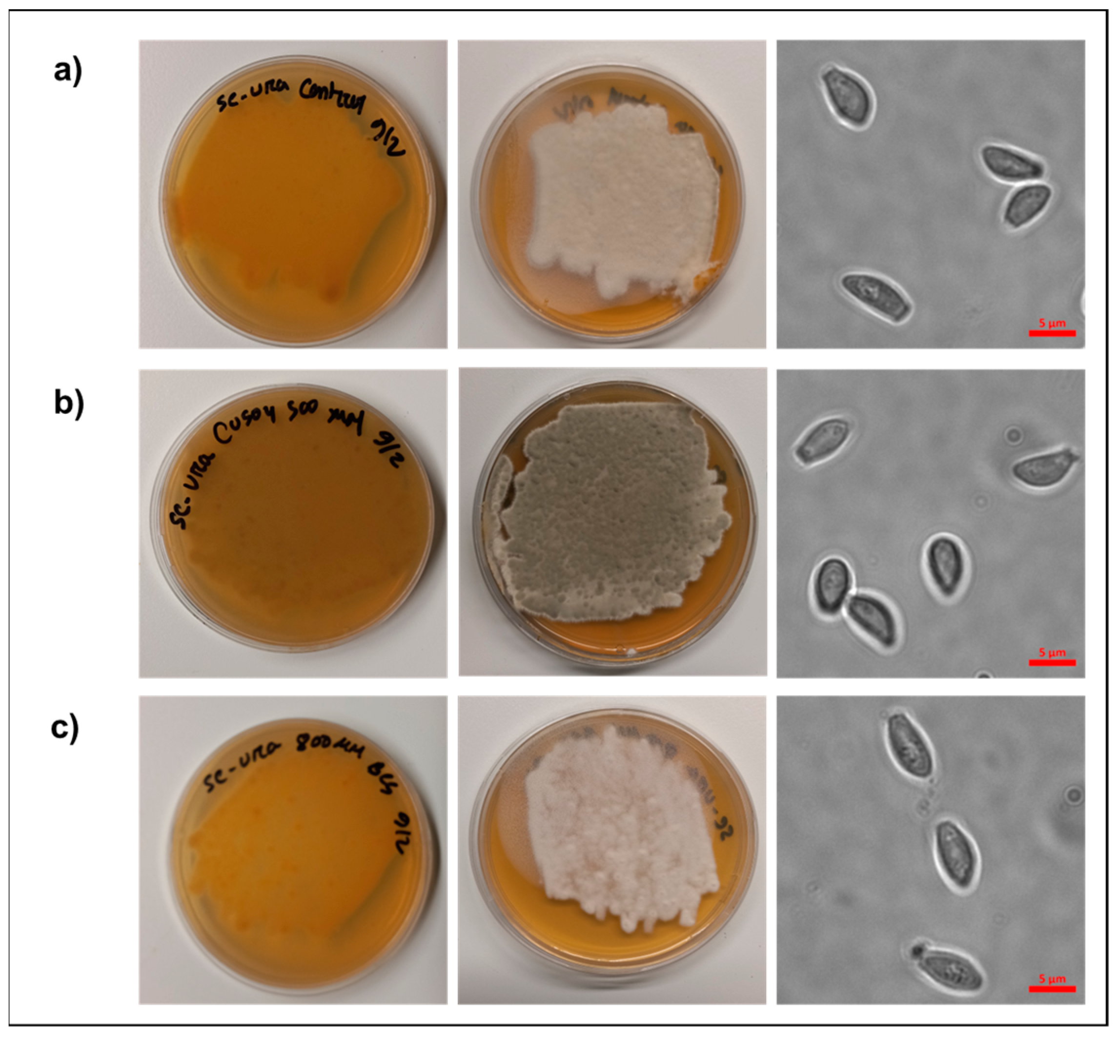
2.1. Media Preparation, Propagation, and Imaging of Pseudogymnoascus destructans

The Pseudogymnoascus destructans strain (MYA4855) was purchased from the American tissue culture collection (ATCC strain number 20631-21) and used for all experiments. The long-term propagation of P. destructans was carried out using solid yeast extract–peptone and glucose (YPD) plates at 15 °C and consisted of 10 g/L of yeast extract, 20 g/L of peptone, 20 g/L glucose, and 15 g/L of agar. Metal growth studies were performed using chemically defined growth plates (SC-Ura) which were made using Milli-Q water and 1.8 g/L of yeast nitrogen base, 5 g/L ammonium sulfate (NH4SO4), 20 g/L glucose, 100 mg/L histidine, 1.7 g/L of the SC-Ura-His amino acid supplement mixture (Sunrise Scientific, Knoxville, TN, USA), and 15 g/L agar. Before autoclaving one pellet (~200 mg) of NaOH was added to the SC-Ura media prior to autoclave sterilization. The addition of sterile Cu-sulfate and Bathocuproine sulfonic acid sodium salt (BCS; Acros Organics, Geel, Belgium) was added to the SC-Ura growth media using a 125 mM stock solution after autoclave sterilization at a temperature of approximately 50 °C to a final BCS concentration of 800 µM or 500 µM unless explicitly noted otherwise.
The inoculation of SC-Ura experimental growth plates was performed as follows: Pseudogymnoascus destructans conidia and mycelium were harvested from a working YPD plate, grown for two weeks at 15 °C by adding 2 mL of 1× TE buffer to the plate and gently rubbing the mycelium mass with an inoculation loop. The resulting cell suspension was transferred and passed through a sterilized pasture pipette containing a plug of Miracloth (EMD Millipore Corp, Burlington, MA, USA) to isolate P. destructans conidia. One hundred microliters of the resulting filtrate was used to inoculate experimental SC-Ura plates.
Imaging of harvested P. destructans was conducted as described above. Fungal spores were mounted with ProLong gold mounting media (Invitrogen, Waltham, MA, USA) and imaged on an inverted Zeiss Axio observer microscope with an Axiocam 820 camera (ZEISS, Oberkochen, Germany) under brightfield illumination.

2.2. Pseudogymnoascus destructans RNA Extraction and Isolation

Transcriptomic studies were performed on P. destructans fungi grown for 10 days at 15 °C on SC-Ura plates alone or supplemented with either the copper chelator BCS (800 µM) or CuSO4 (500 µM). Fungal cells, including conidia and mycelium, were suspended in 2 mL of TE buffer by gently rubbing the mycelium mass with a sterile inoculating loop. The resulting cell suspension was transferred to a 2 mL microcentrifuge tube and pelleted for 5 min at 16,000× g. The cells were washed twice with 1 mL of TE buffer. The resulting cell pellet was snap-frozen in dry ice and stored at −80 °C for later use. Total RNA was extracted from P. destructans cells using TRIZOL reagents using the manufacturer’s recommended procedure (ThermoFisher Scientific, Waltham, MA, USA). Briefly, 1 mL of TRIZOL reagent was added to P. destructans cells along with approximately 100 μL of 0.5 μM zirconium beads (ZB-05; Next. Advanced) and homogenized at 4 °C in a bullet blender (Speed 12 for 5 min). The resulting cell lysate was incubated on ice for 5 min. The cell lysates were then thoroughly mixed with 0.2 mL chloroform and incubated for 2 to 3 min before centrifugation at 12,000× g for 15 min. The upper aqueous layer was transferred to a new microcentrifuge tube, and 500 μL of isopropanol was added. After mixing, the solution was incubated on ice for 10 min and centrifuged at 12,000× g at 4 °C. The supernatant was discarded, and 1 mL of 75% ethanol was added, vortexed, and centrifuged at 7500× g for 5 min at 4 °C. The supernatant was discarded, and the white pellet was air-dried for 5–10 min. The RNA (pellet) was resuspended in 50 µL of 1× TE buffer and incubated for 15 min at 55 °C in a hot water bath. Finally, the concentration of the RNA was determined using nanodrop and analyzed using an Agilent bioanalyzer (Santa Clara, CA, USA). Then, RNA was stored at −80 °C. All RNA samples were sent to Innomics, Inc., (Sunnyvale, CA, USA) for the polyA mRNA enrichment, sequencing library construction, indexing, and RNA sequencing.

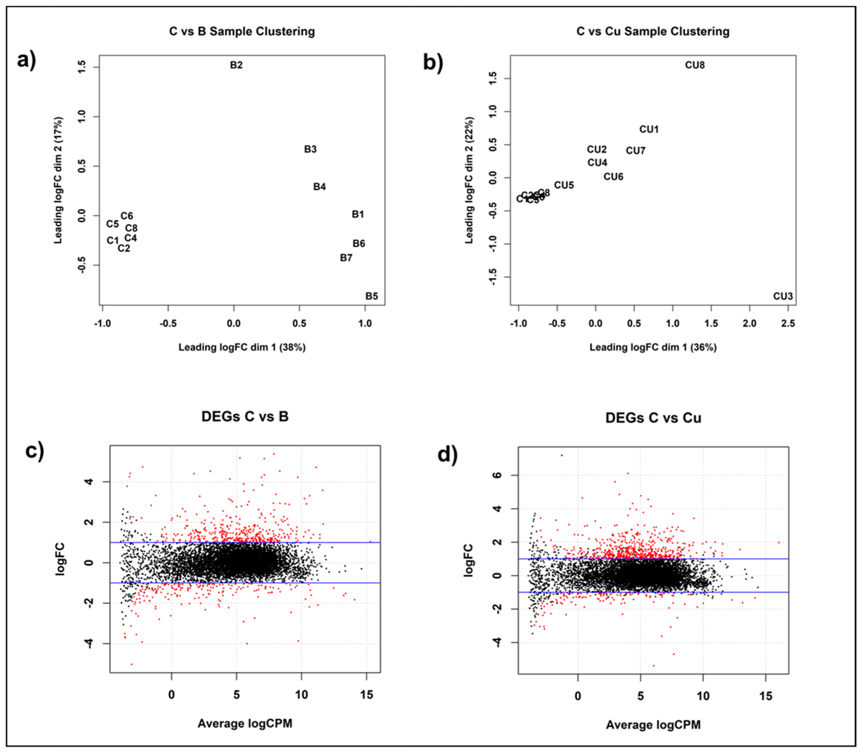
2.3. Differential Gene Expression Under Copper Stress

Sequencing reads were delivered by sequencing facility through a cloud server on Amazon Web Services (AWS). Sequencing reads were processed using ftastx_toolkit (https://www.hannonlab.org/resources/ accessed on 8 November 2023) to trim off any adapter sequences, low-quality base calls, and filter low-quality sequencing reads. Processed sequencing reads were mapped to the P. destructans reference genome (Accession number: GCF_001641265.1_ASM164126v1_genomic) using Tophat2 (version 2.0.13) [36]. Gene expression profiling was subsequently conducted using the featureCounts (subRead package v.1.4.6) [37] function of the subread package to quantify sequencing reads mapped to exons guided by a genome annotation file corresponding to the genome.
The edgeR package (version edgeR_4.0.16) available in Bioconductor was used to identify the total number of differentially expressed genes between biological conditions. Where differentially expressed genes (DEGs) were determined by |log2FC| > 1, FDR < 0.05 (File S1). Subsequential global analysis of DEGs meeting this criterion and a logCPM > 1 were retained for further analysis, ensuring a focus on robustly expressed candidates in the subsequent stages of investigation (File S1).
We used Venny 2.1 to construct intersections, logical relationships, and commonalities within treatment groups in comparison to control groups within P. destructans samples.

2.4. Gene Ontology (GO) and GO Enrichment Analyses

We extracted GO terms associated with biological processes for each DEG from JGI (https://mycocosm.jgi.doe.gov/Pseudest1/Pseudest1.home.html, accessed on 21 March 2024). For each GO term, we counted the number of DEGs that have the GO term, the number of DEGs that have the same particular GO term, the total number of genes that have the GO term, and the number of genes in the genome sharing the same particular GO term, and conducted hypergeometric test to identify over-represented GO terms (p < 0.05).

2.5. Identification of Pseudogymnoascus destructans Gene Homologs

The basic local alignment search tool (BLAST) at the NIH National Center of Biotechnology Information (https://blast.ncbi.nlm.nih.gov/Blast.cgi, accessed on 6 November 2024) was used to identify homologs for P. destructans proteins.

2.6. RNA Sequencing Datasets

All RNA sequencing datasets have been deposited into the NIH GEO database (https://www.ncbi.nlm.nih.gov/geo/, accessed on 29 April 2025) with the accession numbers GSM8964574-GSM8964596.

3. Results

3.1. Pseudogymnoascus destructans Growth Under Copper Stress

To characterize the ability of P. destructans to grow under extremes in Cu bioavailability in SC-Ura growth media, we screened the effect of added Cu sulfate and the copper chelator BCS on P. destructans growth. We tested varying concentrations of supplemental copper sulfate (i.e., 100–500 μM) to simulate Cu-overload growth conditions and added the copper chelator BCS (i.e., 0.2–1 mM) to simulate Cu-withholding growth conditions (Figure S1). We observed that P. destructans had a high tolerance to Cu, with no growth inhibition observed at concentrations up to 500 μM supplemental Cu-sulfate. The gray-green color of the P. destructans hyphal mass when grown under copper-overload conditions may indicate an increase in laccase-produced fungal melanin due to cellular copper homeostasis or a developmental regulator [38], whereas modest P. destructans growth inhibition can be observed with BCS at concentrations > 800 μM. Based on these experiments, we selected the 500 µM CuSO4 to stimulate Cu-overload conditions and 800 µM copper chelator BCS to stimulate Cu-withholding conditions for transcriptomic analysis (vide infra).
Fungi cultured under control and Cu-withholding stress conditions for 10 days had tan mycelium in contact with the growth plate and were off-white on the top of the growth plate (Figure 1a,c). On the other hand, under high copper conditions, P. destructans mycelium turned dark gray-green both on the top and bottom of the growth media plate (Figure 1b). Microscopic examination of P. destructans conidia revealed that copper stress growth conditions did not lead to noticeable changes in their characteristic asymmetric curved conidia morphology [29].

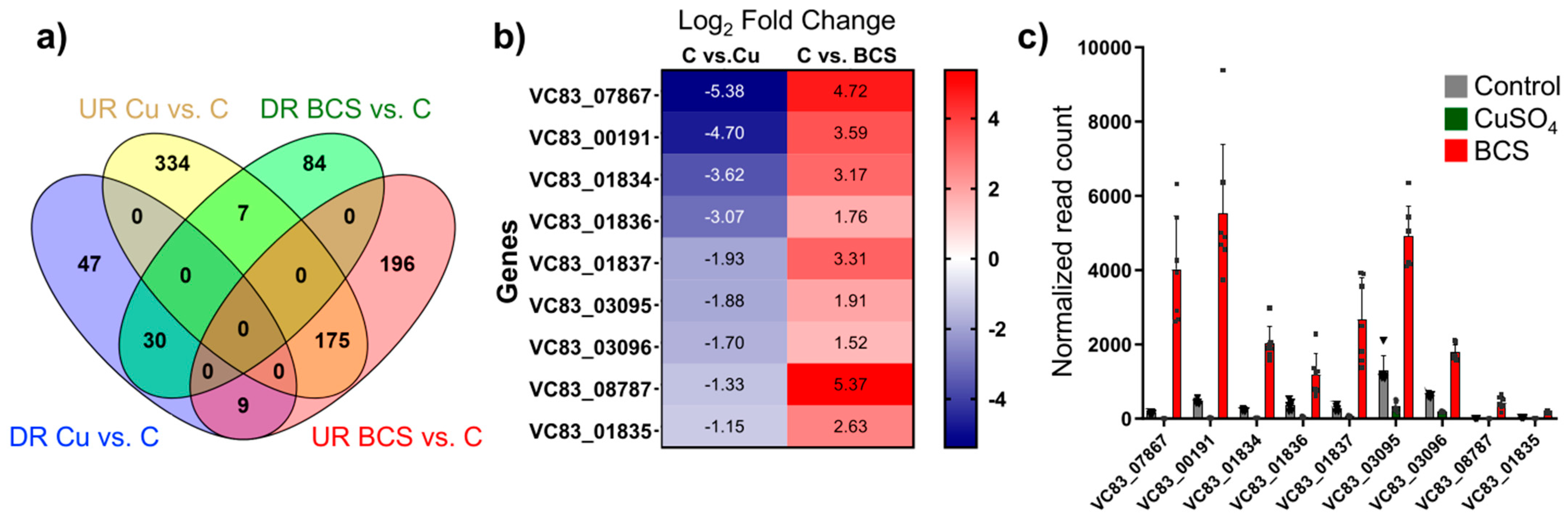
3.2. Differential Gene Expression in Response to Copper Stress

To gain insight into P. destructans copper cellular homeostasis and the pathways modulated by copper withholding and overload stress, we performed global gene expression analysis of mRNA transcripts of P. destructans conidiophores grown under control (i.e., SC-Ura), Cu-withholding (i.e., SC-Ura + 800 µM BCS) and Cu-overload (i.e., SC-Ura + 500 µM CuSO4) conditions. Using clustering by edgeR, we found similar patterns of P. destructans gene expression within each experimental group. Multi-dimensional scaling (MDS) profiles of Cu-withholding versus control and Cu-overload versus control treatments are displayed in Figure 2a,b, respectively. The sample distribution patterns suggest that the differences in gene expression profiles between the two experimental groups are due to variations in P. destructans environmental growth conditions.
Analysis of copper-withholding vs. control transcription profiles revealed a total of 583 genes that were significantly differentially expressed (p < 0.05; |log2FC| > 1) between experimental growth conditions (Figure 2c). Within this dataset, 399 genes were upregulated, and 184 genes were downregulated vs. the control samples. In contrast, analysis of copper-overload vs. control conditions revealed 667 differentially expressed genes (p < 0.05, |log2FC| > 1) that were significant between the two experimental groups (Figure 2d). A total of 542 genes were upregulated, and 125 genes were downregulated between copper-overload and control culture conditions. Further global analysis was performed on differentially expressed genes with p < 0.05, |log2FC| > 1, and total normalized read counts of >10 counts per million (CPM) under at least two growth conditions.
A Venn diagram was constructed to analyze common and unique differentially expressed genes (DEGs) upon Cu-withholding vs. Cu-overload growth conditions (Figure 3a). We observe that 334 genes are uniquely upregulated under copper overload (UR C vs. Cu), while 175 upregulated DEGs are also upregulated under copper-withholding growth conditions (UR C vs. BCS). A unique cluster of seven DEGs is upregulated under Cu-overload and downregulated under Cu-withholding conditions (Figure 3a and Table S1). We anticipate these seven genes may protect for Cu stress or be involved in the P. destructans Cu-detoxification pathways. These seven genes encode for a major facilitator family (MFS) transporter (VC83_08519), a plasma membrane-associated MARVEL Domain protein (VC83_03991), three predicted secreted proteins (VC83_05770; VC83_03107; VC83_06529), and two additional proteins with no clear protein homolog (VC83_07925; VC83_07926) (Table S1).
The cluster of nine shared DEGs that are downregulated upon Cu overload and upregulated under Cu-withholding stress expression bear hallmarks of known genes involved in trace metal ion-acquisition pathways (Figure 3b) [39,40,41]. This behavior suggests that Cu-bioavailability regulates gene transcript levels, and these genes play critical roles in the P. destructans Cu-stress response mechanism. Six of the nine genes in this cluster include homologs to the family of high-affinity copper transporters (CTRs) [42,43,44], FRE metalloreductase [45,46], and the extracellular Cu-scavenging protein C. neoformans CBI1/BIM1 [26,47]. The three genes VC83_01835, VC83_01836, and VC83_01837 encode for a Major Facilitator Superfamily (MSF) transporter, opine dehydrogenase, and S-adenomethionine-utilizing enzyme, respectively. The latter three genes are associated with the production and trafficking of opine metallophores commonly found in bacteria [40,48,49,50]. This cluster of nine genes is consistent with the notion that under low Cu-stress, P. destructans can utilize high-affinity Cu-trafficking proteins and secreted small molecules to acquire trace copper from the environment to fulfill its nutritional copper requirements.

3.3. Comparative Gene Ontology Analysis of P. destructans Under Cu-Withholding and Cu-Overload Conditions

To gain insight into the global function of P. destructans DEGs regulated by Cu-withholding and Cu-overload stress, gene ontology enrichment analysis vs. control growth conditions was performed and are displayed in Files S2 and S3, respectively. Copper-withholding stress leads to 24 biological pathways that are significantly enriched with p < 0.05 (Table 1). In contrast, high copper stress leads to 35 significant enriched biological pathways with p < 0.05 (Table 2). The two growth conditions share 11 enriched pathways. These shared pathways concentrate on the melanin metabolic processes, electron transport, carbohydrate transport, copper transport, and amino acid transport. While some of the gene ontology between the two copper stress conditions is shared, most are unique and are consistent with the notion that P. destructans adapts uniquely to chronic Cu-withholding and Cu-overload stress.
To further understand the P. destructans copper stress response, we performed a targeted comparison analysis of P. destructans DEGs against model fungus Saccharomyces cerevisiae and the three pathogenic fungi Candida albicans, Aspergillus niger, and Cryptococcus neoformans. Our comparison focused on genes associated with the metabolic processes including melanin metabolic processes (GO:0006582), superoxide metabolic processes (GO:0006801), response to oxidative stress (GO:0006979), carbohydrate transport (GO:0008643), copper ion transport (GO:0006825), and electron transport (GO:006118). Significant regulated genes associated with Cu-withholding stress and their predicted subcellular localization are shown in Table S2, while significant regulated genes associated with Cu-overload stress are displayed in Table S3. Our results indicate that P. destructans adapts similarly to other fungal species with more characterized copper stress responses.

3.4. Putative Virulence Factors Controlled by Copper Stress

Previous transcriptomic studies at active sites of P. destructans infections sites on WNS-positive bats by Field and coworkers identified candidate fungal virulence factors [34]. Cross-referencing their list of candidate fungal virulence factors, we find that several genes also respond to copper stress. We identify 15 genes that exhibited significant changes in transcript levels under low copper stress and 13 genes that exhibited significant changes in their transcripts under high-copper-stress conditions (Table 3). Specifically, eight genes (VC83_02553, VC83_01046, VC83_08187, VC83_06435, VC83_04094, VC83_06039, VC83_09074, and VC83_02181) exhibited elevated transcripts under both low and high Cu conditions. Conversely, seven genes (VC83_00970, VC83_01360, VC83_00191, VC83_07867, VC83_00261, VC83_05292, and VC83_01624) showed exclusive upregulation under Cu-withholding conditions. Additionally, two genes, VC83_09076 and VC83_06062, responded uniquely to high-copper-stress conditions. Notable examples of these genes include those involved in the heat shock response, such as VC83_02553 (Alpha-crystallin domain, HSP) and VC83_00970 (Heat shock protein 78, mitochondrial), as well as transporters like VC83_01360 (ZIP Zinc transporter), VC83_00191 (Ctr copper transporter family), VC83_04094 (ATX1; HMA; heavy-metal-associated domain) and VC83_07867 (a Bim1-like protein (BLP)). These findings suggest that P. destructans DEGs that respond to copper stress under laboratory conditions are relevant to the white nose syndrome (WNS) state on its native bat-host.

4. Discussion

Copper is an essential metal micronutrient needed to support eukaryotic life, yet it plays a dual role in fungal pathobiology, acting both as a scarce resource as well as a toxic agent at high concentrations [15,16]. In fungi, two classes of Cu-responsive transcription factors regulate the machinery needed for trace Cu acquisition and Cu detoxification to ensure efficient cellular propagation: the reciprocal pair of Mac1/Ace1 [12,14] transcription factors and single component Cuf1 [51,52] transcription factor. For fungal pathogens, extremes in Cu-bioavailability can be encountered over the course of the mammalian host infection [15], and thus, fungal pathogens must adapt to manage this dynamic metal resource within their host niche. Pseudogymnoascus destructans is a curious fungal pathogen with a unique evolutionary trajectory and is currently thought to be an ancient soil-dwelling plant pathogen that evolved to thrive on susceptible bat hosts. Recent transcriptomic studies by Field and coworkers suggest that trace micronutrient acquisition pathways are associated with bat-host colonization and identified several fungal genes involved in zinc, iron, and copper homeostasis as possible fungal virulence factors [34]. This study aims to describe the P. destructans transcriptional responses to low- and high-copper-stress growth conditions to gain insight into how P. destructans may adapt to changing copper levels at the WNS host–pathogen interface or cave-hibernacula soil niches.

4.1. Copper Stress Metabolic Adaptations

While gross changes in fungal spore morphology have been described in C. neoformans upon BCS-mediated Cu-withholding stress [53], we do not observe these changes in P. destructans spores, which have maintained their size and distinct curved conidia shape. However, transcriptional analysis of the most abundant 50 DEGs (Figure 4a,b) reveals significant modifications within the P. destructans cellular transcriptome, which are unique to copper-withholding and copper-overload stress.

4.2. P. destructans Adaptation Under Cu-Withholding Stress Conditions

The family of high-affinity Cu-transporters (CTRs) facilitates the movement of reduced Cu-ions across biological membranes under Cu-limiting growth conditions. While Ctr1 proteins can act independently to selectively import Cu ions from the extracellular environment, more recently, in C. neoformans, the extracellular scavenging protein, CBi1/BIM1, has been shown to assist in extracellular copper capture, both in in vitro and in vivo models, and has also been shown to participate in fungal cell wall integrity [26,47,54]. The P. destructans genome encodes for two Cu transporters, VC83_00191 (CTR1a) and VC83_04814 (CTR1b), as well as three BIM1-like proteins (BLPs) VC83_02818 (BLP1), VC83_03095 (BLP2), and VC83_07867 (BLP3) [55]. Copper-withholding stress leads to a significant 12.0- and 18.6-fold increase in CTR1a and CTR1b respective transcripts versus control growth conditions. The CTR1a isoform transcripts are approximately four times more abundant in the CTR1b isoform, suggesting that both isoforms may be involved in trace Cu-acquisition. These observations are consistent with previous work by our group, which identified P. destructans CTR1a as a functional high-affinity Cu-transporter that is transcriptionally activated and expressed under Cu-limited growth conditions [55]. We also observe a significant 3.8 and 26.3-fold increase in transcript levels for two of three BLP isoforms, VC83_03095 (BLP2) and VC83_07867 (BLP3), respectively, under copper-withholding growth conditions. However, minimal alterations in the BLP1 isoform (i.e., VC83_02818) transcript levels are observed under the varied Cu-stress growth conditions tested in our study. These observations are consistent with the notion that P. destructans may employ multiple BLP-isoforms to scavenge Cu-ions from is environment.
The three predicted Fre-metalloreductases enzymes (MRed) VC83_03096, VC83_08787, and VC83_01834 are also significantly upregulated upon Cu-withholding stress, which may facilitate the Cu2+ to Cu+ reduction chemistry at the cell surface for the efficient transport of Cu+ ions through CTR channels. In Baker’s yeast, Saccharomyces cerevisiae, FRE1 and FRE2 are the primary genes that facilitate reductive Fe- and Cu-import [56,57]. FRE homologs in other pathogenic fungi such as Fre7 in Candida albicans [58] and Fre4/Fre7 in Cryptococcus neoformans [59] can respond to Cu-withholding stress. An overview of the putative P. destructans CTR/BLP/MRed enzymes participating in high-affinity Cu-import is overviewed in Figure 5. Together, our data are consistent with the notion that the P. destructans copper-withholding stress response involves the transcriptional regulation of both CTR1 isoforms as well as two CBi1/BIM1-homologs to scavenge trace copper ions from their extracellular environment.
  • Identification of a putative novel fungal high-affinity metal acquisition pathway in P. destructans
Pathogenic microbes can secrete metal-binding small molecules to capture essential metal micronutrients. The most well studied are the Fe-binding siderophores [60], although Cu-binding calkophores [61] and Zn-binding zincophores [62,63,64,65] have been reported. We identify a cluster of five genes VC83_01834VC83_01838, which show differential gene regulation displaying reciprocal transcriptional regulation in response to copper stress growth conditions (Figure 3 and Figure S2a). We identify VC83_01834VC83_01838 as the Copper-Responsive gene Cluster (CRC) (Figure 5). The four genes VC83_01834, VC83_01835, VC83_01836, and VC83_01837 segregate in a cluster of DEGs, which are significantly upregulated under Cu-withholding stress and repressed under Cu-overload conditions (Figure 3a,b). We have included the gene VC83_01838, which encodes for a cytochrome P450 monooxygenase enzyme, in the CRC gene cluster because it remains a significant DEG under both Cu-withholding and Cu-overload stress.
The genes encoded in the CRC may participate in the production and trafficking of a calkophore or metallophore small molecule. To our knowledge, P. destructans cannot produce isocyanide Cu-binding calkophores that other fungi can secrete due to the lack of ISP encoded in its genome [28]. However, the P. destructans genes VC83_01835, VC83_01836, and VC83_01837 encode for homologous proteins found in the bacterial operons responsible for the production and trafficking of the opine-metallophores staphylopine [50] and pseudopaline [49], respectively. The presence of the metallo-reductase (VC83_01834) and cytochrome P450 monooxygenase genes (VC83_01838) in this gene cluster is less clear, but may be required for cell surface reduction chemistry needed for metal uptake or the maturation of the small-molecule metallophore. Minor modifications of the opine-metallophore core structure via diversification of amino acids and metabolites are known to generate species-specific metallophore small-molecules [66]. Secreted P. destructans-derived small molecules, including riboflavin [67] and triacetlyfarcine C [68], have been observed at the host–pathogen interface on WNS-positive bat samples. It is possible that P. destructans may have evolved a novel Cu-trafficking pathway involving a small molecule as an alternative or redundant pathway to the resource-intensive protein mediate BLP/CTR pathway (vide supra) [26]. Future studies that focus on the transcriptional regulation of the CRC to other trace metals (i.e., Fe and Zn), as well as validating CRC protein levels under Cu-withholding stress, will be needed to help elucidate its role in P. destructans metal-cell biology and its possible role in virulence.
  • Remodeling of P. destructans superoxide dismutase (SOD) enzymes under Cu-withholding stress
The intracellular bimetallic Cu/Zn SOD can act as a cytosolic copper buffer to facilitate extremes in Cu-homeostasis while protecting against cytoplasm superoxide damage [69]. In the absence of Cu, some animals infecting pathogenic fungi can employ dedicated cytosolic targeting Mn-SODs (i.e., the Mn-SOD3 in Candida albicans) or generate alternative cytosolic targeting Mn-SOD2 transcripts to maintain cytosolic antioxidant activity when cellular Cu-levels are sparse [23,27]. The P. destructans genome encodes for two Cu-containing SODs and two Mn-SOD enzymes. The transcriptional regulation of the four SOD enzymes under Cu-stress growth conditions is shown in Figure S2b. The two Cu-containing SODs variants include the bimetallic cytosolic Cu/Zn SOD (VC83_07077) and a secreted GPI-anchored Cu-only SOD (VC83_08495), whereas the two Mn-SOD isoforms found in the P. destructans genome encode for a mitochondrial-targeting Mn-SOD2 (VC83_07362) variant and cytoplasmic Mn-SOD3 (VC83_02616) (see Table S2 for homology and File S4 for SOD3 localization assignment). In our study, we observe a 2.1-fold reduction in Cu/Zn SOD (VC83_07077) transcripts and a 2.3-fold increase in Mn-SOD3 (VC83_02616) transcripts. We anticipate that this reciprocal SOD transcriptional regulation is needed to divert cellular copper pools to another Cu-enzyme, such as Cytochrome c oxidase or FET3 [70], to maintain efficient cellular respiration or Fe-acquisition. However, no significant change in transcript number or isoform for the mitochondrial localizing Mn-SOD2 (VC83_07362) transcript levels is observed under the conditions tested. The redistribution of intracellular SOD transcripts upon Cu-restrictive growth conditions may be advantageous, allowing P. destructans to maintain cellular redox homeostasis when propagating in Cu-poor soils or allowing it to overcome the metal restrictive host defense mechanisms.
To our surprise, both copper-withholding and copper-overload stress conditions led to significant increases in transcript levels for the extracellular Cu-only SOD (VC83_08495) when compared to control growth conditions, suggesting that this extracellular enzyme is important for adapting to both copper-withholding and copper-overload stress. Cu-only SOD isoform transcripts in other filamentous fungi, such as C. albicans, have been shown to have distinct expression profiles that are associated with their host niche, such as biofilms for SOD6 [71], macrophages for SOD5 [72], or Fe-poor growth conditions for SOD4 [73]. Together, the transcriptional activation patterns of the four superoxide dismutase enzymes in P. destructans appear to mimic that of the pathogenic fungi C. albicans, which adapts its SOD transcriptional levels to cope with bioavailable copper levels and stress [23]. Future studies will be needed to determine if these transcriptional observations are also representative at the SOD protein and activity levels.
  • Notable proteins involved in metal ion homeostasis and Cu-withholding stress adaptations
A putative zinc transporter VC83_01360, a ZRT1 homolog, is also upregulated under Cu-withholding stress. Previous transitional studies have associated VC83_01360 with fungal virulence on infected bat-hosts [34]. In vitro experiments on Candida albicans with the biologically relevant metal scavenger calprotectin in serum-based growth media have shown a decrease in cellular copper levels and have been shown to induce ZRT1 and PRA1 transcripts while preserving cellular Zn levels [15]. This may suggest that Cu-withholding stress may induce Zn-acquisition machinery in P. destructans. However, future systematic studies in P. destructans are still needed to fully elucidate the regulatory crosstalk between Cu and other trace metal ion micronutrients.
Several other biological processes are impacted by Cu-withholding stress, with some genes implemented in P. destructans virulence on bat-hosts. Biological processes associated with electron transport are highly representative, with 15 genes identified as being altered by Cu-withholding stress (Table 1; File S2). Alterations in electron transfer processes may be anticipated due to copper’s role in electron transport protein active sites and the reductive mechanisms for high-affinity Cu-import. The dihydroxynaphthalene (DHN) melanin pathway is one unexpected pathway that was impacted upon both Cu-withholding and Cu-overload stress. Specifically, secreted scytalone dehydratase gene (VC83_07909) transcripts are elevated 35-fold and 6-fold under Cu-withholding and Cu-overload conditions, respectively. The production of melanin under Cu-stress may be a defensive mechanism to protect against the environmental stress of added Cu or nutrient starvation. Melanin produced by the DHN pathway has been shown to protect Aspergillus fumigatus against macrophage and neutrophil killing by interfering with phagolysosome maturation [74]. A similar protective role for DHN was recently described for P. destructans with an immortalized bat keratinocyte model system [75]. We anticipate that the gene VC83_07909 may be involved in DHN melanin production and participate in both copper nutrient stress resistance and bat-host colonization.

4.3. P. destructans Response to Copper Overload Conditions

In Saccharomyces cerevisiae, Ace1 drives the expression of Cu/Zn SOD [76] and the metallothionine-related genes CSR5 [77] and CUP1 [78] which can bind and buffer excess copper ions. Unfortunately, no metallothionine or metallothionine-related genes are predicted for P. destructans within the JGI annotated databank, making it difficult to draw conclusions about this classical Cu-overload biomarker. However, for Cu/Zn SOD (i.e., VC83_07077), we do not observe any significant increase in transcript number upon the Cu-overload growth conditions tested in our study. This may indicate that the additional Cu concentration (i.e., 500 μM CuSO4) used may not be suitable for eliciting the activation of an ACE1 homolog in P. destructans, despite yielding a distinct change in transcripts as indicated in multi-dimensional scaling analysis (Figure 2b). Induction of CUP1/CRS5 in S. cerevisiae can be activated in minutes upon Cu stress [14,77]. Thus, our methodology utilizing chronic exposure to Cu may not be suitable for capturing this Cu-adaptation response.
Significant increases in gene transcripts associated with DNA damage and replication stress upon chronic exposure to elevated levels of copper are observed in our Cu-overload datasets. Notably, we observe an increase in transcripts associated with DNA replication, including the genes for the catalytic domain of DNA Polymerase Pol1 and Pol2, VC83_05453 and VC83_04573, respectively. The induction of increased transcripts for VC83_04943, a subunit of a replication-pausing checkpoint complex CSM3, suggests that Cu-overloaded cells are experiencing replication stress and assistance in maintaining DNA replication in integrity [79,80]. Additionally, the increased transcripts for VC83_08538, which encodes for the ribonucleotide reductase large subunit (RRM1), suggest an active need for dNTP substrates for DNA replication or repair. Ribonucleotide reductase large subunit transcripts have been shown previously to be induced upon exposure to DNA-damaging agents [81]. Our data suggest that P. destructans growth under high-copper conditions may significantly impact genomic stability and DNA replication fidelity.

4.4. Putative P. destructans Secreted Proteases and Virulence Factors Responsive to Cu Stress

What other putative P. destructans virulence factors or genes associated with WNS are responsive to Cu-stress? As P. destructans must scavenge all its essential micronutrients from its infected host, our systemic in vitro study provided opportunities to identify genes associated with overcoming Cu-withholding or -overload stress within bat-host infection conditions. We identify a collection of six heat shock proteins (HSPs) or chaperones/co-chaperones that respond to Cu stress and are associated with the WNS state. HSPs in Candida [82] and Histoplasma [83] species play essential roles in maintaining protein quality control, controlling cellular morphology and drug resistance [84]. Therefore, it is not surprising that infection and nutritional stress lead to elevated transcripts of heat shock and chaperone protein expression.
Early studies on P. destructans implemented the secreted collagen-degrading serine protease, Destructin-1 (i.e., VC83_06062), as a potential virulence factor to promote fungal tissue invasion [34,85]. Two additional secreted homologs to Destructin-1, namely Destructin-2 (i.e., VC83_04892) and Destructin-3 (i.e., VC83_09074), were identified as possible proteases of interest [85]. Our results show that both Cu-withholding and Cu-overload stress led to elevated transcripts vs. control for Destructin-1, Destructin-3, and a third serine peptidase VC83_02181. This suggests that these secreted factors may respond to general environmental stress. Similarly, elevated transcripts for an additional secreted protease VC83_02181 also observed upon both Cu-withholding and Cu-overload stress. However, total transcript levels for VC83_02181 are much less abundant than those for the family of Destructin homologs. This may indicate that VC83_02181 may play a modest role in the P. destructans stress response. Together, our data demonstrate that copper stress can impact the transcription levels of secreted protease and other putative virulence factors associated with the WNS state.

5. Conclusions

This report characterizes P. destructans’ transcriptional response to chronic copper-withholding and copper-overload conditions. Copper-withholding stress leads to the induction of two characteristic pathways that may participate in high-affinity Cu trafficking. The first pathway includes the ubiquitous family of high-affinity Cu transporters and the more recently described Cbi1/Bim1 family of Cu-scavenging proteins. Our data support that both Ctr1a and Ctr1b isoforms, VC83_00191 and VC83_048184, respectively, as well as the two Bim1-like protein (BLP)-encoding genes VC83_03095 (BLP2) and VC83_07867 (BLP3), are significantly induced by Cu-withholding stress. Pseudogymnoascus destructans may also be capable of utilizing a second high-affinity Cu-trafficking pathway involving secreted metallophore small molecules which are encoded by the genes VC83_01834VC83_01838, which we designated the Cu-Responsive gene Cluster (CRC). Future research efforts should be directed at validating the expression levels of CRC genes and identifying metal-binding small molecules in P. destructans growth media.
Additionally, our study found that P. destructans is resilient to high levels of extracellular copper in its growth media. While we do not observe gross differences in morphology, our data demonstrate that elevated copper leads to elevated transcripts for genes associated with maintaining genome stability and DNA-replication fidelity.
While this study describes the first systemic investigation into the transcriptional response of P. destructans to Cu-withholding and Cu-overload stress, it is limited in terms of the functional and metabolic conclusions that can be drawn from transcriptomic analysis alone. As genetic manipulation tools for P. destructans become more widely assessable, there will be opportunities to validate and further interrogate the copper cell biology of P. destructans to better understand the role of copper in WNS propagation with gene knockout fungal strains.

Supplementary Materials

The following supporting information can be downloaded at https://www.mdpi.com/article/10.3390/jof11050372/s1, Figure S1: Growth of Pseudogymnoasus destuctans on synthetic (Sc-Ura) plates supplemented with CuSO4 or BCS. Figure S2: Normalized read counts for Pseudogymnoasus destuctans CRC and SOD genes. Table S1: Functional Annotation and Expression Profile of 7 Gene Cluster Under Copper Stress. Table S2: Comparative blast analysis of selected significantly regulated genes by Cu-withholding stress (BCS) and their fungal homologs. Table S3: Comparative blast analysis of selected significantly regulated genes by Cu-overload stress and their fungal homologs. File S1: Normalized read count table and list of DEG upon Cu stress. File S2: Gene ontology enrichment analysis of genes regulated by Cu-withholding stress. File S3: Gene ontology enrichment analysis of genes regulated by Cu-overload stress. File S4: Localization assignment for Pseudogymnoasus destuctans Mn SOD enzymes.

Author Contributions

Conceptualization, M.R.M., Y.L. and R.L.P.; methodology, S.A., Y.L. and R.L.P.; software, S.A. and Y.L.; validation, S.A., M.R.M., Y.L. and R.L.P.; formal analysis, S.A., Y.L. and R.L.P.; data curation, S.A., Y.L. and R.L.P.; writing—original draft preparation, S.A., Y.L. and R.L.P.; writing—review and editing, S.A., M.R.M., Y.L. and R.L.P.; supervision, Y.L. and R.L.P.; funding acquisition, R.L.P. All authors have read and agreed to the published version of the manuscript.

Funding

This research was supported by grants from the National Institutes of Health (Grant Number R16GM14716; S10GM154304).

Institutional Review Board Statement

Not applicable.

Informed Consent Statement

Not applicable.

Data Availability Statement

Raw data supporting the conclusions of this article will be made available by the authors on request.

Acknowledgments

We would like to thank Daniella Gonzalez for providing technical support throughout this project. S.A., M.R.M., and R.L.P. would like to thank the Texas State University for providing financial support.

Conflicts of Interest

The authors declare no conflicts of interest. The NIH had no role in the design of the study; in the collection, analyses, or interpretation of data; in the writing of the manuscript; or in the decision to publish the results.

Abbreviations

The following abbreviations are used in this manuscript:
PdPseudogymnoascus destructans
BCSBathocuproinedisulfonic acid
GOGene ontology
FDRFalse discovery rate
DEGDifferentially Expressed Gene
RNARibonucleic acid
SODSuperoxide dismutase
CTRCopper transporter
BLPBim1-like protein
CRCCopper-Responsive Gene Cluster
MFSMajor facilitator superfamily
HSPHeat shock protein
SC-UraSynthetic Complete Medium minus Uracil
CPMCounts Per Million
YPDYeast Extract Peptone Dextrose Medium

References

  1. Festa, R.A.; Thiele, D.J. Copper: An essential metal in biology. Curr. Biol. 2011, 21, R877–R883. [Google Scholar] [CrossRef] [PubMed]
  2. Tsang, T.; Davis, C.I.; Brady, D.C. Copper biology. Curr. Biol. 2021, 31, R421–R427. [Google Scholar] [CrossRef] [PubMed]
  3. Solomon, E.I.; Heppner, D.E.; Johnston, E.M.; Ginsbach, J.W.; Cirera, J.; Qayyum, M.; Kieber-Emmons, M.T.; Kjaergaard, C.H.; Hadt, R.G.; Tian, L. Copper active sites in biology. Chem. Rev. 2014, 114, 3659–3853. [Google Scholar] [CrossRef]
  4. Liu, J.; Chakraborty, S.; Hosseinzadeh, P.; Yu, Y.; Tian, S.L.; Petrik, I.; Bhagi, A.; Lu, Y. Metalloproteins Containing Cytochrome, Iron-Sulfur, or Copper Redox Centers. Chem. Rev. 2014, 114, 4366–4469. [Google Scholar] [CrossRef] [PubMed]
  5. Sheng, Y.; Abreu, I.A.; Cabelli, D.E.; Maroney, M.J.; Miller, A.F.; Teixeira, M.; Valentine, J.S. Superoxide dismutases and superoxide reductases. Chem. Rev. 2014, 114, 3854–3918. [Google Scholar] [CrossRef]
  6. Broxton, C.N.; Culotta, V.C. SOD Enzymes and Microbial Pathogens: Surviving the Oxidative Storm of Infection. PLoS Pathog. 2016, 12, 8–13. [Google Scholar] [CrossRef]
  7. Koo, C.W.; Rosenzweig, A.C. Biochemistry of aerobic biological methane oxidation. Chem. Soc. Rev. 2021, 50, 3424–3436. [Google Scholar] [CrossRef]
  8. Klinman, J.P. The copper-enzyme family of dopamine beta-monooxygenase and peptidylglycine alpha-hydroxylating monooxygenase: Resolving the chemical pathway for substrate hydroxylation. J. Biol. Chem. 2006, 281, 3013–3016. [Google Scholar] [CrossRef]
  9. Taylor, A.B.; Stoj, C.S.; Ziegler, L.; Kosman, D.J.; Hart, P.J. The copper-iron connection in biology: Structure of the metallo-oxidase Fet3p. Proc. Natl. Acad. Sci. USA 2005, 102, 15459–15464. [Google Scholar] [CrossRef]
  10. Chen, L.; Min, J.; Wang, F. Copper homeostasis and cuproptosis in health and disease. Signal Transduct. Target. Ther. 2022, 7, 378. [Google Scholar] [CrossRef]
  11. Tsvetkov, P.; Coy, S.; Petrova, B.; Dreishpoon, M.; Verma, A.; Abdusamad, M.; Rossen, J.; Joesch-Cohen, L.; Humeidi, R.; Spangler, R.D.; et al. Copper induces cell death by targeting lipoylated TCA cycle proteins. Science 2022, 375, 1254–1261. [Google Scholar] [CrossRef] [PubMed]
  12. Borghouts, C.; Osiewacz, H.D. GRISEA, a copper-modulated transcription factor from involved in senescence and morphogenesis, is an ortholog of MAC1 in Saccharomyces cerevisiae. Mol. Gen. Genet. 1998, 260, 492–502. [Google Scholar] [CrossRef]
  13. Labbé, S.; Zhu, Z.; Thiele, D.J. Copper-specific transcriptional repression of yeast genes encoding critical components in the copper transport pathway. J. Biol. Chem. 1997, 272, 15951–15958. [Google Scholar] [CrossRef]
  14. Thiele, D.J. Ace1 Regulates Expression of the Saccharomyces-Cerevisiae Metallothionein Gene. Mol. Cell. Biol. 1988, 8, 2745–2752. [Google Scholar] [CrossRef] [PubMed]
  15. Besold, A.N.; Culbertson, E.M.; Culotta, V.C. The Yin and Yang of copper during infection. J. Biol. Inorg. Chem. 2016, 21, 137–144. [Google Scholar] [CrossRef]
  16. García-Santamarina, S.; Thiele, D.J. Copper at the fungal pathogen-host axis. J. Biol. Chem. 2015, 290, 18945–18953. [Google Scholar] [CrossRef]
  17. Rosen, T.; Wang, K.K.A.; Nolan, E.M. Metal sequestration by S100 proteins in chemically diverse environments. Trends Microbiol. 2022, 30, 654–664. [Google Scholar] [CrossRef]
  18. Zackular, J.P.; Chazin, W.J.; Skaar, E.P. Nutritional immunity: S100 proteins at the host-pathogen interface. J. Biol. Chem. 2015, 290, 18991–18998. [Google Scholar] [CrossRef] [PubMed]
  19. Murdoch, C.C.; Skaar, E.P. Nutritional immunity: The battle for nutrient metals at the host-pathogen interface. Nat. Rev. Microbiol. 2022, 20, 657–670. [Google Scholar] [CrossRef]
  20. White, C.; Lee, J.; Kambe, T.; Fritsche, K.; Petris, M.J. A Role for the ATP7A Copper-transporting ATPase in Macrophage Bactericidal Activity. J. Biol. Chem. 2009, 284, 33949–33956. [Google Scholar] [CrossRef]
  21. Bleackley, M.R.; Macgillivray, R.T. Transition metal homeostasis: From yeast to human disease. Biometals 2011, 24, 785–809. [Google Scholar] [CrossRef] [PubMed]
  22. Nevitt, T.; Öhrvik, H.; Thiele, D.J. Charting the travels of copper in eukaryotes from yeast to mammals. Biochim. Biophys. Acta (BBA)—Mol. Cell Res. 2012, 1823, 1580–1593. [Google Scholar] [CrossRef]
  23. Li, C.X.; Gleason, J.E.; Zhang, S.X.; Bruno, V.M.; Cormack, B.P.; Culotta, V.C. Candida albicans adapts to host copper during infection by swapping metal cofactors for superoxide dismutase. Proc. Natl. Acad. Sci. USA 2015, 112, E5336–E5342. [Google Scholar] [CrossRef]
  24. Culbertson, E.M.; Bruno, V.M.; Cormack, B.P.; Culotta, V.C. Expanded role of the Cu-sensing transcription factor Mac1p in Candida albicans. Mol. Microbiol. 2020, 114, 1006–1018. [Google Scholar] [CrossRef]
  25. Raffa, N.; Won, T.H.; Sukowaty, A.; Candor, K.; Cui, C.S.; Halder, S.; Dai, M.J.; Landero-Figueroa, J.A.; Schroeder, F.C.; Keller, N.P. Dual-purpose isocyanides produced by contribute to cellular copper sufficiency and exhibit antimicrobial activity. Proc. Natl. Acad. Sci. USA 2021, 118, e2015224118. [Google Scholar] [CrossRef] [PubMed]
  26. Garcia-Santamarina, S.; Probst, C.; Festa, R.A.; Ding, C.; Smith, A.D.; Conklin, S.E.; Brander, S.; Kinch, L.N.; Grishin, N.V.; Franz, K.J.; et al. A lytic polysaccharide monooxygenase-like protein functions in fungal copper import and meningitis. Nat. Chem. Biol. 2020, 16, 337–344. [Google Scholar] [CrossRef] [PubMed]
  27. Smith, A.D.; Garcia-Santamarina, S.; Ralle, M.; Loiselle, D.R.; Haystead, T.A.; Thiele, D.J. Transcription factor-driven alternative localization of Cryptococcus neoformans superoxide dismutase. J. Biol. Chem. 2021, 296, 100391. [Google Scholar] [CrossRef]
  28. Nickles, G.R.; Oestereicher, B.; Keller, N.P.; Drott, M.T. Mining for a new class of fungal natural products: The evolution, diversity, and distribution of isocyanide synthase biosynthetic gene clusters. Nucleic Acids Res. 2023, 51, 7220–7235. [Google Scholar] [CrossRef]
  29. Gargas, A.; Trest, M.T.; Christensen, M.; Volk, T.J.; Blehert, D.S. Geomyces destructans sp. nov. associated with bat white-nose syndrome. Mycotaxon 2009, 108, 147–154. [Google Scholar] [CrossRef]
  30. Meteyer, C.U.; Buckles, E.L.; Blehert, D.S.; Hicks, A.C.; Green, D.E.; Shearn-Bochsler, V.; Thomas, N.J.; Gargas, A.; Behr, M.J. Histopathologic criteria to confirm white-nose syndrome in bats. J. Vet. Diagn. Investig. 2009, 21, 411–414. [Google Scholar] [CrossRef]
  31. Meteyer, C.U.; Barber, D.; Mandl, J.N. Pathology in euthermic bats with white nose syndrome suggests a natural manifestation of immune reconstitution inflammatory syndrome. Virulence 2012, 3, 583–588. [Google Scholar] [CrossRef] [PubMed]
  32. Meteyer, C.U.; Dutheil, J.Y.; Keel, M.K.; Boyles, J.G.; Stukenbrock, E.H. Plant pathogens provide clues to the potential origin of bat white-nose syndrome. Virulence 2022, 13, 1020–1031. [Google Scholar] [CrossRef] [PubMed]
  33. Field, K.A.; Johnson, J.S.; Lilley, T.M.; Reeder, S.M.; Rogers, E.J.; Behr, M.J.; Reeder, D.A.M. The White-Nose Syndrome Transcriptome: Activation of Anti-fungal Host Responses in Wing Tissue of Hibernating Little Brown Myotis. PLoS Pathog. 2015, 11, e1005168. [Google Scholar] [CrossRef]
  34. Reeder, S.M.; Palmer, J.M.; Prokkola, J.M.; Lilley, T.M.; Reeder, D.A.M.; Field, K.A. Pseudogymnoascus destructans transcriptome changes during white-nose syndrome infections. Virulence 2017, 8, 1695–1707. [Google Scholar] [CrossRef] [PubMed]
  35. Lilley, T.M.; Prokkola, J.M.; Johnson, J.S.; Rogers, E.J.; Gronsky, S.; Kurta, A.; Reeder, D.M.; Field, K.A. Immune responses in hibernating little brown myotis (Myotis lucifugus) with white-nose syndrome. Proc. R. Soc. B 2017, 284, 20162232. [Google Scholar] [CrossRef]
  36. Kim, D.; Pertea, G.; Trapnell, C.; Pimentel, H.; Kelley, R.; Salzberg, S.L. TopHat2: Accurate alignment of transcriptomes in the presence of insertions, deletions and gene fusions. Genome Biol. 2013, 14, R36. [Google Scholar] [CrossRef]
  37. Liao, Y.; Smyth, G.K.; Shi, W. FeatureCounts: An efficient general purpose program for assigning sequence reads to genomic features. Bioinformatics 2014, 30, 923–930. [Google Scholar] [CrossRef]
  38. Upadhyay, S.; Torres, G.; Lin, X.R. Laccases Involved in 1,8-Dihydroxynaphthalene Melanin Biosynthesis in Aspergillus fumigatus Are Regulated by Developmental Factors and Copper Homeostasis. Eukaryot. Cell 2013, 12, 1641–1652. [Google Scholar] [CrossRef]
  39. Smith, A.D.; Logeman, B.L.; Thiele, D.J. Copper Acquisition and Utilization in Fungi. Annu. Rev. Microbiol. 2017, 71, 597–623. [Google Scholar] [CrossRef]
  40. Laffont, C.; Arnoux, P. The ancient roots of nicotianamine: Diversity, role, regulation and evolution of nicotianamine-like metallophores. Metallomics 2020, 12, 1480–1493. [Google Scholar] [CrossRef]
  41. Cain, T.J.; Smith, A.T. Ferric iron reductases and their contribution to unicellular ferrous iron uptake. J. Inorg. Biochem. 2021, 218, 111407. [Google Scholar] [CrossRef] [PubMed]
  42. Dancis, A.; Yuan, D.S.; Haile, D.; Askwith, C.; Eide, D.; Moehle, C.; Kaplan, J.; Klausner, R.D. Molecular characterization of a copper transport protein in S. cerevisiae: An unexpected role for copper in iron transport. Cell 1994, 76, 393–402. [Google Scholar] [CrossRef]
  43. Marvin, M.E.; Williams, P.H.; Cashmore, A.M. The Candida albicans CTR1 gene encodes a functional copper transporter. Microbiology 2003, 149, 1461–1474. [Google Scholar] [CrossRef] [PubMed]
  44. Pena, M.M.O.; Puig, S.; Thiele, D.J. Characterization of the Saccharomyces cerevisiae high affinity copper transporter Ctr3. J. Biol. Chem. 2000, 275, 33244–33251. [Google Scholar] [CrossRef] [PubMed]
  45. Georgatsou, E.; Alexandraki, D. Regulated expression of the Saccharomyces cerevisiae Fre1p/Fre2p Fe/Cu reductase related genes. Yeast 1999, 15, 573–584. [Google Scholar] [CrossRef]
  46. Dancis, A.; Roman, D.G.; Anderson, G.J.; Hinnebusch, A.G.; Klausner, R.D. Ferric reductase of Saccharomyces cerevisiae: Molecular characterization, role in iron uptake, and transcriptional control by iron. Proc. Natl. Acad. Sci. USA 1992, 89, 3869–3873. [Google Scholar] [CrossRef]
  47. Probst, C.; Garcia-Santamarina, S.; Brooks, J.T.; Van der Kloet, I.; Baars, O.; Ralle, M.; Thiele, D.J.; Alspaugh, J.A. Interactions between copper homeostasis and the fungal cell wall affect copper stress resistance. PLoS Pathog. 2022, 18, e1010195. [Google Scholar] [CrossRef]
  48. McFarlane, J.S.; Davis, C.L.; Lamb, A.L. Staphylopine, pseudopaline, and yersinopine dehydrogenases: A structural and kinetic analysis of a new functional class of opine dehydrogenase. J. Biol. Chem. 2018, 293, 8009–8019. [Google Scholar] [CrossRef]
  49. McFarlane, J.S.; Lamb, A.L. Biosynthesis of an Opine Metallophore by Pseudomonas aeruginosa. Biochemistry 2017, 56, 5967–5971. [Google Scholar] [CrossRef]
  50. Ghssein, G.; Brutesco, C.; Ouerdane, L.; Fojcik, C.; Izaute, A.; Wang, S.L.; Hajjar, C.; Lobinski, R.; Lemaire, D.; Richaud, P.; et al. Biosynthesis of a broad-spectrum nicotianamine-like metallophore in Staphylococcus aureus. Science 2016, 352, 1105–1109. [Google Scholar] [CrossRef]
  51. Beaudoin, J.; Labbe, S. The fission yeast copper-sensing transcription factor Cuf1 regulates the copper transporter gene expression through an Ace1/Amt1-like recognition sequence. J. Biol. Chem. 2001, 276, 15472–15480. [Google Scholar] [CrossRef] [PubMed]
  52. Ding, C.; Yin, J.; Tovar, E.M.; Fitzpatrick, D.A.; Higgins, D.G.; Thiele, D.J. The copper regulon of the human fungal pathogen Cryptococcus neoformans H99. Mol. Microbiol. 2011, 81, 1560–1576. [Google Scholar] [CrossRef]
  53. Raja, M.R.; Waterman, S.R.; Qiu, J.; Bleher, R.; Williamson, P.R.; O’Halloran, T.V. A copper hyperaccumulation phenotype correlates with pathogenesis in Cryptococcus neoformans. Metallomics 2013, 5, 363–371. [Google Scholar] [CrossRef]
  54. Brander, S.; Horvath, I.; Ipsen, J.; Peciulyte, A.; Olsson, L.; Hernández-Rollán, C.; Nørholm, M.H.H.; Mossin, S.; Leggio, L.L.; Probst, C.; et al. Biochemical evidence of both copper chelation and oxygenase activity at the histidine brace. Sci. Rep. 2020, 10, 16369. [Google Scholar] [CrossRef]
  55. Anne, S.; Friudenberg, A.D.; Peterson, R.L. Characterization of a High-Affinity Copper Transporter CTR1a in the White-Nose Syndrome Causing Fungal Pathogen Pseudogymnoascus destructans. J. Fungi 2024, 10, 729. [Google Scholar] [CrossRef] [PubMed]
  56. Yun, C.W.; Bauler, M.; Moore, R.E.; Klebba, P.E.; Philpott, C.C. The role of the FRE family of plasma membrane reductases in the uptake of siderophore-iron in Saccharomyces cerevisiae. J. Biol. Chem. 2001, 276, 10218–10223. [Google Scholar] [CrossRef]
  57. Georgatsou, E.; Mavrogiannis, L.A.; Fragiadakis, G.S.; Alexandraki, D. The yeast Fre1p/Fre2p cupric reductases facilitate copper uptake and are regulated by the copper-modulated Mac1p activator. J. Biol. Chem. 1997, 272, 13786–13792. [Google Scholar] [CrossRef] [PubMed]
  58. Woodacre, A.; Mason, R.P.; Jeeves, R.E.; Cashmore, A.M. Copper-dependent transcriptional regulation by Candida albicans Mac1p. Microbiology 2008, 154, 1502–1512. [Google Scholar] [CrossRef]
  59. Garcia-Santamarina, S.; Festa, R.A.; Smith, A.D.; Yu, C.H.; Probst, C.; Ding, C.; Homer, C.M.; Yin, J.; Noonan, J.P.; Madhani, H.; et al. Genome-wide analysis of the regulation of Cu metabolism in Cryptococcus neoformans. Mol. Microbiol. 2018, 108, 473–494. [Google Scholar] [CrossRef]
  60. Schalk, I.J. Bacterial siderophores: Diversity, uptake pathways and applications. Nat. Rev. Microbiol. 2025, 23, 24–40. [Google Scholar] [CrossRef]
  61. Reyes, R.M.; Rosenzweig, A.C. Methanobactins: Structures, Biosynthesis, and Microbial Diversity. Annu. Rev. Microbiol. 2024, 78, 383–401. [Google Scholar] [CrossRef]
  62. Citiulo, F.; Jacobsen, I.D.; Miramón, P.; Schild, L.; Brunke, S.; Zipfel, P.; Brock, M.; Hube, B.; Wilson, D. Scavenges Host Zinc via Pra1 during Endothelial Invasion. PLoS Pathog. 2012, 8, e1002777. [Google Scholar] [CrossRef] [PubMed]
  63. Mastropasqua, M.C.; D’Orazio, M.; Cerasi, M.; Pacello, F.; Gismondi, A.; Canini, A.; Canuti, L.; Consalvo, A.; Ciavardelli, D.; Chirullo, B.; et al. Growth of Pseudomonas aeruginosa in zinc poor environments is promoted by a nicotianamine-related metallophore. Mol. Microbiol. 2017, 106, 543–561. [Google Scholar] [CrossRef]
  64. Grim, K.P.; Francisco, B.S.; Radin, J.N.; Brazel, E.B.; Kelliher, J.L.; Solórzano, P.K.P.; Kim, P.C.; McDevitt, C.A.; Kehl-Fie, T.E. The Metallophore Staphylopine Enables To Compete with the Host for Zinc and Overcome Nutritional Immunity. mBio 2017, 8, e01281-17. [Google Scholar] [CrossRef] [PubMed]
  65. Bellotti, D.; Rowinska-Zyrek, M.; Remelli, M. How Zinc-Binding Systems, Expressed by Human Pathogens, Acquire Zinc from the Colonized Host Environment: A Critical Review on Zincophores. Curr. Med. Chem. 2021, 28, 7312–7338. [Google Scholar] [CrossRef]
  66. Laffont, C.; Brutesco, C.; Hajjar, C.; Cullia, G.; Fanelli, R.; Ouerdane, L.; Cavelier, F.; Arnoux, P. Simple rules govern the diversity of bacterial nicotianamine-like metallophores. Biochem. J. 2019, 476, 2221–2233. [Google Scholar] [CrossRef] [PubMed]
  67. Flieger, M.; Bandouchova, H.; Cerny, J.; Chudiéková, M.; Kolarik, M.; Kovacova, V.; Martínková, N.; Novák, P.; Šebesta, O.; Stodùlková, E.; et al. Vitamin B2 as a virulence factor in Pseudogymnoascus destructans skin infection. Sci. Rep. 2016, 6, 33200. [Google Scholar] [CrossRef]
  68. Mascuch, S.J.; Moree, W.J.; Hsu, C.C.; Turner, G.G.; Cheng, T.L.; Blehert, D.S.; Kilpatrick, A.M.; Frick, W.F.; Meehan, M.J.; Dorrestein, P.C.; et al. Direct detection of fungal siderophores on bats with white-nose syndrome via fluorescence microscopy-guided ambient ionization mass spectrometry. PLoS ONE 2015, 10, e0119668. [Google Scholar] [CrossRef]
  69. Culotta, V.C.; Joh, H.D.; Lin, S.J.; Slekar, K.H.; Strain, J. A physiological role for Saccharomyces cerevisiae copper/zinc superoxide dismutase in copper buffering. J. Biol. Chem. 1995, 270, 29991–29997. [Google Scholar] [CrossRef]
  70. Askwith, C.; Eide, D.; Van Ho, A.; Bernard, P.S.; Li, L.; Davis-Kaplan, S.; Sipe, D.M.; Kaplan, J. The FET3 gene of S. cerevisiae encodes a multicopper oxidase required for ferrous iron uptake. Cell 1994, 76, 403–410. [Google Scholar] [CrossRef]
  71. Nett, J.E.; Lepak, A.J.; Marchillo, K.; Andes, D.R. Time course global gene expression analysis of an in vivo Candida biofilm. J. Infect. Dis. 2009, 200, 307–313. [Google Scholar] [CrossRef] [PubMed]
  72. Frohner, I.E.; Bourgeois, C.; Yatsyk, K.; Majer, O.; Kuchler, K. Candida albicans cell surface superoxide dismutases degrade host-derived reactive oxygen species to escape innate immune surveillance. Mol. Microbiol. 2009, 71, 240–252. [Google Scholar] [CrossRef]
  73. Schatzman, S.S.; Peterson, R.L.; Teka, M.; He, B.; Cabelli, D.E.; Cormack, B.P.; Culotta, V.C. Copper-only superoxide dismutase enzymes and iron starvation stress in Candida fungal pathogens. J. Biol. Chem. 2020, 295, 570–583. [Google Scholar] [CrossRef]
  74. Thywissen, A.; Heinekamp, T.; Dahse, H.M.; Schmaler-Ripcke, J.; Nietzsche, S.; Zipfel, P.F.; Brakhage, A.A. Conidial Dihydroxynaphthalene Melanin of the Human Pathogenic Fungus Aspergillus fumigatus Interferes with the Host Endocytosis Pathway. Front. Microbiol. 2011, 2, 96. [Google Scholar] [CrossRef] [PubMed]
  75. Isidoro-Ayza, M.; Klein, B.S. Pathogenic strategies of Pseudogymnoascus destructans during torpor and arousal of hibernating bats. Science 2024, 385, 194–200. [Google Scholar] [CrossRef]
  76. Gralla, E.B.; Thiele, D.J.; Silar, P.; Valentine, J.S. ACE1, a copper-dependent transcription factor, activates expression of the yeast copper, zinc superoxide dismutase gene. Proc. Natl. Acad. Sci. USA 1991, 88, 8558–8562. [Google Scholar] [CrossRef]
  77. Culotta, V.C.; Howard, W.R.; Liu, X.F. CRS5 encodes a metallothionein-like protein in Saccharomyces cerevisiae. J. Biol. Chem. 1994, 269, 25295–25302. [Google Scholar] [CrossRef] [PubMed]
  78. Peña, M.M.O.; Koch, K.A.; Thiele, D.J. Dynamic Regulation of Copper Uptake and Detoxification Genes in Saccharomyces cerevisiae. Mol. Cell. Biol. 1998, 18, 2514–2523. [Google Scholar] [CrossRef]
  79. Calzada, A.; Hodgson, B.; Kanemaki, M.; Bueno, A.; Labib, K. Molecular anatomy and regulation of a stable replisome at a paused eukaryotic DNA replication fork. Genes. Dev. 2005, 19, 1905–1919. [Google Scholar] [CrossRef]
  80. Bando, M.; Katou, Y.; Komata, M.; Tanaka, H.; Itoh, T.; Sutani, T.; Shirahige, K. Csm3, Tof1, and Mrc1 form a heterotrimeric mediator complex that associates with DNA replication forks. J. Biol. Chem. 2009, 284, 34355–34365. [Google Scholar] [CrossRef]
  81. Elledge, S.J.; Davis, R.W. Two genes differentially regulated in the cell cycle and by DNA-damaging agents encode alternative regulatory subunits of ribonucleotide reductase. Genes. Dev. 1990, 4, 740–751. [Google Scholar] [CrossRef] [PubMed]
  82. Robbins, N.; Cowen, L.E. Roles of Hsp90 in Candida albicans morphogenesis and virulence. Curr. Opin. Microbiol. 2023, 75, 102351. [Google Scholar] [CrossRef] [PubMed]
  83. Cleare, L.G.; Zamith-Miranda, D.; Nosanchuk, J.D. Heat Shock Proteins in Histoplasma and Paracoccidioides. Clin. Vaccine Immunol. 2017, 24, e00221-17. [Google Scholar] [CrossRef] [PubMed]
  84. Nagao, J.; Cho, T.; Uno, J.; Ueno, K.; Imayoshi, R.; Nakayama, H.; Chibana, H.; Kaminishi, H. Candida albicans Msi3p, a homolog of the Saccharomyces cerevisiae Sse1p of the Hsp70 family, is involved in cell growth and fluconazole tolerance. FEMS Yeast Res. 2012, 12, 728–737. [Google Scholar] [CrossRef]
  85. O’Donoghue, A.J.; Knudsen, G.M.; Beekman, C.; Perry, J.A.; Johnson, A.D.; DeRisi, J.L.; Craik, C.S.; Bennett, R.J. Destructin-1 is a collagen-degrading endopeptidase secreted by Pseudogymnoascus destructans, the causative agent of white-nose syndrome. Proc. Natl. Acad. Sci. USA 2015, 112, 7478–7483. [Google Scholar] [CrossRef]
Figure 1. Analysis of P. destructans cultures subjected to different Cu-stress growth conditions. P. destructans was cultured on (a) SC-Ura (control), (b) SC-Ura + 500 µM CuSO4 (Cu-overload), and (c) SC-Ura + 800 µM BCS (Cu-withholding). Right column: Brightfield microscopic images of P. destructans spores grown under each condition are displayed at 1000× total magnification.
Jof 11 00372 g001
Figure 2. Differential gene expression response of Pseudogymnoascus destructans grown under Cu-stress conditions. (a) Multi-dimensional scaling (MDS) plot comparing Cu-withholding (“B”, i.e., SC-Ura + 800 µM BCS) treated versus control (“C”, i.e., SC-Ura) samples displaying the relative similarities in gene expression. (b) MDS plot comparing Cu-overload (“CU”, i.e., SC-Ura + 500 µM CuSO4)-treated versus control (i.e., SC-Ura) samples. (c) Genes differentially expressed under Cu-withholding versus control samples. Significantly upregulated and downregulated genes are indicated as red dots. (d) Genes differentially expressed under Cu-overload versus control samples. Significantly upregulated and downregulated genes are indicated as red dots.
Jof 11 00372 g002
Figure 3. Differential expression of genes in Pseudogymnoascus destructans under copper stress conditions. (a) Venn diagram showing the differential expression of genes that are significantly upregulated (UR) and downregulated (DR) in P. destructans under conditions of Cu-withholding (“BCS”) and Cu-overload (“Cu”) stress. The Venn diagram illustrates the number of genes that are uniquely upregulated or downregulated in each condition, as well as those that are commonly regulated in both stress conditions (p < 0.05; Log2FC > 1; log10CPM > 1). (b) List of the (9) DEG shared between DR Cu vs. C and UR BCS vs. C with their corresponding fold changes. (c) Normalized read counts of the (9) DEGs shared between DR Cu vs. C and UR BCS vs. C. Plots represent the average read counts and standard error for control (gray bars with triangle symbols; n = 6), CuSO4 treatment (green bars with circle symbols; n = 8), and BCS treatment (red bars with square symbols; n = 7).
Jof 11 00372 g003
Figure 4. Comparative heat maps of the most abundant 50 differentially expressed genes versus control upon Cu-withholding (a) and Cu-overload (b) stress conditions. Samples are indicated as C (control, i.e., SC-Ura), B (Cu-withholding;, i.e., SC-Ura + 800 µM BCS), and CU (Cu-overload, i.e., SC-Ura + 500 µM CuSO4) P. destructans genes using scaled logCPM normalized values. Genes were identified as differentially expressed with a false discovery rate (FDR) of less than 0.001, as determined by edgeR. The log2 fold changes (log2FC) for these genes ranged from (a) 2 to 5 under the BCS condition and (b) 2 to 6 under the CuSO4 condition.
Jof 11 00372 g004
Figure 5. Model displaying the predicted subcellular localization of putative proteins involved in trace metal uptake under Cu-withholding stress conditions. See the text for more details.
Jof 11 00372 g005
Table 1. Selected gene ontology biological processes of P. destructans under Cu-withholding stress conditions.
GotermIDgoACCBiological ProcessGOsize 1Fold Enrichment 2p-Value 3DEG 4
4666GO:0006536Glutamate metabolic process116.5001
4712GO:0006582Melanin metabolic process116.5001
9015GO:0015703Chromate transport116.5001
17658GO:0042128Nitrate assimilation116.5001
9010GO:0015698Inorganic anion transport116.5001
6710GO:0009102Biotin biosynthetic process116.5001
19608GO:0045132Meiotic chromosome segregation116.5001
4918GO:0006801Superoxide metabolic process412.371.34 × 10−53
4283GO:0006118Electron transport1042.370.000415
6276GO:0008643Carbohydrate transport104.950.00203
4851GO:0006725Aromatic compound metabolic process104.950.00203
6724GO:0009116Nucleoside metabolic process28.250.00361
4970GO:0006857Oligopeptide transport28.250.00361
24525GO:0051276Chromosome organization and Biogenesis28.250.00361
5077GO:0006979Response to oxidative stress65.500.00382
4977GO:0006865Amino acid transport84.120.00982
4940GO:0006825Copper ion transport35.500.01051
24507GO:0051258Protein polymerization35.500.01051
5112GO:0007017Microtubule-based process35.500.01051
9784GO:0016575Histone deacetylation35.500.01051
6791GO:0009186Deoxyribonucleoside diphosphate Metabolic process35.500.01051
9582GO:0016311Dephosphorylation93.660.01412
5113GO:0007018Microtubule-based movement103.300.01922
24310GO:0051056Regulation of small GTPase mediated signal transduction44.120.02021
1 The number of genes in the GO category represented in the background size; 2 fold enrichment, indicating the degree of over-representation of differentially expressed genes; 3 p-Value indicating the statistical significance of the over-representation of genes in the GO; 4 the number of differentially expressed genes (DEGs) in this category identified by p-value < 0.05.
Table 2. Selected gene ontology biological processes of P. destructans Cu-overload stress conditions.
GotermIDgoACCBiological ProcessGOsize 1Fold Enrichment 2p-Value 3DEG 4
5073GO:0006974Response to DNA damage stimulus114.4201
22806GO:0048478Replication fork protection114.4201
4601GO:0006467Protein thiol-disulfide exchange114.4201
5240GO:0007155Cell adhesion114.4201
4712GO:0006582Melanin metabolic process114.4201
267GO:0000272Polysaccharide catabolic process114.4201
4599GO:0006465Signal peptide processing214.4202
92GO:0000087M phase of mitotic cell cycle114.4201
5135GO:0007040Lysosome organization and biogenesis114.4201
4792GO:0006665Sphingolipid metabolic process114.4201
5022GO:0006916Anti-apoptosis114.4201
4199GO:0006031Chitin biosynthetic process114.4201
4689GO:0006559L-phenylalanine catabolic process114.4201
4700GO:0006570Tyrosine metabolic process114.4201
17658GO:0042128Nitrate assimilation114.4201
9010GO:0015698Inorganic anion transport114.4201
4392GO:0006231dTMP biosynthetic process114.4201
24016GO:0050757Thymidylate synthase biosynthetic process114.4201
6681GO:0009072Aromatic amino acid family metabolic process114.4201
19608GO:0045132Meiotic chromosome segregation114.4201
4429GO:0006270DNA replication initiation67.210.00033
4283GO:0006118Electron transport1042.210.000716
6276GO:0008643Carbohydrate transport104.320.00343
4421GO:0006260DNA replication163.600.00364
5143GO:0007049Cell cycle27.210.00481
4144GO:0005976Polysaccharide metabolic process27.210.00481
4970GO:0006857Oligopeptide transport27.210.00481
4928GO:0006811Ion transport27.210.00481
24525GO:0051276Chromosome organization and biogenesis27.210.00481
4484GO:0006334Nucleosome assembly133.320.00983
4940GO:0006825Copper ion transport34.810.01371
5112GO:0007017Microtubule-based process34.810.01371
24507GO:0051258Protein polymerization34.810.01371
9350GO:0016051Carbohydrate biosynthetic process34.810.01371
4977GO:0006865Amino acid transport83.610.01422
1 The number of genes in the GO category represented in the background size; 2 fold enrichment, indicating the degree of over-representation of differentially expressed genes; 3 p-Value indicating the statistical significance of the over-representation of genes in the GO; 4 The number of differentially expressed genes (DEG) in this category identified by p-value < 0.05.
Table 3. Selected protease genes and putative virulence factors that are differential expressed in P. destructans under Cu-stress conditions (see text for more details).
Control vs. BCS
Gene IDConserved DomainsLog2FClogCPMp-ValueFDR
VC83_02553Alpha-crystallin domain (HSP)4.528.321.16 × 10−84.36 × 10⁻⁷
VC83_00970Heat shock protein 78, mitochondrial1.147.743.75 × 10−50.0004443
VC83_01046Heat shock protein 78, mitochondrial1.2111.404.61 × 10−50.0005284
VC83_08187Heat shock protein 78, mitochondrial1.6010.555.41× 10−68.85 × 10⁻⁵
VC83_06435Hsp901.398.271.37× 10−50.0001892
VC83_01360Zip; ZIP Zinc transporter1.485.583.15 × 10−65.59 × 10⁻⁵
VC83_00191Ctr; Ctr copper transporter family3.5811.621.54 × 10−573.70 × 10−54
VC83_04094ATX1;HMA; Heavy-metal-associated domain (HMA)1.177.671.22 × 10−73.53 × 10⁻⁶
VC83_07867BLP34.7111.122.13 × 10−681.03 × 10−64
VC83_00261Endo Mannanase, GH76 Family1.316.560.00130.0087237
VC83_05292N/A1.029.591.38 × 10−50.0001895
VC83_06039Ascorbate peroxidases and cytochrome C peroxidases1.425.804.94 × 10−102.73 × 10⁻⁸
VC83_01624ABC-type multidrug transport system1.706.783.02 × 10−155.10 × 10⁻¹³
VC83_09074Destructin-31.464.990.00030.0030929
VC83_02181Peptidases_S532.512.792.53 × 10−76.44 × 10⁻⁶
Control vs. Cu
VC83_02553Alpha-crystallin domain (HSP)3.707.571.59 × 10−60.0001101
VC83_01046Hsp70 chaperone1.3511.510.00020.0054754
VC83_08187Alpha-crystallin domain (HSP)1.6610.602.65 × 10−50.0009256
VC83_06435STI1 Hsp90 cochaperone1.248.168.33 × 10−50.0022095
VC83_00191Ctr; Ctr copper transporter family−4.697.651.20 × 10−453.84 × 10−42
VC83_04094ATX1; HMA; Heavy-metal-associated domain (HMA)1.537.943.65 × 10−111.30 × 10⁻⁸
VC83_07867BLP3−5.386.041.65 × 10−1451.58 × 10−141
VC83_09076Pectate_lyase_1.606.451.98 × 10−106.13 × 10⁻⁸
VC83_06039Ascorbate peroxidases and cytochrome C peroxidases1.595.940.00120.0148732
VC83_08771MFS_1; Major Facilitator Superfamily−1.056.824.13 × 10−85.45 × 10⁻⁶
VC83_09074Destructin-32.906.241.35 × 10−139.26 × 10⁻¹¹
VC83_06062Destructin-12.0110.950.00220.0227017
VC83_02181Peptidases_S532.432.740.00070.0111273
Disclaimer/Publisher’s Note: The statements, opinions and data contained in all publications are solely those of the individual author(s) and contributor(s) and not of MDPI and/or the editor(s). MDPI and/or the editor(s) disclaim responsibility for any injury to people or property resulting from any ideas, methods, instructions or products referred to in the content.

