Article

Simultaneous Monitoring of Mutation and Chimerism Using Next-Generation Sequencing in Myelodysplastic Syndrome

1
Department of Laboratory Medicine, College of Medicine, The Catholic University of Korea, Seoul 06591, Korea
2
Catholic Genetic Laboratory Center, Seoul St. Mary’s Hospital, College of Medicine, The Catholic University of Korea, Seoul 06591, Korea
3
Department of Hematology, Seoul St. Mary’s Hematology Hospital, College of Medicine, The Catholic University of Korea, Seoul 06591, Korea
*
Authors to whom correspondence should be addressed.
J. Clin. Med. 2019, 8(12), 2077; https://doi.org/10.3390/jcm8122077
Submission received: 23 September 2019 / Revised: 2 November 2019 / Accepted: 21 November 2019 / Published: 28 November 2019
(This article belongs to the Section Hematology)

Abstract

:
Monitoring minimal residual disease (MRD) provides important information during treatment of hematologic malignancies. Chimerism analysis also provides key information after allogeneic hematopoietic stem cell transplantation (allo-HSCT). Recent advances in next-generation sequencing (NGS) have enabled identification of various mutations and quantification of mutant allele burden. In this study, we developed a new analytic algorithm to monitor chimerism applicable to NGS multi-gene panel in use to identify mutations of myelodysplastic syndrome (MDS). We enrolled patients who were diagnosed with MDS and received allo-HSCT and their corresponding donors. Monitoring MRD by NGS assay was performed using 53 DNA samples by calculating mutant allele burden after treatment. For monitoring chimerism by NGS, we selected 121 single nucleotide polymorphisms (SNPs) after careful stepwise evaluation and calculated average donor allele burden. Data obtained from NGS were compared with bone marrow findings, chromosome analysis and short tandem repeat (STR)-based chimerism. SNP-based NGS chimerism analysis was accurate and even superior to conventional STR method by overcoming the various technical limitations of STR. In addition, simultaneous monitoring of mutation and chimerism using NGS could implement comprehensive pre- and post-HSCT monitoring of various clinical conditions such as complete donor chimerism, persistent mixed chimerism, early relapse, and even donor cell-derived diseases.

1. Introduction

Monitoring minimal residual disease (MRD) provides important information during treatment of hematologic malignancies. Various techniques can be used to analyze genetic alterations in hematologic malignancies; therefore, selecting the appropriate technique for each patient is the initial step of MRD monitoring. Reverse transcription quantitative PCR can be used to measure fusion transcripts, but it cannot be applied in patients without gene fusion. Recurrent mutations such as those in the NPM1 gene are also MRD markers [1]. However, the majority of mutations vary by patient, so analyzing individual mutations via quantitative PCR in a patient-specific setting remains challenging. Recent advances in next-generation sequencing (NGS) have enabled identification of various mutations and quantification of allele burden in acute myeloid leukemia (AML). Thus, using NGS for both identification and MRD monitoring is becoming increasingly feasible [2,3]. Myelodysplastic syndrome (MDS) is a genetically heterogeneous disorder similar to AML, and various associated somatic mutations have been identified in more than 50 genes [4]. Because recurrent gene fusions or mutations are rare in MDS, multi-gene targeting NGS is a suitable technique for MRD monitoring [5]. In addition, monitoring of chimerism provides key information after allogeneic hematopoietic stem cell transplantation (allo-HSCT). Multiplex PCR amplification of short tandem repeat (STR) markers is a commonly used technique because of its high applicability and accuracy with 1–5% sensitivity [6]. Real-time quantitative PCR of insertion/deletion markers showed higher sensitivity with a 0.1% threshold compared with that of STR analysis [7]. Recently, a custom NGS chimerism panel using single-nucleotide polymorphism (SNP) markers has been applied to chimerism and showed a high sensitivity of 0.5–1%, which was higher than that of STR analysis [8,9]. Hence, the optimal method for monitoring MRD in MDS both pre- and post- HSCT may require more than one technology [10]. In this study, we developed a new analytic algorithm using a clinically used NGS myeloid panel that simultaneously monitors mutation and chimerism in MDS. Chimerism was calculated from allele burden of SNPs included in the NGS panel after careful stepwise evaluation.

2. Experimental Section

2.1. Ethics Statement

This study was performed in accordance with the Declaration of Helsinki and approved by the Institutional Review Board at Seoul St. Mary’s Hospital (KC16SISI0395). The IRB waived the requirement to obtain informed consent from participants, because this is the retrospective study of the clinical cases involving minimal risk to the patients.

2.2. Subject and DNA Isolation

We enrolled 14 patients who were diagnosed with MDS and received allo-HSCT at Seoul St. Mary’s Hematology Hospital and their corresponding donors. To investigate the most effective approach for the simultaneous detection of the mutation and chimersim, we have included all possible conditions after allo-HSCT including complete donor chimerism, mixed chimerism and donor cell-derived MDS. Medical records of the patients were carefully reviewed including bone marrow pathologic findings and chromosomal analysis. Fifty three samples were obtained from donors (n = 14), patients at the time of diagnosis (n = 14) and after HSCT (n = 25). DNA was extracted from peripheral blood or bone marrow (BM) aspirates using a column-based DNA isolation technique (QIAamp DNA Blood mini kit, QIAGEN, Hilden, Germany). DNA concentration and purity was checked by ND-1000 spectrophotometry (Nanodrop Technologies, Wilmington, DE, USA).

2.3. STR Analysis

STR analysis was performed using AmpFlSTR Identifier PCR Amplification (Applied Biosystems, Warrington, UK) as previously reported [11]. Briefly, 16 STR markers were amplified; D8S1179 at chromosome 8, D21S11 at 21q11.2–q21, D7S820 at 7q11.2–22, CSF1PO at 5q33.3–34, D3S1358 at 3p, TH01 at 11p15.5, D13S317 at 13q22–31, D16S539 at 16q24-qter, D2S1338 at 2q35–37.1, D19S433 at 19q12–13.1, vWA at 12p12-pter, TPOX at 2p23-2per, D18S51 at 18q21.3, D5S818 at 5q21–31, and FGA at 4q28, the amelogenin locus at X (p22.1–22.3) and Y (p11.2) chromosomes. PCR was performed using a C1000 Touch™ Thermal Cycler (Bio-Rad laboratories Inc., Hercules, CA, USA). Amplified PCR products were analyzed by capillary electrophoresis using an ABI 3130xl genetic analyzer (Applied Biosystems, Foster City, CA, USA). GeneMapper ID Software Version 4.1 (Applied Biosystems, Foster City, CA, USA) was used for automated genotyping and quantification of peak areas.

2.4. Customised NGS Panel Analysis

NGS was performed using a customized myeloid panel containing 87 genes frequently mutated in patients with MDS and myeloproliferative neoplasia (Supplementary Table S1). Target capture sequencing was performed using a customized target kit (3039061, Agilent Technologies, Santa Clara, CA, USA) according to the manufacturer’s instructions. DNA libraries were constructed according to the protocol, and the customized target kit was performed using an Illumina HiSeq4000 platform to generate 101 bp paired-end reads. We used cutadapt [12] and sickle (https://github.com/najoshi/sickle, accessed on 29 October 2015) for removing adapter sequences and low-quality sequence reads. Burrows-Wheeler aligner [13] was used to align the sequencing reads onto the human reference genome (hg19). We used a Genome Analysis ToolKit (GATK) [14] for local realignment, score recalibration, and filtering of sequence data. Picard (https://github.com/broadinstitute/picard, accessed on 18 May 2015) and Samtools [15] were also used for basic processing and management of the sequencing data and generated mpileup file. VarScan v.2.3.9. (http://varscan.sourceforge.net/, accessed on 16 September 2015) was used to call variants. The donor chimerism and mutant allele burden (MAB) were calculated using reads counts for the four bases (A,C,G,T) on target sites retrieved by Samtools mpileup and sequenza [16]. Figure 1 illustrates the analytical work flow.

2.5. Minimal Residual Disease Monitoring

Annotated variants were further classified into four tiers according to the Standards and Guidelines by the Association for Molecular Pathology (AMP) [17]. All the variants with minor allele frequency >0.01 were filtered out based on the Exome Aggregation Consortium (ExAC, http://exac.broadinstitute.org/) and genome aggregation database (gnomAD, https://gnomad.broadinstitute.org/), as well as an ethnic-specific Korean Variant Archive (KOVA, http://kobic.re.kr /kova/). The variants, reported more than three times in the hematopoietic tissues in the Catalogue Of Somatic Mutations In Cancer database (COSMIC, https://cancer.sanger.ac.uk/cosmic) were included. In addition, nonsense, frameshift, or splice site variants were included when the known mechanism of the mutation was loss-of function. All of the variants were manually verified using the Integrative Genomic Viewer. We finally selected the leukemia-associated mutations from detected variants as MRD markers. Limit of blank was determined by mean % background error (BE) + 3 standard deviations (SD) loci (Table 1) [18].

2.6. SNP-Based NGS Chimerism Analysis

For NGS chimerism analysis, we reviewed all the identified SNPs as the frequency of heterozygosity in the general Korean population (the Korean reference genome database investigated 1722 Korean individuals). The homozygous and heterozygous alleles were determined by base count frequency of 90–100% and 45–60% [8], respectively. We examined 153 SNPs with a frequency of heterozygosity ranging from 0.2 to 0.8 in the Korean database and selected optimal SNPs for NGS chimerism analysis based on the following criteria: (1) >500 mean read depth, (2) ≤0.2% BE, and (3) <10% measurement error of heterozygous alleles (ME, difference of read count between reference and alternative alleles) [19]. Finally, 121 SNPs were selected for NGS chimerism analysis (Supplementary Table S2), and the average read depth, %BE, and %ME of selected SNPs were 1398.3 ± 538.9, 0.082% ± 0.035%, and 3.31% ± 2.27%, respectively. Donor allele burden of each SNP was calculated according to the following formula; A/(A+a) [Donor(AA) and Recipient(aa)], a/(A+a) [Donor(aa) and Recipient(AA)], 1-2x{a/(A+a)} [Donor(AA) and Recipient(Aa)], 1-2x{A/(A+a)} [Donor(aa) and Recipient(Aa)]. Donor chimerism was defined as the average donor allele burden [19].

3. Results

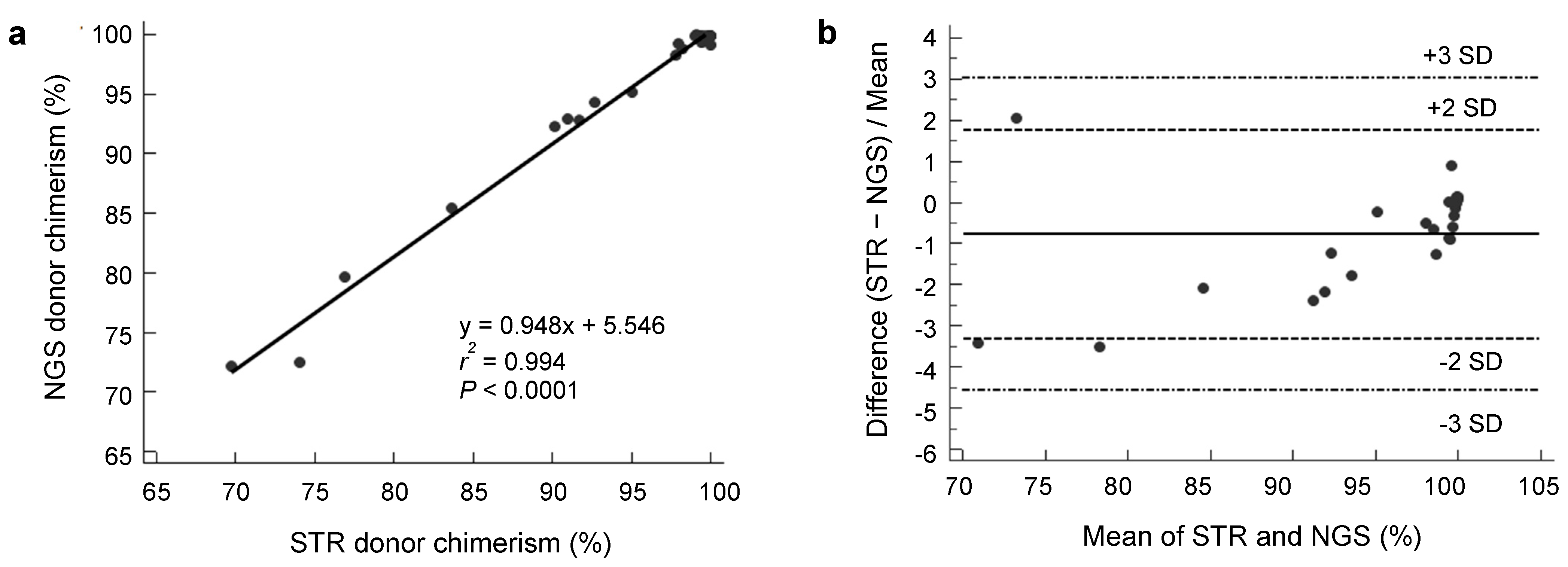
3.1. Analytical Performance of NGS Donor Chimerism

Mean on-target read depth ranged from 1406.81× to 2052.61×, and the 50× coverage rates ranged from 99.34% to 99.75%. We did not detect any donor carrying a clonal hematopoiesis-related mutation [20]. The number of informative SNPs was variable (median 25.5, range 9–41) and higher than that of STR (median 10.5, range 5–14). Donor chimerism from NGS analysis highly correlated with results from STR analysis, with r2 = 0.994 (p < 0.0001) by least-squares analysis (Figure 2a), and all data points were within 3 SDs of the mean % difference on Blant-Altman plot (Figure 2b). In addition, we calculated positive (PPV) and negative predictive value (NPV) for our test to diagnose complete donor chimerism with three cut-off values including 99%, 95% and 90% [21,22,23]. The overall agreements between the NGS and STR analyses were 100% when we applied the 95% and 90% cut-off values. When applying 99% cut-off value, NGS analysis showed 100% sensitivity, 91.7% specificity, 92.3% PPV and 100% NPV. Because our laboratory uses 95% cut-off value for complete chimerism, current agreement between two methods was 100% in qualitative comparison.

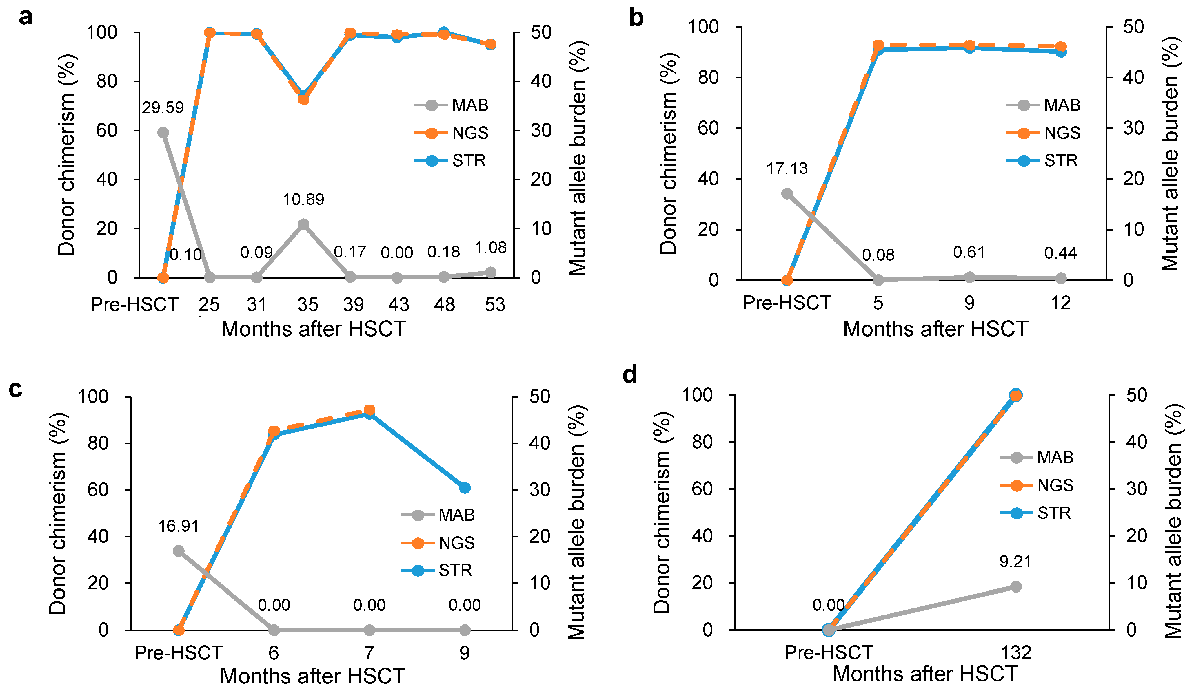
3.2. Clinical Usefulness of Simultaneous Monitoring

Nine patients (1–9) showed complete donor chimerism (STR 99.29 ± 0.76%, NGS 99.60 ± 0.58%) with 0.03 ± 0.05% MAB (Figure 3).
Patient 10 maintained mixed chimerism with normal hematologic findings. Among the 14 patients, three patients (11–13) relapsed after allo-HSCT. We detected increased MAB and decreased donor chimerism at relapse (patient 11, Figure 4a). Patient 12 showed mixed chimerism at the first year follow-up and persistent SF3B1 mutation at a low level (0.44 ± 0.16, Figure 4b). Patient 13 showed mixed chimerism until 7 months after allo-HSCT. NRAS mutation was observed at diagnosis, but it disappeared after allo-HSCT. He relapsed to AML at 9 months after allo-HSCT with decreased donor chimerism. Notably, a new cytogenetic aberration was obtained without reappearing NRAS mutation (Figure 4c). Patient 14 verified the effect of the analysis algorithm. She was treated for AML and received allo-HSCT 11 years ago. During regular follow-up, pancytopenia occurred, and BM examination was performed for hematopathologic evaluation and chimerism analysis, revealing 3-lineage dysplasia and increased blasts (5%). Notably, complete donor chimerism (99.8%) along with PHF6 mutation was identified by NGS, which suggested that the mutation originated from donor cells (Figure 4d). This impression was further supported by cytogenetic analysis as //46,XY,+1,der(1;7)(q10;q10)[5]/46,XY[5], which demonstrated donor-cell derived MDS accompanied by 1q gain and 7q loss. Patient characteristics and the results of donor chimerism and mutant burden are described in Table 2.

4. Discussion

In hematologic malignancies, risk stratification and clinical decision are made based on monitoring disease after treatment. Among available technologies for MRD monitoring, multi-parameter flow cytometry and measuring mutant burden by quantitative PCR are sensitive and specific. But they are restricted for patients with particular aberrant expression or molecular markers and do not detect evolving clonal aberrations [24,25,26]. NGS has great potential for MRD monitoring because it has ability to determine multiple mutations simultaneously with clonal burden [10,27]. After allo-HSCT, patients are monitored by chimerism analysis and routine hematology test at regular intervals. Chimerism analysis is highly applicable to most patients after allo-HSCT, but commonly used STR assay is less sensitive and specific because leukemic cells are not directly targeted. CD34-positive cell sorted chimerism may overcome some part of the limitations, but CD34 expression is variable and cell sorting process is laborious. Another important consideration is multiple meaning of mixed chimerism. The mixed chimerism can have various clinical implications including disease relapse, graft failure or rejection. However, previous studies showed that mixed chimerism may remain stable over long time and be compatible with prolonged remission [28]. Moreover, there are increasing states of mixed chimerism, especially after reduced intensity conditioning regimens and after T-cell depletion [28]. Therefore, results from chimerism analysis should be comprehensively interpreted and it is desirable to combine the different methods to strengthen the strength and make up for the weakness of them.
In this study, we developed a new analytic algorithm to monitor chimerism applicable to NGS multi-gene panel in use to identify mutations of MDS through careful stepwise evaluation. This is the first attempt to analyze both chimerism and mutation simultaneously, especially through a novel but simple analytical algorithm. Through this algorithm, we could implement comprehensive pre- and post-HSCT monitoring of various clinical conditions such as complete donor chimerism, persistent mixed chimerism, early relapse, and donor cell-derived MDS. Patients with complete donor chimerism showed MAB less than threshold. Among patient with mixed chimerism, three relapsed showed increased MAB and decreased donor chimerism. The other patient relapsed to AML with decreased donor chimerism without reappearing initially detected NRAS mutation. This result was in line with a previous study that showed some mutations to be effectively eliminated through HSCT [24]. Another patient maintained stable mixed chimerism. These findings indicated that the individual disease process is effectively demonstrated through simultaneous analysis. Notably, we successfully detected a donor-cell derived MDS patient showing complete donor chimerism and evolving clonal mutation. Donor cell-derived hematologic malignancy is an infrequent complication after all-HSCT. Its diagnosis can be delayed until blasts emerge in peripheral blood because STR shows complete donor chimerism. Previous report reviewed literatures and identified more than 70 cases of donor cell-derived leukemia (DCL). Time between allo-HSCT and occurrence of DCL was various (median 30 months, range 1-279) [29]. Although DCL can be diagnosed by chmerism analysis, it is more important to detect the genetic landscape and factors which contribute to DLC such as germline mutations [30]. It can help us to understand pathogenesis of DCL and to make better therapeutic plans. The other important interesting condition is pre-existing clonal hematopoiesis in donor [20]. We did not detect clonal hematopoeisis –related mutations in donor, however, it is necessary to monitor if the mutations were detected in donor cells. Our simultaneous analysis using NGS is very useful to evaluate these particular conditions after allo-HSCT.
This method is very convenient and cost-effective because it can be applied to any NGS panel for hematologic malignancies after selection and evaluation of informative SNPs. The turn-around time of the simultaneous analysis was not longer than that of mutation analysis using NGS (about 4 days) because SNP-based chimerism calculation can be performed simply with minimal time (less than 30 min). The number of informative SNPs was enough to analyze chimerism and the NGS chimerism was concordant with STR assay result. The overall agreements between the NGS and STR analyses to diagnose complete chimerism were excellent. We postulated that SNP-based NGS chimerism analysis would be superior to STR assay because NGS overcomes the various technical limitations of STR assay, such as stutter peak, peak height imbalance, non-template adenine addition, dye interference, voltage spikes, and allele dropouts [9,31]. Although we tried to minimize the bias of conventional NGS methodology through excluding SNPs with low read depth, high background error rate and allelic imbalance, there are still technical limitations such as repetitive amplification of the same reads. This can be overcome by barcoded error-corrected sequencing methods such as single molecule molecular inversion probes (smMIPs) [32,33]. Development of improved error-corrected sequencing method to increase the sensitivity and specificity of NGS technology coupled with automated calculation system would potentiate the usefulness of this novel analytic algorithm. And it is worthy to validate the analytical and clinical performance of this improved method through a large number of cases in a prospective manner.

5. Conclusions

In this study, we developed a new analytic algorithm using a clinically used NGS myeloid panel that simultaneously monitors mutation and chimerism. This method is applicable to any NGS panels and allows chimerism analysis from allele burdens of SNPs included in the NGS panel. This approach showed excellent performance and provided useful information to understand various clinical status after allo-HSCT.

Supplementary Materials

The following are available online at https://www.mdpi.com/2077-0383/8/12/2077/s1, Table S1: List of target genes of the NGS panel; Table S2: List of SNPs included in the NGS chimerism calculation.

Author Contributions

Conception and design: Y.K. and M.K.; patient data and samples: S.-S.P., Y.-J.K. and M.K.; experiments, collection and assembly of data: J.-M.L., E.H. and M.K.; data analysis and interpretation: J.-M.L., E.H., Y.K. and M.K..; manuscript writing and editing: J.-M.L., Y.K. and M.K.

Acknowledgments

This research was supported by the Bio & Medical Technology Development Program of the National Research Foundation (NRF) funded by the Ministry of Science & ICT (2018M3A9E8020866) and by a grant (18172MFDS182) from Ministry of Food and Drug Safety in 2019.

Conflicts of Interest

The authors declare no conflict of interests.

References

  1. Petrova, L.; Vrbacky, F.; Lanska, M.; Zavrelova, A.; Zak, P.; Hrochova, K. IDH1 and IDH2 mutations in patients with acute myeloid leukemia: Suitable targets for minimal residual disease monitoring? Clin. Biochem. 2018, 61, 34–39. [Google Scholar] [CrossRef]
  2. Kim, T.; Moon, J.H.; Ahn, J.S.; Kim, Y.K.; Lee, S.S.; Ahn, S.Y.; Jung, S.H.; Yang, D.H.; Lee, J.J.; Choi, S.H. Next-generation sequencing-based posttransplant monitoring of acute myeloid leukemia identifies patients at high risk of relapse. Blood 2018, 132, 1604–1613. [Google Scholar] [CrossRef]
  3. Morita, K.; Kantarjian, H.M.; Wang, F.; Yan, Y.; Bueso-Ramos, C.; Sasaki, K.; Issa, G.C.; Wang, S.; Jorgensen, J.; Song, X. Clearance of Somatic Mutations at Remission and the Risk of Relapse in Acute Myeloid Leukemia. J. Clin. Oncol. Off. J. Am. Soc. Clin. Oncol. 2018, 36, 1788–1797. [Google Scholar] [CrossRef]
  4. Swerdlow, S.H.; Campo, E.; Harris, N.L.; Jaffe, E.S.; Pileri, S.A.; Stein, H.; Thiele, J. WHO Classification Tumours of Haematopoietic and Lymphoid Tissues; International Agency for Research on Cancer (IARC): Lyon, France, 2017; Volume 4, pp. 104–105. [Google Scholar]
  5. Roloff, G.W.; Lai, C.; Hourigan, C.S.; Dillon, L.W. Technical advances in the measurement of residual disease in acute myeloid leukemia. J. Clin. Med. 2017, 6, 87. [Google Scholar] [CrossRef]
  6. Bader, P.; Niethammer, D.; Willasch, A.; Kreyenberg, H.; Klingebiel, T. How and when should we monitor chimerism after allogeneic stem cell transplantation? Bone Marrow Transplant. 2005, 35, 107. [Google Scholar] [CrossRef] [PubMed]
  7. Ahci, M.; Stempelmann, K.; Buttkereit, U.; Crivello, P.; Trilling, M.; Heinold, A.; Steckel, N.K.; Koldehoff, M.; Horn, P.A.; Beelen, D.W. Clinical utility of quantitative PCR for chimerism and engraftment monitoring after allogeneic stem cell transplantation for hematologic malignancies. Biol. Blood Marrow Transplant. 2017, 23, 1658–1668. [Google Scholar] [CrossRef]
  8. Aloisio, M.; Licastro, D.; Caenazzo, L.; Torboli, V.; D’eustacchio, A.; Severini, G.M.; Athanasakis, E.A. Technical application of quantitative next generation sequencing for chimerism evaluation. Mol. Med. Rep. 2016, 14, 2967–2974. [Google Scholar] [CrossRef] [PubMed]
  9. Kim, J.; Hwang, I.S.; Shin, S.; Choi, J.R.; Lee, S.T. SNP-based next-generation sequencing reveals low-level mixed chimerism after allogeneic hematopoietic stem cell transplantation. Ann. Hematol. 2018, 97, 1731–1734. [Google Scholar] [CrossRef]
  10. Shapiro, R.M.; Kim, D.D.H. Next-generation sequencing-based minimal residual disease monitoring in patients receiving allogeneic hematopoietic stem cell transplantation for acute myeloid leukemia or myelodysplastic syndrome. Curr. Opin. Hematol. 2018, 25, 425–432. [Google Scholar] [CrossRef]
  11. Han, E.; Kim, M.; Kim, Y.; Han, K.; Lim, J.; Kang, D.; Lee, G.D.; Kim, J.R.; Lee, J.W.; Chung, N.G. Practical informativeness of short tandem repeat loci for chimerism analysis in hematopoietic stem cell transplantation. Clin. Chim. Acta 2017, 468, 51–59. [Google Scholar] [CrossRef]
  12. Martin, M. Cutadapt removes adapter sequences from high-throughput sequencing reads. EMBnet. J. 2011, 17, 10–12. [Google Scholar] [CrossRef]
  13. Li, H.; Durbin, R. Fast and accurate short read alignment with Burrows–Wheeler transform. Bioinformatics 2009, 25, 1754–1760. [Google Scholar] [CrossRef] [PubMed]
  14. McKenna, A.; Hanna, M.; Banks, E.; Sivachenko, A.; Cibulskis, K.; Kernytsky, A.; Garimella, K.; Altshuler, D.; Gabriel, S.; Daly, M. The Genome Analysis Toolkit: A MapReduce framework for analyzing next-generation DNA sequencing data. Genome Res. 2010, 20, 1297–1303. [Google Scholar] [CrossRef] [PubMed]
  15. Li, H.; Handsaker, B.; Wysoker, A.; Fennell, T.; Ruan, J.; Homer, N.; Marth, G.; Abecasis, G.; Durbin, R.; Subgroup, G.P.D.P. The Sequence Alignment/Map format and SAMtools. Bioinformatics 2009, 25, 2078–2079. [Google Scholar] [CrossRef]
  16. Favero, F.; Joshi, T.; Marquard, A.M.; Birkbak, N.J.; Krzystanek, M.; Li, Q.; Szallasi, Z.; Eklund, A.C. Sequenza: Allele-specific copy number and mutation profiles from tumor sequencing data. Ann. Oncol. 2015, 26, 64–70. [Google Scholar] [CrossRef]
  17. Li, M.M.; Datto, M.; Duncavage, E.J.; Kulkarni, S.; Lindeman, N.I.; Roy, S.; Tsimberidou, A.M.; Vnencak-Jones, C.L.; Wolff, D.J.; Younes, A. Standards and guidelines for the interpretation and reporting of sequence variants in cancer: A joint consensus recommendation of the Association for Molecular Pathology, American Society of Clinical Oncology, and College of American Pathologists. J. Mol. Diagn. 2017, 19, 4–23. [Google Scholar] [CrossRef]
  18. Thol, F.; Gabdoulline, R.; Liebich, A.; Klement, P.; Schiller, J.; Kandziora, C.; Hambach, L.; Stadler, M.; Koenecke, C.; Flintrop, M. Measurable residual disease monitoring by NGS before allogeneic hematopoietic cell transplantation in AML. Blood 2018, 132, 1703–1713. [Google Scholar] [CrossRef]
  19. Kristt, D.; Klein, T. Reliability of quantitative chimerism results: Assessment of sample performance using novel parameters. Leukemia 2006, 20, 1169. [Google Scholar] [CrossRef]
  20. Frick, M.; Chan, W.; Arends, C.M.; Hablesreiter, R.; Halik, A.; Heuser, M.; Michonneau, D.; Blau, O.; Hoyer, K.; Christen, F. Role of donor clonal hematopoiesis in allogeneic hematopoietic stem-cell transplantation. J. Clin. Oncol. 2018, 37, 375–385. [Google Scholar] [CrossRef]
  21. Scharf, S.J.; Smith, A.G.; Hansen, J.A.; McFarland, C.; Erlich, H.A. Quantitative determination of bone marrow transplant engraftment using fluorescent polymerase chain reaction primers for human identity markers. Blood 1995, 85, 1954–1963. [Google Scholar] [CrossRef]
  22. Dubovsky, J.; Daxberger, H.; Fritsch, G.; Printz, D.; Peters, C.; Matthes, S.; Gadner, H.; Lion, T.; Muller-Berat, N. Kinetics of chimerism during the early post-transplant period in pediatric patients with malignant and non-malignant hematologic disorders: Implications for timely detection of engraftment, graft failure and rejection. Leukemia 1999, 13, 2060–2069. [Google Scholar] [CrossRef]
  23. Antin, J.H.; Childs, R.; Filipovich, A.H.; Giralt, S.; Mackinnon, S.; Spitzer, T.; Weisdorf, D. Establishment of complete and mixed donor chimerism after allogeneic lymphohematopoietic transplantation: Recommendations from a workshop at the 2001 Tandem Meetings of the International Bone Marrow Transplant Registry and the American Society of Blood and Marrow Transplantation. Biol. Blood Marrow Transplant. 2001, 7, 473–485. [Google Scholar] [PubMed]
  24. Ding, L.; Ley, T.J.; Larson, D.E.; Miller, C.A.; Koboldt, D.C.; Welch, J.S.; Ritchey, J.K.; Young, M.A.; Lamprecht, T.; McLellan, M.D. Clonal evolution in relapsed acute myeloid leukaemia revealed by whole-genome sequencing. Nature 2012, 481, 506. [Google Scholar] [CrossRef] [PubMed]
  25. Rautenberg, C.; Germing, U.; Haas, R.; Kobbe, G.; Schroeder, T. Relapse of acute myeloid leukemia after allogeneic stem cell transplantation: Prevention, detection, and treatment. Int. J. Mol. Sci. 2019, 20, 228. [Google Scholar] [CrossRef]
  26. Schuurhuis, G.J.; Heuser, M.; Freeman, S.; Béné, M.C.; Buccisano, F.; Cloos, J.; Grimwade, D.; Haferlach, T.; Hills, R.K.; Hourigan, C.S. Minimal/measurable residual disease in AML: A consensus document from the European LeukemiaNet MRD Working Party. Blood 2018, 131, 1275–1291. [Google Scholar] [CrossRef]
  27. Hamilton, B.K.; Rybicki, L.; Hirsch, C.; Przychodzen, B.; Nazha, A.; Gerds, A.T.; Hanna, R.; Kalaycio, M.; Sekeres, M.A.; Sobecks, R. Mutation clonal burden and allogeneic hematopoietic cell transplantation outcomes in acute myeloid leukemia and myelodysplastic syndromes. Bone Marrow Transplant. 2019, 54, 1281–1286. [Google Scholar] [CrossRef]
  28. Schaap, N.; Schattenberg, A.; Mensink, E.; Preijers, F.; Hillegers, M.; Knops, R.; Pennings, A.; Boezeman, J.; Geurts van Kessel, A.; de Pauw, B.; et al. Long-term follow-up of persisting mixed chimerism after partially T cell-depleted allogeneic stem cell transplantation. Leukemia 2002, 16, 13–21. [Google Scholar] [CrossRef]
  29. Bouvier, A.; Ribourtout, B.; François, S.; Orvain, C.; Paz, D.L.; Beucher, A.; Guérard, A.; Guardiola, P.; Ugo, V.; Blanchet, O. Donor cell-derived acute promyelocytic leukemia after allogeneic hematopoietic stem cell transplantation. Eur. J. Haematol. 2018, 101, 570–574. [Google Scholar] [CrossRef]
  30. Kobayashi, S.; Kobayashi, A.; Osawa, Y.; Nagao, S.; Takano, K.; Okada, Y.; Tachi, N.; Teramoto, M.; Kawamura, T.; Horiuchi, T. Donor cell leukemia arising from preleukemic clones with a novel germline DDX41 mutation after allogenic hematopoietic stem cell transplantation. Leukemia 2017, 31, 1020. [Google Scholar] [CrossRef]
  31. Clark, J.R.; Scott, S.D.; Jack, A.L.; Lee, H.; Mason, J.; Carter, G.I.; Pearce, L.; Jackson, T.; Clouston, H.; Sproul, A. Monitoring of chimerism following allogeneic haematopoietic stem cell transplantation (HSCT): Technical recommendations for the use of short tandem repeat (STR) based techniques, on behalf of the United Kingdom National External Quality Assessment Service for Leucocyte Immunophenotyping Chimerism Working Group. Br. J. Haematol. 2015, 168, 26–37. [Google Scholar]
  32. Hiatt, J.B.; Pritchard, C.C.; Salipante, S.J.; O’Roak, B.J.; Shendure, J. Single molecule molecular inversion probes for targeted, high-accuracy detection of low-frequency variation. Genome Res. 2013, 23, 843–854. [Google Scholar] [CrossRef] [PubMed]
  33. Waalkes, A.; Penewit, K.; Wood, B.L.; Wu, D.; Salipante, S.J. Ultrasensitive detection of acute myeloid leukemia minimal residual disease using single molecule molecular inversion probes. Haematologica 2017, 102, 1549–1557. [Google Scholar] [CrossRef] [PubMed] [Green Version]
Figure 1. Algorithm for simultaneous analysis of chimerism and mutation. The A,C,G,T counts at the positions of mutation markers and informative single nucleotide polymorphisms (SNPs) were extracted from Mpileup file using Sequenza and mpileup2acgt. BAM: Binary version of Sequence Alignment Map, VCF: Variant Calling Format, SNP: Single Nucleotide Polymorphism, MRD: Minimal Residual Disease.
Jcm 08 02077 g001
Figure 2. (a) Scatter plot of donor chimerism values obtained by next-generation sequencing (NGS) and STR shows excellent correlation. (b) Blant-Altman plot of the two chimerism assays.
Jcm 08 02077 g002
Figure 3. Representative integrative genomic viewer at SETBP1 region of pre- (upper chart) and post- hematopoietic stem cell transplantation (HSCT) (lower chart) samples from patient 1. Somatic mutation (6.96%, green area in circle) disappeared and single nucleotide polymorphisms (arrows) changed after HSCT.
Jcm 08 02077 g003
Figure 4. Kinetics of engraftment monitoring using NGS, STR and mutant allele burden (MAB) on longitudinal follow-up samples before and after hematopoeitc stem cell transplantation (HSCT) of patient 11 (a), 12 (b), 13 (c) and 14 (d).
Jcm 08 02077 g004
Table 1. Determination of limit of blank for monitoring minimal residual disease.
MutationLocationRead Depth%BE%SD
NRAS:c.35G>AChr1:1152587472048.7 ± 416.20.0260.041
SF3B1:c.2098A>GChr2:1982668341930.5 ± 365.20.0180.031
SETBP1:c.2608G>AChr18:425319131598.5 ± 313.40.0410.064
U2AF1:c.101C>A,TCh21:445244561410.7 ± 351.40.0260.055
PHF6:c.820C>TChrX:1335491361184.0 ± 455.80.0230.052
BE, background error; Chr, chromosome; Read depth described as mean ± SD.
Table 2. Characteristics of patients with donor chimerism and mutant burden.
Case TypeNo.Gender/AgeType of HSCTStatusSampleF/U MonthsBM Findings Cellularity (Blasts)Chromosome Analysis%Donor STRNo. IRA STR%Donor NGSNo. IRA NGSMAB (%) Mutation
Complete donor chimerism1M/61Unrelated matched - PBSCTPrePB RCMD *46,XY[20] * 22.40U2AF1:c.101C>T
6.60SETBP1:c.2608G>A
Post 1PB30NANA99.41499.94410.00U2AF1:c.101C>T
0.00SETBP1:c.2608G>A
2F/39Family mismatched -PBSCTPrePB Hypo-MDS *46,XX,t(2;3)(p23;q29)[14]/
46,XX[6] *
10.33SF3B1:c.2098A>G
Post 1BM2530% (1%)46,XX[20]100899.87240.00
3M/29Family mismatched -PBSCTPrePB MDS EB-1 *46,XY[20] * 31.78U2AF1:c.101C>T
Post 1PB46NANA99.11199.97310.00
4F/29Sibling matched -PBSCTPrePB RCMD *47,XX,+8,del(11)(q21)[8]/
46,XX[12] *
37.64U2AF1:c.101C>A
Post 1PB54NANA99.9599.91170.15
5M/45Sibling matched -PBSCTPrePB MDS EB-2 *46,XY[15] * 26.22U2AF1:c.101C>T
Post 1PB37NANA100699.93270.00
6F/26Sibling matched -PBSCTPrePB RCMD *47,XX,+8[3]/46,XX[17] * 6.88U2AF1:c.101C>A
Post 1PB32NANA97.81498.28300.10
7M/58Family mismatched -PBSCTPrePB RCMD *46,XY[20] * 46.74U2AF1:c.101C>T
3.69NRAS:c.35G>A
Post 1BM1050% (<1%)46,XY[20]98.21398.81280.00U2AF1:c.101C>T
0.00NRAS:c.35G>A
8M/24Family mismatched -PBSCTPrePB RCMD *47,XY,+8[20] * 7.10U2AF1:c.101C>A
Post 1PB37NANA99.6799.87110.08
9F/31Sibling matched -PBSCTPrePB MDS EB-1 *46,XX[20] * -None
Post 1PB30NANA99.71099.8620-
Mixed chimerism10M/3Sibling matched - BMTPrePB RCC *46,XY[20] * -None
Post 1PB120% (1%) *46,XY[2]//46,XX[28] *76.9980.569-
Post 2PB380% (<1%) *46,XY[8]//46,XX[12] *69.7672.0010-
11F/59Unrelated matched - PBSCTPrePB RCMD *46,XX[20] * 29.59U2AF1:c.101C>T
Post 1PB25NANA99.91499.88370.10
Post 2PB31NANA99.41499.39370.09
Post 3BM3570% (18%)47,XX,+21[9]//46,XY[11]74.01472.492810.89
Post 4PB39NANA99.01499.85370.17
Post 5BM4330% (5%)//46,XY[30]98.01499.23370.00
Post 6PB48NANA1001499.09370.18
Post 7PB535% (18%) *47,XX,+21[10]//46,XY[10] *95.01495.23371.08
12M/54Sibling matched - BMTPrePB RARS *46,XY[12] * 17.13SF3B1:c.2098A>G
Post 1PB515% (<1%) *46,XY[20] *90.9792.91190.08
Post 2PB930% (<1%) *46,XY[20] *91.7892.84190.61
Post 3PB1210% (3%) *46,XY[10] *90.2892.35190.44
13M/52Sibling matched - BMTPrePB MDS EB-2 *46,XY[20] * 16.91NRAS:c.35G>A
Post 1PB630% (<1%) *46,XY[20] *83.7985.4490.00
Post 2BM715% (1%)46,XY[20]92.7894.3790.00
Post 3BM940% (5%)46,XY,t(1;21)(p36.3;q11.2)[19]/
46,XY[1]
60.99NANA0.00
Donor-cell derived MDS14F/59Sibling matched - BMTPrePB MDS EB-1 *NA * 0.00PHF6:c.820C>T
Post 1BM13215% (5%)//46,XY,+1,der(1;7)(q10;q10)[5]/46,XY[5]100599.80139.21
* Data within 3 months of chimerism analysis. Values above threshold of minimal residual disease were written in bold. HSCT, hematopoietic stem cell transplantation; PBSCT peripheral blood stem cell transplantation; BMT, bone marrow transplantation; RCMD refractory cytopenia with multilineage dysplasia; NA, not available; Hypo-MDS, hypocellular myelodysplastic syndrome; EB, excess blasts; RCC, refractory cytopenia of childhood; RARS, refractory anemia with ring sideroblasts; F/U months, months after HSCT; %Donor, Donor chimerism; IRA, informative recipient alleles; MAB, mutant allele burden.

