Article

A Cold-Induced LEA3 Protein, DohD, Confers Cryoprotective Protection Against Low-Temperature Stress in Deinococcus radiodurans

by
Wenxiu Wang
1,2,
Zhi Qi
2,
Chunxia Yan
2,
Zhengfu Zhou
1,* and
Jin Wang
1,*
1
National Key Laboratory of Agricultural Microbiology, Biotechnology Research Institute, Chinese Academy of Agricultural Sciences, Beijing 100081, China
2
Key Laboratory of Forage and Endemic Crop Biology, Ministry of Education, School of Life Science, Inner Mongolia University, Hohhot 010021, China
*
Authors to whom correspondence should be addressed.
Int. J. Mol. Sci. 2025, 26(8), 3511; https://doi.org/10.3390/ijms26083511
Submission received: 7 March 2025 / Revised: 4 April 2025 / Accepted: 7 April 2025 / Published: 9 April 2025
(This article belongs to the Section Molecular Biology)

Abstract

Deinococcus radiodurans is a remarkably unique microorganism, exhibiting extraordinary tolerance to extreme conditions such as ionizing radiation, ultraviolet light, and desiccation. However, the response mechanisms of D. radiodurans under low-temperature stress remain largely unexplored and have yet to be fully elucidated. The DohD protein is a hydrophilic member of the late embryogenesis abundant 3 (LEA3) family of D. radiodurans, playing a pivotal role in abiotic stress adaptation. Bioinformatics analysis revealed that DohD contains tandem repeats and disordered domains, with a remarkably high α-helix content (91.41%). Furthermore, DohD exhibits extremely low homology with other proteins, highlighting its uniqueness to D. radiodurans. Under low-temperature stress (15 °C), the expression of dohD was significantly upregulated (5-fold), regulated by a dual mechanism involving positive control by DrRRA and negative regulation by Csp. Circular dichroism spectroscopy unveiled temperature-dependent structural plasticity: as the temperature increased from 0 °C to 50° C, the α-helix content decreased from 23.5% to 18.7%, while the antiparallel β-sheet content increased from 31.3% to 50.8%. This suggests an α-helix to β-sheet interconversion mechanism as a strategy for thermal adaptation. Additionally, deletion of dohD impaired the tolerance of D. radiodurans to cold, desiccation, oxidative, and high-salt stresses, accompanied by the reduced activities of antioxidant enzymes (SOD, CAT, POD) and the downregulation of related gene expression. This study elucidates the multifunctional role of DohD in stress resistance through structural dynamics, transcriptional regulation, and redox homeostasis, providing valuable insights into the adaptation mechanisms of extremophiles.

1. Introduction

Cold-induced proteins (CIPs), a class of biomolecules uniquely evolved to maintain structural integrity and catalytic efficiency under low-temperature stress, serve as critical mediators of cold adaptation across diverse organisms. As pivotal regulators of cellular homeostasis, metabolic reprogramming, and ecological resilience, CIPs play essential roles in enabling organisms to thrive in thermally challenging environments [1,2,3]. CIPs are broadly categorized into functional subgroups based on their mechanisms of action. The first class includes chaperone-like proteases, exemplified by the PSME-3 activator in Caenorhabditis elegans, which extends the lifespan at 15 °C by enhancing the proteolytic degradation of misfolded proteins—a mechanism conserved in humans to mitigate neurodegenerative protein aggregation [4]. A second category comprises cryoprotective agents, such as ice-binding proteins, which inhibit ice crystal growth in frozen food systems, preserving macromolecular stability and improving post-thawing quality [5]. CIPs are further stratified into photosynthetic regulators, which sustain carbon fixation under cold stress by modulating Calvin cycle enzymes [6,7], and metabolic adjustors, which rewire nitrogen assimilation pathways through the dynamic expression of amino acid synthases [8]. Emerging evidence also highlights a distinct subclass of membrane-stabilizing CIPs that enhance lipid bilayer fluidity and cold-responsive signaling, crucial for maintaining cellular integrity [9]. Functional studies reveal CIPs as molecular “Swiss Army knives”, integrating structural plasticity with context-dependent activity. Their cold-adaptive traits—such as flexible catalytic domains, increased surface hydrophobicity, and cold-inducible post-translational modifications—enable enzymatic function under conditions of reduced kinetic energy [2,3].
In extreme environments, diverse microbial communities thrive, many of which possess cold-adapted proteins to cope with low-temperature conditions [10]. These specialized proteins play critical roles in maintaining cellular integrity by regulating membrane fluidity and permeability, while also facilitating essential metabolic processes, including methane oxidation and sulfate reduction [11,12]. Among these extremophiles, D. radiodurans stands out for its remarkable resilience to multiple environmental stressors, including high levels of ionizing radiation, ultraviolet exposure, desiccation, chemical toxicity, and extreme temperature fluctuations [13,14,15,16]; late embryogenesis abundant (LEA) proteins play a pivotal role in alleviating low-temperature-induced damage [17,18]. Characterized by their exceptional hydrophilicity and desiccation tolerance, these proteins have been shown to protect human oocytes during vitrification preservation through mechanisms involving the suppression of FOS gene expression and the modulation of oxidative stress pathways [19]. Notably, D. radiodurans contains three desiccation stress-related proteins (DR1172, DR1372, and DR0105) that exhibit homology to LEA proteins [20,21,22].
Our research focuses on the DR0105 protein, which is predicted to contain a unique intrinsically disordered domain designated as DohD (D. radiodurans orphan genes hydrophilic intrinsically disordered protein). Late embryogenesis abundant (LEA) proteins have been extensively characterized as stress protectants in plants and certain extremophilic organisms, yet their evolutionary mechanisms and functional diversification in bacterial systems remain poorly understood. Specifically, while the cold adaptation strategies of D. radiodurans have been well documented, the molecular mediators enabling its enzymatic machinery to maintain functionality under suboptimal temperatures have not been systematically investigated. This study aims to address two critical knowledge gaps: the evolutionary trajectory of LEA-like proteins in radiation-resistant bacteria, and the specific mechanisms through which these proteins confer cryoprotection. By integrating phylogenetic analysis with functional characterization of DohD—a newly identified cold-induced protein—we seek to establish a mechanistic framework explaining how structural adaptations in bacterial LEA homologs contribute to extreme environmental resilience.

2. Results

2.1. DohD Is a Hydrophilic LEA3 Protein Containing a Disordered Domain

The dohD gene is situated on chromosome I of D. radiodurans, spanning a total length of 492 base pairs (bp). The encoded DohD protein comprises 163 amino acids and features five tandem repeats, each consisting of 16 amino acids (Table 1). Structural analysis reveals that these repeats share significant similarity with the characteristic 11-amino-acid tandem repeats commonly observed in the LEA3 protein family.
Phylogenetic analysis demonstrated that the DohD protein exhibits the highest sequence similarity to a homologous protein from Lachancea lanzarotensis (Figure 1). However, comprehensive sequence alignment and comparative analysis revealed significant divergences between these proteins. Based on these findings, we propose that the DohD protein represents a unique functional protein specifically evolved in D. radiodurans.
To further investigate the functional role of the DohD protein in low-temperature stress resistance, we conducted a comprehensive structural analysis. Secondary structure prediction revealed that 99% of the DohD protein adopts an α-helical conformation (Figure 2A). This predominant α-helical structure may play a crucial role in maintaining the structural stability of RNA or single-stranded DNA under low-temperature conditions. Specifically, the α-helical structure could prevent the formation of detrimental secondary structures in nucleic acids, thereby ensuring the proper progression of essential biological processes in D. radiodurans during cold stress.
Analysis of the Ramachandran plot for the DohD protein revealed that the dihedral angle distribution of φ (phi) and ψ (psi) indicates that most residues reside within the allowed regions, corresponding to secondary structures such as α-helices and β-sheets, which suggests high overall conformational stability (Figure 2B). Notably, some residues exhibit φ or ψ angles approaching 180°, likely reflecting extended conformations characteristic of β-sheets. This structural feature aligns with the β-barrel folding motif observed in typical cold-shock proteins, such as members of the Csp family, implying that DohD may maintain functional activity at low temperatures through a rigid β-sheet framework. In summary, the structural stability of DohD is closely associated with its low-temperature stress resistance, and the features of its Ramachandran plot further support its potential cold adaptation mechanism.

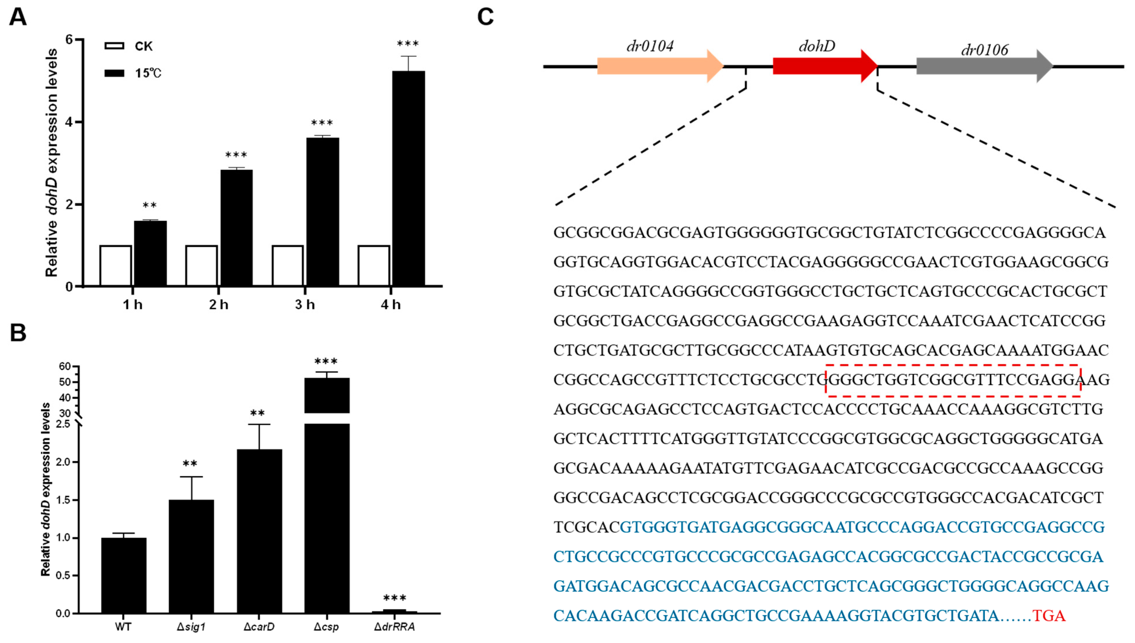
2.2. Low-Temperature Stress Induces DohD Expression

To investigate the transcriptional expression of the orphan gene dohD in Deinococcus radiodurans under abiotic stress conditions, wild-type D. radiodurans cells in the early logarithmic growth phase were exposed to normal and low-temperature treatments. The expression level of the dohD gene was quantified using qRT-PCR (Figure 3A). The results revealed that the most significant induction occurred after 4 h of stress treatment at 15 °C, with the expression of the dohD gene upregulated by approximately 5-fold. At other time points, the expression of the dohD gene also exhibited an increase. These findings suggest that the dohD gene can rapidly respond to low-temperature stress and may play a critical role in the low-temperature stress resistance mechanism of D. radiodurans.
Sig1, CarD, Csp, and DrRRA are key transcriptional regulators in D. radiodurans, and exert their effects through stress adaptation mechanisms such as oxidative stress response (Sig1, CarD), cold adaptation (Csp), ionizing radiation, and osmotic pressure adaptation (DrRRA), respectively. Therefore, they were selected as candidates to verify the upstream regulatory factors of the dohD gene. To elucidate the regulatory pathway of the DohD protein under low-temperature conditions, the expression of dohD was analyzed in the deletion mutants Δsig1, ΔcarD, Δcsp, and ΔdrRRA at 15 °C using qRT-PCR (Figure 3B). Notably, dohD expression displayed contrasting regulatory patterns in Δcsp and ΔdrRRA mutants: the deletion of csp resulted in a 53-fold upregulation of dohD, while the knockout of drRRA led to a 70-fold downregulation. In contrast, Δsig1 and ΔcarD exhibited no significant changes in dohD expression, indicating their non-involvement in its regulation. These findings reveal a dual regulatory mechanism in which dohD is positively controlled by DrRRA and negatively regulated by Csp, establishing a balanced transcriptional network under cold stress.
To determine whether DrRRA and Csp directly regulate the dohD gene, this study examined the transcription factor binding sites within the promoter region of dohD. As shown in Figure 3C, the promoter region of dohD in D. radiodurans contains a conserved sequence motif, GGGCTGGTCGGCGTTTCCGAGG, which forms an inverted repeat interspersed with non-conserved bases. This sequence motif is likely to represent the core recognition site for the binding of transcription factors DrRRA and Csp to the promoter.

2.3. The Role of Secondary Structural Changes in the DohD Protein in Low-Temperature Stress Resistance

To further investigate the structural changes of the DohD protein under low-temperature conditions, this study systematically analyzed the dynamic alterations in its secondary structure across a temperature gradient ranging from 0 °C to 50 °C using circular dichroism spectroscopy. These analyses revealed the low-temperature adaptive structural characteristics of the protein. Experimental data demonstrated that temperature variations significantly influence the secondary structure composition of the DohD protein [23]. Specifically, as the temperature increased from 0 °C to 50 °C, the α-helix content (Helix%) gradually decreased (23.5% → 18.7%), while the antiparallel β-sheet content (Antiparallel%) exhibited an inverse growth trend (31.3% → 50.8%). In contrast, the fluctuations in β-turn (Turn%) and random coil (Random coil%) content remained within a range of 3.2% (Figure 4). This pattern of structural reorganization suggests that temperature regulation may be mediated through an “α-helix ↔ β-sheet” interconversion mechanism.
Under low-temperature conditions (0 °C), the DohD protein exhibits characteristic structural stability, marked by a higher proportion of α-helix (23.5%) and a lower proportion of antiparallel β-sheet (31.3%) content, which together establish a structural steady-state equilibrium. The α-helix, as a rigid structural element, likely maintains its abundance through a stable hydrogen bond network, thereby enhancing the protein’s structural order. This feature aligns closely with the structural strategies employed by psychrophilic proteins to preserve functional activity at low temperatures. When the temperature increases to 50 °C, the significant enrichment of antiparallel β-sheet (50.8%) suggests a new structural stabilization mechanism, i.e., the formation of an interlocking network through the expansion of the β-sheet structure, compensating for the loss of rigidity resulting from the unwinding of the α-helices (Table 2).
Notably, the proportions of β-turn and random coil structures remain relatively stable across the temperature range (with a fluctuation of 3.2%), suggesting that these structural elements may function as molecular “hinges”, facilitating localized conformational adjustments rather than driving the overall structural reorganization. This observation aligns with established theories of protein conformational dynamics, which propose that flexible regions enable the overall structure to adapt to environmental perturbations through limited conformational changes.
This study reveals that the DohD protein possesses a unique temperature-responsive structural regulation mechanism: it maintains a high α-helix content to ensure structural rigidity at low temperatures, while initiating β-sheet reorganization to achieve structural restabilization at elevated temperatures. This dual-mode regulatory strategy likely underpins its molecular basis for low-temperature stress resistance.
Subcellular localization analysis using laser confocal microscopy demonstrated the temperature-dependent redistribution of DohD. At 30 °C, DohD exhibited a uniform cytoplasmic distribution (Figure 5, left), indicating its potential role in fundamental cellular processes. Under cold stress conditions (15 °C), DohD underwent significant relocalization, characterized by the formation of dense membrane-associated clusters, which predominated over the diffusely distributed cytoplasmic pools (Figure 5, right). This membrane enrichment suggests potential interactions with stress-sensing or signaling components at the membrane interface during cold adaptation.

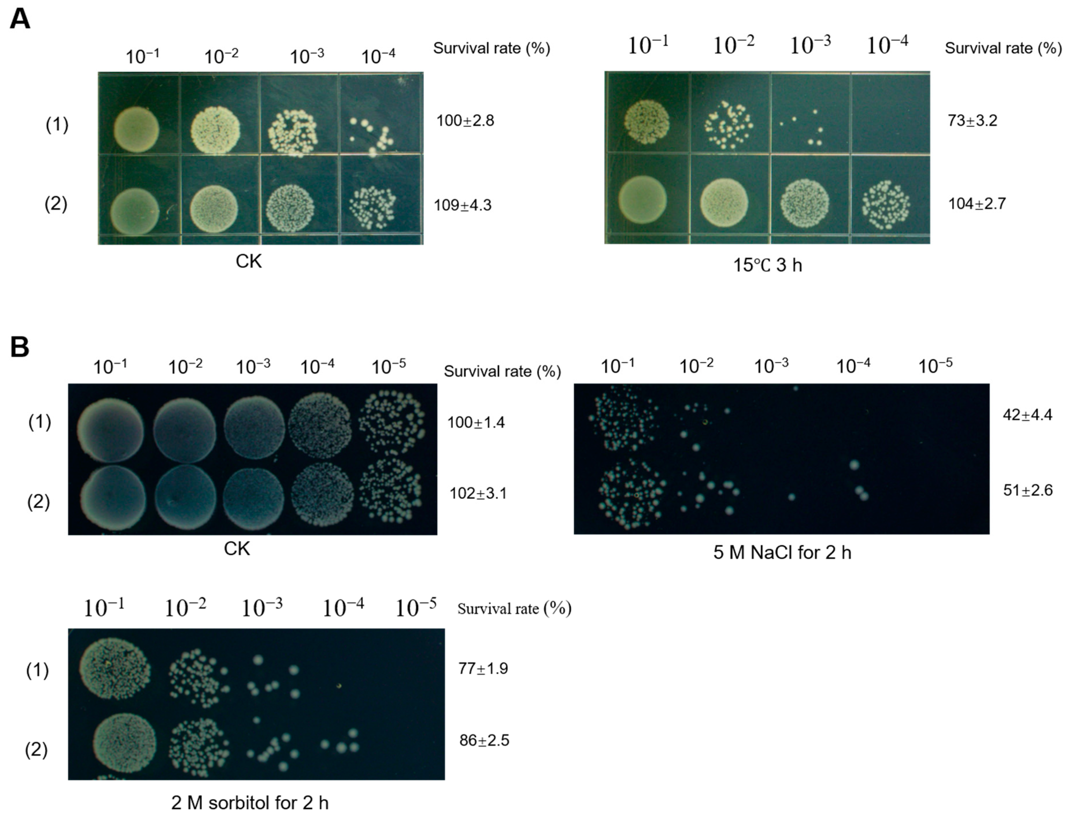
2.4. Deletion of dohD Decreases Abiotic Stress Tolerances of D. radiodurans

To determine whether the deletion of dohD affects the growth of D. radiodurans R1, growth curves were analyzed under standard culture conditions (30 °C, 220 rpm, TGY medium). As illustrated in Figure 6A, the wild-type strain (DR-WT), the dohD deletion mutant (∆dohD), and the complementation strain (com-dohD) displayed nearly identical S-shaped growth curves, with no significant differences in OD600 values during either the logarithmic or stationary phases. These results demonstrate that the deletion of dohD does not impair cellular proliferation under optimal growth conditions.
To investigate the role of dohD in abiotic stress resistance, we systematically assessed the viability of D. radiodurans strains under various stress conditions. Under cold stress (15 °C for 5 h), the colony-forming units (CFUs) of the dohD deletion mutant (∆dohD) decreased by one order of magnitude compared to the results for the wild-type strain (WT), accompanied by a 45% reduction in survival rate (Figure 6C,E). Time-dependent survival assays further revealed that after 60 h at 15 °C, ∆dohD exhibited a 25% growth decline relative to that of WT, while the complementation strain (com-dohD) showed partial recovery (15% decline, Figure 6B). These findings demonstrate that dohD plays a critical role in cold stress adaptation.
In addition to temperature sensitivity, the dohD deletion mutant (∆dohD) exhibited reduced tolerance to oxidative and high-salt stresses. The mutant strain demonstrated a significant decrease in antioxidant capacity (Figure 6F) and a two-order-of-magnitude reduction in survival under high-salt conditions compared to the results for the wild-type strain (WT, Figure 6G). Furthermore, under desiccation stress, ∆dohD displayed severely impaired growth and colony formation, while the complementation strain (com-dohD) partially restored the wild-type phenotype (Figure 6D).
Collectively, these findings demonstrate that the deletion of dohD significantly impairs the abiotic stress tolerance of D. radiodurans, encompassing cold, desiccation, oxidative, and high-salt stresses. The gene’s specific regulatory role in cold and desiccation stress responses, combined with its auxiliary functions in antioxidant defense and osmoregulation, underscores its multifunctional contribution to environmental adaptation. The partial phenotypic restoration observed in the complementation strain (com-dohD) further corroborates the functional specificity of dohD in the stress signaling pathways.

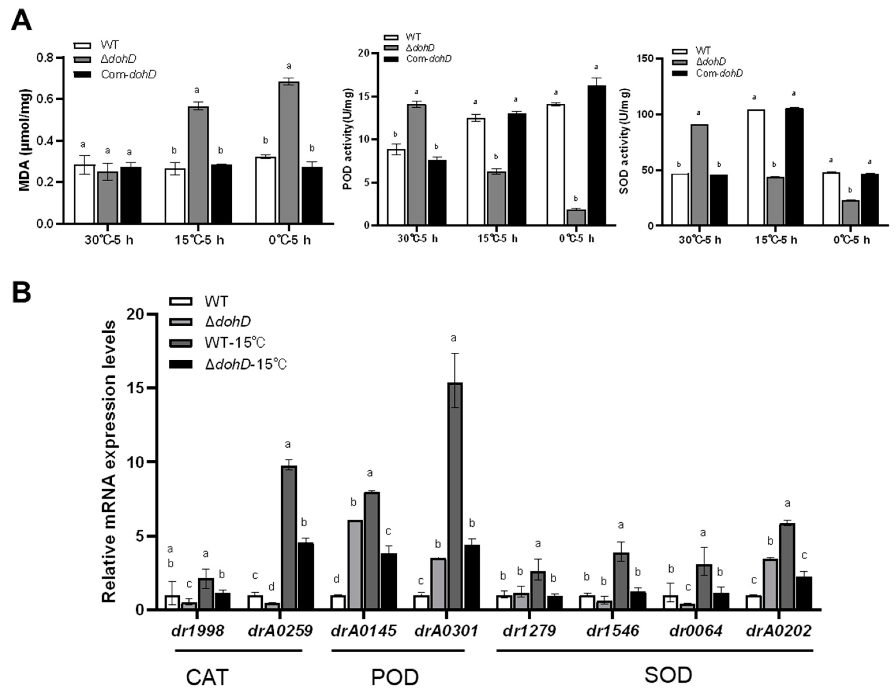
2.5. DohD Alleviates Cold-Induced Oxidative Damage by Maintaining Antioxidant Enzyme Activity

Low-temperature stress induces the accumulation of reactive oxygen species (ROS) within cells, leading to lipid peroxidation of the cell membrane and the production of malondialdehyde (MDA). The elevated MDA content is widely used as a key indicator to evaluate the extent of cellular oxidative damage. In response to low-temperature stress, D. radiodurans maintains intracellular redox homeostasis by activating detoxifying enzymes such as superoxide dismutase (SOD) and catalase (CAT), thereby mitigating oxidative damage caused by low temperatures. To investigate the role of dohD in this process, we assessed the effects of dohD deletion on the expression and activity of ROS-scavenging enzymes, including CAT, peroxidase (POD), and SOD. Consistent with the low-temperature-sensitive phenotype of the ΔdohD mutant strain, the expression and activity of these ROS-scavenging enzymes were significantly lower than those in the wild-type (WT) strain after 5 h of low-temperature treatment (Figure 7A). Specifically, after 5 h at 15 °C, POD and SOD activities decreased by 2.2-fold and 2.1-fold, respectively, while at 0 °C, the reductions were 7.1-fold and 4.1-fold, respectively. Furthermore, the expression of genes encoding CAT (dr1998 and drA0259), POD (drA0145 and drA0301), and SOD (dr1279, dr1546, dr0644, and drA0202) was significantly downregulated in the ΔdohD mutant strain under low-temperature stress, consistent with the observed decline in antioxidant enzyme activities (Figure 7B). Collectively, these findings demonstrate that DohD is essential for the expression and activity of ROS-scavenging enzymes and likely plays a critical role in the extreme low-temperature tolerance of D. radiodurans.

2.6. DohD Enhances the Low-Temperature Stress Tolerance of E. coli

To investigate the role of DohD in Escherichia coli tolerance to low-temperature stress, we performed bacterial survival assays. Cells grown to the logarithmic phase (OD600≈0.6) were exposed to 30 °C (control) or 15 °C (low-temperature stress) for 3 h. Serial dilutions (10−4) of the cultures were spotted (7 µL/dot) onto LB plates and incubated overnight at 37 °C. At 30 °C, all strains exhibited comparable growth, confirming the absence of baseline growth defects (Figure 8A, left). Notably, under 15 °C stress, the DohD-expressing strain demonstrated significantly enhanced survival compared to that of the control (Figure 8A, right), highlighting the critical role of DohD in low-temperature adaptation.
Remarkably, DohD also conferred cross-protection against osmotic stress. The DohD-expressing strain demonstrated significantly enhanced survival under high-salt conditions (Figure 8B), indicating its dual role in mitigating both thermal and osmotic stress. This multifunctional capability aligns with its stress-responsive localization dynamics, suggesting a coordinated mechanism for environmental adaptation.

3. Discussion

The extremophilic bacterium D. radiodurans employs a sophisticated arsenal of molecular strategies to withstand extreme environments. This study demonstrates that DohD, functioning as a molecular chaperone, enhances the strain’s resilience to stress. By integrating structural dynamics, transcriptional regulation, and antioxidant defense mechanisms, DohD orchestrates a multilayered adaptive response, providing novel insights into the stress adaptation strategies of D. radiodurans.

3.1. Structural Plasticity of the Cold-Inducible Protein DohD Drives Its Cold Adaptation Mechanism

Intrinsically disordered regions (IDRs) in stress-response proteins are increasingly recognized as mediators of structural flexibility [24], which may explain DohD’s ability to adopt multiple conformations (Figure S3). DohD exhibits pronounced transcriptional upregulation under cold stress (5-fold induction at 15 °C), regulated by the transcription factors DrRRA (positive regulation) and Csp (negative regulation). This induction aligns with functional necessity, as ΔdohD mutants displayed severe cold sensitivity, including reduced survival and impaired antioxidant enzyme activity. Central to its adaptive function is DohD’s structural plasticity, characterized by temperature-dependent α-helix to β-sheet interconversion. At low temperatures, the high α-helix content (23.5%) stabilizes RNA or single-stranded DNA through rigid hydrogen-bonded frameworks [25,26,27]. Conversely, under elevated temperatures (50 °C), the enrichment of antiparallel β-sheets (50.8%) suggests a compensatory mechanism to restore structural stability [28], highlighting DohD’s dual-mode conformational regulation. This demonstrates how bacterial LEA homologs achieve dual thermostability through conformational dynamics—a functional strategy unreported in eukaryotic systems. Notably, its conserved β-barrel folding pattern, reminiscent of that of cold-shock proteins (Csps) [29,30], underscores DohD’s evolutionary linkages to cold adaptation.

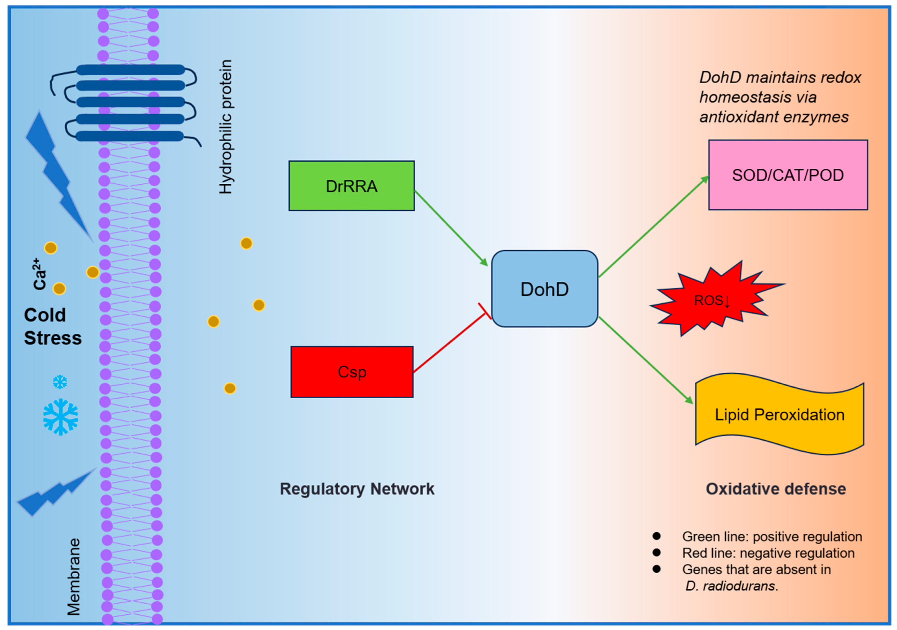
3.2. Regulatory Circuitry and Oxidative Defense

To elucidate how extremophiles coordinate stress-responsive gene expression, we identified a novel dual regulatory axis: DrRRA activates dohD (70-fold downregulation in ΔdrRRA), while Csp suppresses it (53-fold upregulation in Δcsp) (Figure 3B). This counterbalanced system differs fundamentally from the single-factor regulation of plant LEA genes [31,32], suggesting evolutionary specialization in extremophiles. Crucially, this regulatory architecture directly links to DohD’s antioxidant function. ΔdohD mutants showed a 4.1-fold reduced SOD activity at 0 °C (Figure 7A), exceeding the 2.5-fold decline observed in Synechocystis LEA knockouts under similar conditions, underscoring DohD’s central role in redox homeostasis, a trait shared with extremophile-specific chaperones like Deinococcus DrwH [33], yet distinct in its transcriptional coordination. The above results quantitatively validate our hypothesis that bacterial LEA proteins integrate transcriptional control with redox homeostasis [34].

3.3. DohD Integrates Antioxidant Defense with Membrane Stabilization

DohD uniquely couples antioxidant defense (2.1-fold higher SOD/POD activities in WT vs. ΔdohD, Figure 7A) with membrane stabilization—a dual mechanism evidenced by its temperature-dependent relocalization (30 °C: cytoplasmic; 15 °C: membrane clusters, Figure 5). Cold stress induces ROS accumulation, posing a threat to cellular components through lipid peroxidation [35,36,37,38]. DohD mitigates this damage by sustaining the activities of antioxidant enzymes (SOD, CAT, POD) and the expression of their encoding genes. In ΔdohD mutants, SOD activity decreased by 4.1-fold at 0 °C (Figure 6A), which correlated with elevated MDA levels—a key marker of oxidative membrane damage [39,40]. This phenotype aligns with the role of Arabidopsis LEA proteins in scavenging ROS under dehydration stress [31,32,41], suggesting a conserved mechanism across diverse domains of life. In Figure 9, the regulatory mechanism of D. radiodurans’ tolerance to cold stress is summarized.

3.4. Cross-Species Functionality Highlights Evolutionary Conservation

Addressing the evolutionary conservation of cryoprotective mechanisms, heterologous DohD expression in E. coli conferred a 15% higher survival at 15 °C (Figure 8A)—a functionality absent in plant LEA3. Despite low sequence homology (Figure 1), this cross-kingdom compatibility suggests that α-helix clustering (Figure 2A) represents a universal structural motif for bacterial cryoprotection. Furthermore, the improved salt tolerance in E. coli (Figure 7B) underscores DohD’s multifunctionality, akin to the osmoprotective roles of Bacillus cold-shock proteins (CSPs). Such versatility aligns with the “moonlighting” capability of intrinsically disordered proteins, which adopt context-dependent conformations to fulfill diverse biological roles.

4. Materials and Methods

4.1. Bacterial Strains, Plasmids, and Media

The strains and plasmids used in this study are described in Supplementary Table S1. D. radiodurans WT and its derivatives were cultured aerobically at 30 °C in TGY medium (1% tryptone, 0.5% yeast extract, and 0.1% glucose) in the presence of antibiotics, as required. E. coli strains were grown in Luria–Bertani (LB) broth at 37 °C, with appropriate antibiotics. Solid media contained 1.5% agar.

4.2. Construction of DohD Deletion Mutants and Complementary Strains

The mutant strains were constructed according to the principle of homologous recombination (see Supplementary Figure S1), with primers designed according to the full sequence of the dohD gene; see Table S2 in the Supplementary Materials. Using the D. radiodurans R1 genome as a template, P1/P2 amplified the upstream fragment of dohD at 921 bp (U), and P5/P6 amplified the downstream fragment of dohD at 915 bp (D). Using the pKatAPH3 plasmid as a template, P3/P4 amplified the kanamycin resistance gene nptII fragment 970 bp (K). A homologous recombinase was used to catalyze the fusion of the three fragments, UKD, in a 1:1:1 molar ratio. P1/P6 primers were used as templates to amplify the fusion fragment UKD (3596 bp). Finally, the fusion fragments were imported into D. radiodurans R1, according to the experimental method previously reported [42]. Single colonies were screened for kanamycin resistance (10 µg/mL). The results confirmed that the nptII gene was inserted in the correct position, replacing the dohD gene. The obtained dohD deletion mutant was named ∆dohD and was used for further studies. The dohD gene was inserted between the nucleic acid endonuclease BamH I and Spe I sites of the pRADZ3 plasmid, and the recombinant plasmid was then transformed into the ∆dohD mutant strain for the construction of the complementary strain com-dohD.

4.3. Bioinformatics Analysis

Bioinformatics analyses were conducted on the DohD protein using various tools. The Kyte–Doolittle hydropathy prediction website (https://www.novoprolabs.com/tools/protein-hydropathy; accessed on 15 March 2023) was employed to analyze its hydrophobicity. The VSL2 website (http://www.pondr.com/; accessed on 10 August 2023) was used to predict the ordered and disordered regions. Transmembrane features were investigated using the TMHMM (transmembrane hidden Markov model) website (https://www.novopro.cn/tools/tmhmm.html; accessed on 17 April 2024). Phylogenetic analysis was carried out by aligning DohD sequences with homologs from different sources, including Lachancea lanzarotensis, archaeal CSPs, and bacterial Csp members, using sequence alignment tools and neighbor-joining methods. All protein sequences containing LEA3 domains were retrieved from the UniProt and NCBI databases using the keywords “LEA3” and “hydrophilic stress protein”. Phylogenetic analysis was performed for homologous proteins with sequence similarity ≥ 30% and coverage ≥ 70% (E value < 1 × 10⁻⁵) by BLASTP. Secondary structure prediction was performed with PSIPRED. Structural stability was assessed through Ramachandran plot analysis.

4.4. Real-Time Fluorescence Quantitative PCR for Gene Expression

D. radiodurans R1 organisms were collected under various abiotic stress conditions using the Ambion®RNA extraction kit from Life (Austin, TX, USA). The 16S gene was used as the internal reference, and qRT-PCR was performed for the dohD, dr1998, drA0259, drA0145, drA0301, dr1279, dr1546, dr0644, drA0202, sig1, carD, csp, and drRRA genes under different temperature conditions using the ΔΔCt method. The qRT-PCR was carried out with an AB 7500 Real-Time PCR System, according to the manufacturer’s recommendations. The primer pairs used in this experiment are listed in Supplementary Table S2.

4.5. Activity Measurement of Major Antioxidant Enzymes

The D. radiodurans WT and ΔdohD mutant cells (OD600 ≈ 2, treated with 15 °C for 2 h and 30 °C for 2 h) were harvested by centrifugation, washed and resuspended with sterile PBS (pH 7.0), and disrupted on ice with an ultrasonicator. The debris was removed by centrifugation at 13,000× g at 4 °C for 20 min. Protein concentrations of the supernatants were determined by the Bradford method using BSA as the standard. The total SOD activity detection (WST-8 method) and lipid oxidation (MDA) detection kits were purchased from Shanghai Biyuntian Company (Shanghai, China), and the peroxidase (POD) detection kit was purchased from Nanjing, China. All experiments were repeated at least three times, independently.

4.6. Abiotic Stress-Resistance Assays

Low temperature resistance assay. A 1 mL sample of D. radiodurans R1 and its derivatives (with appropriate antibiotics) was incubated in TGY medium until the early stages of logarithmic growth (OD600 = 2) and cultured in a shaker at 15 °C and 30 °C (blank control group) for 1 to 5 h at 220 rpm, and a tube containing the sample was removed every 1 h and diluted to a final concentration of 10−1~10−5. An appropriate amount of the diluted sample was dropped onto the surface of the TGY solid medium and incubated in an incubator at 30 °C for 2–3 days to record the growth phenotype.
Desiccation resistance assay. A 1 mL sample of D. radiodurans R1 and its derivatives (with appropriate antibiotics) was incubated in TGY medium until the early stages of stable growth (OD600 = 8) and stored in a desiccator at room temperature for 60 days at relative humidity of less than 5%, following the reported method [33]. It was stored at room temperature in a desiccator at less than 5% relative humidity for 20 days. Samples were taken at 10-day intervals for cell recovery and diluted to a final concentration of 10−1~10−5. An appropriate amount of the diluted sample was dropped onto the surface of the TGY solid medium and incubated in an incubator at 30 °C for 2–3 days to record the growth phenotype.

4.7. DohD Expression and Purification

The dohD gene was amplified by PCR using genomic DNA from D. radiodurans R1 as the template, following the cloning strategy described in Reference [43]. The amplified fragment was directionally ligated into the pET28a expression vector via BamH I/Hind III double digestion to construct the recombinant plasmid, which was subsequently introduced into E. coli BL21(DE3) competent cells via electroporation. Protein expression was induced overnight at 16 °C with 0.5 mM isopropyl-β-D-thiogalactoside (IPTG). Bacterial cells were harvested under low-temperature conditions and lysed by ice-bath sonication. The supernatant and pellet fractions were separated by centrifugation and analyzed by SDS-PAGE using a Bio-Rad electrophoresis system from Bio-Rad Laboratories, Inc. Company (Hercules, CA, USA).
For protein purification, the nickel-affinity chromatography method, reported by Fredriksen [44], was modified as follows: The supernatant was filtered through a 0.22 μm membrane and loaded onto a His Trap HP nickel column (5 mL bed volume; GE Healthcare, Chicago, IL, USA) using an ÄKTA pure chromatography system. Fractions containing the target protein were pooled based on SDS-PAGE analysis, followed by desalting and buffer exchange using ultrafiltration concentrators. To prevent oxidative damage, the storage buffer was supplemented with 5 mM reduced glutathione and 2 mM dithiothreitol (DTT). The purified His-tagged DohD protein was stored at 4 °C for subsequent experiments, using the purification process visualized in Supplementary Figure S2.

4.8. Microscopy

For imaging of the D. radiodurans cells, a colony grown on a medium plate was inoculated into liquid medium and cultured overnight. The bacterial solution was cultured in an incubator at different temperatures. A total of 5 microliters of the bacterial solution was placed on a slide and covered with a coverslip. Imaging was performed on an AXR-NSPARC super-resolution confocal microscope(Carl Zeiss AG; Jena, Germany). GFP was excited at 488 nm and detected at 491–535 nm. Images were acquired with a ×100/1.46-NA oil objective.

5. Conclusions

DohD exemplifies D. radiodurans’ evolutionary ingenuity in merging structural disorder with functional precision. Through α/β structural switching, redox balancing, and dynamic localization, it orchestrates a multilayered defense against cold stress. Its integration into the DrRRA–Csp regulatory network further underscores the complexity of D. radiodurans’ adaptive mechanisms.

Supplementary Materials

The following supporting information can be downloaded at: https://www.mdpi.com/article/10.3390/ijms26083511/s1.

Author Contributions

Conceptualization, W.W., Z.Z., and J.W.; methodology, W.W., Z.Z., and J.W.; software, W.W.; validation, Z.Q.; formal analysis, Z.Q. and C.Y.; resources, W.W.; data curation, W.W.; writing—original draft preparation, W.W., Z.Z., and J.W.; writing—review and editing, W.W.; visualization, W.W.; supervision, Z.Z. and J.W.; project administration, Z.Z. and J.W.; funding acquisition, Z.Z. and J.W. All authors have read and agreed to the published version of the manuscript.

Funding

This research was funded by the major agricultural breeding project “Frontier Basic Research on Biological Nitrogen Fixation”, including the component titled “Artificial Design and Combination Optimization of an Efficient Associative Nitrogen-Fixing System” (2024ZD0407903), and the Third Xinjiang Scientific Expedition (2022xjkk020602).

Institutional Review Board Statement

Not applicable.

Informed Consent Statement

Not applicable.

Data Availability Statement

All data underlying the results are included as part of the published article and its Supplementary Materials.

Conflicts of Interest

The authors declare no conflicts of interest.

References

  1. Guo, P.; Liu, A.; Qi, Y.; Wang, X.; Fan, X.; Guo, X.; Yu, C.; Tian, C. Genome-wide identification of cold shock proteins (CSPs) in sweet cherry (Prunus avium L.) and exploring the differential responses of PavCSP1 and PavCSP3 to low temperature and salt stress. Genes Genom. 2024, 46, 1023–1036. [Google Scholar] [CrossRef]
  2. Derzelle, S.; Hallet, B.; Ferain, T.; Delcour, J.; Hols, P. Improved adaptation to cold-shock, stationary-phase, and freezing stresses in Lactobacillus plantarum overproducing cold-shock proteins. Appl. Environ. Microb. 2003, 69, 4285–4290. [Google Scholar] [CrossRef]
  3. Lindquist, J.A.; Mertens, P.R. Cold shock proteins: From cellular mechanisms to pathophysiology and disease. Cell Commun. Signal. 2018, 16, 63. [Google Scholar] [CrossRef]
  4. Lee, H.J.; Alirzayeva, H.; Koyuncu, S.; Rueber, A.; Noormohammadi, A.; Vilchez, D. Cold temperature extends longevity and prevents disease-related protein aggregation through PA28gamma-induced proteasomes. Nat. Aging 2023, 3, 546–566. [Google Scholar] [CrossRef]
  5. Liu, M.; Bai, S.; Jiang, Z.; Li, H.; Tu, Z.; Liao, T.; Yu, W.; Qiu, L. Identification and isolation of a novel antifreeze peptide from crayfish shells. LWT 2024, 198, 116030. [Google Scholar] [CrossRef]
  6. Yu, M.; Luobu, Z.; Zhuoga, D.; Wei, X.; Tang, Y. Advances in plant response to low-temperature stress. Plant Growth Regul. 2024, 105, 167–185. [Google Scholar] [CrossRef]
  7. Liu, X.; Li, L.; Zhao, G.; Xiong, P. Optimization strategies for CO2 biological fixation. Biotechnol. Adv. 2024, 73, 108364. [Google Scholar] [CrossRef]
  8. Xiong, R.; Wang, H.; Mao, F.; Tao, L.; Tan, X.; Pan, X.; Zeng, Y.; Zeng, Y. The mechanism of carbon and nitrogen metabolism under low temperature and low light stress after heading in late indica rice. Plant Physiol. Biochem. 2025, 218, 109316. [Google Scholar] [CrossRef]
  9. Bo, Y.; Zhang, H.; Tong, Y.; Jia, Y.; Liu, X.; Yang, L.; Zuo, Z.; Wang, Y. Growth, antioxidant enzyme activity and transcriptome response to low-temperature induction of flowering in cultivated strawberry. Plant Stress 2024, 12, 100453. [Google Scholar] [CrossRef]
  10. Rekadwad, B.N.; Shouche, Y.S.; Jangid, K. Exploring glaciers and glacier environments as potential habitats for cold-loving bacterial taxa with diverse industrial and environmental implications. Environ. Sustain. 2024, 7, 279–286. [Google Scholar] [CrossRef]
  11. Shen, L.; Liu, Y.; Chen, L.; Lei, T.; Ren, P.; Ji, M.; Song, W.; Lin, H.; Su, W.; Wang, S.; et al. Genomic basis of environmental adaptation in the widespread poly-extremophilic Exiguobacterium group. ISME J. 2024, 18, wrad020. [Google Scholar] [CrossRef]
  12. Margesin, R.; Collins, T. Microbial ecology of the cryosphere (glacial and permafrost habitats): Current knowledge. Appl. Microbiol. Biot. 2019, 103, 2537–2549. [Google Scholar] [CrossRef]
  13. Shen, L.; Hu, J.; Zhang, L.; Wu, Z.; Chen, L.; Adhikari, N.P.; Ji, M.; Chen, S.; Peng, F.; Liu, Y. Genomics-based identification of a cold adapted clade in Deinococcus. BMC Biol. 2024, 22, 145. [Google Scholar] [CrossRef]
  14. Slade, D.; Radman, M. Oxidative stress resistance in Deinococcus radiodurans. Microbiol. Mol. Biol. Rev. 2011, 75, 133–191. [Google Scholar] [CrossRef]
  15. Wang, L.; Xu, G.; Chen, H.; Zhao, Y.; Xu, N.; Tian, B.; Hua, Y. DrRRA: A novel response regulator essential for the extreme radioresistance of Deinococcus radiodurans. Mol. Microbiol. 2008, 67, 1211–1222. [Google Scholar] [CrossRef]
  16. Battista, J.R. Against all odds: The survival strategies of Deinococcus radiodurans. Annu. Rev. Microbiol. 1997, 51, 203–224. [Google Scholar] [CrossRef]
  17. Aziz, M.A.; Sabeem, M.; Kutty, M.S.; Rahman, S.; Alneyadi, M.K.; Alkaabi, A.B.; Almeqbali, E.S.; Brini, F.; Vijayan, R.; Masmoudi, K. Enzyme stabilization and thermotolerance function of the intrinsically disordered LEA2 proteins from date palm. Sci. Rep. 2023, 13, 11878. [Google Scholar] [CrossRef]
  18. Liu, Y.; Liang, J.; Sun, L.; Yang, X.; Li, D. Group 3 LEA Protein, ZmLEA3, Is Involved in Protection from Low Temperature Stress. Front. Plant Sci. 2016, 7, 1011. [Google Scholar] [CrossRef]
  19. Li, L.; Bi, X.; Wu, X.; Chen, Z.; Cao, Y.; Zhao, G. Improving vitrification efficiency of human in vitro matured oocytes by the addition of LEA proteins. Hum. Reprod. 2024, 39, 1275–1290. [Google Scholar] [CrossRef]
  20. Makarova, K.S.; Aravind, L.; Wolf, Y.I.; Tatusov, R.L.; Minton, K.W.; Koonin, E.V.; Daly, M.J. Genome of the extremely radiation-resistant bacterium Deinococcus radiodurans viewed from the perspective of comparative genomics. Microbiol. Mol. Biol. Rev. 2001, 65, 44–79. [Google Scholar] [CrossRef]
  21. Omelchenko, M.V.; Wolf, Y.I.; Gaidamakova, E.K.; Matrosova, V.Y.; Vasilenko, A.; Zhai, M.; Daly, M.J.; Koonin, E.V.; Makarova, K.S. Comparative genomics of Thermus thermophilus and Deinococcus radiodurans: Divergent routes of adaptation to thermophily and radiation resistance. BMC Evol. Biol. 2005, 5, 57. [Google Scholar] [CrossRef] [PubMed]
  22. Im, S.; Joe, M.; Kim, D.; Park, D.; Lim, S. Transcriptome analysis of salt-stressed Deinococcus radiodurans and characterization of salt-sensitive mutants. Res. Microbiol. 2013, 164, 923–932. [Google Scholar] [CrossRef] [PubMed]
  23. Jones, D.T. Protein secondary structure prediction based on position-specific scoring matrices. J. Mol. Biol. 1999, 292, 195–202. [Google Scholar] [CrossRef]
  24. Oldfield, C.J.; Dunker, A.K. Intrinsically disordered proteins and intrinsically disordered protein regions. Annu. Rev. Biochem. 2014, 83, 553–584. [Google Scholar] [CrossRef] [PubMed]
  25. Li, L.; Zhou, X.; Chen, Z.; Cao, Y.; Zhao, G. The Group 3 LEA proteins of Artemia franciscana for cryopreservation. Cryobiology 2022, 106, 1–12. [Google Scholar] [CrossRef]
  26. Ujjainkar, N.R.; Panpatil, A.U.; Chimote, V.P.; Pawar, B.D.; Kale, A.A. Molecular characterization of late embryogenesis abundant proteins encoding genes from wild sorghum genotype IS-18,909 expressed under drought stress. Vegetos 2024. [Google Scholar] [CrossRef]
  27. Koubaa, S.; Brini, F. Functional analysis of a wheat group 3 late embryogenesis abundant protein (TdLEA3) in Arabidopsis thaliana under abiotic and biotic stresses. Plant Physiol. Biochem. 2020, 156, 396–406. [Google Scholar] [CrossRef]
  28. Huang, P.S.; Oberdorfer, G.; Xu, C.; Pei, X.Y.; Nannenga, B.L.; Rogers, J.M.; DiMaio, F.; Gonen, T.; Luisi, B.; Baker, D. High thermodynamic stability of parametrically designed helical bundles. Science 2014, 346, 481–485. [Google Scholar] [CrossRef]
  29. Cardoza, E.; Singh, H. Involvement of CspC in response to diverse environmental stressors in Escherichia coli. J. Appl. Microbiol. 2022, 132, 785–801. [Google Scholar] [CrossRef]
  30. von Konig, K.; Kachel, N.; Kalbitzer, H.R.; Kremer, W. RNA and DNA Binding Epitopes of the Cold Shock Protein TmCsp from the Hyperthermophile Thermotoga maritima. Protein J. 2020, 39, 487–500. [Google Scholar] [CrossRef]
  31. Li, S.; Meng, H.; Yang, Y.; Zhao, J.; Xia, Y.; Wang, S.; Wang, F.; Zheng, G.; Li, J. Overexpression of AtruLEA1 from Acer truncatum Bunge Enhanced Arabidopsis Drought and Salt Tolerance by Improving ROS-Scavenging Capability. Plants 2025, 14, 117. [Google Scholar] [CrossRef]
  32. Jia, H.; Wang, X.; Shi, Y.; Wu, X.; Wang, Y.; Liu, J.; Fang, Z.; Li, C.; Dong, K. Overexpression of Medicago sativa LEA4-4 can improve the salt, drought, and oxidation resistance of transgenic Arabidopsis. PLoS ONE 2020, 15, e234085. [Google Scholar] [CrossRef]
  33. Jiang, S.; Wang, J.; Liu, X.; Liu, Y.; Guo, C.; Zhang, L.; Han, J.; Wu, X.; Xue, D.; Gomaa, A.E.; et al. DrwH, a novel WHy domain-containing hydrophobic LEA5C protein from Deinococcus radiodurans, protects enzymatic activity under oxidative stress. Sci. Rep. 2017, 7, 9281. [Google Scholar] [CrossRef]
  34. Turell, L.; Zeida, A.; Trujillo, M. Mechanisms and consequences of protein cysteine oxidation: The role of the initial short-lived intermediates. Essays Biochem. 2020, 64, 55–66. [Google Scholar] [CrossRef]
  35. Huang, Y.; Xiong, K.; Wang, A.; Wang, Z.; Cui, Q.; Xie, H.; Yang, T.; Fan, X.; Jiang, W.; Tan, X.; et al. Cold stress causes liver damage by inducing ferroptosis through the p38 MAPK/Drp1 pathway. Cryobiology 2023, 113, 104563. [Google Scholar] [CrossRef]
  36. Ali, S.; Tyagi, A.; Bae, H. ROS interplay between plant growth and stress biology: Challenges and future perspectives. Plant Physiol. Biochem. 2023, 203, 108032. [Google Scholar] [CrossRef]
  37. Xiang, Z.; Zhang, L.; Zhang, M.; Yao, Y.; Qian, Q.; Wei, Z.; Cui, B.; Wang, D.; Quan, C.; Lu, M.; et al. OsNCED5 confers cold stress tolerance through regulating ROS homeostasis in rice. Plant Physiol. Biochem. 2025, 220, 109455. [Google Scholar] [CrossRef]
  38. Zhao, Q.; Li, S.; Wang, F.; Lu, J.; Tan, G.; Wang, N.; Qi, F.; Zhang, C.; Deyholos, M.K.; Zang, Z.; et al. Transcriptome analysis and physiological response to heat and cold stress in flax (Linum usitatissimum L.) at the seedling stage. Environ. Exp. Bot. 2025, 229, 106076. [Google Scholar] [CrossRef]
  39. Cordiano, R.; Di Gioacchino, M.; Mangifesta, R.; Panzera, C.; Gangemi, S.; Minciullo, P.L. Malondialdehyde as a Potential Oxidative Stress Marker for Allergy-Oriented Diseases: An Update. Molecules 2023, 28, 5979. [Google Scholar] [CrossRef] [PubMed]
  40. Nabih, G.A.; Sheshtawy, N.E.; Mikkawy, D.M.E.E.; Kamel, M.A. Serum malondialdehyde as a marker of oxidative stress in rheumatoid arthritis. Egypt. Rheumatol. Rehabil. 2024, 51, 43. [Google Scholar] [CrossRef]
  41. Zhao, P.; Liu, F.; Ma, M.; Gong, J.; Wang, Q.; Jia, P.; Zheng, G.; Liu, H. Overexpression of AtLEA3-3 confers resistance to cold stress in Escherichia coli and provides enhanced osmotic stress tolerance and ABA sensitivity in Arabidopsis thaliana. Mol. Biol. 2011, 45, 851–862. [Google Scholar] [CrossRef]
  42. Xu, G.; Wang, L.; Chen, H.; Lu, H.; Ying, N.; Tian, B.; Hua, Y. RecO Is Essential for DNA Damage Repair in Deinococcus radiodurans. J. Bacteriol. 2008, 190, 2624–2628. [Google Scholar] [CrossRef] [PubMed]
  43. Bradshaw, W.J.; Kirby, J.M.; Roberts, A.K.; Shone, C.C.; Acharya, K.R. The molecular structure of the glycoside hydrolase domain of Cwp19 from Clostridium difficile. FEBS J. 2017, 284, 4343–4357. [Google Scholar] [CrossRef]
  44. Fredriksen, L.; Stokke, R.; Jensen, M.S.; Westereng, B.; Jameson, J.K.; Steen, I.H.; Eijsink, V. Discovery of a Thermostable GH10 Xylanase with Broad Substrate Specificity from the Arctic Mid-Ocean Ridge Vent System. Appl. Environ. Microbiol. 2019, 85, e02970-18. [Google Scholar] [CrossRef] [PubMed]
Figure 1. Phylogenetic analysis of DohD protein homologs. The evolutionary relationships were reconstructed using the neighbor-joining method implemented in MEGA 11.0 software, with branch lengths representing evolutionary distances between sequences. Ijms 26 03511 i001: The target protein of this study.
Ijms 26 03511 g001
Figure 2. Structural analysis of the DohD protein. (A) Homology modeling of the predicted DohD protein structure; (B) Ramachandran plot of the DohD protein structure.
Ijms 26 03511 g002
Figure 3. Transcriptional analysis of dohD in D. radiodurans. (A) Relative expression levels of dohD at 15 °C over different time intervals. (B) Relative expression levels of dohD in different mutant strains under 15 °C stress conditions. (C) Structural analysis of the dohD gene promoter in D. radiodurans. The sequence highlighted in the red box represents the predicted binding site for transcription factors DrRRA and Csp. The termination codon of the dohD gene is indicated in bold red letter. The dohD gene sequence is shown in blue and the dohD gene promoter region is shown in black. Statistical significance was determined using one-way ANOVA, followed by Dunnett’s multiple comparison test. A probability value of p ≤ 0.05 was considered statistically significant. Data are presented as means ± SEM. **: p ≤ 0.01; ***: p ≤ 0.001.
Ijms 26 03511 g003
Figure 4. Secondary structure transition of DohD monitored by circular dichroism spectroscopy at different temperatures.
Ijms 26 03511 g004
Figure 5. Subcellular localization of DohD protein under normal (30 °C) and low-temperature stress (15 °C) conditions. Green fluorescence represents DohD fused with GFP. Scale bar: 1 µm.
Ijms 26 03511 g005
Figure 6. The deletion of the dohD gene reduced the low temperature and desiccation stress tolerance of D. radiodurans. (A) The growth curves of different D. radiodurans strains under normal culture temperature conditions. (B) The growth curves of different D. radiodurans strains under low temperature conditions. (C) Temperature sensitivity assays of different D. radiodurans strains. Spotted agar plates after temperature treatment and serial dilution: (left) 30 °C for 5 h.; (right) 15 °C for 5 h. (D) Desiccation sensitivity assays of different D. radiodurans strains. Spotted agar plates after desiccation treatment and serial dilution: (left) desiccation for 10 days; (right) desiccation for 20 days. (E) Statistics of colony number of different D. radiodurans strains at 15 °C for 5 h. One-way ANOVA and Dunnett’s multiple comparison test; a probability value of p ≤ 0.05 was considered significant. Data are presented as averages ± SEM. ***: p ≤ 0.001; ns: non-signifAicant. (F) Growth upon oxidative stress of D. radiodurans WT and mutant ΔdohD, under 80 mM H2O2 for 30 min and then spotted on TGY agar plates. (G) Growth upon high-salt stress of D. radiodurans WT and mutant ΔdohD under 4 M NaCl for 4 h and then spotted on TGY agar plates.
Ijms 26 03511 g006
Figure 7. Expression and catalytic activities of ROS scavenging enzymes in D. radiodruans WT and ΔdohD mutant strains under low-temperature stress. (A) Effects of the dohD deletion on the enzyme activities after treatment with different temperatures for 5 h. (B) Effects of dohD deletion on the expression of antioxidant enzyme genes after treatment with different temperatures for 5 h. All experiments were performed in triplicate and are represented as mean ± standard deviation. Different letters indicate significant differences (p < 0.05).
Ijms 26 03511 g007
Figure 8. Phenotypes of DohD-expressing strain and control strain growth under various culture conditions. (A) Effect of culture at 15 °C on the growth of DohD-expressing strain and control strain. (B) Effect of culture at NaCl and sorbitol on the growth of DohD-expressing strain and control strain. (1) Control strain: pet28a/E. coli BL21(DE3); (2) DohD-expressing strain: pet28a+dohD/E. coli BL21(DE3).
Ijms 26 03511 g008
Figure 9. DohD regulatory network and low-temperature defense mechanism. This figure systematically illustrates the transcriptional regulatory pathway of the DohD protein under low-temperature stress and the oxidative defense mechanism it orchestrates.
Ijms 26 03511 g009
Table 1. Repeated motifs of DohD protein.
LocalizationSequence
59–74ADTQEAAQN⋯⋯AREKAQD
77–92ANVHESAQD⋯⋯FRAGAQE
99–114ADARDAAAQ⋯⋯ARDHAQN
117–136TDVHEKAQD⋯⋯AHETAQN
150–163ADVREI⋯⋯D⋯⋯RRSKNQD
Table 2. Percentage of helix, antiparallel, parallel, β-turn, and random coil structures in DohD protein obtained by far-UV CD spectrometry and calculated with CDNN software 2.1.
TreatmentHelix %Antiparallel %Parallel %β-Turn %Random Coil %
0 °C23.531.39.121.125.7
15 °C20.441.110.122.327.2
25 °C20.142.110.322.427.9
35 °C19.645.110.322.827.2
50 °C18.750.810.323.626.3
Disclaimer/Publisher’s Note: The statements, opinions and data contained in all publications are solely those of the individual author(s) and contributor(s) and not of MDPI and/or the editor(s). MDPI and/or the editor(s) disclaim responsibility for any injury to people or property resulting from any ideas, methods, instructions or products referred to in the content.

