Article

Bioinformatics Analysis of the Glutamate-Gated Chloride Channel Family in Bursaphelenchus xylophilus

1
Key Laboratory of National Forestry and Grassland Administration on Prevention and Control Technology of Pine Wilt Disease, Anhui Academy of Forestry, Hefei 230088, China
2
Laboratory of Forest Disaster Warning and Control in Yunnan Province, Southwest Forestry University, Kunming 650224, China
*
Authors to whom correspondence should be addressed.
Int. J. Mol. Sci. 2025, 26(8), 3477; https://doi.org/10.3390/ijms26083477
Submission received: 4 February 2025 / Revised: 21 March 2025 / Accepted: 29 March 2025 / Published: 8 April 2025
(This article belongs to the Special Issue Parasite Biology and Host-Parasite Interactions: 2nd Edition)

Abstract

:
Glutamate-gated chloride channels (GluCls), a class of ion channels found in the nerve and muscle cells of invertebrates, are involved in vital life processes. Bursaphelenchus xylophilus, the pathogen of pine wilt disease, has induced major economic and ecological losses in invaded areas of Asia and Europe. We identified 33 GluCls family members by sequence alignment analysis. A subsequent bioinformatic analysis revealed the physicochemical properties, protein structure, and gene expression patterns in different developmental stages. The results showed that GluCls genes are distributed across all six chromosomes of B. xylophilus. These proteins indicated a relatively conserved structure by NCBI-conserved domains and InterPro analysis. A gene structure analysis revealed that GluCls genes consist of 5 to 14 exons. Expression pattern analysis revealed BxGluCls were extensively involved in the development of second instar larvae of B. xylophilus. Furthermore, BxGluCls15, BxGluCls25, and BxGluCls28 were mainly associated with the development of eggs of B. xylophilus. BxGluCls12, BxGluCls18, and BxGluCls32 were predominantly linked to nematode resistance and adaptation. Investigation the structure and expression patterns of BxGluCls is crucial to understand the developmental trends of B. xylophilus. It also helps identify molecular targets for the development of biopesticides or drugs designed to control this nematode.

1. Introduction

Bursaphelenchus xylophilus, the causal agent of pine wilt disease (PWD), is widely recognized as a critical quarantine pest globally [1]. This pathogen induces rapid wilting and mortality in infected pine, typically resulting in tree death within 2 to 3 months. The swift dissemination of the disease significantly complicates its management and control [2]. By 2024, PWD had spread to 620 counties in 18 provinces of China, posing a severe threat to forest ecosystems and biodiversity [3]. Effective control strategies remain a priority due to the pathogen’s ecological and economic impacts.
Glutamic acid functions as a critical neurotransmitter in the central nervous system of both vertebrates and invertebrates [4]. It mediates nerve signal transmission by interacting with specific glutamate receptors located on the cell membrane. In vertebrates, ligand-gated glutamate receptors facilitate excitatory signaling through cation channel activation. By contrast, in invertebrates, glutamate exhibits dual functionality, serving either as an excitatory or inhibitory neurotransmitter, depending on the receptor subtype and physiological context [5]. A notable feature of invertebrate systems is the presence of glutamate-gated chloride channels (GluCls), which represent a unique subclass of ionotropic receptors within the cysteine-loop superfamily, also referred to as Cys-loop ligand-gated ion channels (Cys-loop LGICs). These channels play a pivotal role in mediating inhibitory neurotransmission [6]. When glutamate binds to ionotropic inhibitory glutamate receptors (iGluRs) in invertebrates, it triggers the opening of GluCls, resulting in chloride ion influx and postsynaptic inhibition. This mechanism highlights the versatility of glutamate as a neurotransmitter and underscores the evolutionary divergence of it signaling pathways across different phyla. The structural and functional diversity of glutamate receptors reflects their crucial roles in synaptic communication. In vertebrates, the primary role of glutamate is excitatory, mediated predominantly through ionotropic receptors such as AMPA, NMDA, and kainate receptors. Conversely, in invertebrates, glutamate’s inhibitory actions via GluCls underscore the adaptation of this amino acid to diverse neuronal functions [7]. The precise regulation of these channels is essential for maintaining synaptic homeostasis and preventing pathological conditions associated with excessive excitation or inhibition.
GluCls have not been detected in vertebrates; rather, they are exclusively present in the nerve and muscle cells of invertebrates, making these ion channels an ideal target for insecticides [8]. In nematodes, including Caenorhabditis elegans and Haemonchus contortus, GluCls are widely distributed throughout the nervous system [9,10]. These channels play critical roles not only in mediating rapid inhibitory effects within neural cells but also in regulating essential life activities such as movement and swallowing. For insects, GluCls are vital for various physiological functions, including sensation, locomotion, and feeding [11]. In nematodes, GluCls are extensively expressed in both neuronal and muscular tissues. To date, a total of eight distinct GluCls have been identified, each consisting of six α subunits, one β subunit, and one γ subunit. Interestingly, in insects, only a single GluCl subunit has been characterized thus far [12]. The first insect GluCls were cloned from Drosophila melanogaster and exhibited significant homology to both the α and β subunits of nematode GluCls, specifically those from C. elegans [13,14]. This conservation highlights the functional importance of GluCls across diverse invertebrate species, despite their structural variability.
GluCls were reported to be targets of macrolides, indole diterpenoids, and phenyl pyrazoles [15]. The direct action of ivermectin, an antihelminthic drug, on GluCls in locust myofibrils was first demonstrated by Janssen and co-workers [16]. The full-length sequence of the GluClα subunit from Plutella xylostella was cloned, followed by a sequence alignment analysis. This analysis demonstrated a 73% sequence similarity between the P. xylostella GluClα subunit and its orthologs in Tribolium castaneum (red flour beetle) and D. melanogaster (fruit fly). This finding underlines the conservation of GluCls across various insects and nematodes, which is a molecular foundation for developing selective nematicide against B. xylophilus [17]. To sum up, due to their exclusive presence in invertebrates and their potential as highly selective targets, GluCls play a crucial role in the quest for neuroactive insecticides with inherent selectivity.
To elucidate the role of GluCls in the development of B. xylophilus, this study utilized genomic data to identify and bioinformatically analyze members of BxGluCls gene family. Given the critical role of GluCls in nematode physiology and their potential as targets for nematicides, we also explored the possibility that these channels might contribute to drug resistance in B. xylophilus. A functional analysis of BxGluCls informed the development of innovative biopesticides, which were designed to work in tandem with existing potent drugs. The study commenced with the screening of BxGluCls family members from genomic data, proceeding to analyze their gene structure, physicochemical properties, conserved motifs, and protein structures. Functional predictions were made using molecular docking techniques. Specifically, the main goal of our study was to perform screening of BxGluCls family members from genomic data, analyzing the structure of their encoding genes, and assessing the physicochemical properties, conserved motifs, and protein structures of BxGluCls. Furthermore, we attempted to predict potential drug binding sites on GluCls through molecular docking.

2. Results

2.1. Identification of BxGluCls

After conducting a BLASTX search in the Wormbase database and validating the conserved domains using Conserved Domains and InterPro, 33 different GluCls were identified in B. xylophilus, which were named BxGluCls1 to BxGluCls33, based on their genomic numbering order (Table 1). Then, we compared the protein sequences of GluCls discovered in BxGluCls1 to BxGluCls33 with the sequences of the relevant proteins from other nematodes in order to assess the extent of similarity between these. The comparison revealed that while GluCls in B. xylophilus share significant sequence similarity with those from other nematodes, there are notable differences that may reflect species-specific adaptations or functional diversification. These findings highlight the importance of understanding the evolutionary and functional context of GluCls in different nematode species, which could have implications for targeted interventions or therapeutic strategies.
The calculation formula for the E-value is:
E = K × m × n × e−λS
m is the length of the query sequence, n is the total length of all sequences in the database, K and λ are parameters related to the scoring system and the composition of the database, S is the alignment score.

2.2. Physicochemical Analysis of BxGluCls

In order to assess the extent of similarity among the different GluCls, we determined their physichochemical properties, including the number of amino acids, molecular weight, isoelectric point (PI), aliphatic index, hydropathicity and instability index. This was accomplished by means of Expasy 3.0 software.
The physicochemical property assay of BxGluCls family members revealed that the average number of amino acids in BxGluCls proteins was 535.33. The average molecular mass of the BxGluCls protein family was 61.63 kDa, with the smallest being BxGluCls8 at 27.29 kDa and the largest being BxGluCls17 at 105.70 kDa. Additionally, the theoretical isoelectric point distribution of the BxGluCls protein family ranged from 4.79 to 9.76. The proteins BxGluCls5, BxGluCls8, BxGluCls22, BxGluCls23, BxGluCls24, and BxGluCls31 are hydrophobic, while all other proteins are hydrophilic. BxGluCls3, BxGluCls5, BxGluCls17, BxGluCls22, and BxGluCls26 were found to be stable, while the remaining proteins were unstable (Table 2).

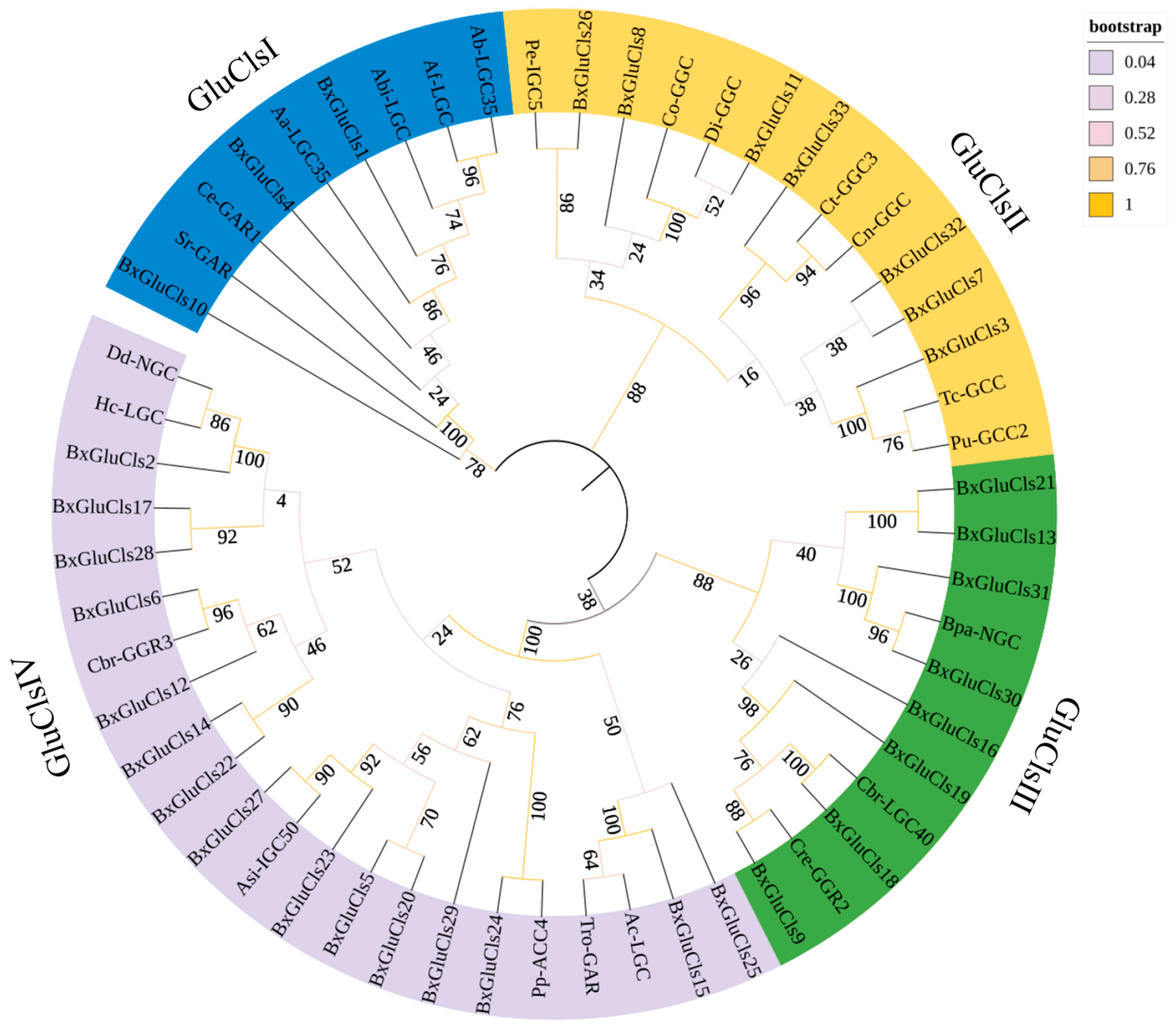
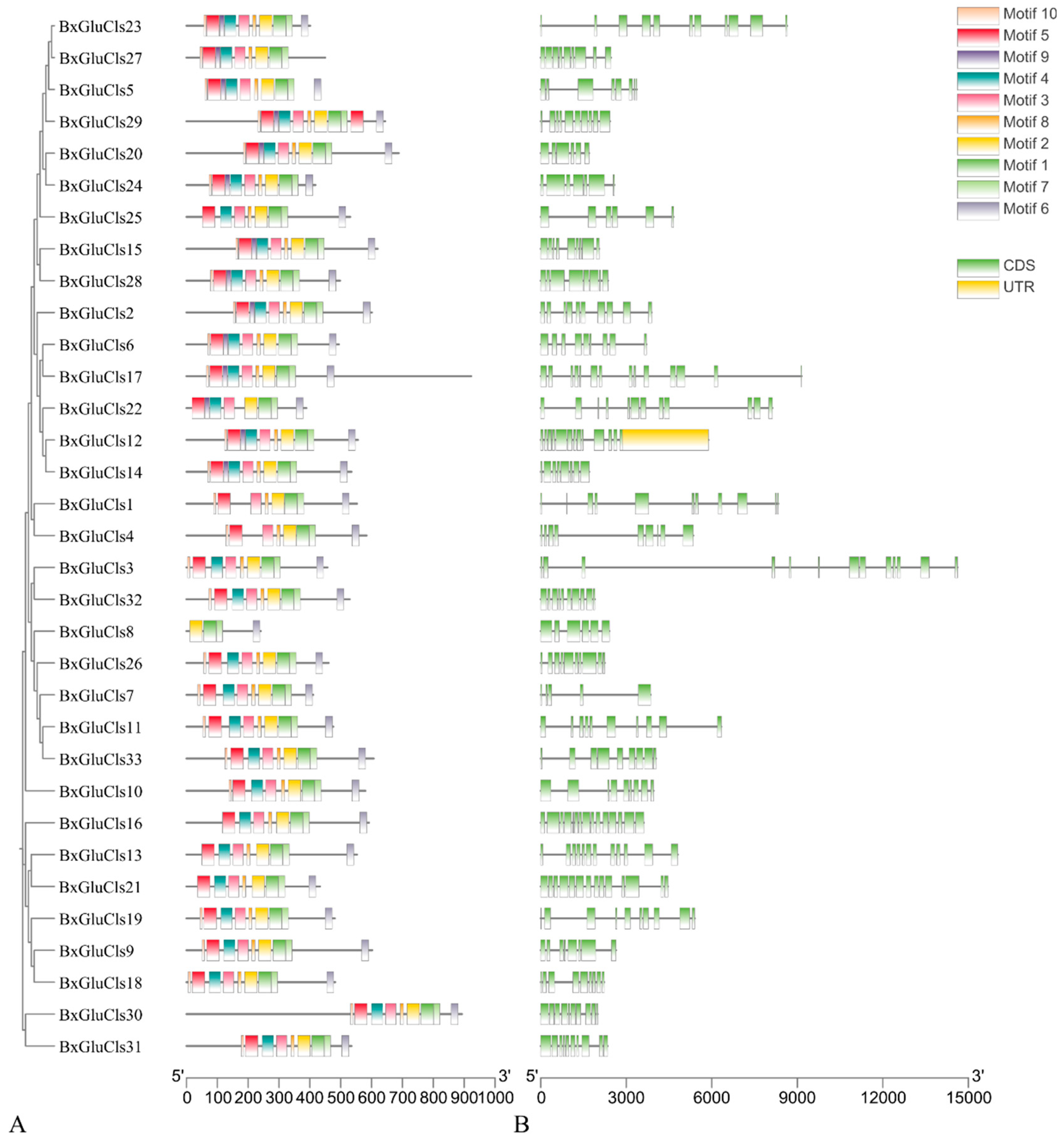
2.3. Phylogenetic and Conserved Motif Analysis of BxGluCls

In order to assess the extent of phylogenetic similarity among the 33 protein sequences, we conducted phylogenetic and conserved motif analyses of BxGluCls. We performed multiple alignments of 33 BxGluCls sequences. The optimal model was chosen according to the maximum likelihood tree. The maximum likelihood evolutionary tree was constructed using the bootstrap test.
Based on the prediction results of the maximum likelihood tree in the best model, the LG+G+F model was selected to construct the phylogenetic evolutionary tree of the BxGluCls family proteins. This model had a BIC (Bayesian Information Criterion) value of 47,331.640 and an AICc (Corrected Akaike Information Criterion) value of 46,368.567. This model incorporates the LG substitution matrix, gamma-distributed rate heterogeneity, and empirical base frequencies. The model was chosen because it provided the lowest BIC value (47,331.640), indicating that it is the best fit for the data among the tested models. Bootstrap values (1000 replicates) were shown at the nodes, representing the statistical support for each branch. The different colors (e.g., blue, green, pink) represent different clades or groups of related sequences within the tree. These colors help to highlight the evolutionary relationships and groupings among the BxGluCls family proteins and their orthologs from other nematodes. Specifically, blue represents GluClsI family, yellow represents GluClsII family, and green and pink represents GluClsIII family and GluClsIV family, indicating distinct evolutionary lineages within the GluCls family (Figure 1).
The phylogenetic tree showed that the BxGluCls family proteins were divided into several major clades. GluCls1, GluCls4, GluCls10 of B. xylophilus, LGC35 of Aphelenchoides besseyi, LGC of Ap. fujianensis, LGC of Ap. bicaudatus, LGC35 of Ap. avenae, GAR1 of Ca. elegans, and GAR of Strongyloides ratti form a well-supported clade, suggesting a close evolutionary relationship. Similarly, GluCls2, GluCls5, GluCls6, GluCls12, GluCls14, GluCls15, GluCls17, GluCls20, GluCls22, GluCls23, GluCls24, GluCls25, GluCls27 of B. xylophilus, NGC of Ditylenchus destructor, LGC of Haemonchus contortus, GGR3 of Ca. briggsae, IGC50 of Anisakis simplex, ACC4 of Pristionchus pacificus, GAR of Trichostrongylus colubriformis and LGC of Ancylostoma caninum clustered together in another distinct clade, indicating potential gene duplication events within this group. GluCls3, GluCls7, GluCls11, GluCls26, GluCls32, GluCls33 of B. xylophilus, IGC5 of Parascaris equorum, GGC of Cooperia oncophora, GGC of Dirofilaria immitis, GGC3 of Cyathostomum tetracanthum, GGC of Cylicocyclus nassatus, GCC of Toxocara canis, and GCC2 of Pa. univalens were grouped together with high sequence similarity and bootstrap support, indicating that they may have arisen from a recent duplication event. Additionally, GluCls9, GluCls13, GluCls16, GluCls18, GluCls19, GluCls21, GluCls30, GluCls31 of B. xylophilus, NGC of Brugia pahangi, LGC40 of Ca. briggsae, and GGR2 of Ca. remanei form a separate clade with a relatively high number of exons, further supporting the idea of gene duplication and subsequent functional diversification. These clades were also supported by high bootstrap values, further validating their robustness.
The compositions of the introns and exons in the BxGluCls family genes were determined by analysis. The results indicated that the number of exons in the 33 BxGluCls genes ranged from 5 to 14. Specifically, BxGluCls7 had only 5 exons, while BxGluCls16, BxGluCls17, and BxGluCls21 had the highest number of exons, totaling 14. A phylogenetic tree illustrates the evolutionary relationships among GluCls family genes from different nematode species, providing insights into the conservation and divergence of intron-exon structures. The analysis of structural motifs in the BxGluCls proteins revealed that these proteins shared similar motifs. The shared structural motifs were confirmed by the analysis of conserved motif structures (Figure 2). The figure illustrates the conserved motifs identified within the BxGluCls family, highlighting the regions of sequence similarity across the proteins. The clustering of these motifs indicated regions of high conservation, suggesting functional importance. The presence of 5’ and 3’ regions in the figure refers to the untranslated regions (UTR) of the gene sequences, while CDS denotes the coding sequence. These elements were included to provide a comprehensive view of the gene structure and to highlight the conserved motifs within the context of the full gene sequence.

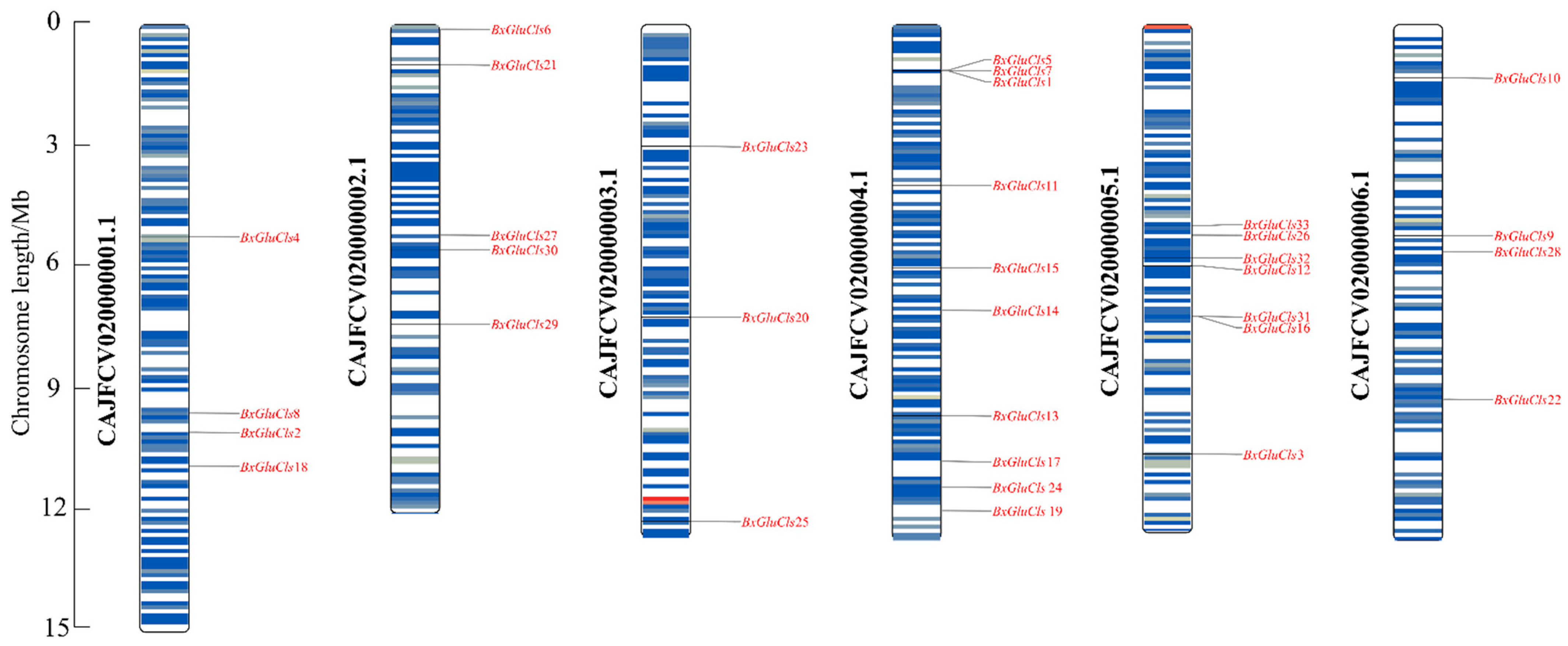
2.4. Chromosomal Distribution of BxGluCls

The chromosomal localization of the 33 BxGluCls genes in B. xylophilus was determined using TBtools v2.056, which identified the precise positions of these channel-encoding genes across different chromosomes (Figure 3). Chromosomal localization analysis revealed that most BxGluCls family genes were widely distributed in chromosomes. For example, BxGluCls4 was located in the middle of chromosome 1, while BxGluCls2, BxGluCls8, and BxGluCls18 were positioned in the 3′ region of chromosome 1. On chromosome 2, BxGluCls6 and BxGluCls21 were found in the 5′ region; BxGluCls27 and BxGluCls30 may be gene duplication events. However, BxGluCls20, BxGluCls23, and BxGluCls25 were dispersed on chromosome 3. On chromosome 4, BxGluCls1, BxGluCls5, and BxGluCls7 were located in the 5′ region, and tandem arrays may exist between them. BxGluCls11, BxGluCls14, and BxGluCls15 were in the central region; and BxGluCls13, BxGluCls17, BxGluCls19, and BxGluCls24 were positioned in the 3′ region. There were possible gene duplication events between BxGluCls26 and BxGluCls33, BxGluCls12 and BxGluCls32, BxGluCls16 and BxGluCls31, in the centromeric region of chromosome 5. BxGluCls9, BxGluCls10, BxGluCls22, and BxGluCls28 were located on chromosome 6. Notably, chromosome 4 contained the highest number of BxGluCls genes, followed by chromosomes 2 and 5, whereas chromosome 3 had the fewest. This distribution pattern suggests potential functional clustering or regulatory mechanisms associated with chromosomal architecture. These findings suggest that the BxGluCls family genes are not only widely distributed across chromosomes but also exhibit complex evolutionary dynamics, including tandem duplication and gene clustering.

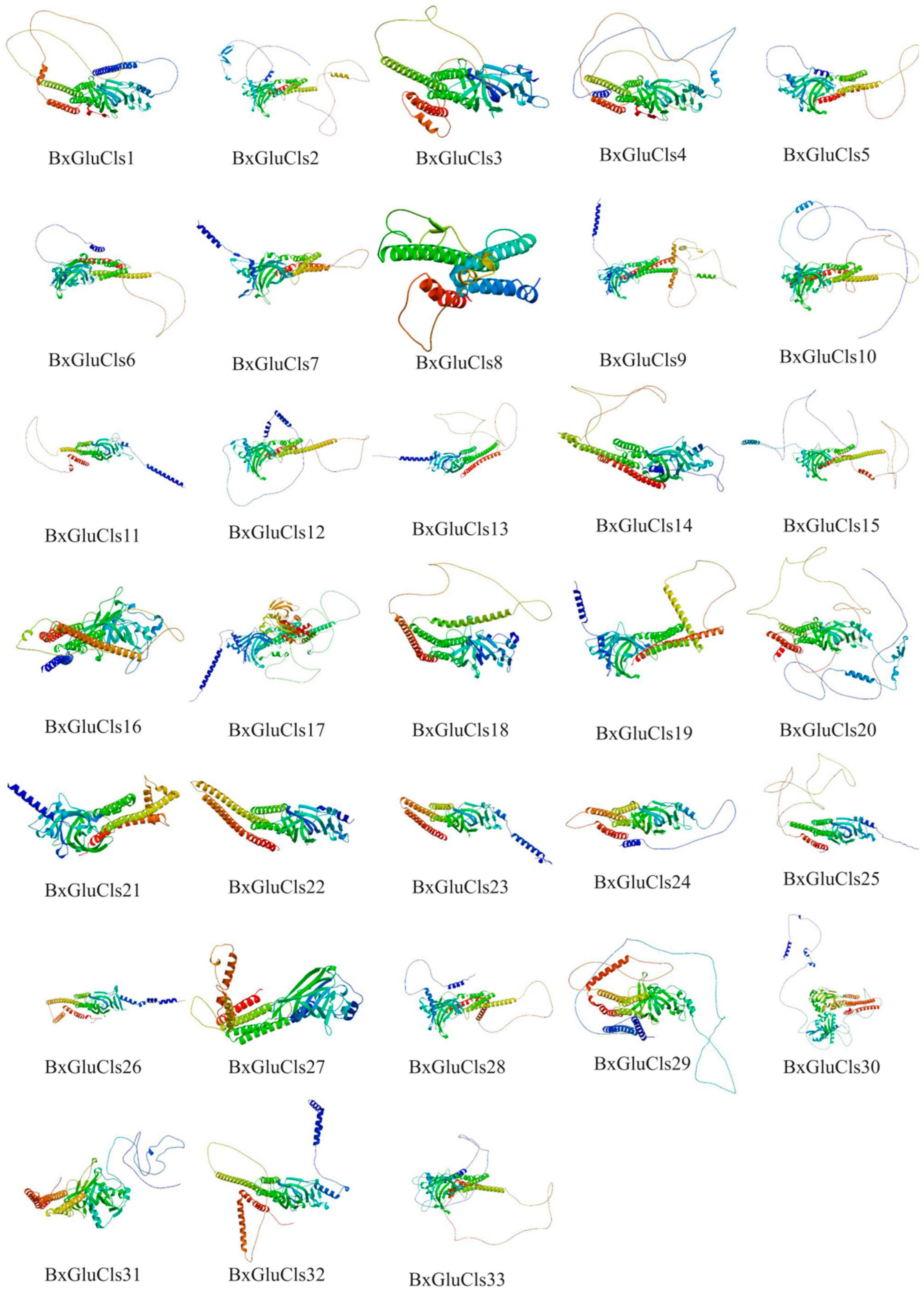
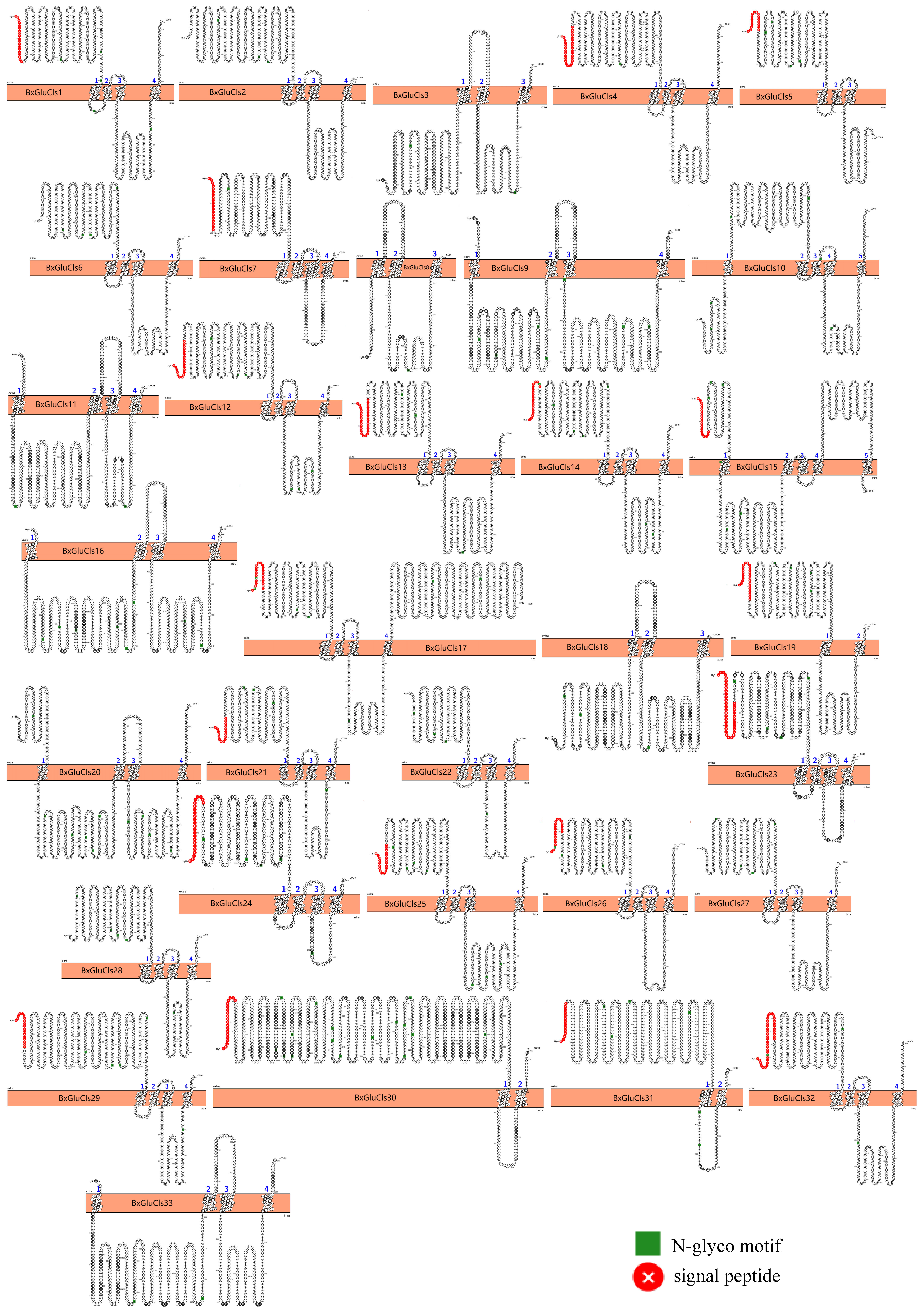
2.5. Protein Structure Analysis of BxGluCls

Our analysis of the secondary structures of 33 BxGluCls proteins revealed that they had similar percentages of α-helices (21.46%–37.70%), β-folds (15.99%–27.51%), β-turs (1.50%–5.53%), and irregular coils (35.97%–59.16%). Moreover, through protein 3D structure prediction, we suggest that BxGluCls family proteins have small structural differences, i.e., their conformations are very similar. This implies that their functions are also similar. A detailed 3D structural analysis of BxGluCls proteins provides a comprehensive view of their key structural elements and functional domains. The high conservation of the pore region, ligand-binding pocket, and Cys-loop supports their classification as Cys-loop ligand-gated ion channels. These findings further validate our motif analysis and phylogenetic clustering results, highlighting the functional similarity and potential unique adaptations of BxGluCls proteins. The 33 BxGluCls proteins exhibited high sequence similarity. Despite this overall similarity, the number of potential N-glycosylation sites varied from one to 16 among the proteins, indicating functional diversity. Proteins with higher numbers of N-glycosylation sites may have additional roles in cellular signaling or structural stability (Table S1, Figure 4 and Figure 5).

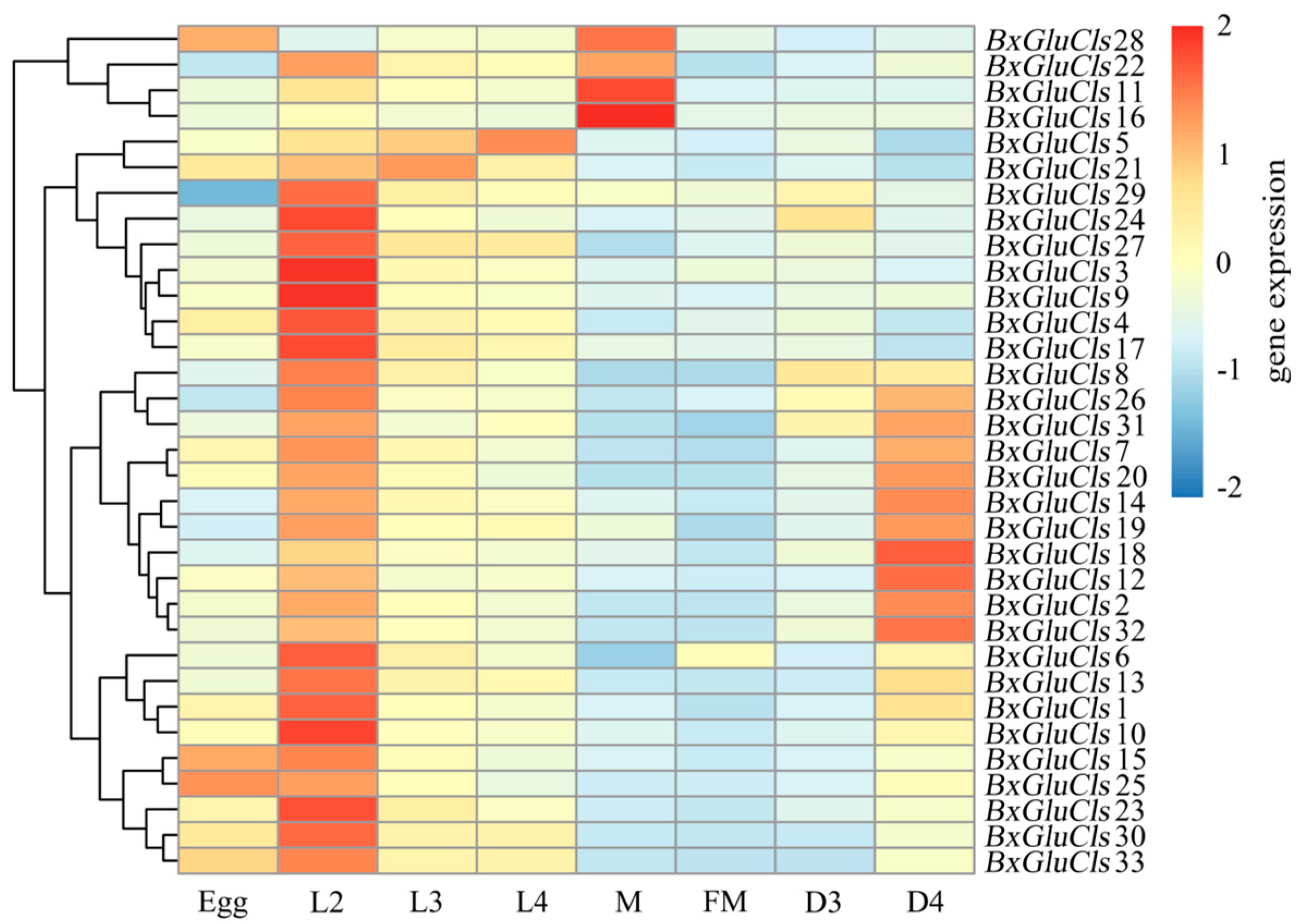
2.6. Expression Profiles of BxGluCls in Different Stages

We were interested to see whether the gene expression of BxGluCls alters under different stages of the worm life cycle. To analyze gene expression in different developmental stages of B. xylophilus, we employed semi-quantitative reverse transcription polymerase chain reaction (RT-qPCR). We have also investigated the alterations in the gene expression of BxGluCls under the stress conditions. This approach allowed us to visualize the expression patterns of the 33 BxGluCls genes across different developmental stages and conditions. By analyzing gene expression data from B. xylophilus under various nematode states and stress conditions, the expression data of 33 BxGluCls genes were obtained. Heat map analysis (Figure 6) revealed significant variations in BxGluCls gene expression across different nematode states. Specifically, BxGluCls15, BxGluCls25, and BxGluCls28 were highly expressed in nematode eggs, suggesting their involvement in egg development. Except for BxGluCls5, BxGluCls11, BxGluCls16, and BxGluCls28, all other BxGluCls genes exhibited high expression in the second-instar larvae of adult B. xylophilus. Conversely, except for BxGluCls5, BxGluCls21, the expression of other BxGluCls genes were lower in the third and fourth-instar larvae during the growth period. The findings indicated that BxGluCls played a more significant role in the developmental process of B. xylophilus and had a reduced influence on the third and fourth instar larva. BxGluCls12, BxGluCls18, and BxGluCls32 were highly expressed in dauer fourth-instar larva (D4) B. xylophilus, suggesting their role in nematode resistance and adaptation to adverse environments. BxGluCls11, BxGluCls16, BxGluCls22, and BxGluCls28 were highly expressed in male adult nematodes (M), indicating their association with sex differentiation, particularly in male formation. Regarding the gene expression, the egg stage was totally different than the L2 stage, i.e., the first larvae stage. So, the transition from an egg to a larva was marked by a significant change in the expression pattern of 33 GluCls forms. L3 and L4 were quite similar, displaying no or very few visible changes in the GluCls gene expression pattern. However, when a larva became an adult, male or female, the transition was accompanied by a marked change in the gene expression pattern.

3. Discussion

Since its initial discovery in China in 1982, B. xylophilus has caused numerous major plant epidemics, spreading rapidly and posing a serious threat to China’s forestry production and ecological security [18]. Controlling B. xylophilus remains a significant challenge due to the emergence of pesticide resistance and environmental concerns associated with chemical treatments [19]. The invasion of alien species into new environments often leads to significant changes in gene families, which are crucial for their adaptation and evolution [20]. This study suggests that targeting GluCls could offer a novel and selective approach to managing PWD. Previous studies have demonstrated that GluCls are essential ion channels found exclusively in invertebrates, making them ideal targets for selective insecticides and nematicides. These channels are widely distributed in the nervous systems of nematodes, where they regulate various physiological processes, including movement, feeding, and sensation [21]. However, few studies have investigated the function of GluCls as an insecticide target in plant-parasitic nematodes [22]. A GluCls subunit database, based on information from 125 nematodes, has been published [23]. Additionally, the expression of GluCls genes has been shown to be up-regulated in B. xylophilus [24]. Therefore, elucidating the role of GluCls as a potential target for B. xylophilus is of paramount scientific and practical significance. In this study, bioinformatics analysis of the GluCls family in B. xylophilus provides valuable insights into its role in the nematode’s development, stress response, and potential as a target for novel biopesticides.
GluCls are known to play a critical role in neural signaling and muscle function in nematodes, with their selective expression patterns suggesting potential involvement in specific developmental processes. The expression dynamics of GluCls across developmental stages in B. xylophilus highlight their regulatory roles in nematode ontogeny, particularly in larval maturation and reproductive processes. The high expression of BxGluCls15, BxGluCls25, and BxGluCls28 in eggs indicates their potential contribution to early embryonic development. This finding aligns with studies in C. elegans, which demonstrate GluCls are essential for embryonic development and larval growth [25,26]. In nematodes, GluCls modulate ion gradients critical for oocyte maturation and embryonic viability, likely through calcium signaling pathways associated with eggshell formation [27]. Notably, the temporal specificity of these genes suggests stage-dependent regulatory mechanisms, potentially coordinated with hormonal cues such as ecdysteroids, which are pivotal in arthropod molting and may share conserved signaling cascades in ecdysozoans [28]. Similarly, the elevated expression of BxGluCls12, BxGluCls18, and BxGluCls32 in dauer fourth-instar larva (D4) implies their role in resistance and adaptation to environmental stressors. The lower expression of some GluCls genes in adult stages suggests a diminished role in mature nematodes, consistent with findings in Haemonchus contortus, where certain GluCls subunits are predominantly expressed in larva [29]. Although sexual dimorphism in B. xylophilus remains underexplored, the differential expression of GluCls in larval stages hints at their potential involvement in sex-specific neural circuit maturation. The enrichment of BxGluCls25 in early larvae may influence gender-specific behaviors or gonad development, as observed in insects, where glutamate signaling regulates sex-determination pathways [30]. Future studies integrating single-cell transcriptomics could clarify whether specific GluCls isoforms are localized to reproductive tissues or sexually dimorphic neurons, providing insights into their roles in gametogenesis or mating behaviors.
Phylogenetic analysis revealed distinct clades within the BxGluCls family, indicating evolutionary conservation and divergence. Conserved motif analysis further highlighted the importance of specific structural elements for channel function, with variations in motifs potentially linked to functional adaptations [31]. This suggested that while some GluCls retain conserved functions, others have specialized roles in specific developmental stages or stress responses. The conserved structural features of BxGluCls proteins, as evidenced by their similar physicochemical properties and gene structure, suggested a high degree of functional conservation within this family. This conservation was consistent with the critical role of GluCls in neural signaling and muscle function across nematodes. The stage-specific expression patterns of BxGluCls genes further supported the hypothesis that these channels are involved in specific developmental processes and stress responses. The topological prediction of BxGluCls proteins revealed a canonical structure consistent with Cys-loop ligand-gated ion channels, with four transmembrane domains and extracellular N- and C-termini [32]. The unique features suggested potential functional adaptations specific to these proteins. These findings provided a structural basis for understanding the function and regulation of BxGluCls proteins.
By elucidating the structure, function, and expression patterns of BxGluCls, future research could focus on developing biopesticides that selectively target these channels, thereby reducing the survival and reproductive capabilities of B.xylophilus. This study provides a comprehensive characterization of the BxGluCls family in B. xylophilus, highlighting their conserved structural features and distinct expression patterns. These findings not only advance our understanding of the molecular mechanisms underlying the development and stress response of B. xylophilus but also offer valuable insights for the development of targeted control strategies. Furthermore, investigating the mechanisms responsible for the stage-specific expression and resistance-associated functions of BxGluCls genes could further clarify their role in the biology of B. xylophilus.

4. Materials and Methods

4.1. Identification of BxGluCls Family Members and Analysis of Their Physicochemical Properties

Firstly, we queried and downloaded the GluCls gene sequences of Ap. bicaudatus, Ap. fujianensis, Ap. besseyi, Ap. avenae, Anisakis simplex, C. elegans, Brugia malayi, Ca. bimaculatus, Ca. teleostei, Ditylenchus destructor and Parascaris equorum from NCBI database (https://www.ncbi.nlm.nih.gov/gene, accessed on 21 March 2025). Then we employed BLASTX to align the obtained BxGluCls gene sequences in B. xylophilus genome (PRJEB40022) by Wormbase (version: WBPS19) (https://parasite.wormbase.org, accessed on 21 March 2025) [33]. Then we characterized these candidate sequences used NCBI-conserved domains 3.21 (https://www.ncbi.nlm.nih.gov/Structure/cdd, accessed on 21 March 2025) [34] and InterPro 101.0 (https://www.ebi.ac.uk/ Interpro, accessed on 21 March 2025) [35]. This process helped to remove redundant gene sequences and obtain the protein sequences of BxGluCls. The physicochemical properties of BxGluCls were further analyzed by Expasy 3.0 (https://web.expasy.org/protparam, accessed on 21 March 2025) [36].

4.2. Phylogenetic Analysis of the BxGluCls Family and Analysis of Conserved Motifs

To elucidate the evolutionary relationships of BxGluCls, MEGA 11.0 software was employed to conduct multiple sequence alignments of 33 BxGluCls. The optimal model for the maximum likelihood tree was identified, and a maximum likelihood evolutionary tree was constructed, with the Bootstrap test performed 1000 times [37]. Furthermore, gene information for BxGluCls was extracted from the gene structure annotation file. Gene structure visualization and analysis were performed using the online tool GSDS 2.0 (http://gsds.gao-lab.org/, accessed on 21 March 2025) [38]. In parallel, MEME Suite 5.5.6 (https://meme-suite.org/meme/, accessed on 21 March 2025) [39] was used to analyze the distribution of conserved motifs in BxGluCls proteins, with the motif value set to 10 and other parameters set to default. The results from MEME were integrated with the maximum likelihood tree to generate gene structure maps annotated with conserved motif structures.

4.3. Chromosomal Distribution and Protein Structure Analysis of the BxGluCls Family

We screened the gene family members from gene structure annotation files. BxGluCls annotation information was extracted for information including chromosome location, start and termination sites, and gene length. Based on this information, a distribution density map of BxGluCls on B. xylophilus chromosomes was created by TBtools [40,41]. Subsequently, the secondary structure analysis of BxGluCls was conducted using online tools such as Prabi (last modification on September 2022, accessed on 21 March 2025) (https://doua.prabi.fr/software/cap3) [42], NetPhos 3.1 (https://services.healthtech.dtu.dk/services/NetPhos-3.1/, accessed on 21 March 2025) [43], and Protter 1.0 (http://wlab.ethz.ch/protter/start/, accessed on 21 March 2025) [44]. Additionally, Swiss-Model (https://swissmodel.expasy.org/interactive, accessed on 21 March 2025) [45] was utilized to predict the protein tertiary structure.

4.4. Expression Profiles of BxGluCls Family in Different Developmental Stages

To analyze gene expression in different developmental stages using semi-quantitative reverse transcription polymerase chain reaction (RT-qPCR), B. xylophilus at various developmental stages were synchronized. For embryos, synchronized eggs were obtained by placing mixed-stage B. xylophilus in petri dishes at 25 °C in the dark for 10 min. This allowed the eggs laid by pregnant females to adhere to the bottom due to surface glycoproteins. The upper layer of water and nematodes was carefully removed to collect the synchronized eggs. To elucidate the molecular response mechanisms of BxGluCls genes to different treatments, expression values of 33 BxGluCls were obtained from various insect states of B. xylophilus, including eggs. The resulting expression data were normalized and visualized using the Heatmap Plots tool in R-Studio 4.4.3 (http://cran.r-project.org, accessed on 21 March 2025) [46]. The Heatmap was constructed using the Complex Heatmap package in R, which supports hierarchical clustering with great flexibility. To obtain second instar larvae (L2), the synchronized eggs were placed in a food-free environment and allowed to hatch at 25 °C in the dark. The synchronized L2 were transferred onto a lawn of B. cinerea on a PDA plate. Nematodes were collected after 24, 48, and 72 h to obtain third instar larva (L3), fourth instar larvae (L4), and adults, respectively. The dauer third-instar larva (D3) and the dauer fourth-instar larva (D4) were isolated from infected pines. The male and female nematodes were obtained by manually picking them under a microscope using worm pickers.

5. Conclusions

In this study, we screened and characterized the BxGluCls family based on the GluCls family within the B. xylophilus genome, identifying 33 BxGluCls family members. Our analysis of the physicochemical properties of these proteins revealed that the number of amino acids ranged from 241 to 923, with molecular masses ranging from 27.29 to 105.70 kDa. As shown in Figure 3, a higher number of BxGluCls genes were distributed on chromosomes 2, 4, and 5. Gene structure and conserved motif analyses indicated that BxGluCls family members exhibited highly similar structures, suggesting evolutionary conservation within nematodes. Predictions of protein secondary and tertiary structures revealed minimal structural differences among BxGluCls family proteins, indicating functional stability and high synergy. We also analyzed the expression patterns of BxGluCls genes across various developmental stages of the nematode. These findings demonstrate that GluCls play a crucial role in the growth, development, and resistance mechanisms of B. xylophilus. This study provides a foundation for further research into the functional roles of BxGluCls. In summary, this study confirms that BxGluCls gene family members are essential for nematode embryonic development, growth, and sexual differentiation. However, their role in the immune processes of B. xylophilus requires further investigation.

Supplementary Materials

The following supporting information can be downloaded at: https://www.mdpi.com/article/10.3390/ijms26083477/s1.

Author Contributions

Conceptualization, X.H. and J.C.; methodology, R.W.; software, H.L.; validation, H.L., R.W. and J.C.; formal analysis, H.L.; investigation, J.P.; resources, J.P.; data curation, H.L.; writing—original draft preparation, H.L. and X.H.; writing—review and editing, J.P.; visualization, R.W. and J.C.; supervision, J.C.; project administration, J.P.; funding acquisition, J.P. All authors have read and agreed to the published version of the manuscript.

Funding

The study supported by Key Laboratory of National Forestry and Grassland Administration on Prevention and Control Technology of Pine Wilt Disease (202403); Yunnan Fundamental Research Projects (202401BD070001-115); Southwest Forestry University Forestry major in Yunnan Province First-Class Construction Discipline (LXXK-2023M06, LXXK-2024Z04).

Institutional Review Board Statement

Not applicable.

Informed Consent Statement

Not applicable.

Data Availability Statement

No data was used for the research described in the article.

Conflicts of Interest

The authors declare no conflicts of interest.

References

  1. Nickle, W.R.; Golden, A.M.; Mamiya, Y.; Wergin, W.P. On the Taxonomy and morphology of the pine wood nematode, Bursaphelenchus xylophilus (Steiner &Buhrer 1934) Nickle 1970. J. Nematol. 1970, 13, 385–392. [Google Scholar]
  2. Nickle, W.R. A taxonomic review of the genera of the Aphelenchoidae (Fuchs, 1937) Thorne, 1949 (Nematoda: Tylenchida). J. Nematol. 1970, 2, 374–392. [Google Scholar]
  3. Li, S.; Sun, H.; Zhou, Y.; Li, X.; Yu, Z.; Dong, Z. Occurrence of major forest pests in China in 2021 and prediction of occurrence trend in 2022. For. Pest Dis. 2022, 41, 44–47. [Google Scholar]
  4. Zhou, Y.; Danbolt, N.C. Glutamate as a neurotransmitter in the healthy brain. J. Neural Transm. 2014, 121, 799–817. [Google Scholar] [CrossRef] [PubMed]
  5. Cleland, T.A. Inhibitory glutamate receptor channels. Mol. Neurobiol. 1996, 13, 97–136. [Google Scholar] [CrossRef]
  6. Wolstenholme, A.J. Glutamate-gated chloride channels. J. Biol. Chem. 2012, 287, 40233–40238. [Google Scholar] [CrossRef]
  7. Zhang, X.; Zhang, L.; Si, Y.; Wen, X.; Wang, L.; Song, L. Unveiling the functional diversity of ionotropic glutamate receptors in the Pacific Oyster (Crassostrea gigas) by systematic studies. Front. Physiol. 2023, 14, 1280553. [Google Scholar] [CrossRef]
  8. Jones, A.K.; Sattelle, D.B. The cys-loop ligand-gated ion channel gene superfamily of the red flour beetle, Tribolium castaneum. BMC Genom. 2007, 8, 327. [Google Scholar] [CrossRef]
  9. Yates, D.M.; Wolstenholme, A.J. An ivermectin-sensitive glutamate-gated chloride channel subunit from Dirofilaria immitis. Int. J. Parasitol. 2004, 34, 1075–1081. [Google Scholar] [CrossRef]
  10. Cheeseman, C.L.; Delany, N.S.; Woods, D.J.; Wolstenholme, A.J. High-affinity Ivermectin Binding to Recombinant Subunits of the Haemonchus contortus Glutamate-gated Chloride Channel. Mol. Biochem. Parasitol. 2001, 114, 161–168. [Google Scholar] [CrossRef]
  11. Wolstenholme, A.J.; Rogers, A.T. Glutamate-gated chloride channels and the mode of action of the avermectin/milbemycin an-thelmintics. Parasitology 2005, 131, S85–S95. [Google Scholar] [PubMed]
  12. Tandon, R.; LePage, K.T.; Kaplan, R.M. Cloning and characterization of genes encoding α and β subunits of glutamate-gated chloride channel protein in Cylicocyclus nassatus. Mol. Biochem. Parasitol. 2006, 150, 46–55. [Google Scholar] [CrossRef] [PubMed]
  13. Cully, D.F.; Vassilatis, D.K.; Liu, K.K.; Paress, P.S.; Van der Ploeg, L.H.; Schaeffer, J.M.; Arena, J.P. Cloning of an avermectin-sensitive glutamate-gated chloride channel from Caenorhabditis elegans. Nature 1994, 371, 707–711. [Google Scholar] [CrossRef]
  14. Cully, D.F.; Wilkinson, H.; Vassilatis, D.K.; Etter, A.; Arena, J.P. Molecular biology and electrophysiology of glutamate-gated chloride channels of invertebrates. Parasitology 1996, 113, S191–S200. [Google Scholar] [CrossRef]
  15. Kane, N.S.; Hirschberg, B.; Qian, S.; Hunt, D.; Thomas, B.; Brochu, R.; Ludmerer, S.W.; Zheng, Y.; Smith, M.; Arena, J.P.; et al. Drug-resistance Drosophila indicate glutamate-gated chloride channels are targets for antiparasitics nodulisporic acid and ivermectin. Proc. Natl. Acad. Sci. USA 2000, 97, 13949–13954. [Google Scholar]
  16. Vassilatis, D.K.; Arena, J.P.; Plasterk, R.H.; Wilkinson, H.A.; Schaeffer, J.M.; Cully, D.F.; Van der Ploeg, L.H. Genetic and biochemical evidence for a novel avermectin-sensitive chloride channel in Caenorhabditis elegans isolation and characterization. J. Biol. Chem. 1997, 272, 33167–33174. [Google Scholar]
  17. Guo, L.; Tang, B.; Dong, W.; Liang, P.; Gao, X. Cloning and sequence analysis of cDNA of GluCl receptor alpha gene from the Diamondback moth, Plutella xylostella. Plant Prot. 2010, 36, 49–54. [Google Scholar]
  18. Feng, Y.; Jian, W.; Ding, X.; Ye, J. Genetic Diversity and Population Structure of Bursaphelenchus xylophilus in Guangdong, Guangxi, and Jiangsu Provinces in China. Forests 2024, 15, 934. [Google Scholar] [CrossRef]
  19. Blomme, T.; Vandepoele, K.; De Bodt, S.; Simillion, C.; Maere, S.; Van de Peer, Y. The gain and loss of genes during 600 million years of vertebrate evolution. Genome Biol. 2006, 7, R43. [Google Scholar] [CrossRef]
  20. Meng, F.; Tian, C. Gene Family Expansion during the Adaptation of Colletotrichum gloeosporioides to Woody Plants. J. Fungi 2023, 9, 1185. [Google Scholar] [CrossRef]
  21. Qian, K.; Jiang, C.; Guan, D.; Zhuang, A.; Meng, X.; Wang, J. Characterization of Glutamate-Gated Chloride Channel in Tribolium castaneum. Insects 2023, 14, 580. [Google Scholar] [CrossRef]
  22. Huang, X.; Li, Q.; Chen, J.; Liu, W.; Guo, K.; Hu, J.; Shao, H. Molecular characterization and functional analysis of genes mediating emamectin benzoate action to the pinewood nematode (Bursaphelenchus xylophilus). Pestic. Biochem. Physiol. 2024, 205, 106148. [Google Scholar] [CrossRef] [PubMed]
  23. O’halloran, D.M. Database of glutamate-gated chloride (GluCl) subunits across 125 nematode species: Patterns of gene accretion and sequence diversification. G3 Genes Genomes Genet. 2022, 12, jkab438. [Google Scholar] [CrossRef] [PubMed]
  24. Lu, F.; Guo, K.; Chen, A.; Chen, S.; Lin, H.; Zhou, X. Transcriptomic profiling of effects of emamectin benzoate on the pine wood nematode Bursaphelenchus xylophilus. Pest. Manag. Sci. 2020, 76, 747–757. [Google Scholar] [CrossRef]
  25. Dent, J.A.; Smith, M.M.; Vassilatis, D.K.; Avery, L. The genetics of ivermectin resistance in Caenorhabditis elegans. Proc. Natl. Acad. Sci. USA. 2000, 97, 2674–2679. [Google Scholar] [CrossRef] [PubMed]
  26. Cook, A.; Aptel, N.; Portillo, V.; Siney, E.; Sihota, R.; Holden-Dye, L.; Wolstenholme, A. Caenorhabditis elegans ivermectin receptors regulate locomotor behaviour and are functional orthologues of Haemonchus contortus receptors. Mol. Biochem. Parasitol. 2006, 147, 118–125. [Google Scholar] [CrossRef]
  27. Knouse, M.C.; Deutschmann, A.U.; Nenov, M.N.; Wimmer, M.E.; Briand, L.A. Sex differences in pre- and post-synaptic glutamate signaling in the nucleus accumbens core. Biol. Sex Differ. 2023, 14, 52. [Google Scholar] [CrossRef]
  28. Louro, M.; Kuzmina, T.A.; Bredtmann, C.M.; Diekmann, I.; de Carvalho, L.M.M.; von Samson-Himmelstjerna, G.; Krücken, J. Genetic variability, cryptic species and phylogenetic relationship of six cyathostomin species based on mitochondrial and nuclear sequences. Sci. Rep. 2021, 11, 8245. [Google Scholar] [CrossRef]
  29. Jagannathan, S.; Laughton, D.L.; Critten, C.L.; Skinner, T.M.; Horoszok, L.; Wolstenholme, A.J. Ligand-gated chloride channel subunits encoded by the Haemonchus contortus and Ascaris suum orthologues of the Caenorhabditis elegans gbr-2 (avr-14) gene. Mol. Biochem. Parasitol. 1999, 103, 129–140. [Google Scholar] [CrossRef]
  30. Lord, A.; Cunha, T.J.; Medeiros, B.A.S.d.; Sato, S.; E Khost, D.; Sackton, T.B.; Giribet, G. Expanding on Our Knowledge of Ecdysozoan Genomes: A Contiguous Assembly of the Meiofaunal Priapulan Tubiluchus corallicola. Genome Biol. Evol. 2023, 15, 103. [Google Scholar] [CrossRef]
  31. Vandenhole, M.; Mermans, C.; De Beer, B.; Xue, W.; Zhao, Y.; Ozoe, Y.; Liu, G.; Dermauw, W.; Van Leeuwen, T. A glutamate-gated chloride channel as the mite-specific target-site of dicofol and other diphenyl carbinol acaricides. Commun. Biol. 2023, 6, 1160. [Google Scholar] [CrossRef] [PubMed]
  32. Wong, M.G.; The, N.L.; Glastras, S. Maternal obesity and offspring risk of chronic kidney disease. Nephrology 2018, 4, 84–87. [Google Scholar]
  33. Howe, K.L.; Bolt, B.J.; Shafie, M.; Kersey, P.; Berriman, M. WormBase ParaSite: A comprehensive resource for helminth genomics. Mol. Biochem. Parasitol. 2017, 215, 2–10. [Google Scholar] [PubMed]
  34. Lu, S.; Wang, J.; Chitsaz, F.; Derbyshire, M.K.; Geer, R.C.; Gonzales, N.R.; Gwadz, M.; I Hurwitz, D.; Marchler, G.H.; Song, J.S.; et al. CDD/SPARCLE: The conserved domain database in 2020. Nucleic Acids Res. 2020, 48, D265–D268. [Google Scholar]
  35. Cantelli, G.; Bateman, A.; Brooksbank, C.; I Petrov, A.; Malik-Sheriff, R.S.; Ide-Smith, M.; Hermjakob, H.; Flicek, P.; Apweiler, R.; Birney, E.; et al. The European Bioinformatics Institute (EMBL-EBI) in 2021. Nucleic Acids Res. 2022, 50, D11–D19. [Google Scholar] [CrossRef]
  36. Duvaud, S.; Gabella, C.; Lisacek, F.; Stockinger, H.; Ioannidis, V.; Durinx, C. Expasy, the Swiss Bioinformatics Resource Portal, as designed by its users. Nucleic Acids Res. 2021, 49, W216–W227. [Google Scholar] [CrossRef]
  37. Tamura, K.; Stecher, G.; Kumar, S. MEGA11: Molecular Evolutionary Genetics Analysis Version 11. Mol. Biol. Evol. 2021, 38, 3022–3027. [Google Scholar] [CrossRef]
  38. Hu, B.; Jin, J.; Guo, A.-Y.; Zhang, H.; Luo, J.; Gao, G. GSDS 2.0: An upgraded gene feature visualization server. Bioinformatics 2015, 31, 1296–1297. [Google Scholar] [CrossRef]
  39. Bailey, T.L.; Johnson, J.; Grant, C.E.; Noble, W.S. The MEME suite. Nucleic Acids Res. 2015, 43, 39–49. [Google Scholar]
  40. Chen, C.; Chen, H.; Zhang, Y.; Thomas, H.R.; Frank, M.H.; He, Y.; Xia, R. TBtools: An integrative toolkit developed for interactive analyses of big biological data. Mol. Plant 2020, 13, 1194–1202. [Google Scholar] [CrossRef]
  41. Chen, C.; Wu, Y.; Li, J.; Wang, X.; Zeng, Z.; Xu, J.; Liu, Y.; Feng, J.; Chen, H.; He, Y.; et al. TBtools-II: “A one for all, all for one” bioinformatics platform for biological big-data mining. Mol. Plant 2023, 16, 1733–1742. [Google Scholar] [CrossRef] [PubMed]
  42. Guirimand, T.; Delmotte, S.; Navratil, V. VirHostNet 2.0: Surfing on the web of virus/host molecular interactions data. Nucleic Acids Res. 2015, 43, 583–587. [Google Scholar] [CrossRef] [PubMed]
  43. Blom, N.; Gammeltoft, S.; Brunak, S. Sequence and structure-based prediction of eukaryotic protein phosphorylation sites 11 Edited by F. E. Cohen. J. Mol. Biol. 1999, 294, 1351–1362. [Google Scholar] [CrossRef]
  44. Omasits, U.; Ahrens, C.H.; Müller, S.; Wollscheid, B. Protter: Interactive protein feature visualization and integration with experimental proteomic data. Bioinformatics 2014, 30, 884–886. [Google Scholar] [CrossRef] [PubMed]
  45. Biasini, M.; Bienert, S.; Waterhouse, A.; Arnold, K.; Studer, G.; Schmidt, T.; Kiefer, F.; Cassarino, T.G.; Bertoni, M.; Bordoli, L.; et al. SWISS-MODEL: Modelling protein tertiary and quaternary structure using evolutionary information. Nucleic Acids Res. 2014, 42, W252–W258. [Google Scholar] [CrossRef]
  46. Gu, Z.G. Complex heatmap visualization. iMeta 2022, 1, e43. [Google Scholar] [CrossRef]
Figure 1. Phylogenetic tree of BxGluCls and GluCls in other nematodes. Note: Bx represents Bursaphelenchus xylophilus, Aa represents Aphelenchoides avenae, Abi represents Aphelenchoides bicaudatus, Ab represents Aphelenchoides besseyi, Ac represents Ancylostoma caninum, Af represents Aphelenchoides fujianensis, Asi represents Anisakis simplex, Bpa represents Brugia pahangi, Cbr represents Caenorhabditis briggsae, Ce represents Caenorhabditis elegans, Cn represents Cylicocyclus nassatus, Co represents Cooperia oncophora, Cre represents Caenorhabditis remanei, Ct represents Cyathostomum tetracanthum, Dd represents Ditylenchus destructor, Di represents Dirofilaria immitis, Hc represents Haemonchus contortus, Pe represents Parascaris equorum, Pp represents Pristionchus pacificus, Pu represents Parascaris univalens, Sr represents Strongyloides ratti, Tc represents Toxocara canis, and Tro represents Trichostrongylus colubriformis.
Ijms 26 03477 g001
Figure 2. Comparison of the structure motifs distribution across the 33 BxGluCls proteins. Note: (A) Motif Distribution: This panel illustrates the distribution of conserved motifs across the 33 BxGluCls proteins. (B) Exon-Intron Structure: This panel shows the exon-intron structure of the BxGluCls genes, depicted in green. CDS (Coding Sequence): This refers to the portion of the gene sequence that is translated into the protein. It includes the exons and excludes the introns. UTR (Untranslated Region): These are regions of the mRNA that are not translated into protein. The 5’ UTR is located upstream of the coding sequence, while the 3’ UTR is located downstream. These regions can play important roles in gene regulation. Motifs: These are short, conserved sequences that often have functional significance. In the context of our study, the motifs refer to conserved sequence patterns within the BxGluCls proteins.
Ijms 26 03477 g002
Figure 3. Chromosomal position of BxGluCls. Note: Each column represents a different chromosome, labeled from 1 to 6. The vertical axis represents the relative positions of genes along the chromosomes, with 5’ and 3’ regions indicated.
Ijms 26 03477 g003
Figure 4. Predicted three-dimensional structures of BxGluCls, showing α-helices, β-folded structures, β-turns, and irregular(random) coils.
Ijms 26 03477 g004
Figure 5. Predicted topologies of 33 BxGluCls proteins. Note: Glycosylation sites are indicated in green, the cell membrane is shown in orange, and signal peptides are highlighted in red.
Ijms 26 03477 g005
Figure 6. Expression pattern of BxGluCls in different ages. Note: Egg was egg stage of B. xylophilus; M was male adult of B. xylophilus; FM was female adult of B. xylophilus; L2 was second instar larva; L3 was third instar larva; L4 was fourth instar larva; D3 was dauer third-instar larva; D4 was dauer fourth-instar larva.
Ijms 26 03477 g006
Table 1. Comparison of glutamate-gated chloride ion channels (GluCls) from B. xylophilus and those from other nematodes.
Protein ID of B. xylophilusProtein NameSequence ID in NCBIProtein Name in Other NematodesOther Nematodes NameScores of BlastP 1Similarity E-Value 2
BXYJ5.040019200BxGluCls1KAI6177746.1Ligand-Gated ion ChannelAphelenchoides bicaudatus6830
BXYJ5.010220700BxGluCls2CRZ22203.1BMA-AVR-14, isoform bBrugia malayi4050
BXYJ5.050236700BxGluCls3KAI6190451.1Ligand-gated ion channel 50Aphelenchoides bicaudatus7960
BXYJ5.010094300BxGluCls4KAI6171507.1Glutamate-gated chloride channel subunit betaAphelenchoides bicaudatus5750
BXYJ5.040019000BxGluCls5KAI1719268.1Neurotransmitter-gated ion-channel ligand binding domain-containing proteinDitylenchus destructor5960
BXYJ5.020004500BxGluCls6KAI1720087.1Neurotransmitter-gated ion-channel ligand binding domain-containing proteinDitylenchus destructor5550
BXYJ5.040019100BxGluCls7KAI1720087.1Neurotransmitter-gated ion-channel ligand binding domain-containing proteinDitylenchus destructor5550
BXYJ5.010211400BxGluCls8KAI6226376.1Glutamate-gated chloride channelAphelenchoides fujianensis2479 × 10−80
BXYJ5.060084300BxGluCls9KAI6175995.1Protein CBR-GGR-2Aphelenchoides bicaudatus7210
BXYJ5.060015200BxGluCls10KAI6207390.1Unc-49B proteinAphelenchoides besseyi7660
BXYJ5.040069900BxGluCls11KAI6226375.1BMA-AVR-14, isoform bAphelenchoides fujianensis7370
BXYJ5.050134500BxGluCls12KAI6192191.1Ligand-Gated ion ChannelAphelenchoides bicaudatus6470
BXYJ5.040223500BxGluCls13KAI6176054.1Glycine receptor subunit beta-type 4Aphelenchoides bicaudatus7010
BXYJ5.040164200BxGluCls14KAI1717202.1Neurotransmitter-gated ion-channel ligand binding domain-containing proteinDitylenchus destructor6440
BXYJ5.040127700BxGluCls15KAI6236273.1Ligand-gated ion channel 50Aphelenchoides besseyi7620
BXYJ5.050172600BxGluCls16KAI6199839.1Glycine receptor subunit beta-type 4Aphelenchoides besseyi6800
BXYJ5.040241100BxGluCls17KAI1719624.1Neurotransmitter-gated ion-channel ligand binding domain-containing proteinDitylenchus destructor5740
BXYJ5.010233100BxGluCls18KAI6173198.1Ligand-Gated ion ChannelAphelenchoides besseyi5700
BXYJ5.040257200BxGluCls19KAI1722739.1Neurotransmitter-gated ion-channel ligand binding domain-containing proteinDitylenchus destructor5320
BXYJ5.030152800BxGluCls20KAH7724515.1CRE-LGC-46 proteinAphelenchoides avenae7640
BXYJ5.020020400BxGluCls21KAI6171345.1Glycine receptor subunit beta-type 4Aphelenchoides bicaudatus4400
BXYJ5.060201900BxGluCls22KAI6179964.1Cation transporter family proteinAphelenchoides besseyi4822 × 10−166
BXYJ5.030045000BxGluCls23KAI6189254.1ACC-1 proteinAphelenchoides besseyi5830
BXYJ5.040248800BxGluCls24KAI6202533.1Acc-4Aphelenchoides besseyi6290
BXYJ5.030239900BxGluCls25KAI1716061.1Neurotransmitter-gated ion-channel ligand binding domain-containing proteinDitylenchus destructor6020
BXYJ5.050106900BxGluCls26QBZ81966.1Ivermectin-sensitive glutamate-gated chloride channel subunit 5Parascaris equorum5230
BXYJ5.020097700BxGluCls27AVV64053.1Ligand-gated ion channel 50Anisakis simplex6700
BXYJ5.060095300BxGluCls28KAI6189662.1Cation transporter family proteinAphelenchoides bicaudatus6270
BXYJ5.020169700BxGluCls29KAI6191605.1Cation transporter family proteinAphelenchoides bicaudatus5630
BXYJ5.020110300BxGluCls30KAI6193159.1Glycine receptor subunit alphaZ1Aphelenchoides besseyi12000
BXYJ5.050172500BxGluCls31KAI1705856.1Neurotransmitter-gated ion-channel ligand binding domain-containing proteinDitylenchus destructor6040
BXYJ5.050127700BxGluCls32KAI1727299.1Neurotransmitter-gated ion-channel ligand binding domain-containing proteinDitylenchus destructor7320
BXYJ5.050098400BxGluCls33KAI6190334.1Glutamate-gated chloride channelAphelenchoides bicaudatus7090
1 BLASTP Scores: BLASTP is a protein sequence alignment tool based on the BLAST 2.16 (Basic Local Alignment Search Tool) algorithm. Scores are important indicators for measuring the similarity between the query sequence and the target sequences in the database. 2 Similarity E-value: Similarity E-value is a statistical measure used to assess the significance of sequence alignment results.
Table 2. Physicochemical properties analysis of BxGluCls.
Protein NameNumber of Amino AcidsMolecular Weight /kDaIsoelectric PointAliphatic Index 1Hydropathicity 2Instability Index 3Stability
BxGluCls155364.596.6883.74−0.18851.19Unstable
BxGluCls260168.068.6582.06−0.17650.16Unstable
BxGluCls345752.288.9085.75−0.03038.30Stable
BxGluCls458466.256.5890.15−0.02841.05Unstable
BxGluCls544851.567.4688.300.02339.46Stable
BxGluCls649456.978.6689.90−0.15844.23Unstable
BxGluCls741146.705.6698.32−0.01748.06Unstable
BxGluCls824127.299.76103.530.10147.60Unstable
BxGluCls960270.008.7083.87−0.40341.37Unstable
BxGluCls1058066.688.9779.10−0.28945.70Unstable
BxGluCls1147655.448.9882.48−0.22447.19Unstable
BxGluCls1255663.446.7487.90−0.22046.22Unstable
BxGluCls1355363.609.1276.00−0.29656.18Unstable
BxGluCls1453561.096.4384.06−0.22653.42Unstable
BxGluCls1562071.018.9384.69−0.16141.35Unstable
BxGluCls1659268.648.3578.24−0.25050.47Unstable
BxGluCls17923105.708.7982.99−0.27237.64Stable
BxGluCls1848255.836.5081.35−0.21341.91Unstable
BxGluCls1948155.519.0284.74−0.14053.86Unstable
BxGluCls2068878.868.5577.02−0.32057.41Unstable
BxGluCls2143350.495.7888.24−0.06450.73Unstable
BxGluCls2238945.718.7694.420.02039.73Stable
BxGluCls2340147.408.8495.790.06440.84Unstable
BxGluCls2441848.007.0598.110.18345.42Unstable
BxGluCls2553160.668.7078.17−0.19559.36Unstable
BxGluCls2646153.179.3895.14−0.09237.57Stable
BxGluCls2746754.156.3486.57−0.04946.27Unstable
BxGluCls2849858.036.2282.93−0.21654.99Unstable
BxGluCls2964474.165.5383.82−0.21248.58Unstable
BxGluCls30893102.024.7985.69−0.37042.28Unstable
BxGluCls3153561.846.4596.690.02347.45Unstable
BxGluCls3252961.579.1289.91−0.22250.42Unstable
BxGluCls3359067.249.0882.58−0.33144.58Unstable
1 Aliphatic index: The Aliphatic Index is a measure that describes the relative volume occupied by aliphatic side chains in a protein. 2 Hydropathicity: Hydropathicity is a relative value used to measure the hydrophobicity or hydrophilicity of a molecule. Positive values indicate hydrophobicity, while negative values indicate hydrophilicity. 3 Instability index: The Instability Index is a metric used to evaluate the stability of proteins. If the Instability Index is less than 40, the protein is considered stable. If the Instability Index is greater than 40, the protein is unstable.
Disclaimer/Publisher’s Note: The statements, opinions and data contained in all publications are solely those of the individual author(s) and contributor(s) and not of MDPI and/or the editor(s). MDPI and/or the editor(s) disclaim responsibility for any injury to people or property resulting from any ideas, methods, instructions or products referred to in the content.

