
Coordination Complexes as a New Generation Photosensitizer for Photodynamic Anticancer Therapy


Abstract
:
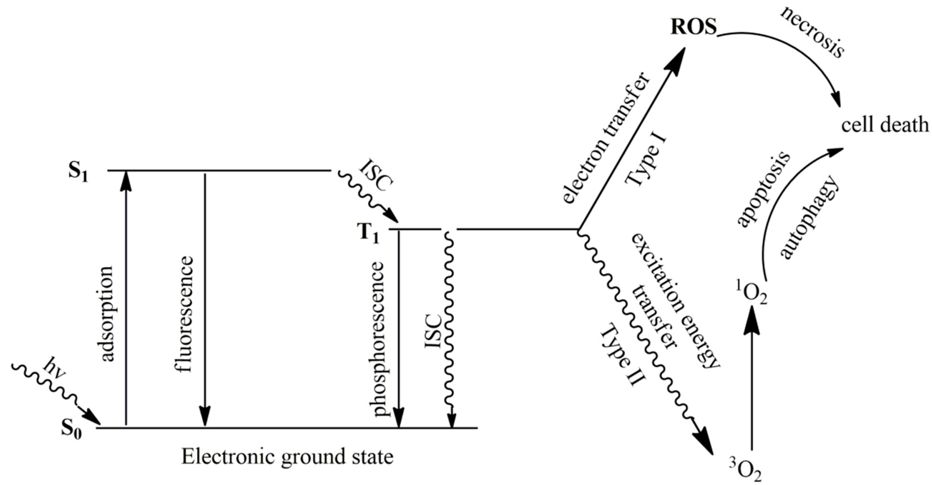
1. Introduction
2. Structure and Physicochemical Characteristics of Coordination Complexes as Photosensitizers for Photodynamic Anticancer Therapy
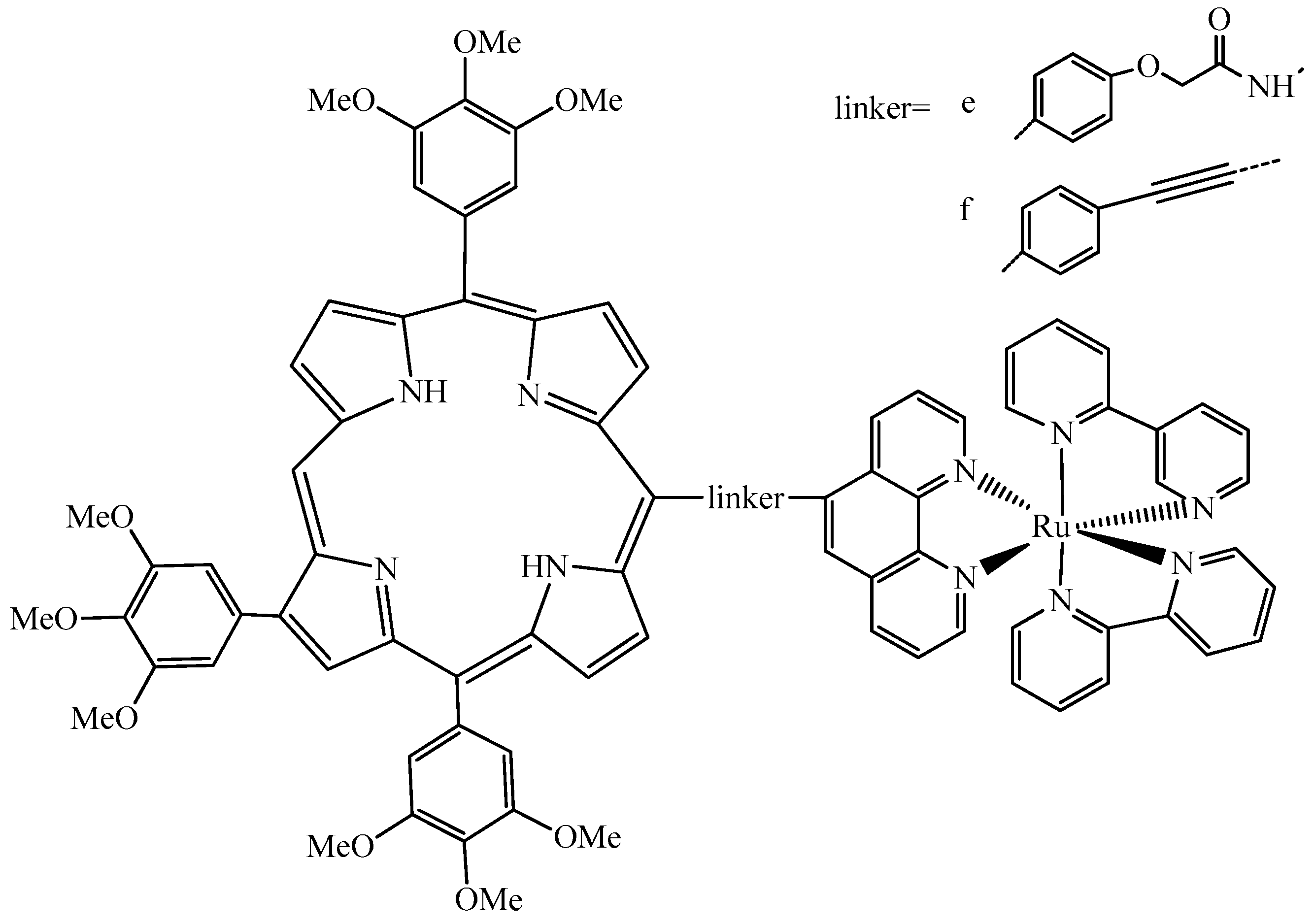
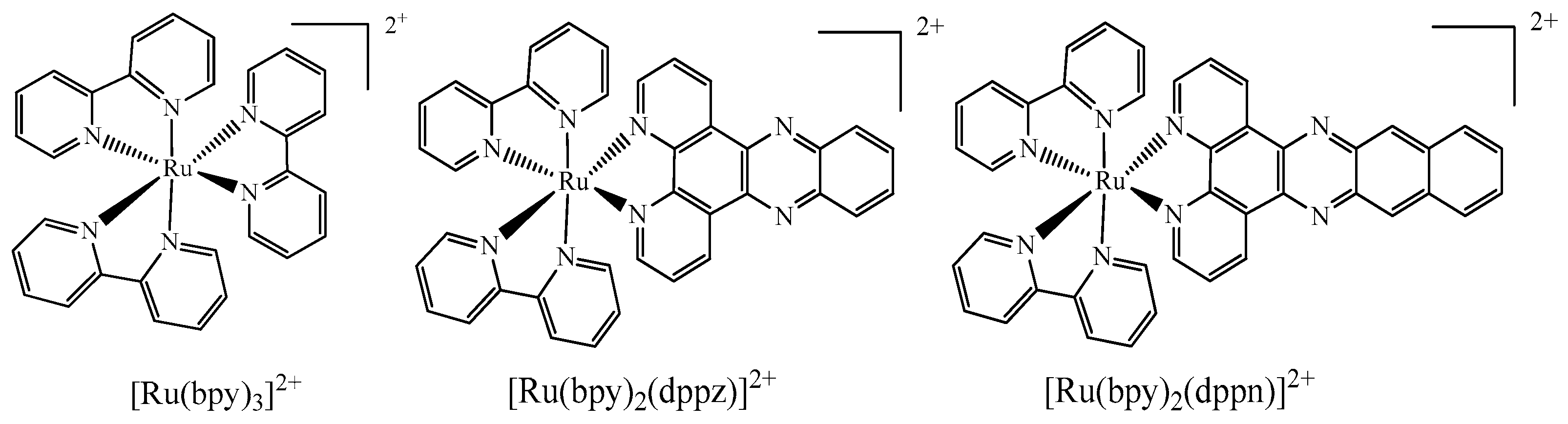
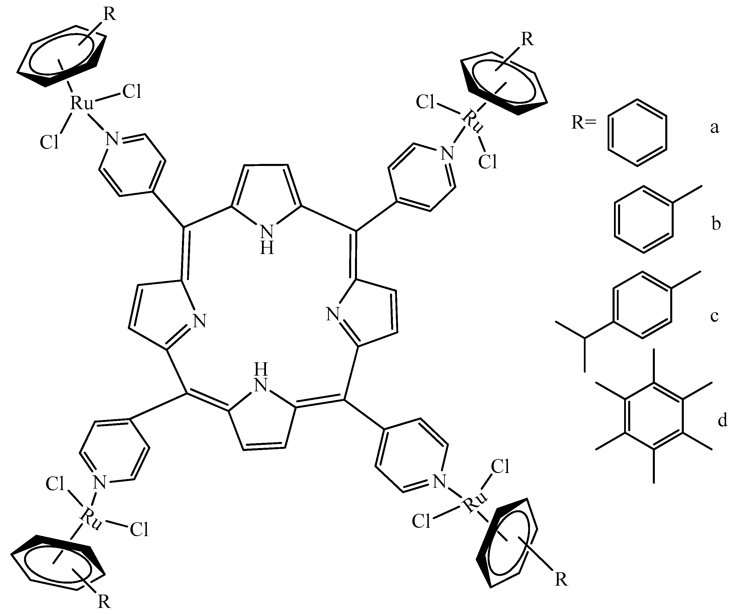
2.1. Ruthenium(II)-Based Complexes
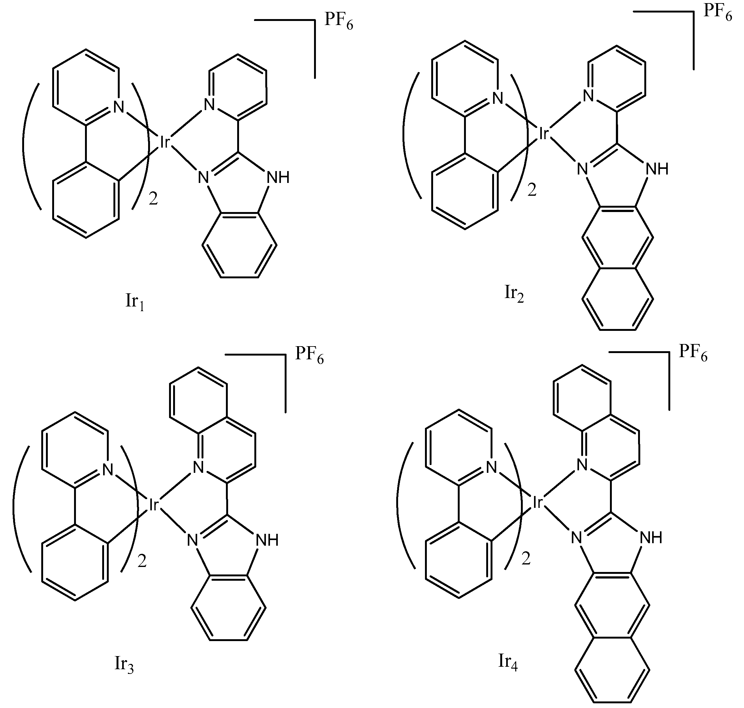
2.2. Iridium(III)-Based Complexes
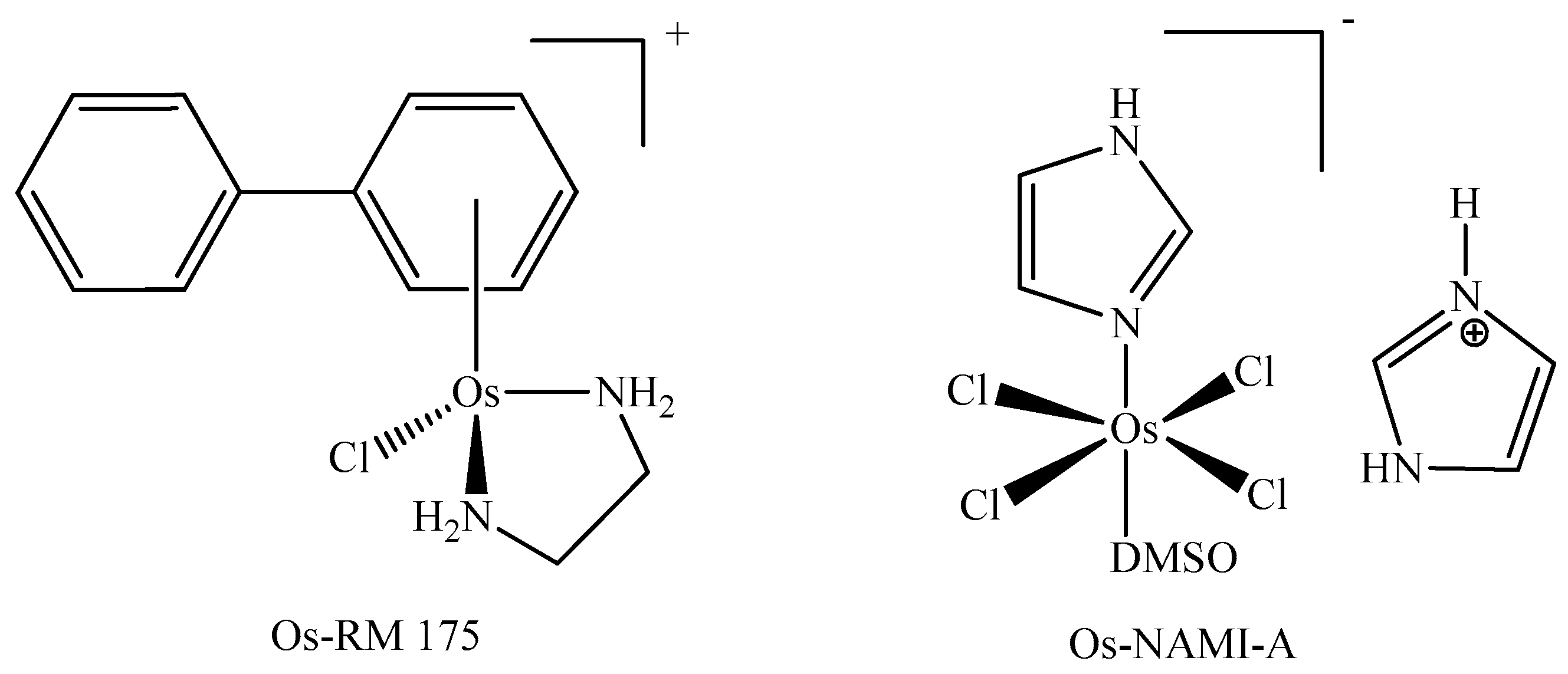
2.3. Osmium(0,II)-Based Complexes
2.4. Copper(0,I)-Based Complexes
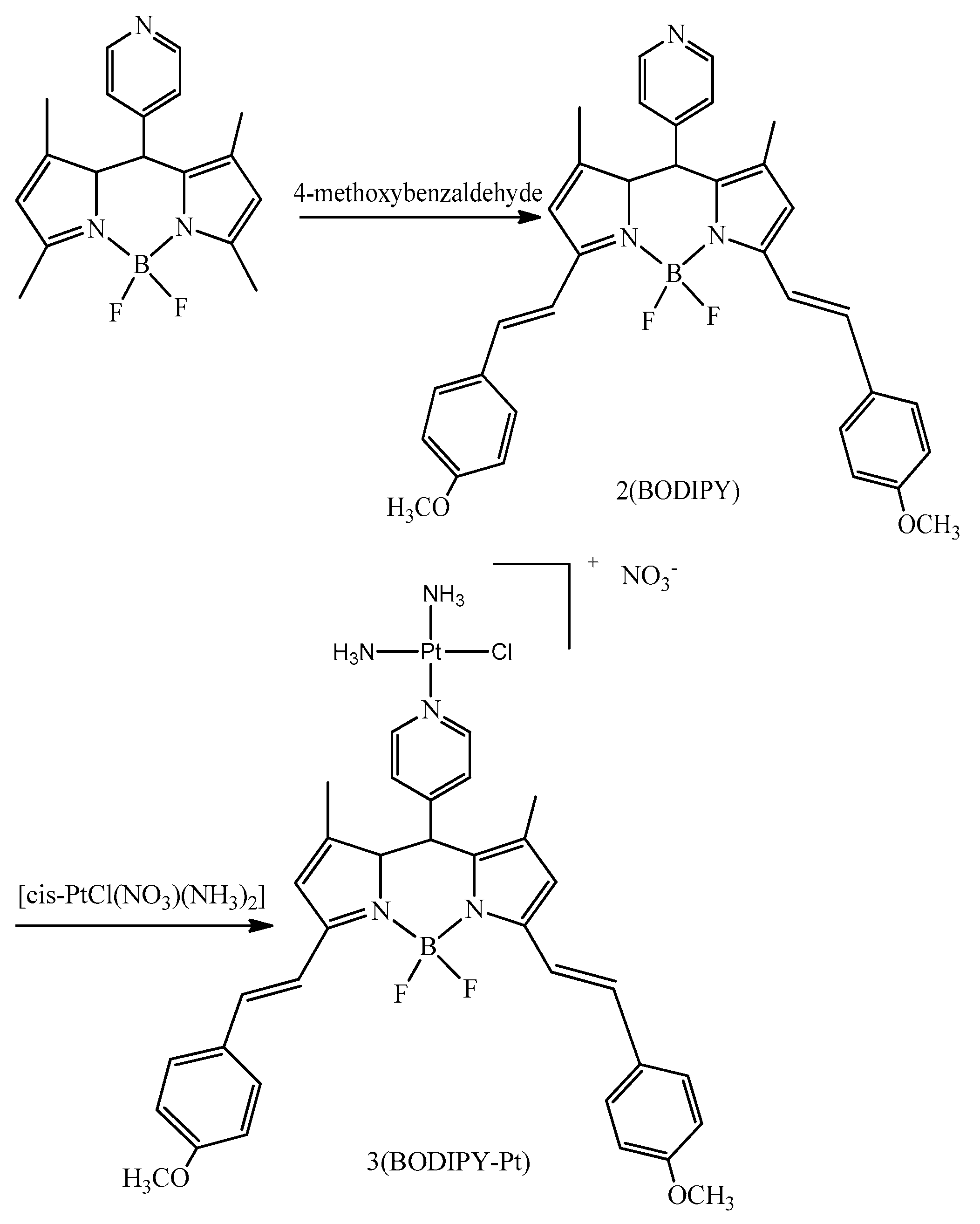
2.5. Platinum(II)-Based Complexes
3. Anti-Cancer Activities Comparison of Ru(II), Ir(III), Os(0,II), Cu(0,I), Pt(II) Coordination Complexes in Photodynamic Anticancer Therapy
4. Designing New Complexes Increasing the Efficiency of Photodynamic Therapy in Multimodal Oncology
5. Conclusions and Future Perspectives
Author Contributions
Funding
Informed Consent Statement
Data Availability Statement
Acknowledgments
Conflicts of Interest
References
- Sundaram, P.; Abrahamse, H. Effective photodynamic therapy for colon cancer cells using chlorin e6 coated hyaluronic acid-based carbon nanotubes. Int. J. Mol. Sci. 2020, 21, 4745. [Google Scholar] [CrossRef] [PubMed]
- Park, S.; Kim, K.E.; Park, H.J.; Cho, D. The Role of Erythroid Differentiation Regulator 1 (ERDR1) in the Control of Proliferation and Photodynamic Therapy (PDT) Response. Int. J. Mol. Sci. 2020, 21, 2603. [Google Scholar] [CrossRef] [PubMed] [Green Version]
- Mokoena, D.; George, B.; Abrahamse, H. Enhancing breast cancer treatment using a combination of cannabidiol and gold nanoparticles for photodynamic therapy. Int. J. Mol. Sci. 2019, 20, 4771. [Google Scholar] [CrossRef] [PubMed] [Green Version]
- Abdel-Kader, M.H. The journey of PDT throughout history: PDT from Pharos to present. In Photodynamic Medicine: From Bench to Clinic; Royal Society of Chemistry: Washington, DC, USA, 2016; pp. 1–21. [Google Scholar]
- Lin, S.; Liu, C.; Han, X.; Zhong, H.; Cheng, C. Viral Nanoparticle System: An Effective Platform for Photodynamic Therapy. Int. J. Mol. Sci. 2021, 22, 1728. [Google Scholar] [CrossRef]
- de Oliveira, A.B.; Ferrisse, T.M.; Marques, R.S.; de Annunzio, S.R.; Brighenti, F.L.; Fontana, C.R. Effect of photodynamic therapy on microorganisms responsible for dental caries: A systematic review and meta-analysis. Int. J. Mol. Sci. 2019, 20, 3585. [Google Scholar] [CrossRef] [PubMed] [Green Version]
- Hsieh, Y.H.; Zhang, J.H.; Chuang, W.C.; Yu, K.H.; Huang, X.B.; Lee, Y.C.; Lee, C.I. An in vitro study on the effect of combined treatment with photodynamic and chemical therapies on Candida albicans. Int. J. Mol. Sci. 2018, 19, 337. [Google Scholar] [CrossRef] [Green Version]
- Dai, T.; He, W.; Yao, C.; Ma, X.; Ren, W.; Mai, Y.; Wu, A. Applications of inorganic nanoparticles in the diagnosis and therapy of atherosclerosis. Biomater. Sci. 2020, 8, 3784–3799. [Google Scholar] [CrossRef]
- Jin, Y.; Guan, Z.; Wang, X.; Wang, Z.; Zeng, R.; Xu, L.; Cao, P. ALA-PDT promotes HPV-positive cervical cancer cells apoptosis and DCs maturation via miR-34a regulated HMGB1 exosomes secretion. Photodiagnosis. Photodyn. Ther. 2018, 24, 27–35. [Google Scholar] [CrossRef] [PubMed]
- Castro, K.A.D.F.; Moura, N.M.M.; Figueira, F.; Ferreira, R.I.; Simões, M.M.Q.; Cavaleiro, J.A.S.; Faustino, M.A.F.; Silvestre, A.J.D.; Freire, C.S.; Tomé, J.P.C.; et al. New Materials Based on Cationic Porphyrins Conjugated to Chitosan or Titanium Dioxide: Synthesis, Characterization and Antimicrobial Efficacy. Int. J. Mol. Sci. 2019, 20, 2522. [Google Scholar] [CrossRef] [Green Version]
- Chang, J.E.; Liu, Y.; Lee, T.H.; Lee, W.K.; Yoon, I.; Kim, K. Tumor Size-Dependent Anticancer Efficacy of Chlorin Derivatives for Photodynamic Therapy. Int. J. Mol. Sci. 2018, 19, 1596. [Google Scholar] [CrossRef] [Green Version]
- Sun, Y.; Ma, X.; Hu, H. Application of Nano-Drug Delivery System Based on Cascade Technology in Cancer Treatment. Int. J. Mol. Sci. 2021, 22, 5698. [Google Scholar] [CrossRef] [PubMed]
- Staron, J.; Boron, B.; Karcz, D.; Szczygiel, M.; Fiedor, L. Recent progress in chemical modifications of chlorophylls and bacteriochlorophylls for the applications in photodynamic therapy. Curr. Med. Chem. 2015, 22, 3054–3074. [Google Scholar] [CrossRef] [PubMed]
- Fiedor, J.; Fiedor, L.; Kammhuber, N.; Scherz, A.; Scheer, H. Photodynamics of the Bacteriochlorophyll–Carotenoid System. 2. Influence of Central Metal, Solvent and β-Carotene on Photobleaching of Bacteriochlorophyll Derivatives. Photochem. Photobiol. 2002, 76, 145–152. [Google Scholar] [CrossRef]
- Drzewiecka-Matuszek, A.; Skalna, A.; Karocki, A.; Stochel, G.; Fiedor, L. Effects of heavy central metal on the ground and excited states of chlorophyll. JBIC J. Biol. Inorg. 2005, 10, 453–462. [Google Scholar] [CrossRef]
- Karcz, D.; Boroń, B.; Matwijczuk, A.; Furso, J.; Staroń, J.; Ratuszna, A.; Fiedor, L. Lessons from chlorophylls: Modifications of porphyrinoids towards optimized solar energy conversion. Molecules 2014, 19, 15938–15954. [Google Scholar] [CrossRef] [Green Version]
- Vallejo, M.C.S.; Moura, N.M.M.; Gomes, A.T.P.C.; Joaquinito, A.S.M.; Faustino, M.A.F.; Almeida, A.; Gonçalves, I.; Serra, V.V.; Neves, M.G.P.M.S. The Role of Porphyrinoid Photosensitizers for Skin Wound Healing. Int. J. Mol. Sci. 2021, 22, 4121. [Google Scholar] [CrossRef]
- Dash, B.S.; Jose, G.; Lu, Y.J.; Chen, J.P. Functionalized Reduced Graphene Oxide as a Versatile Tool for Cancer Therapy. Int. J. Mol. Sci. 2021, 22, 2989. [Google Scholar] [CrossRef]
- Dang, J.; He, H.; Chen, D.; Yin, L. Manipulating tumor hypoxia toward enhanced photodynamic therapy (PDT). Biomater. Sci. 2017, 5, 1500–1511. [Google Scholar] [CrossRef]
- Wang, K.; Zhang, Y.; Wang, J.; Yuan, A.; Sun, M.; Wu, J.; Hu, Y. Self-assembled IR780-loaded transferrin nanoparticles as an imaging, targeting and PDT/PTT agent for cancer therapy. Sci. Rep. 2016, 6, 1–11. [Google Scholar] [CrossRef]
- McFarland, S.A.; Mandel, A.; Dumoulin-White, R.; Gasser, G. Metal-based photosensitizers for photodynamic therapy: The future of multimodal oncology? Curr. Opin. Chem. Biol. 2020, 56, 23–27. [Google Scholar] [CrossRef] [PubMed]
- Pucelik, B.; Sułek, A.; Drozd, A.; Stochel, G.; Pereira, M.M.; Pinto, S.; Dąbrowski, J.M. Enhanced cellular uptake and photodynamic effect with amphiphilic fluorinated porphyrins: The role of sulfoester groups and the nature of reactive oxygen species. Int. J. Mol. Sci. 2020, 21, 2786. [Google Scholar] [CrossRef] [Green Version]
- Khonkarn, R.; Mankhetkorn, S.; Talelli, M.; Hennink, W.E.; Okonogi, S. Cytostatic effect of xanthone-loaded mPEG-bp (HPMAm-Lac2) micelles towards doxorubicin sensitive and resistant cancer cells. Colloids Surf. B 2012, 94, 266–273. [Google Scholar] [CrossRef]
- Rijcken, C.J.; Hofman, J.W.; van Zeeland, F.; Hennink, W.E.; van Nostrum, C.F. Photosensitiser-loaded biodegradable polymeric micelles: Preparation, characterisation and in vitro PDT efficacy. J. Control. Release 2007, 124, 144–153. [Google Scholar] [CrossRef] [PubMed]
- Higgins, S.L.H.; Tucker, A.J.; Winkel, B.S.; Brewer, K.J. Metal to ligand charge transfer induced DNA photobinding in a Ru (II)–Pt (II) supramolecule using red light in the therapeutic window: A new mechanism for DNA modification. Chem. Comm. 2012, 48, 67–69. [Google Scholar] [CrossRef] [PubMed] [Green Version]
- Synatschke, C.V.; Nomoto, T.; Cabral, H.; Förtsch, M.; Toh, K.; Matsumoto, Y.; Kataoka, K. Multicompartment micelles with adjustable poly (ethylene glycol) shell for efficient in vivo photodynamic therapy. ACS Nano 2014, 8, 1161–1172. [Google Scholar] [CrossRef] [PubMed]
- Guo, M.; Mao, H.; Li, Y.; Zhu, A.; He, H.; Yang, H.; Chen, H. Dual imaging-guided photothermal/photodynamic therapy using micelles. Biomaterials 2014, 35, 4656–4666. [Google Scholar] [CrossRef] [Green Version]
- Kramer-Marek, G. Spectroscopic properties and photodynamic effects of new lipophilic porphyrin derivatives: Efficacy, localisation and cell death pathways. J. Photoch. Photobiol. B 2006, 84, 1–14. [Google Scholar] [CrossRef] [Green Version]
- Nackiewicz, J.; Kliber-Jasik, M.; Skonieczna, M. A novel pro-apoptotic role of zinc octacarboxyphthalocyanine in melanoma me45 cancer cell’s photodynamic therapy (PDT). J. Photoch. Photobiol. B 2019, 190, 146–153. [Google Scholar] [CrossRef] [PubMed]
- Kuś, P.; Kozik, V.; Rojkiewicz, M.; Sochanik, A.; Szurko, A.; Kempa, M.; Sakowicz, J. The synthesis of new potential photosensitizers. Part 3. Tetraphenylporphyrin esters of profens. Dyes Pigm. 2015, 116, 46–51. [Google Scholar] [CrossRef]
- Dabrowski, J.M.; Arnaut, L.G.; Pereira, M.M.; Monteiro, C.J.; Urbanska, K.; Simões, S.; Stochel, G. New halogenated water-soluble chlorin and bacteriochlorin as photostable PDT sensitizers: Synthesis, spectroscopy, photophysics, and in vitro photosensitizing efficacy. ChemMedChem 2010, 5, 1770–1780. [Google Scholar] [CrossRef]
- Nawalany, K.; Rusin, A.; Kepczynski, M.; Filipczak, P.; Kumorek, M.; Kozik, B.; Nowakowska, M. Novel nanostructural photosensitizers for photodynamic therapy: In vitro studies. Int. J. Pharm. 2012, 430, 129–140. [Google Scholar] [CrossRef]
- Karges, J.; Heinemann, F.; Maschietto, F.; Patra, M.; Blacque, O.; Ciofini, I.; Gasser, G. A Ru(II) polypyridyl complex bearing aldehyde functions as a versatile synthetic precursor for long-wavelength absorbing photodynamic therapy photosensitizers. Bioorg. Med. Chem. 2019, 27, 2666–2675. [Google Scholar] [CrossRef] [PubMed]
- Dąbrowski, J.M.; Arnaut, L.G. Photodynamic therapy (PDT) of cancer: From local to systemic treatment. Photochem. Photobiol. Sci. 2015, 14, 1765–1780. [Google Scholar] [CrossRef] [PubMed]
- Lobo, S.; Catarina, A.; Gomes-da-Silva, L.C.; Rodrigues-Santos, P.; Cabrita, A.; Santos-Rosa, M.; Arnaut, L.G. Immune Responses after Vascular Photodynamic Therapy with Redaporfin. J. Clin. Med. 2020, 9, 104. [Google Scholar] [CrossRef] [Green Version]
- Pucelik, B.; Arnaut, L.G.; Dąbrowski, J.M. Lipophilicity of bacteriochlorin-based photosensitizers as a determinant for PDT optimization through the modulation of the inflammatory mediators. J. Clin. Med. 2020, 9, 8. [Google Scholar] [CrossRef] [Green Version]
- Ghazal, B.; Kaya, E.N.; Husain, A.; Ganesan, A.; Durmuş, M.; Makhseed, S. Biotinylated-cationic zinc(II) phthalocyanine towards photodynamic therapy. J. Porphyr. Phthalocyanines 2019, 23, 46–55. [Google Scholar] [CrossRef]
- Kielmann, M.; Prior, C.; Senge, M.O. Porphyrins in troubled times: A spotlight on porphyrins and their metal complexes for explosives testing and CBRN defense. New J. Chem. 2018, 42, 7529–7550. [Google Scholar] [CrossRef]
- Carneiro, J.; Gonçalves, A.; Zhou, Z.; Griffin, K.E.; Kaufman, N.E.; Vicente, M.D.G.H. Synthesis and in vitro PDT evaluation of new porphyrins containing meso-epoxymethylaryl cationic groups. Lasers Surg. Med. 2018, 50, 566–575. [Google Scholar] [CrossRef]
- Wang, Y.; Xie, Y.; Li, J.; Peng, Z.H.; Sheinin, Y.; Zhou, J.; Oupický, D. Tumor-penetrating nanoparticles for enhanced anticancer activity of combined photodynamic and hypoxia-activated therapy. ACS Nano 2017, 11, 2227–2238. [Google Scholar] [CrossRef] [PubMed]
- Rizvi, I.; Obaid, G.; Bano, S.; Hasan, T.; Kessel, D. Photodynamic therapy: Promoting in vitro efficacy of photodynamic therapy by liposomal formulations of a photosensitizing agent. Lasers Surg. Med. 2018, 50, 499–505. [Google Scholar] [CrossRef]
- Moret, F.; Reddi, E. Strategies for optimizing the delivery to tumors of macrocyclic photosensitizers used in photodynamic therapy (PDT). J. Porphyr. Phthalocyanines 2017, 21, 239–256. [Google Scholar] [CrossRef] [Green Version]
- Staroń, J.; Dąbrowski, J.M.; Cichoń, E.; Guzik, M. Lactose esters: Synthesis and biotechnological applications. Crit. Rev. Biotechnol. 2018, 38, 245–258. [Google Scholar] [CrossRef]
- Pucelik, B.; Gürol, I.; Ahsen, V.; Dumoulin, F.; Dąbrowski, J.M. Fluorination of phthalocyanine substituents: Improved photoproperties and enhanced photodynamic efficacy after optimal micellar formulations. Eur. J. Med. Chem. 2016, 124, 284–298. [Google Scholar] [CrossRef]
- Heinemann, F.; Karges, J.; Gasser, G. Critical overview of the use of Ru(II) polypyridyl complexes as photosensitizers in one-photon and two-photon photodynamic therapy. Acc. Chem. Res. 2017, 50, 2727–2736. [Google Scholar] [CrossRef]
- Boerner, J.K.L.; Zaleski, J.M. Metal complex–DNA interactions: From transcription inhibition to photoactivated cleavage. Curr. Opin. Chem. Biol. 2005, 9, 135–144. [Google Scholar] [CrossRef] [PubMed]
- Monro, S.; Colón, K.L.; Yin, H.; Roque III, J.; Konda, P.; Gujar, S.; McFarland, S.A. Transition metal complexes and photodynamic therapy from a tumor-centered approach: Challenges, opportunities, and highlights from the development of TLD1433. Chem. Rev. 2018, 119, 797–828. [Google Scholar] [CrossRef] [PubMed]
- Mari, C.; Pierroz, V.; Ferrari, S.; Gasser, G. Combination of Ru(II) complexes and light: New frontiers in cancer therapy. Chem. Sci. 2015, 6, 2660–2686. [Google Scholar] [CrossRef] [Green Version]
- Day, A.H.; Ubler, M.H.; Best, H.L.; Lloyd-Evans, E.; Mart, R.J.; Fallis, I.A.; Allemann, R.K.; Al-Wattar, E.A.H.; Keymer, N.I.; Buurma, N.J.; et al. Targeted cell imaging properties of a deep red luminescent iridium(III) complex conjugated with a c-Myc signal peptide. Chem. Sci. 2020, 11, 1599–1606. [Google Scholar] [CrossRef] [PubMed] [Green Version]
- Ma, D.; Zhang, C.; Liu, R.; Qiu, Y.; Duan, L. Controlling ion distribution for high performance organic light emitting diodes based on sublimable cationic iridium(III) complexes. ACS Appl. Mater. Interfaces 2018, 10, 29814–29823. [Google Scholar] [CrossRef]
- Sheet, S.J.; Sen, B.; Khatua, S. Organoiridium(III) complexes as luminescence color switching probes for selective detection of nerve agent simulation in solution and vapor phase. Inorg. Chem. 2019, 58, 3635–3645. [Google Scholar] [CrossRef]
- Guo, J.; Zhou, J.; Fu, G.; He, Y.; Li, W.; Lu, X. Two efficient near-infrared (NIR) luminescent [Ir(C^N)2(N^O)] characteristic complexes with 8-hydroxyquinoline (8-HQ) as the ancillary ligand. Inorg. Chem. Commun. 2019, 101, 69–73. [Google Scholar] [CrossRef]
- Bevernaegie, R.; Wehlin, S.A.M.; Elias, B.; Troian-Gautier, L. A Roadmap Towards Visible Light Mediated Electron Transfer Chemistry with Iridium(III) Complexes. ChemPhotoChem 2021, 5, 217–234. [Google Scholar]
- Curtin, P.N.; Tinker, L.L.; Burgess, C.M.; Cline, E.D.; Bernhard, S. Structure−activity correlations among iridium(III) photosensitizers in a robust water-reducing system. Inorg. Chem. 2009, 48, 10498–10506. [Google Scholar] [CrossRef] [PubMed]
- Wang, F.X.; Chen, M.H.; Lin, Y.N.; Zhang, H.; Tan, C.P.; Ji, L.N.; Mao, Z.W. Dual functions of cyclometalated iridium(III) complexes: Anti-metastasis and lysosome-damaged photodynamic therapy. ACS Appl. Mater. 2017, 9, 42471–42481. [Google Scholar] [CrossRef] [PubMed]
- Huang, H.; Banerjee, S.; Sadler, P.J. Recent advances in the design of targeted iridium(III) photosensitizers for photodynamic therapy. ChemBioChem 2018, 19, 1574–1589. [Google Scholar] [CrossRef] [PubMed]
- Alessio, E.; Messori, L. Anticancer drug candidates face-to-face: A case story in medicinal inorganic chemistry. Molecules 2019, 24, 1995. [Google Scholar] [CrossRef] [PubMed] [Green Version]
- Buil, M.L.; Cardo, J.J.F.; Esteruelas, M.A.; Fernández, I.; Oñate, E. An entry to stable mixed phosphine-Osmium-NHC polyhydrides. Inorg. Chem. 2016, 55, 5062–5070. [Google Scholar] [CrossRef] [PubMed]
- Esteruelas, M.A.; Larramona, C.; Oñate, E. Osmium-mediated direct C-H bond activation at the 8-position of quinolines. Organometallics 2016, 35, 1597–1600. [Google Scholar] [CrossRef] [Green Version]
- Meier-Menches, S.M.; Gerner, C.; Berger, W.; Hartinger, C.G.; Keppler, B.K. Structure–activity relationships for ruthenium and osmium anticancer agents–towards clinical development. Chem. Soc. Rev. 2018, 47, 909–928. [Google Scholar] [CrossRef]
- Smitten, K.L.; Scattergood, P.A.; Kiker, C.; Thomas, J.A.; Elliott, P.I. Triazole-based osmium(II) complexes displaying red/near-IR luminescence: Antimicrobial activity and super-resolution imaging. Chem. Sci. 2020, 11, 8928–8935. [Google Scholar] [CrossRef] [PubMed]
- Mehvash, Z.; Suboot, H.; Elham, A. Scope of organometallic compounds based on transition metal-arene systems as anticancer agents: Starting from the classical paradigm to targeting multiple strategies. RSC Adv. 2019, 9, 3239–3278. [Google Scholar]
- Giereth, R.L.; Reim, I.; Frey, W.; Junge, H.; Tschierlei, S.; Karnahl, M. Remarkably long-lived excited states of copper photosensitizers containing an extended π-system based on an anthracene moiety. Sustain. Energy Fuels 2019, 3, 692–700. [Google Scholar] [CrossRef]
- McCullough, B.J.; Neyhouse, B.J.; Schrage, B.R.; Reed, D.T.; Osinski, A.J.; Ziegler, C.J. Visible-light-driven photosystems using heteroleptic Cu(I) photosensitizers and Rh(III) catalysts to produce H2. Inorg. Chem. 2018, 57, 2865–2875. [Google Scholar] [CrossRef]
- Sah, B.; Wu, J.; Vanasse, A.; Pandey, N.K.; Chudal, L.; Huang, Z.; Antosh, M.P. Effects of Nanoparticle Size and Radiation Energy on Copper-Cysteamine Nanoparticles for X-ray Induced Photodynamic Therapy. Nanomaterials 2020, 10, 1087. [Google Scholar] [CrossRef] [PubMed]
- Shrestha, S.; Wu, J.; Sah, B.; Vanasse, A.; Cooper, L.N.; Ma, L.; Antosh, M.P. X-ray induced photodynamic therapy with copper-cysteamine nanoparticles in mice tumors. Proc. Natl. Acad. Sci. USA 2019, 116, 16823–16828. [Google Scholar] [CrossRef] [Green Version]
- Tekin, V.; Aweda, T.; Guldu, O.K.; Muftuler, F.Z.B.; Bartels, J.; Lapi, S.E.; Unak, P. A novel anti-angiogenic radio/photo sensitizer for prostate cancer imaging and therapy: 89Zr-Pt@ TiO2-SPHINX, synthesis and in vitro evaluation. Nucl. Med. Biol. 2021, 94, 20–31. [Google Scholar] [CrossRef]
- Zhu, S.; Yao, S.; Wu, F.; Jiang, L.; Wong, K.L.; Zhou, J.; Wang, K. Platinated porphyrin as a new organelle and nucleus dual-targeted photosensitizer for photodynamic therapy. Org. Biomol. Chem. 2017, 15, 5764–5771. [Google Scholar] [CrossRef]
- Lazarides, T.; McCormick, T.M.; Wilson, K.C.; Lee, S.; McCamant, D.W.; Eisenberg, R. Sensitizing the sensitizer: The synthesis and photophysical study of bodipy−Pt(II)(diimine)(dithiolate) conjugates. J. Am. Chem. Soc. 2011, 133, 350–364. [Google Scholar] [CrossRef]
- Sun, T.; Guan, X.; Zheng, M.; Jing, X.; Xie, Z. Mitochondria-localized fluorescent BODIPY-platinum conjugate. ACS Med. Chem. Lett. 2015, 6, 430–433. [Google Scholar] [CrossRef] [Green Version]
- Qi, F.; Yuan, H.; Chen, Y.; Guo, Y.; Zhang, S.; Liu, Z.; Guo, Z. BODIPY-based monofunctional Pt(II) complexes for specific photocytotoxicity against cancer cells. J. Inorg. Biochem. 2021, 218, 111394. [Google Scholar] [CrossRef] [PubMed]
- Mengqian, Y.; Deng, J.; Guo, G.; Zhang, J.; Yang, L.; Wu, F. A folate-conjugated platinum porphyrin complex as a new cancer-targeting photosensitizer for photodynamic therapy. Org. Biomol. Chem. 2019, 21, 5367–5374. [Google Scholar]
- Lainé, P.; Bedioui, F.; Loiseau, F.; Chiorboli, C.; Campagna, S. Conformationally Gated Photoinduced Processes within Photosensitizer Acceptor Dyads Based on Osmium(II) Complexes with Triarylpyridinio-Functionalized Terpyridyl Ligands: Insights from Experimental Study. J. Am. Chem. Soc. 2006, 128, 7510–7521. [Google Scholar] [CrossRef]
- Peng, Z.; Gharavi, A.R.; Yu, L. Synthesis and characterization of photorefractive polymers containing transition metal complexes as photosensitizer. J. Am. Chem. Soc. 1997, 119, 4622–4632. [Google Scholar] [CrossRef]
- Yao, M.; Ma, L.; Li, L.; Zhang, J.; Lim, R.X.; Chen, W.; Zhang, Y. A new modality for cancer treatment—nanoparticle mediated microwave induced photodynamic therapy. J. Biomed. Nanotechnol. 2016, 12, 1835–1851. [Google Scholar] [CrossRef]
- Foresto, E.; Gilardi, P.; Ibarra, L.E.; Cogno, I.S. Light-activated green-drugs: How we can use them in Photodynamic therapy and mass-produce them with biotechnological tools. Phytomed. Plus 2021, 1, 100044. [Google Scholar] [CrossRef]
- Lee, S.; Yoon, J. Supramolecular photosensitizers rejuvenate photodynamic therapy. Chem. Soc. Rev. 2018, 47, 1174–1188. [Google Scholar]
- Grzybowski, A.; Sak, J.; Pawlikowski, J. A brief report on the history of phototherapy. Clin. Dermatol. 2016, 34, 532–537. [Google Scholar] [CrossRef] [PubMed]
- Gomes, J.; Leal, I.; Bednarczyk, K.; Gmurek, M.; Stelmachowski, M.; Zaleska-Medynska, A.; Bastos, F.C.; Quinta-Ferreira, M.E.; Costa, R.; Quinta-Ferreira, R.M. Detoxification of Parabens Using UV-A enhanced by Noble Metals—TiO2 Supported Catalysts. J. Environ. Chem. Eng. 2017, 5, 3065–3074. [Google Scholar] [CrossRef]
- Ramandi, S.; Entezari, M.H.; Ghows, N. Sono-synthesis of solar light responsive S-N-C-tri doped TiO2 photo-catalyst under optimized conditions for degradation and mineralization of Diclofenac. Ultrason. Sonochem. 2017, 38, 234–245. [Google Scholar] [CrossRef]
- Wang, X.H.; Peng, H.S.; Yang, W.; Ren, Z.D.; Liu, X.M.; Liu, Y.A. Indocyanine green-platinum porphyrins integrated conjugated polymer hybrid nanoparticles for near-infrared-triggered photothermal and two-photon photodynamic therapy. J. Mater. Chem. B 2017, 5, 1856–1862. [Google Scholar] [CrossRef] [PubMed]


| HeLa | A459 | A549R | |||||
|---|---|---|---|---|---|---|---|
| Complex | IC50 (μM) | PI a | IC50 (μM) | PI | IC50 (μM) | PI | |
| Ir1 | Dark | 11.69 | 8.4 | 7.64 | 8.0 | 5.98 | 4.2 |
| Ir1 | Light b | 1.39 | 0.95 | 1.43 | |||
| Ir2 | Dark | 15.13 | 63 | 5.76 | 24 | 4.26 | 9.7 |
| Ir2 | Light | 0.24 | 0.24 | 0.44 | |||
| Ir3 | Dark | >100 | >200 | >100 | >137 | >100 | >83 |
| Ir3 | Light | 0.50 | 0.73 | 1.20 | |||
| Ir4 | Dark | >100 | >476 | >100 | >322 | >100 | >139 |
| Ir4 | Light | 0.21 | 0.31 | 0.72 | |||
| Cisplatin | Dark | 12.04 | 1.1 | 8.57 | 1.0 | 73.42 | 1.0 |
| Cisplatin | Light | 10.56 | 8.42 | 69.38 | |||
| NAMI-A | Dark | >100 | >100 | >100 | |||
| NAMI-A | Light | >100 | >100 | >100 | |||
Publisher’s Note: MDPI stays neutral with regard to jurisdictional claims in published maps and institutional affiliations. |
© 2021 by the authors. Licensee MDPI, Basel, Switzerland. This article is an open access article distributed under the terms and conditions of the Creative Commons Attribution (CC BY) license (https://creativecommons.org/licenses/by/4.0/).
Share and Cite
Pobłocki, K.; Drzeżdżon, J.; Kostrzewa, T.; Jacewicz, D. Coordination Complexes as a New Generation Photosensitizer for Photodynamic Anticancer Therapy. Int. J. Mol. Sci. 2021, 22, 8052. https://doi.org/10.3390/ijms22158052
Pobłocki K, Drzeżdżon J, Kostrzewa T, Jacewicz D. Coordination Complexes as a New Generation Photosensitizer for Photodynamic Anticancer Therapy. International Journal of Molecular Sciences. 2021; 22(15):8052. https://doi.org/10.3390/ijms22158052
Chicago/Turabian StylePobłocki, Kacper, Joanna Drzeżdżon, Tomasz Kostrzewa, and Dagmara Jacewicz. 2021. "Coordination Complexes as a New Generation Photosensitizer for Photodynamic Anticancer Therapy" International Journal of Molecular Sciences 22, no. 15: 8052. https://doi.org/10.3390/ijms22158052
APA StylePobłocki, K., Drzeżdżon, J., Kostrzewa, T., & Jacewicz, D. (2021). Coordination Complexes as a New Generation Photosensitizer for Photodynamic Anticancer Therapy. International Journal of Molecular Sciences, 22(15), 8052. https://doi.org/10.3390/ijms22158052