Review

Anti-NASH Drug Development Hitches a Lift on PPAR Agonism

Department of In Vitro Toxicology & Dermato-Cosmetology (IVTD), Faculty of Medicine and Pharmacy, Vrije Universiteit Brussel (VUB); Laarbeeklaan 103, 1090 Brussels, Belgium
*
Author to whom correspondence should be addressed.
These authors contributed equally to this work.
Cells 2020, 9(1), 37; https://doi.org/10.3390/cells9010037
Submission received: 25 November 2019 / Revised: 15 December 2019 / Accepted: 17 December 2019 / Published: 21 December 2019
(This article belongs to the Special Issue Recent Advances in Liver Repair Strategies)

Abstract

:
Non-alcoholic fatty liver disease (NAFLD) affects one-third of the population worldwide, of which a substantial number of patients suffer from non-alcoholic steatohepatitis (NASH). NASH is a severe condition characterized by steatosis and concomitant liver inflammation and fibrosis, for which no drug is yet available. NAFLD is also generally conceived as the hepatic manifestation of the metabolic syndrome. Consequently, well-established drugs that are indicated for the treatment of type 2 diabetes and hyperlipidemia are thought to exert effects that alleviate the pathological features of NASH. One class of these drugs targets peroxisome proliferator-activated receptors (PPARs), which are nuclear receptors that play a regulatory role in lipid metabolism and inflammation. Therefore, PPARs are now also being investigated as potential anti-NASH druggable targets. In this paper, we review the mechanisms of action and physiological functions of PPARs and discuss the position of the different PPAR agonists in the therapeutic landscape of NASH. We particularly focus on the PPAR agonists currently under evaluation in clinical phase II and III trials. Preclinical strategies and how refinement and optimization may improve PPAR-targeted anti-NASH drug testing are also discussed. Finally, potential caveats related to PPAR agonism in anti-NASH therapy are stipulated.

1. Introduction

Non-alcoholic steatohepatitis (NASH) is an advanced form of non-alcoholic fatty liver disease (NAFLD) in which intrahepatic lipid accumulation in at least 5% of the hepatocytes is accompanied by hepatic inflammation and eventually fibrosis. NASH can further progress to life-threatening cirrhosis and hepatocellular carcinoma, and as such represents an emerging cause for liver transplantation. NAFLD is projected to affect 33.5% of the adult population by 2030, of which 27% will suffer from NASH. Currently, no effective therapies exist that can resolve NASH, yet considerable efforts have been made not only to understand the mechanisms by which this disease progresses but also to develop a suitable therapy [1,2,3].
The occurrence of NASH is strongly associated with the diagnosis of metabolic syndrome and obesity. As a consequence, introducing a balanced lifestyle seems to be a prime intervention when treating NASH. Weight loss of at least 10% of the body weight has been shown to solve NASH within one year [4]. In addition, bariatric surgery also proved to resolve NASH in 85% of obese patients who failed to lose weight through lifestyle modifications [5]. However, it was more efficient in patients with mild NASH than in those suffering from a more severe form of the disease. Intensive diets, lifestyle changes, or weight-loss surgery are often not attainable by the patients, and it has been shown that also a number of NASH patients is lean. These patients do not suffer from the metabolic syndrome, nor are they obese, but they do often carry specific genetic polymorphisms that have been associated with an increased propensity for developing NAFLD and NASH, such as PNPLA3 rs738409 [6]. Yet, the vast majority of NASH patients are obese and suffer from insulin resistance, type 2 diabetes, and hyperlipidemia. Therefore, drugs that target pathways disturbed in the metabolic syndrome are thought to hold anti-NASH properties as well. A major drug class administered to patients suffering from the metabolic syndrome targets peroxisome proliferator-activated receptors (PPARs). These multi-regulatory, ligand-activated, nuclear transcription factors regulate a multitude of processes that are mainly related to lipid metabolism, glucose homeostasis, and insulin signaling [7]. Therefore, PPARs are also attractive targets to tackle NAFLD. Distinct isotypes of PPARs can get activated by endogenous molecules (i.e., fatty acids and phospholipids) as well as synthetic ligands [8]. Synthetic PPAR-γ agonists, which are typically referred to as thiazolidinediones, sensitize the liver for insulin, and are indicated as a treatment for type 2 diabetes. Fibrates, which are agonists of PPAR-α, exert anti-hyperlipidemic effects. Fenofibrate was shown to additionally reduce the so-called ‘regulated on activation normal T-cell expressed and secreted’ (RANTES) serum levels in type 2 diabetes patients with hypertriglyceridemia [9], also indicating anti-inflammatory properties.
Nonetheless, up to now, none of the PPAR agonists on the market has demonstrated satisfactory efficacy in the resolution of NASH. Yet, PPAR agonists represent one of the most advanced classes of anti-NASH molecules currently in the pipeline of drug development [3,10,11].
This review discusses the application of PPAR agonists as a potential treatment of NASH. Based on the underlying molecular mechanisms of this therapy, possible caveats related to the administration of PPAR agonists are described. Furthermore, a novel strategy that potentially could improve PPAR-targeted preclinical anti-NASH drug testing is presented.

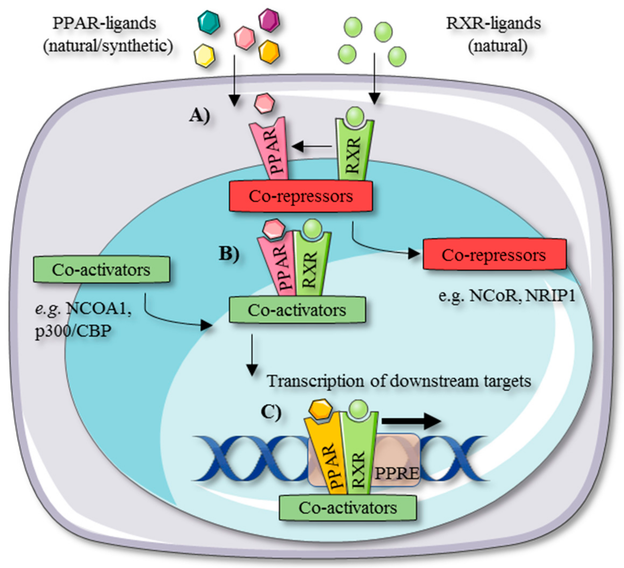
2. PPAR Tissue Distribution and Working Mechanism

The PPAR family consists of three isotypes, indicated by PPAR-α (NR1C1), -δ (syn. -β) (NR1C2), and -γ (NR1C3) [12,13]. PPAR-α was the first discovered isotype and was named after its ability to induce peroxisome proliferation in rodents [7]. All PPAR isotypes control lipid metabolism and are as such mainly expressed in tissues with high metabolic activity. Despite sharing high sequence homology and key functions, the PPAR isotypes are encoded by different genes, which are located on different chromosomes, and more or less specifically expressed in the body [14]. They regulate comparable as well as different processes and are activated by distinct synthetic ligands, resulting in the transcription of both overlapping and distinct downstream target genes [8,14]. PPAR-α and PPAR-δ are largely expressed in tissues with high mitochondrial and peroxisomal β-oxidative activity. PPAR-α is primarily expressed in liver, heart, kidneys, and brown adipose tissue, whereas PPAR-δ occurs ubiquitously. PPAR-γ is mainly expressed in white adipose tissue and is essential for adipocyte differentiation. It is also present in macrophages [8], including the liver-specific Kupffer cells.
The PPAR isotypes share a similar working mechanism. Upon the binding of an activating ligand to a particular PPAR isotype, the PPAR concerned heterodimerizes with a retinoid X receptor (RXR) to bind specific peroxisome proliferator response elements (PPREs) on the DNA, resulting in the transcription of its downstream target genes (Figure 1) [8,15].
Coactivators (e.g., p300/CREB-binding protein (p300/CBP) and nuclear receptor coactivator 1 (NCOA1)) and corepressors (e.g., nuclear receptor co-repressor 1 (NCoR1) and nuclear receptor-interacting protein 1 (NRIP1)) tightly and selectively control these transcriptional inductions and repressions. This also lays at the structural basis of partial agonists that exhibit altered pharmacological properties [16,17,18,19]. Corepressors suppress the transcription of PPAR-related genes in the unliganded state. When a PPAR is activated by a specific ligand, coactivators translocate the corepressors, resulting in downstream transcription. Another mechanism by which PPARs modulate gene transcription is transrepression, which occurs independently from PPREs. Multiple molecular mechanisms have been described that regulate PPAR-mediated transrepression. In a ‘direct’ model of PPAR transrepression, PPARs bind to inflammatory transcription factors, such as nuclear factor-κB (NF-κB), activator protein-1 (AP-1), and signal transducer and activator of transcription (STAT), to prevent protein–protein interaction and therefore binding to response elements. Second, PPARs regulate the mitogen-activated protein kinase (MAPK) pathway. PPARs also interact with coactivators that regulate PPAR activation and repression. Yet, these coregulators are not specific to PPARs and therefore competition among coactivators can occur between PPARs and inflammatory transcription factors such as NF-κB and AP-1. Transrepression can occur as well through the action of corepressors on the promotor regions of inflammatory genes such as NF-κB, AP-1, and STAT 5–7. For example, PPAR-δ agonism in macrophages leads to the release of corepressors that will exert on their turn repressing activity on NF-κB, balancing the distribution of these cofactors. Furthermore, PPAR-α agonists induce the inhibitor of kappa B (IκB)α in hepatocytes to prevent the nuclear transfer of NF-κB subunits from the cytoplasm [20,21].
The most important target genes of PPAR-α are related to peroxisomal and mitochondrial β-oxidative catabolism of fatty acids, ketogenesis, and nuclear transcription factors linked to inflammation and lipogenesis [22]. PPAR-α activates carnitine palmitoyl-CoA transferase 1α (CPT1A), an importer of fatty acids into mitochondria. It also stimulates more downstream enzymes in the mitochondrial β-oxidation pathway, such as acyl-CoA dehydrogenase medium chain (ACADM) and acyl-CoA dehydrogenase very long chain (ACADVL) [23]. Consequently, PPAR-α fulfills a key role during fasting due to its ability of enhancing cellular energy production through ATP production and ketogenesis [24]. The anti-inflammatory effects of PPAR-α stimulation are attributed to its link with NF-κB and AP-1 [21,25]. Using the carbohydrate-responsive element-binding protein (ChREBP)−/− and PPAR-α knockout mice, it has been shown that PPAR-α cross-talks with ChREBP, a glucose-sensing lipogenic transcription factor, to regulate fibroblast growth factor (FGF)21 expression. The latter is a hepatokine that improves insulin sensitivity and lipid metabolism and controls the preference for sucrose [24,26]. Sterol regulatory element-binding protein (SREBP)1, which is the insulin-sensing variant of ChREBP, is apart from the liver X receptor, which is also regulated by PPAR-α. The chronic activation of PPAR-α in mice results in the upregulation of SREBP1 downstream genes, while this could not be observed in SREBP1−/− mice [23,27]. Consequently, PPAR-α functions as a critical sensor for maintaining cellular energy homeostasis through both catabolic and anabolic pathways.
As shown for PPAR-α, PPAR-δ controls processes related to fatty acid metabolism and inflammation. It is the least well studied PPAR isotype despite of its ubiquitous expression [28]. Using PPAR-δ knockout mice, it could be demonstrated that PPAR-δ exhibits anti-atherogenic properties by reducing very-low density lipoproteins (VLDLs). This is also a consequence from FGF21 signaling [29], which also forms a link to PPAR-α [24,26]. PPAR-δ activation counters angiotensin II-induced adipocyte growth and lipid accumulation. As such, it also reduces the angiotensin II-mediated development of reactive oxygen species, which are implicated in the multiple hit pathogenesis of NASH. Furthermore, the activation of PPAR-δ favors the development of smaller adipocytes, which results in a better adipokine profile. Additionally, the hematopoietic deficiency of PPAR-δ in obese mice attenuates the activation of Kupffer cells that are important for NASH development and stellate cell activation. PPAR-δ also occurs in stellate cells [12,15,26].
PPAR-γ is a critical regulator of adipocyte differentiation and lipogenesis. Chronic stimulation can cause weight gain and obesity, which can eventually lead to related diseases. Hence, PPAR-γ seems to fulfill a developmental function by maximizing energy storage [30]. Paradoxically, PPAR-γ stimulation lessens free fatty acid levels (through adipogenesis), attenuates hepatic glucose production, and increases glucose uptake by the muscles as a result of improved insulin sensitivity [31,32]. Furthermore, PPAR-γ activation reduces inflammation. It attenuates the activation of interferon-γ-stimulated mouse peritoneal macrophages through AP-1, NF-κB, and STAT-mediated mechanisms and blocks tumor necrosis factor-α (TNF-α) production in human monocyte cultures [33,34].

3. Dysregulation of PPARs during NASH

The strong correlation between the prevalence of the metabolic syndrome and NAFLD has evoked attention for studying the dysregulation of hepatic PPARs in patients suffering from NASH. Francque and co-workers discovered a correlation between decreased hepatic PPARA levels and increased insulin resistance and NASH severity. In addition, a negative correlation with adiponectin levels, an anti-inflammatory adipokine, was measured along with increased visceral obesity. Interestingly, histologic evaluation showed that NASH resolution was associated with an upregulation of PPARA together with its target genes, indicating a clear link between PPAR-α function and NASH pathology, thereby opening perspectives for specific PPAR-α-targeted anti-NASH drug development [35]. The expression of the two other PPAR isotypes remained unchanged [35,36]. Nonetheless, a recent study found a correlation between severe, but not moderate, hepatic steatosis and decreased hepatic PPAR-δ mRNA levels. This was accompanied by a decrease in the amount of VLDL receptors in humans. Decreased VLDL receptor levels were as well observed in PPAR-δ knocked-down primary mouse hepatocytes and in the liver of PPAR-δ null mice, confirming the observations in human [29]. Moreover, high-fat diet (HFD)-fed mice show as well decreased hepatic PPAR-δ mRNA levels, invigorating the outcome of the latter study [37].

4. PPAR Agonists as Potential Anti-NASH Treatment

4.1. PPAR-α Agonists

The interest for the use of PPAR-α agonists (mainly fibrates [38]) in anti-NASH treatment arose already two decades ago, when gemfibrozil was tested in patients suffering from NASH. Gemfibrozil reduced aspartate aminotransferase (AST), alanine aminotransferase (ALT), and gamma-glutamyl transferase (GGT) levels, as well as VLDL triglyceride production. Gemfibrozil also lowered fatty acid mobilization from the adipose tissue and induced lipid clearance in the liver [39]. Fenofibrate treatment in type 2 diabetes patients, suffering from hypertriglyceridemia, decreased serum RANTES, which is also known as the C–C chemokine ligand 5 (CCL5) levels [9], indicating beneficial anti-inflammatory properties related to the NAFLD pathology. Yet, although it improved metabolic syndrome-related parameters, it did not improve liver histology in a 48-week trial with biopsy-proved NAFLD patients. Due to the small trial size of only 16 patients and the lack of a control group [40], further studies should clarify whether fenofibrate might be indicated for anti-NASH therapy. In addition, clofibrate was tested for its possible anti-NASH properties. After one-year treatment, clofibrate did not show any histological improvements in steatosis, inflammation, or fibrosis, nor a reduction in ALT, AST, GGT, bilirubin, or cholesterol, which led to the discontinuation of its evaluation [41]. Pemafibrate, a novel selective PPAR-α agonist, showed to ameliorate liver dysfunction in type 2 diabetes patients, and it was later demonstrated to also improve the NASH pathology in rodents by the stimulation of lipid metabolism and reducing inflammation [42,43].

4.2. PPAR-δ Agonists

Activators of PPAR-δ have been mainly investigated for the treatment of dyslipidemia [44,45,46,47]. One selective PPAR-δ agonist, named seladelpar, was recently suspended from a phase II trial due to unexpected histological findings. Nevertheless, seladelpar improved the serum lipid profile in dyslipidemic patients and also reduced liver enzyme levels [44,45]. In a foz/foz mouse model, seladelpar also reversed the hepatic storage of lipotoxic lipids and improved insulin sensitivity and serum lipid profile, indicating beneficial properties for treating NASH [48].

4.3. PPAR-γ Agonists

A multitude of studies has evaluated the use of insulin-sensing agents for the treatment of NASH [49,50]. Thiazolidinediones, which are PPAR-γ agonists, are such a class of insulin sensing drugs [30,51]. The potential anti-NASH efficacy of pioglitazone (and vitamin E) has been evaluated in the so-called PIVENS (Pioglitazone, Vitamin E, or Placebo for Nonalcoholic Steatohepatitis) trial. Here, it was found that pioglitazone and vitamin E reduce hepatic steatosis and lobular inflammation, but not concomitant fibrosis (Table 1) [52].
Vitamin E alone met the primary outcome, which was a histologic improvement of NASH based on a scoring system [52,54]. Nevertheless, in a later trial, Cusi and co-workers observed the resolution of NASH in 51% of the studied subjects suffering from prediabetes and type 2 diabetes with NASH upon long-term pioglitazone treatment [55]. Pioglitazone also improved liver injury and fibrosis in non-diabetic NASH patients [56]. As a result, the American Association for the Study of Liver Diseases (AASLD) and the European Association for the Study of the Liver (EASL) now recommend the use of pioglitazone and vitamin E for the treatment of biopsy-proven NASH. Albeit, it should be noted that the use of pioglitazone in subjects that do not suffer from type 2 diabetes is off-label and that thiazolidinediones typically cause weight gain [57,58,59]. Furthermore, pioglitazone was shown to cause a 63% increased risk for developing bladder cancer in a population-based cohort study. This was not the case for rosiglitazone, another thiazolidinedione that is/was also administered as an insulin sensitizer to type 2 diabetes patients [60]. Unfortunately, in a two-year follow-up study, rosiglitazone did only show anti-steatogenic effects in the first year of treatment, nor additional anti-NASH effects upon longer administration [61,62]. Yet, long-term rosiglitazone treatment has been associated with an increased risk of myocardial infarction and heart failure, although without influencing cardiovascular mortality [63]. Therefore, rosiglitazone has been withdrawn from the market in Europe, and its use is highly restricted in the US [64]. Lobeglitazone, one of the latest developed PPAR-γ agonists, has also been evaluated in type 2 diabetes patients suffering from NAFLD (Table 1). Twenty-four-week treatment improved glucose homeostasis, lipid profile, and hepatic steatosis, with a moderate weight gain in comparison to pioglitazone. Decisive anti-NASH efficacy of lobeglitazone should be assessed in further randomized controlled trials using liver histology as a primary endpoint [51,65,66].

4.4. PPAR-α/δ Agonists

Elafibranor is currently under clinical phase III evaluation and one of the most promising anti-NASH drugs. It is a first-in-class dual PPAR-α/δ agonist that has shown to resolve NASH after a 52-week treatment by means of reduced liver enzymes, steatosis, and markers of systemic inflammation. Furthermore, a reduction in fibrosis could be observed as well compared to the non-responders. It does not cause weight gain [67,68].

4.5. PPAR-α/γ Agonists

Given the overlapping and distinct metabolic effects of the thiazolidinediones and fibrates, drugs targeting both receptors were thought to be ideal for treating diseases related to the metabolic syndrome [18,69]. Only one dual PPAR-α/γ agonist has been (experimentally) tested for the treatment of NASH. Saroglitazar, which is already authorized in India for the treatment of diabetic dyslipidemia [70], has predominant PPAR-α activity but also agonizes PPAR-γ. This compound induced a reduction of NASH characteristics in both in vitro (palmitate-exposed HepG2 and HepG2-LX2 co-cultures) and in vivo (choline-deficient HFD-fed mice) models, where it seemed to be more effective than pioglitazone and fenofibrate [71]. Saroglitazar is currently being evaluated in a clinical phase II study (Table 1), but no clinical results have been divulgated. Several (older) PPAR-α/γ agonists exist, but much less is known about their potential anti-NASH efficacies. A major issue of this drug class, despite improving glucose and lipid metabolism, is the development of multiple adverse effects, such as edema, cardiovascular events, weight gain, renal dysfunction, and abnormal liver enzyme tests. Their further development has consequently been discontinued [72,73].

4.6. PPAR-pan Agonists

Lanifibranor, one of the most recently developed potential anti-NASH drugs, is the first PPAR-pan agonist, targeting all three PPAR isotypes. Lanifibranor was found to reduce liver steatosis, inflammation, and hepatocyte ballooning in different mouse models of NASH (methionine- and choline-deficient (MCD) diet-fed and foz/foz mice). An attenuated fibrotic response was also observed in the MCD diet-fed mouse model and CCl4-induced fibrosis in mice. In addition, it showed the inhibition of fibrotic genes in HFD-fed mice and inhibited the activation and proliferation of primary human hepatic stellate cells [74].

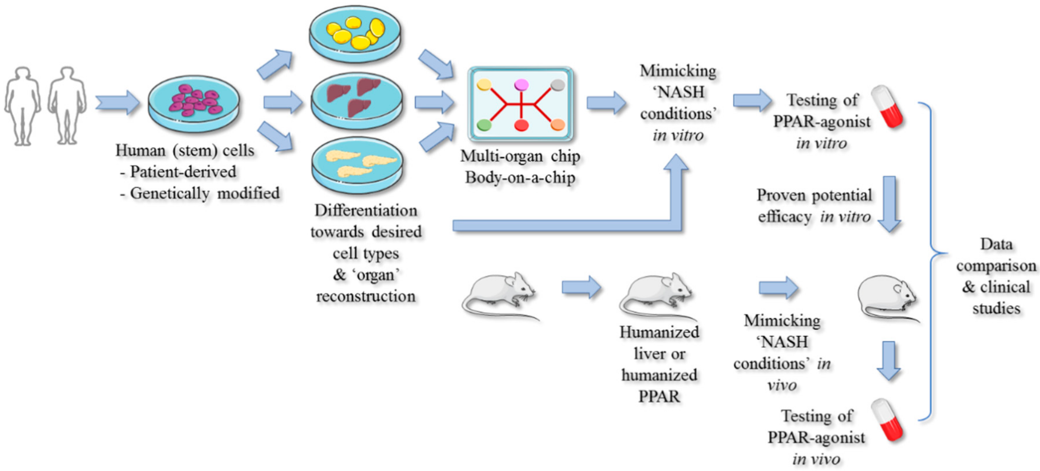
5. Strategies for Improving PPAR-Targeted Anti-NASH Drug Testing and Therapy

Based on the lack of an approved anti-NASH therapy and the current models that are used, it can be argued that (i) current disease models fail in representing the heterogeneity of the disease, and as such only illuminate a small facet of the NAFLD pathology, (ii) targeting nuclear receptors with broad and diffuse working mechanisms might not be sufficient for treating NASH, and (iii) personalized targeting of the underlying comorbidities might be the key adjuvant therapy in a heterogeneous population of NASH patients.

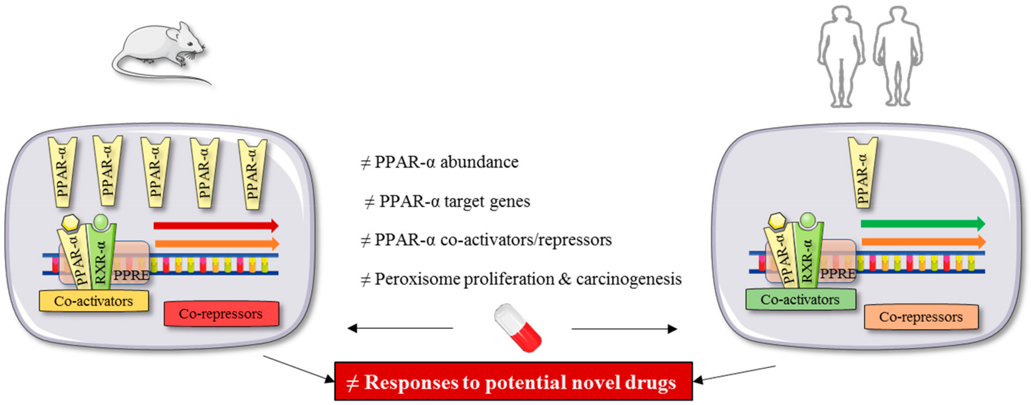
PPAR-Targeted Preclinical Drug Testing

From the abovementioned studies, it is clear that murine models can significantly contribute to the assessment of potential anti-NASH characteristics of novel drugs targeting different PPAR isotypes. Nevertheless, these agents could be tested in a more efficient way when interspecies differences regarding (i) the PPAR biology, (ii) responses to PPAR agonism, and (iii) specific polymorphisms related to NASH pathology could be taken out of the equation.
It is estimated that primary rodent hepatocytes express 10 to 20 times higher levels of PPAR-α than observed in human cells [75]. Furthermore, rodent and human PPAR-α show differences in responsiveness toward PPAR agonists [76]. For example, the exposure of HepG2 and CV-1 cells transfected with the human or rat PPAR-α to PPAR-α agonists (clofibrate, ETYA, and WY-14) show differential levels of PPAR-α activation [77]. On top, endogenous PPAR-α ligands differentially activate the mouse and human PPAR-α in transfected COS-7 cells. This might, apart from the higher basal PPAR-α abundance in mouse liver, explain why rodents develop liver cancer upon the administration of fibrates, which does not occur in humans [76,78]. Such differences are of major importance, since the PPAR isotypes need to be agonized in a specific manner and not only in the most potent way possible. This seems rather impossible to evaluate when only rodent models are being used (Figure 2).
This issue was addressed by Yang and co-workers, who generated PPAR-α humanized transgenic mice by using a P1 phage artificial chromosome (PAC) genomic clone bred onto a PPAR-α-null mouse background. When fenofibrate was administered to these transgenic mice, they did not show hepatomegaly and hepatocyte proliferation compared to wild-type mice. In addition, the miRNA MIRLET7C, which is involved in cell growth and regulation of the proto-oncogene MYC, was differentially regulated between the transgenic and wild-type mice, indicating a divergent regulation of the rodent and human PPAR-α. However, the authors pointed to the fact that these transgenic mice still show peroxisome proliferation upon fenofibrate administration. Yet, they did not take into account the possible regulatory differences of coactivators/repressors of PPAR-α, rendering unique responses to (partial) PPAR agonists [17,79]. This concern was later addressed by Tateno and co-workers, who created a mouse model, possessing a humanized liver containing more than 70% human hepatocytes and expressing more than 82% of the human genes. It could be shown that fenofibrate did not induce peroxisome proliferation in humanized livers compared to wild-type mice. It was assumed that human coactivators/repressors regulate the action of PPAR-α in a humanized way [80,81]. Later, it was shown that PPAR-α activation in human hepatocytes occurred in a more moderate way than in the residual mouse hepatocytes, confirming the abovementioned hypotheses [82].
Apart from these sophisticated refinements in animal experimentation and successful application, more practical tools exist to test the efficacy of new compounds with potential anti-NASH efficacy. Rogue et al. tested a battery of PPAR agonists (troglitazone, rosiglitazone, muraglitazar, tesaglitazar, fenofibrate, and bezafibrate) in oleic acid-overloaded HepaRG cells. Only troglitazone did not reduce the oleic acid-induced increased lipid load, demonstrating the potential of this model for the preclinical assessment of new anti-NAFLD compounds [83]. Yet, although harboring great applicability potential, none of the successfully tested drugs in the in vitro model showed convincing results during clinical studies [40,61]. Here, one should keep in mind that the complexity and inflammatory aspects of the NASH pathology are absent in the above-mentioned in vitro system. Feaver and co-workers constructed a human co-culture model containing primary human hepatocytes, stellate cells, and macrophages in a lipotoxic medium consisting of elevated insulin, glucose, and fatty acid levels [84]. Elafibranor lowered the lipid load in this system and attenuated the inflammatory response by downregulating interleukin (IL)-6 and CCL2 secretion [85], which correlates with clinical observations [67]. Our group could recently make similar observations using a human-derived adult stem cell-based model of NASH induced by fatty acids, insulin, glucose, and inflammatory cytokines. In this in vitro model, elafibranor potently reduced the increased lipid load and attenuated the inflammatory response [86].
The lack of approved (PPAR-targeted) therapies against NASH is at least partly ascribed to the lack of validated in vitro and in vivo models. Major issues are important interspecies differences and a relatively high cost [87]. Furthermore, given the multi-regulatory functions and broad tissue distribution of PPARs, it is clear that studies using non-human-based species should be interpreted with caution. Mice hosting humanized hepatic PPARs-α [79] or liver [81] address this issue only partly, since NAFLD is a multi-organ disease. In addition, PPAR(-α) target genes may be different between humans and rodents [23]. Therefore, we believe that human-based in vitro models could greatly contribute to select compounds with suitable PPAR isotype activity as well in simple [86,88] as in more complex co-cultures [84,85]. These models could ultimately be fine-tuned to specific ‘NASH subtypes’, mimicking the heterogeneity within the population of the molecular driving mechanisms of the disease and being suitable to evaluate different (combination) therapies. The lack of multiple cell types and/or organs for testing novel drug candidates as PPAR agonists is a drawback of most in vitro models. Yet, the advancements made to the development of ‘human-on-a-chip’ models seem promising [89]. Although still much has to be realized [90], the interconnection of in vitro surrogate organs such as liver, pancreas, and adipose tissue, merged with data of humanized animal models, could enable more reliable PPAR-targeted anti-NASH drug testing and thus improve clinical outcomes. Furthermore, by implementing in these chips patient-derived (induced-pluripotent) stem cells carrying high-risk genetic backgrounds for developing NASH, the assessment of personalized therapies might ever become a reality (Figure 3) [91].

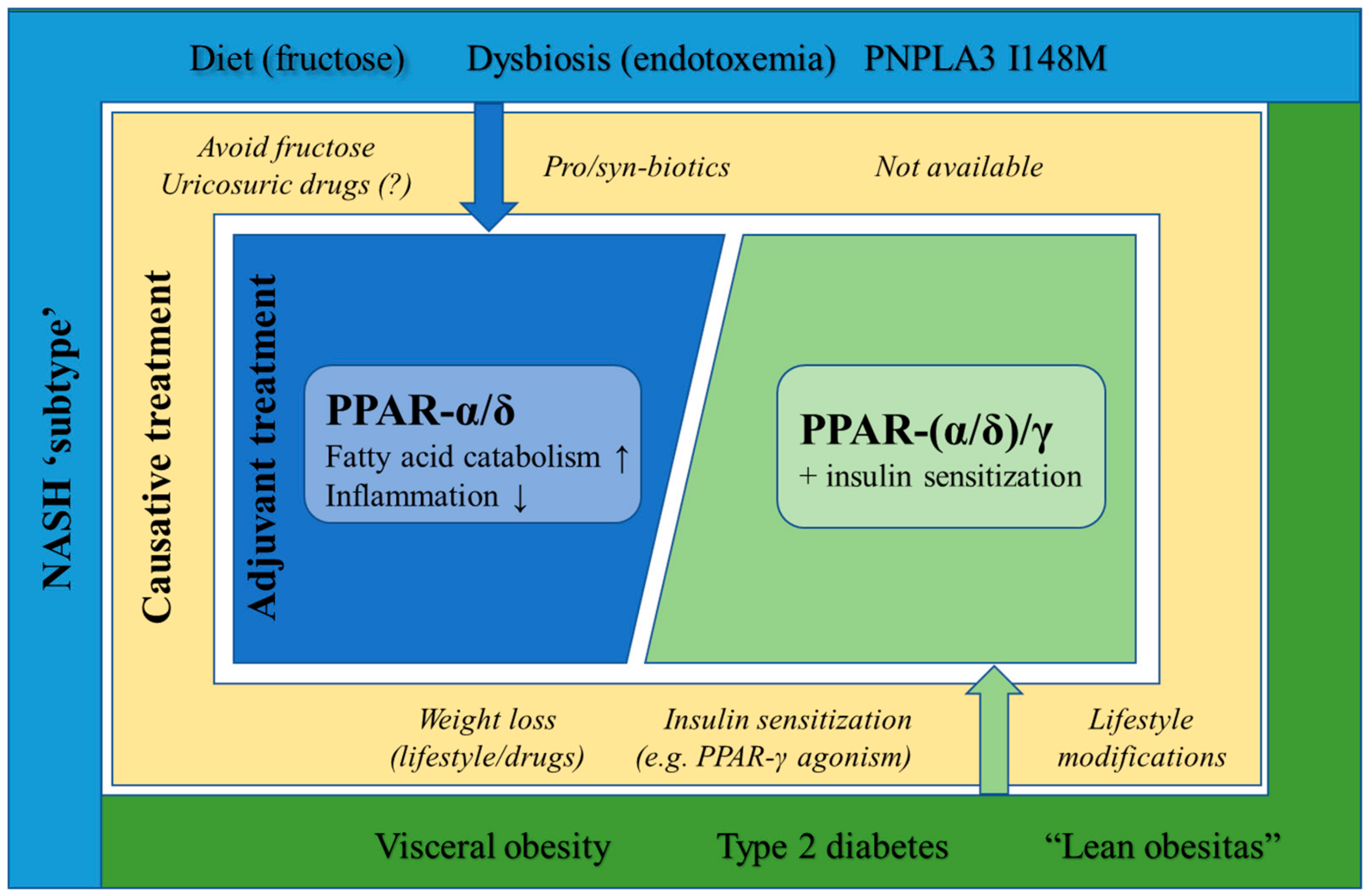
6. Targeted PPAR Agonism as NASH-Specific Therapy

Only a minority of steatosis subjects progresses toward NASH, indicating that environmental, hereditary, or other secondary causes (e.g., certain infections and extrahepatic conditions) lay at the basis of NAFLD progression [92]. This has also been outlined in the ‘multiple hit hypothesis’ for the pathogenesis of NASH [93]. In addition, the moderate success rates that have been obtained so far during clinical trials raise the question whether the current strategies for anti-NASH therapy are sufficiently addressing the complexity and the diverse pathogeneses of NASH [10]. It seems reasonable to conclude that PPAR-targeted anti-NASH therapy should preferably also consider the specific underlying etiologies and causative factors of the disease.

6.1. Diet-Induced NASH

Nutritional habits are often uniformly classified as a major cause of the metabolic syndrome and obesity. Nevertheless, different carbohydrates and lipid species can govern and drive divergent metabolic processes [94,95]. However, this is often neglected in the development of novel drugs against diseases with heterogeneous pathogeneses.
A high-carbohydrate diet in combination with low-energy expenditure induces de novo lipogenesis that results in the generation of fatty acids and a variety of lipid species. Glucose is generally regarded as the main sugar that drives de novo lipogenesis. Yet, fructose is also widely present in many drinks and foods and can induce as well de novo lipogenesis and fat accumulation [96]. Fructose may activate PPAR-γ coactivator-1β, which is a coactivator of SREBP-1c, triggering the lipogenic cascade [97]. It has also been reported in rat studies that fructose inhibits the function of PPAR-α, resulting in decreased β-oxidation and increased NF-κB activity [98,99]. Fructose may activate an inflammatory response due to its specific metabolism that only slightly differs from glucose catabolism. Glucose is first metabolized by glucokinase/hexokinase, while fructose is metabolized by fructokinase C. The latter enzyme phosphorylates fructose, followed by the formation of glyceraldehyde and dihydroxyacetone phosphate by the action of aldolase B. The persistent phosphorylation of fructose requires ATP and phosphate, resulting in the deposition of uric acid, which acts as a danger-associated molecular pattern (DAMP) [95]. DAMPs on their turn activate inflammasomes to produce IL-1β, which triggers an inflammatory response and thereby lay at the basis of NASH [100]. PPAR agonism for fructose-driven NASH might prove effective because of the specific action of fructose on PPAR(-α) activity. Nonetheless, it is unlikely that PPAR-α agonism can also capture the unique metabolism of fructose that results in the formation of uric acid that in turn drives the inflammatory reaction. The deposition of uric acid might remain present, resulting in a lean but inflamed liver.
Moreover, serum uric acid levels in NAFLD patients have been proposed as potential predictors of liver damage severity [101]. It has been shown in vitro, using HepG2 cells, that fructose induces lipid accumulation and uric acid formation. This is prevented by allopurinol [102], which is a drug that is indicated for the treatment of gout. In this view, PPAR-(γ) agonists with uricosuric properties could be eminent anti-NASH drugs. For example, arhalofenate, a selective partial PPAR-γ modulator, has been developed as a lipid-lowering drug, yet has also been found to block the reabsorption of uric acid through the inhibition of URAT1 in the proximal tubules of the kidney, thereby lowering serum uric acid levels [103,104]. African Americans show the lowest NAFLD prevalence among different ethnicities and exhibit fructose malabsorption, possibly rendering hepatoprotective effects [105,106].

6.2. Obesity and Type 2 Diabetes-Induced NASH

Obesity increases the risk for developing NAFLD and metabolic syndrome-related complications, in which the waist circumference is a key determinant [106]. In obesity, white adipose tissue produces a plethora of inflammatory mediators [107], leading to low-grade chronic inflammation. This effect is mediated by adipose tissue macrophages secreting pro-inflammatory mediators (TNF-α, IL-6), and detrimentally affecting insulin signaling in adipose tissue and liver [108]. TNF-α, but also lipotoxic lipid species such as free fatty acids, provoke the phosphorylation of serine residues of the insulin receptor substrate 1, which prevents signal transmission from the insulin receptor [107,109]. As such, inflammation in obesity-driven NAFLD is mainly derived from adipose tissue, instead of the liver. Impressively, the bariatric surgery of morbidly obese NASH patients resolved NASH in up to 85% of the patients [5], and it has been further observed that TNF-α levels decline with weight loss [110,111]. This points to the fact that adipose tissue is a major driver of the disease.
PPAR-γ agonists can reduce obesity-induced inflammation and improve insulin signaling [21]. Yet, the use of (full) PPAR-γ-agonists is limited because of weight gain [30]. Although partial PPAR-γ agonism could be efficient for obesity-induced NASH, it is clear that weight loss through lifestyle modification, bariatric surgery, or pharmacological therapy is of major importance in this subgroup of NASH patients [4,5]. It efficiently may reduce the source of inflammation and (whole-body) insulin resistance.

6.3. Lean NASH

Ethnicity is another risk factor for NAFLD. Several studies reported that a single nucleotide polymorphism (SNP) in PNPLA3 (rs738409) confers a higher susceptibly for developing NASH [112,113,114]. As this genetic trait is most common in Hispanics, they carry the highest risk. On the contrary, African Americans are the least susceptible for developing NAFLD [107,112].
PNPLA3 or adiponutrin is an adipose triglyceride lipase in which the rs738409 variant structurally prevents substrate binding [115]. The PNPLA3 rs738409 variant does not modify the disease through changes in insulin sensitivity, metabolic syndrome, body mass index, or dyslipidemia [116,117,118]. This subtype of NASH patients would theoretically not benefit from insulin-sensing PPAR agonists, yet they might do from PPAR agonists with strong β-oxidative capacity. On the contrary, the loss-of-function SNP Gly972Arg in the insulin receptor substrate-1 diminishes insulin signaling and increases the susceptibility for NAFLD progression [119]. Therefore, insulin sensitizing agents could be indicated as anti-NASH therapy when carrying this specific SNP [120]. However, lean NAFLD is not only caused by genetic alterations. Up to 5% of the Western population is ‘metabolically obese’, in which insulin resistance, hypertriglyceridemia, and hyperuricemia occurs [121]. In contrast to some (e.g., PNPLA3 rs738409) genetically-driven lean NASH patients, ‘metabolically obese’ NASH patients might favor from insulin-sensing PPAR(-(α)/γ) agonists. Indeed, 80% of the non-obese type 2 diabetes patients exhibit liver steatosis [122]. Other causes of lean NASH, such as HIV treatment, endocrine disorders (e.g., polycystic ovarian syndrome, hypothyroidism), total parenteral nutrition, jejunoileal bypass, or the use of NASH-inducing drugs (e.g., valproic acid, amiodarone, and methotrexate) [106,123,124], all ask for the individual identification and evaluation of the specific NASH pathogenesis and potential subsequent therapy.

6.4. Microbiome-Induced NASH

The microbiome is a highly dynamic population of microbiota subject to nutrition, environment, and immunity [125]. Alterations in composition in this often-called ‘forgotten organ’ can lead to dysbiosis, which may result in a plethora of diseases [126,127]. NAFLD has been associated with an increased gut permeability, small intestinal bacterial overgrowth [128], and endotoxemia (in which lipopolysaccharide (LPS) plays a prominent role) [129,130]. LPS is a component of Gram-negative bacteria, which are elevated in the gut of NASH patients [131]. LPS affects insulin signaling through the Toll-like receptor 4–monocyte differentiation antigen CD14 system, leading to insulin resistance [132]. It also holds the capacity to trigger inflammasome activation, leading to sustained hepatic inflammation [133]. Much more complex alterations in the gut microbiome of NASH patients have been reported [125,134], but it is clear that PPAR-targeted therapy might not be effective in the long-term for these patients, since the real cause of the disease is not targeted. Multiple trials are running to specifically target and restore the gut microbiome (e.g., using probiotics) in NASH patients, in which already satisfactory results have been obtained [135].

7. Outlook and Conclusions

A multitude of agents targeting one, two, or all three PPAR isotypes has been evaluated and is under evaluation for anti-NASH treatment. Although theoretically promising, none of the on-the-market anti-hyperglycemic and anti-hyperlipidemic drugs have so far proven to adequately tackle NASH. Hitherto, elafibranor, currently being evaluated in a clinical phase III trial, seems to pose the best outbalanced activity on the PPAR-α and -δ isotypes to treat NASH [67]. Nonetheless, two other PPAR agonists, lanifibranor [74] and saroglitazar [71], respectively a PPAR-pan and PPAR-α/γ agonist, are both being tested in clinical phase II trials and seem promising as well.
As the origin of NASH is not as clear-cut as previously thought [136,137], and NASH patient trials are generally obtaining unsatisfactory low success rates [10], the rationale behind the development of novel anti-NASH drug candidates and/or combination therapies could perhaps benefit from a subdivision into ‘NASH types’, paralleling the ‘multiple hit hypothesis’ for the pathogenesis of NASH [93] (Figure 4).
The PPAR agonists under development all target consequences of pre-existing, underlying morbidities, rendering them the potential to rapidly intervene in case of liver steatosis and inflammation. Yet, apart from the often insulin-sensing effects of these compounds, no other disease drivers and/or modifiers are targeted, leaving the cause behind NAFLD progression untouched. Therefore, it can be questioned whether subdividing NASH and diagnosing into subtypes could not increase the efficiency to target the causes that lay at the basis of the disease state and improve clinical outcomes. Furthermore, it is plausible that combination therapies, targeting different aspects of the disease (e.g., probiotics together with a PPAR agonist), are necessary to both target the initial disease driver and relieve the existing hepatic steatosis and inflammation. Indeed, combination therapies are already being explored to also target uncovered disturbed pathways. This has been outlined by the Food and Drug Administration as well [138]. Although elafibranor seems to be one of the most promising compounds for anti-NASH treatment, a clinical program to evaluate combination therapies with a glucagon-like peptide-1 receptor agonist and a sodium-glucose transporter-2 inhibitor has been recently announced [139]. Nevertheless, the transition of benign hepatic steatosis to NASH could be delayed or prevented by PPAR-(α/δ)-agonists. By lowering the hepatic lipid load at the early steatosis stage, the liver might regain physiological functions before adaptive mechanisms are exhausted.
These considerations in anti-NASH therapy could be translated into the early drug developmental stages, in which human-based approaches can provide information with respect to potential efficacy. The main reason behind the use of human-based models in PPAR-targeted anti-NASH drug development is connected with the important inter-species differences in the biology of PPARs [86,140]. Yet, mice carrying humanized (hepatic) PPARs could be key for the evaluation of multi-organ targeted anti-NASH therapies [80,81]. In the future, human-based body-on-a chip methodologies might be considered as suitable human-relevant test models [86,89,91].

Author Contributions

Conceptualization, J.B., T.V. and R.M.R.; methodology, J.B. and R.M.R.; writing—original draft preparation, J.B. and R.M.R.; writing—review and editing, J.B., A.N., M.R., K.B., V.R., J.D.K., T.V. and R.M.R.; visualization, J.B. and R.M.R. supervision, R.M.R., V.R. and T.V.; project administration, R.M.R., V.R. and T.V.; funding acquisition, J.B., R.M.R., V.R. and T.V. All authors have read and agreed to the published version of the manuscript.

Funding

This work was funded by grants of Research Foundation Flanders (1S10518N, 12H2216N, 1S73019N and G042019N), University Hospital of the Vrije Universiteit Brussel-Belgium (Willy Gepts Fonds UZ-VUB), Onderzoeksraad Vrije Universiteit Brussel and the Research Chair Mireille Aerens for Alternatives to Animal Testing.

Acknowledgments

Figures were partially made using Servier Medical Art.

Conflicts of Interest

The authors declare no conflicts of interest.

References

  1. Younossi, Z.M.; Marchesini, G.; Pinto-Cortez, H.; Petta, S. Epidemiology of Nonalcoholic Fatty Liver Disease and Nonalcoholic Steatohepatitis: Implications for Liver Transplantation. Transplantation 2019, 103, 22–27. [Google Scholar] [CrossRef]
  2. Estes, C.; Razavi, H.; Loomba, R.; Younossi, Z.; Sanyal, A.J. Modeling the Epidemic of Nonalcoholic Fatty Liver Disease Demonstrates an Exponential Increase in Burden of Disease. Hepatology 2018, 67, 123–133. [Google Scholar] [CrossRef] [PubMed]
  3. Sanyal, A.J. Past, Present and Future Perspectives in Nonalcoholic Fatty Liver Disease. Nat. Rev. Gastroenterol. Hepatol. 2019, 16, 377–386. [Google Scholar] [CrossRef] [PubMed]
  4. Vilar-Gomez, E.; Martinez-Perez, Y.; Calzadilla-Bertot, L.; Torres-Gonzalez, A.; Gra-Oramas, B.; Gonzalez-Fabian, L.; Friedman, S.L.; Diago, M.; Romero-Gomez, M. Weight Loss through Lifestyle Modification Significantly Reduces Features of Nonalcoholic Steatohepatitis. Gastroenterology 2015, 149, 367–378. [Google Scholar] [CrossRef] [PubMed]
  5. Lassailly, G.; Caiazzo, R.; Buob, D.; Pigeyre, M.; Verkindt, H.; Labreuche, J.; Raverdy, V.; Leteurtre, E.; Dharancy, S.; Louvet, A.; et al. Bariatric Surgery Reduces Features of Nonalcoholic Steatohepatitis in Morbidly Obese Patients. Gastroenterology 2015, 149, 379–388. [Google Scholar] [CrossRef] [Green Version]
  6. Younossi, Z.; Anstee, Q.M.; Marietti, M.; Hardy, T.; Henry, L.; Eslam, M.; George, J.; Bugianesi, E. Global Burden of NAFLD and NASH: Trends, Predictions, Risk Factors and Prevention. Nat. Rev. Gastroenterol. Hepatol. 2018, 15, 11–20. [Google Scholar] [CrossRef] [PubMed]
  7. Kota, B.P.; Huang, T.H.-W.; Roufogalis, B.D. An Overview on Biological Mechanisms of PPARs. Pharmacol. Res. 2005, 51, 85–94. [Google Scholar] [CrossRef] [PubMed]
  8. Poulsen, L.I.; Siersbæk, M.; Mandrup, S. PPARs: Fatty Acid Sensors Controlling Metabolism. Semin. Cell Dev. Biol. 2012, 23, 631–639. [Google Scholar] [CrossRef]
  9. Feng, X.; Gao, X.; Jia, Y.; Zhang, H.; Xu, Y.; Wang, G. PPAR-α Agonist Fenofibrate Decreased RANTES Levels in Type 2 Diabetes Patients with Hypertriglyceridemia. Med. Sci. Monit. 2016, 22, 743–751. [Google Scholar] [CrossRef]
  10. Friedman, S.L.; Neuschwander-Tetri, B.A.; Rinella, M.; Sanyal, A.J. Mechanisms of NAFLD Development and Therapeutic Strategies. Nat. Med. 2018, 24, 908–922. [Google Scholar] [CrossRef]
  11. Connolly, J.J.; Ooka, K.; Lim, J.K. Future Pharmacotherapy for Non-Alcoholic Steatohepatitis (NASH): Review of Phase 2 and 3 Trials. J. Clin. Transl. Hepatol. 2018, 6, 1–12. [Google Scholar] [CrossRef] [PubMed] [Green Version]
  12. Tanaka, N.; Aoyama, T.; Kimura, S.; Gonzalez, F.J. Targeting Nuclear Receptors for the Treatment of Fatty Liver Disease. Pharmacol. Ther. 2017, 179, 142–157. [Google Scholar] [CrossRef] [PubMed]
  13. Burn, K.A.; Vanden Heuvel, J.P. Modulation of PPAR Activity via Phosphorylation. Biochim Biophys Acta 2009, 1771, 952–960. [Google Scholar] [CrossRef] [PubMed] [Green Version]
  14. Bugge, A.; Mandrup, S. Molecular Mechanisms and Genome-Wide Aspects of PPAR Subtype Specific Transactivation. PPAR Res. 2010, 2010, 169506. [Google Scholar] [CrossRef] [Green Version]
  15. Liss, K.H.H.; Finck, B.N. PPARs and Nonalcoholic Fatty Liver Disease. Biochimie 2017, 136, 65–74. [Google Scholar] [CrossRef] [Green Version]
  16. Oberfield, J.L.; Collins, J.L.; Holmes, C.P.; Goreham, D.M.; Cooper, J.P.; Cobb, J.E.; Lenhards, J.M.; Hull-Ryde, E.A.; Mohr, C.P.; Blanchard, S.G.; et al. A Peroxisome Proliferator-Activated Receptor γ Ligand Inhibits Adipocyte Differentiation. Proc. Natl. Acad. Sci. USA 1999, 96, 6102–6106. [Google Scholar] [CrossRef] [Green Version]
  17. Berger, J.; Moller, D.E. The Mechanisms of Action of PPARs. Annu. Rev. Med. 2002, 53, 409–435. [Google Scholar] [CrossRef] [Green Version]
  18. Gross, B.; Pawlak, M.; Lefebvre, P.; Staels, B. PPARs in Obesity-Induced T2DM, Dyslipidaemia and NAFLD. Nat. Rev. Endocrinol. 2017, 13, 36–49. [Google Scholar] [CrossRef]
  19. Giby, V.G.; Ajith, T.A. Role of Adipokines and Peroxisome Proliferator-Activated Receptors in Nonalcoholic Fatty Liver Disease. World J. Hepatol. 2014, 6, 570–579. [Google Scholar] [CrossRef]
  20. Capelli, D.; Cerchia, C.; Montanari, R.; Loiodice, F.; Tortorella, P.; Laghezza, A.; Cervoni, L.; Pochetti, G.; Lavecchia, A. Structural Basis for PPAR Partial or Full Activation Revealed by a Novel Ligand Binding Mode. Sci. Rep. 2016, 6, 1–12. [Google Scholar] [CrossRef]
  21. Ricote, M.; Glass, C.K. PPARs and Molecular Mechanisms of Transrepression. Biochim. Biophys. Acta 2007, 1171, 926–935. [Google Scholar] [CrossRef] [PubMed] [Green Version]
  22. Schupp, M.; Lazar, M.A. Endogenous Ligands for Nuclear Receptors: Digging Deeper. J. Biol. Chem. 2010, 285, 40409–40415. [Google Scholar] [CrossRef] [PubMed] [Green Version]
  23. Rakhshandehroo, M.; Knoch, B.; Müller, M.; Kersten, S. Peroxisome Proliferator-Activated Receptor Alpha Target Genes. PPAR Res. 2010, 2010, 1–20. [Google Scholar] [CrossRef] [PubMed] [Green Version]
  24. Piccinin, E.; Moschettal, A. Hepatic-Specific PPARa-FGF21 Action in NAFLD. Gut 2016, 65, 1075–1076. [Google Scholar] [CrossRef]
  25. Zhang, N.; Chu, E.S.H.; Zhang, J.; Li, X.; Liang, Q.; Chen, J.; Chen, M.; Teoh, N.; Farrell, G.; Sung, J.J.Y.; et al. Peroxisome Proliferator Activated Receptor Alpha Inhibits Hepatocarcinogenesis through Mediating NF-KB Signaling Pathway. Oncotarget 2014, 5, 8330–8340. [Google Scholar] [CrossRef]
  26. Iroz, A.; Montagner, A.; Benhamed, F.; Levavasseur, F.; Polizzi, A.; Anthony, E.; Régnier, M.; Fouché, E.; Lukowicz, C.; Cauzac, M.; et al. A Specific ChREBP and PPARα Cross-Talk Is Required for the Glucose-Mediated FGF21 Response. Cell Rep. 2017, 21, 403–416. [Google Scholar] [CrossRef] [Green Version]
  27. Oosterveer, M.H.; Grefhorst, A.; van Dijk, T.H.; Havinga, R.; Staels, B.; Kuipers, F.; Groen, A.K.; Reijngoud, D.J. Fenofibrate Simultaneously Induces Hepatic Fatty Acid Oxidation, Synthesis, and Elongation in Mice. J. Biol. Chem. 2009, 284, 34036–34044. [Google Scholar] [CrossRef] [Green Version]
  28. Palomer, X.; Barroso, E.; Pizarro-Delgado, J.; Peña, L.; Botteri, G.; Zarei, M.; Aguilar, D.; Montori-Grau, M.; Vázquez-Carrera, M. PPARβ/δ: A Key Therapeutic Target in Metabolic Disorders. Int. J. Mol. Sci. 2018, 19, 913. [Google Scholar] [CrossRef] [Green Version]
  29. Zarei, M.; Barroso, E.; Palomer, X.; Dai, J.; Rada, P.; Quesada-López, T.; Escolà-Gil, J.C.; Cedó, L.; Zali, M.R.; Molaei, M.; et al. Hepatic Regulation of VLDL Receptor by PPARβ/δ and FGF21 Modulates Non-Alcoholic Fatty Liver Disease. Mol. Metab. 2018, 8, 117–131. [Google Scholar] [CrossRef]
  30. Heikkinen, S.; Auwerx, J.; Argmann, C.A. PPARγ in Human and Mouse Physiology. Biochim. Biophys. Acta 2007, 1771, 999–1013. [Google Scholar] [CrossRef] [Green Version]
  31. Kim, H.; Ahn, Y. Role of Peroxisome Proliferator–Activated Receptor-γ in the Glucose-Sensing Apparatus of Liver and β-Cells. Biochemistry 2004, 53, 1–6. [Google Scholar] [CrossRef] [PubMed] [Green Version]
  32. Pais, R.; Moraru, I.; Ratziu, V. Glitazones for Human Nonalcoholic Steatohepatitis. Therap. Adv. Gastroenterol. 2011, 4, 325–334. [Google Scholar] [CrossRef] [PubMed] [Green Version]
  33. Ricote, M.; Li, A.C.; Willson, T.M.; Kelly, C.J.; Glass, C.K. The Peroxisome Proliferator-Activated Receptor-γ Is a Negative Regulator of Macrophage Activation. Nature 1998, 391, 79–82. [Google Scholar] [CrossRef] [PubMed]
  34. Chengyu, J.; Ting, A.T.; Seed, B. PPAR-γ Agonists Inhibit Production Of monocyte Inflammatory Cytokine. Nature 1999, 391, 82–86. [Google Scholar]
  35. Francque, S.; Verrijken, A.; Caron, S.; Prawitt, J.; Paumelle, R.; Derudas, B.; Lefebvre, P.; Taskinen, M.R.; Van Hul, W.; Mertens, I.; et al. PPAR-α Gene Expression Correlates with Severity and Histological Treatment Response in Patients with Non-Alcoholic Steatohepatitis. J. Hepatol. 2015, 63, 164–173. [Google Scholar] [CrossRef]
  36. Nagaya, T.; Tanaka, N.; Suzuki, T.; Sano, K.; Horiuchi, A.; Komatsu, M.; Nakajima, T.; Nishizawa, T.; Joshita, S.; Umemura, T.; et al. Down-Regulation of SREBP-1c Is Associated with the Development of Burned-out NASH. J. Hepatol. 2010, 53, 724–731. [Google Scholar] [CrossRef] [Green Version]
  37. Li, X.; Wang, Z.; Klaunig, J.E. Modulation of Xenobiotic Nuclear Receptors in High-Fat Diet Induced Non-Alcoholic Fatty Liver Disease. Toxicology 2018, 1, 199–213. [Google Scholar] [CrossRef]
  38. Botta, M.; Audano, M.; Sahebkar, A.; Sirtori, C.R.; Mitro, N.; Ruscica, M. PPAR Agonists and Metabolic Syndrome: An Established Role? Int. J. Mol. Sci. 2018, 19, 1197. [Google Scholar] [CrossRef] [Green Version]
  39. Basaranoglu, M.; Acbay, O.; Sonsuz, A. A Controlled Trial of Gemfibrozil in the Treatment of Patients with Nonalcoholic Steatohepatitis. J. Hepatol. 1999, 31, 384. [Google Scholar] [CrossRef]
  40. Fernández-Miranda, C.; Pérez-Carreras, M.; Colina, F.; López-Alonso, G.; Vargas, C.; Solís-Herruzo, J.A. A Pilot Trial of Fenofibrate for the Treatment of Non-Alcoholic Fatty Liver Disease. Dig. Liver Dis. 2008, 40, 200–205. [Google Scholar] [CrossRef]
  41. Laurin, J.; Lindor, K.D.; Crippin, J.S.; Gossard, A.; Gores, G.J.; Ludwig, J.; Rakela, J.; McGill, B.M. Ursodeoxycholic Acid or Clofibrate in the Treatment of Non-Alcohol- Induced Steatohepatitis: A Pilot Study. Hepatology 1996, 23, 1464–1467. [Google Scholar] [CrossRef] [PubMed]
  42. Ishibashi, S.; Arai, H.; Yokote, K.; Araki, E.; Suganami, H.; Yamashita, S. Efficacy and Safety of Pemafibrate (K-877), a Selective Peroxisome Proliferator-Activated Receptor α Modulator, in Patients with Dyslipidemia: Results from a 24-Week, Randomized, Double Blind, Active-Controlled, Phase 3 Trial. J. Clin. Lipidol. 2018, 12, 173–184. [Google Scholar] [CrossRef] [PubMed] [Green Version]
  43. Honda, Y.; Kessoku, T.; Ogawa, Y.; Tomeno, W.; Imajo, K.; Fujita, K.; Yoneda, M.; Takizawa, T.; Saito, S.; Nagashima, Y.; et al. Pemafibrate, a Novel Selective Peroxisome Proliferator-Activated Receptor Alpha Modulator, Improves the Pathogenesis in a Rodent Model of Nonalcoholic Steatohepatitis. Sci. Rep. 2017, 7, 1–11. [Google Scholar] [CrossRef] [PubMed]
  44. Bays, H.E.; Schwartz, S.; Littlejohn, T.; Kerzner, B.; Krauss, R.M.; Karpf, D.B.; Choi, Y.J.; Wang, X.; Naim, S.; Roberts, B.K. MBX-8025, a Novel Peroxisome Proliferator Receptor-Δagonist: Lipid and Other Metabolic Effects in Dyslipidemic Overweight Patients Treated with and without Atorvastatin. J. Clin. Endocrinol. Metab. 2011, 96, 2889–2897. [Google Scholar] [CrossRef] [Green Version]
  45. Choi, Y.J.; Roberts, B.K.; Wang, X.; Geaney, J.C.; Naim, S.; Wojnoonski, K.; Karpf, D.B.; Krauss, R.M. Effects of the PPAR-δ Agonist MBX-8025 on Atherogenic Dyslipidemia. Atherosclerosis 2012, 220, 470–476. [Google Scholar] [CrossRef]
  46. Ooi, E.M.M.; Watts, G.F.; Sprecher, D.L.; Chan, D.C.; Barrett, P.H.R. Mechanism of Action of a Peroxisome Proliferator-Activated Receptor (PPAR)-δ Agonist on Lipoprotein Metabolism in Dyslipidemic Subjects with Central Obesity. J. Clin. Endocrinol. Metab. 2011, 96, 1568–1576. [Google Scholar] [CrossRef] [Green Version]
  47. Risérerus, U.; Sprecher, D.; Johnson, T.; Olson, E.; Hirschberg, S.; Liu, A.; Fang, Z.; Hegde, P.; Richards, D.; Sarov-Blat, L.; et al. Activation of Peroxisome Proliferator–δ Activated Receptor (PPAR) Promotes Reversal of Multiple Metabolic Abnormalities, Reduces Oxidative Stress, and Increases Fatty Acid Oxidation in Moderately Obese Men. Diabetes 2008, 57, 332–339. [Google Scholar] [CrossRef] [Green Version]
  48. Haczeyni, F.; Wang, H.; Barn, V.; Mridha, A.R.; Yeh, M.M.; Haigh, W.G.; Ioannou, G.N.; Choi, Y.-J.; McWherter, C.A.; Teoh, N.C.-H.; et al. The Selective Peroxisome Proliferator-Activated Receptor-Delta Agonist Seladelpar Reverses Nonalcoholic Steatohepatitis Pathology by Abrogating Lipotoxicity in Diabetic Obese Mice. Hepatol. Commun. 2017, 1, 663–674. [Google Scholar] [CrossRef] [Green Version]
  49. Oseini, A.M.; Sanyal, A.J. Therapies in Non-Alcoholic Steatohepatitis (NASH). Liver Int. 2017, 37, 97–103. [Google Scholar] [CrossRef] [Green Version]
  50. Ozturk, Z.A.; Kadayifci, A. Insulin Sensitizers for the Treatment of Non-Alcoholic Fatty Liver Disease. World J. Hepatol. 2014, 6, 199–206. [Google Scholar] [CrossRef]
  51. Yong-ho, L.; Jae, H.K.; So, R.K.; Heung, J.Y.; Eun-Jung, R.; Young, M.C.; Byung-Wan, L. Lobeglitazone, a Novel Thiazolidinedione, Improves Non-Alcoholic Fatty Liver Disease in Type 2 Diabetes: Its Efficacy and Predictive Factors Related to Responsiveness Study Patients. Endocrinol. Nutr. Metab. 2017, 32, 60–69. [Google Scholar]
  52. Sanyal, A.J.; Chalasani, N.; Kowdley, K.V.; McCullough, A.; Diehl, A.M.; Bass, N.M.; Neuschwander-Tetri, B.A.; Lavine, J.E.; Tonascia, J.; Unalp, A.; et al. Pioglitazone, Vitamin E, or Placebo for Nonalcoholic Steatohepatitis. N. Engl. J. Med. 2010, 362, 1675–1685. [Google Scholar] [CrossRef] [PubMed] [Green Version]
  53. Yan, H.M.; Xia, M.F.; Wang, Y.; Chang, X.X.; Yao, X.Z.; Rao, S.X.; Zeng, M.S.; Tu, Y.F.; Feng, R.; Jia, W.P.; et al. Efficacy of Berberine in Patients with Non-Alcoholic Fatty Liver Disease. PLoS ONE 2015, 10, 1–16. [Google Scholar] [CrossRef] [PubMed] [Green Version]
  54. Sanyal, A.J.; Mofrad, P.S.; Contos, M.J.; Sargeant, C.; Luketic, V.A.; Sterling, R.K.; Stravitz, R.T.; Shiffman, M.L.; Clore, J.; Mills, A.S. A Pilot Study of Vitamin E Versus Vitamin E and Pioglitazone for the Treatment of Nonalcoholic Steatohepatitis. Clin. Gastroenterol. Hepatol. 2004, 2, 1107–1115. [Google Scholar] [CrossRef]
  55. Cusi, K.; Orsak, B.; Bril, F.; Lomonaco, R.; Hecht, J.; Ortiz-Lopez, C.; Tio, F.; Hardies, J.; Darland, C.; Musi, N.; et al. Long-Term Pioglitazone Treatment for Patients with Nonalcoholic Steatohepatitis and Prediabetes or Type 2 Diabetes Mellitus a Randomized Trial. Ann. Intern. Med. 2016, 165, 305–315. [Google Scholar] [CrossRef] [PubMed]
  56. Aithal, G.P.; Thomas, J.A.; Kaye, P.V.; Lawson, A.; Ryder, S.D.; Spendlove, I.; Austin, A.S.; Freeman, J.G.; Morgan, L.; Webber, J. Randomized, Placebo-Controlled Trial of Pioglitazone in Nondiabetic Subjects With Nonalcoholic Steatohepatitis. Gastroenterology 2008, 135, 1176–1184. [Google Scholar] [CrossRef] [PubMed] [Green Version]
  57. Chalasani, N.; Younossi, Z.; Lavine, J.E.; Diehl, A.M.; Brunt, E.M.; Cusi, K.; Charlton, M.; Sanyal, A.J. The Diagnosis and Management of Non-Alcoholic Fatty Liver Disease: Practice Guideline by the American Association for the Study of Liver Diseases, American College of Gastroenterology, and the American Gastroenterological Association. Hepatology 2012, 55, 2005–2023. [Google Scholar] [CrossRef]
  58. Marchesini, G.; Day, C.P.; Dufour, J.F.; Canbay, A.; Nobili, V.; Ratziu, V.; Tilg, H.; Roden, M.; Gastaldelli, A.; Yki-Jarvinen, H.; et al. EASL-EASD-EASO Clinical Practice Guidelines for the Management of Non-Alcoholic Fatty Liver Disease. J. Hepatol. 2016, 64, 1388–1402. [Google Scholar] [CrossRef] [Green Version]
  59. Mahady, S.E.; Webster, A.C.; Walker, S.; Sanyal, A.; George, J. The Role of Thiazolidinediones in Non-Alcoholic Steatohepatitis—A Systematic Review and Meta Analysis. J. Hepatol. 2011, 55, 1383–1390. [Google Scholar] [CrossRef]
  60. Tuccori, M.; Filion, K.B.; Yin, H.; Yu, O.H.; Platt, R.W.; Azoulay, L. Pioglitazone Use and Risk of Bladder Cancer: Population Based Cohort Study. Br. Med. J. 2016, 352, i1541. [Google Scholar] [CrossRef] [Green Version]
  61. Ratziu, V.; Charlotte, F.; Bernhardt, C.; Giral, P.; Halbron, M.; Lenaour, G.; Hartmann-Heurtier, A.; Bruckert, E.; Poynard, T. Long-Term Efficacy of Rosiglitazone in Nonalcoholic Steatohepatitis: Results of the Fatty Liver Improvement by Rosiglitazone Therapy (FLIRT 2) Extension Trial. Hepatology 2010, 51, 445–453. [Google Scholar] [CrossRef] [PubMed]
  62. Ratziu, V.; Giral, P.; Jacqueminet, S.; Charlotte, F.; Hartemann-Heurtier, A.; Serfaty, L.; Podevin, P.; Lacorte, J.M.; Bernhardt, C.; Bruckert, E.; et al. Rosiglitazone for Nonalcoholic Steatohepatitis: One-Year Results of the Randomized Placebo-Controlled Fatty Liver Improvement With Rosiglitazone Therapy (FLIRT) Trial. Gastroenterology 2008, 135, 100–110. [Google Scholar] [CrossRef] [PubMed]
  63. Singh, S.; Loke, Y.K.; Furberg, C.D. Long-Term Risk of Cardiovascular Events With Rosiglitazone. Am. Med. Assoc. 2007, 298, 1189–1195. [Google Scholar] [CrossRef] [PubMed]
  64. Pladevall, M.; Riera-Guardia, N.; Margulis, A.V.; Varas-Lorenzo, C.; Calingaert, B.; Perez-Gutthann, S. Cardiovascular Risk Associated with the Use of Glitazones, Metformin and Sufonylureas: Meta-Analysis of Published Observational Studies. BMC Cardiovasc. Disord. 2016, 16, 14. [Google Scholar] [CrossRef] [Green Version]
  65. Sin, G.K.; Doo, M.K.; Jeong-Teak, W.; Hak, C.J.; Choon, H.C.; Kyung, S.K.; Jeong, H.P.; Yong, S.P.; Sang, J.K.; Dong, S.C. Efficacy and Safety of Lobeglitazone Monotherapy in Patients with Type 2 Diabetes Mellitus over 24-Weeks: A Multicenter, Randomized, Double-Blind, Parallel-Group, Placebo Controlled Trial. PLoS ONE 2014, 9, e92843. [Google Scholar]
  66. Sun, H.K.; Sin, G.K.; Doo, M.K.; Jeong-Teak, W.; Hak, C.J.; Choon, H.C.; Kyung, S.K.; Jeong, H.P.; Yong, S.P.; Sang, J.K.; et al. Safety and Efficacy of Lobeglitazone Monotherapy in Patients with Type 2 Diabetes Mellitus over 52 Weeks: An Open-Label Extension Study. Diabetes Res. Clin. Pract. 2015, 110, e27–e30. [Google Scholar]
  67. Ratziu, V.; Harrison, S.A.; Francque, S.; Bedossa, P.; Lehert, P.; Serfaty, L.; Romero-Gomez, M.; Boursier, J.; Abdelmalek, M.; Caldwell, S.; et al. Elafibranor, an Agonist of the Peroxisome Proliferator-Activated Receptor-α and -δ, Induces Resolution of Nonalcoholic Steatohepatitis Without Fibrosis Worsening. Gastroenterology 2016, 150, 1147–1159. [Google Scholar] [CrossRef] [Green Version]
  68. Hanf, R.; Millatt, L.J.; Cariou, B.; Noel, B.; Rigou, G.; Delataille, P.; Daix, V.; Hum, D.W.; Staels, B. The Dual Peroxisome Proliferator-Activated Receptor Alpha/Delta Agonist GFT505 Exerts Anti-Diabetic Effects in Db/Db Mice without Peroxisome Proliferator-Activated Receptor Gamma-Associated Adverse Cardiac Effects. Diabetes Vasc. Dis. Res. 2014, 11, 440–447. [Google Scholar] [CrossRef]
  69. Fiévet, C.; Fruchart, J.C.; Staels, B. PPARα and PPARγ Dual Agonists for the Treatment of Type 2 Diabetes and the Metabolic Syndrome. Curr. Opin. Pharmacol. 2006, 6, 606–614. [Google Scholar] [CrossRef]
  70. Kaul, U.; Parmar, D.; Manjunath, K.; Shah, M.; Parmar, K.; Patil, K.P.; Jaiswal, A. New Dual Peroxisome Proliferator Activated Receptor Agonist—Saroglitazar in Diabetic Dyslipidemia and Non-Alcoholic Fatty Liver Disease: Integrated Analysis of the Real World Evidence. Cardiovasc. Diabetol. 2019, 18, 80. [Google Scholar] [CrossRef] [Green Version]
  71. Jain, M.R.; Giri, S.R.; Bhoi, B.; Trivedi, C.; Rath, A.; Rathod, R.; Ranvir, R.; Kadam, S.; Patel, H.; Swain, P.; et al. Dual PPARα/γ Agonist Saroglitazar Improves Liver Histopathology and Biochemistry in Experimental NASH Models. Liver Int. 2018, 38, 1084–1094. [Google Scholar] [CrossRef] [PubMed] [Green Version]
  72. Rubin, C.J.; Viraswami-Appanna, K.; Fiedorek, F.T. Efficacy and Safety of Muraglitazar: A Double-Blind, 24-Week, Dose-Ranging Study in Patients with Type 2 Diabetes. Diabetes Vasc. Dis. Res. 2009, 6, 205–215. [Google Scholar] [CrossRef] [PubMed] [Green Version]
  73. Aravind, S.; Banshi, S.; Bhavana, S. Saroglitazar for the Treatment of Hypertriglyceridemia in Patients with Type 2 Diabetes: Current Evidence. DiabetesMetab. Syndr. Obes. Targets Ther. 2015, 8, 189–196. [Google Scholar] [CrossRef] [PubMed] [Green Version]
  74. Wettstein, G.; Luccarini, J.-M.; Poekes, L.; Faye, P.; Kupkowski, F.; Adarbes, V.; Defrene, E.; Estivalet, C.; Gawronski, X.; Jantzen, I.; et al. The New-Generation Pan-Peroxisome Proliferator-Activated Receptor Agonist IVA337 Protects the Liver From Metabolic Disorders and Fibrosis. Hepatol. Commun. 2017, 1, 524–537. [Google Scholar] [CrossRef]
  75. Holden, P.R.; Tugwood, J.D. Peroxisome Proliferator-Activated Receptor Alpha: Role in Rodent Liver Cancer and Species Differences. J. Mol. Endocrinol. 1999, 22, 1–8. [Google Scholar] [CrossRef] [Green Version]
  76. Yoon, M. The Role of PPARα in Lipid Metabolism and Obesity: Focusing on the Effects of Estrogen on PPARα Actions. Pharmacol. Res. 2009, 60, 151–159. [Google Scholar] [CrossRef]
  77. Mukherjee, R.; Jow, L.; McDonnell, D.P. Human and Rat Peroxisome Proliferator Activated Receptors (PPARs) Demonstrate Similar Tissue Distribution but Different Responsiveness to PPAR Activators. J. Steroid Biochem. Mol. Biol. 1994, 51, 157–166. [Google Scholar] [CrossRef]
  78. Peters, J.M.; Cheung, C.; Gonzalez, F.J. Peroxisome Proliferator-Activated Receptor- α and Liver Cancer: Where Do We Stand? J. Mol. Med. 2005, 83, 774–785. [Google Scholar] [CrossRef]
  79. Yang, Q.; Nagano, T.; Shah, Y.; Cheung, C.; Ito, S.; Gonzalez, F.J. The PPARα-Humanized Mouse: A Model to Investigate Species Differences in Liver Toxicity Mediated by PPARα. Toxicol. Sci. 2008, 101, 132–139. [Google Scholar] [CrossRef] [Green Version]
  80. Tateno, C.; Miya, F.; Wake, K.; Kataoka, M.; Ishida, Y.; Yamasaki, C.; Yanagi, A.; Kakuni, M.; Wisse, E.; Verheyen, F.; et al. Morphological and Microarray Analyses of Human Hepatocytes from Xenogeneic Host Livers. Lab. Investig. 2013, 93, 54–71. [Google Scholar] [CrossRef] [Green Version]
  81. Tateno, C.; Yamamoto, T.; Utoh, R.; Yamasaki, C.; Ishida, Y.; Myoken, Y.; Oofusa, K.; Okada, M.; Tsutsui, N.; Yoshizato, K. Chimeric Mice with Hepatocyte-Humanized Liver as an Appropriate Model to Study Human Peroxisome Proliferator-Activated Receptor-α. Toxicol. Pathol. 2015, 43, 233–248. [Google Scholar] [CrossRef] [PubMed]
  82. de la Rosa Rodriguez, M.A.; Sugahara, G.; Hooiveld, G.J.E.J.; Ishida, Y.; Tateno, C.; Kersten, S. The Whole Transcriptome Effects of the PPARα Agonist Fenofibrate on Livers of Hepatocyte Humanized Mice. BMC Genom. 2018, 19, 1–16. [Google Scholar] [CrossRef] [PubMed]
  83. Rogue, A.; Anthérieu, S.; Vluggens, A.; Umbdenstock, T.; Claude, N.; De la Moureyre-Spire, C.; Weaver, R.J.; Guillouzo, A. PPAR Agonists Reduce Steatosis in Oleic Acid-Overloaded HepaRG Cells. Toxicol. Appl. Pharmacol. 2014, 276, 73–81. [Google Scholar] [CrossRef] [PubMed] [Green Version]
  84. Feaver, R.E.; Cole, B.K.; Lawson, M.J.; Hoang, S.A.; Marukian, S.; Blackman, B.R.; Figler, R.A.; Sanyal, A.J.; Wamhoff, B.R.; Dash, A. Development of an in Vitro Human Liver System for Interrogating Nonalcoholic Steatohepatitis. J. Clin. Invest. 2016, 1, e90954. [Google Scholar] [CrossRef]
  85. Feaver, R.E.; Cole, B.K. Comparison of Obeticholic Acid and GFT505 (Elafibranor) for Treatment of Non-Alcoholic Steatohepatitis (NASH) in a Human in Vitro Surrogate System. Poster presented at ‘The Liver Meeting’, Boston, MA, USA, 11–15 November 2016. [Google Scholar]
  86. Boeckmans, J.; Buyl, K.; Natale, A.; Vandenbempt, V.; Branson, S.; De Boe, V.; Rogiers, V.; De Kock, J.; Rodrigues, R.M.; Vanhaecke, T. Elafibranor Restricts Lipogenic and Inflammatory Responses in a Human Skin Stem Cell-Derived Model of NASH. Pharmacol. Res. 2019, 144, 377–389. [Google Scholar] [CrossRef]
  87. Santhekadur, P.K.; Kumar, D.P.; Sanyal, A.J. Preclinical Models of Non-Alcoholic Fatty Liver Disease. J. Hepatol. 2018, 68, 230–237. [Google Scholar] [CrossRef]
  88. Parafati, M.; Kirby, R.J.; Khorasanizadeh, S.; Rastinejad, F.; Malany, S. A Nonalcoholic Fatty Liver Disease Model in Human Induced Pluripotent Stem Cell-Derived Hepatocytes, Created by Endoplasmic Reticulum Stress-Induced Steatosis. Dis. Model. Mech. 2018, 11, dmm033530. [Google Scholar] [CrossRef] [Green Version]
  89. Sung, J.H.; Wang, Y.I.; Narasimhan Sriram, N.; Jackson, M.; Long, C.; Hickman, J.J.; Shuler, M.L. Recent Advances in Body-on-a-Chip Systems. Anal. Chem. 2019, 91, 330–351. [Google Scholar] [CrossRef]
  90. Kimura, H.; Sakai, Y.; Fujii, T. Organ/Body-on-a-Chip Based on Microfluidic Technology for Drug Discovery. Drug Metab. Pharmacokinet. 2018, 33, 43–48. [Google Scholar] [CrossRef]
  91. Skardal, A.; Shupe, T.; Atala, A. Organoid-on-a-Chip and Body-on-a-Chip Systems for Drug Screening and Disease Modeling. Drug Discov. Today 2016, 21, 1399–1411. [Google Scholar] [CrossRef]
  92. Della Corte, C.; Ferrari, F.; Villani, A.; Nobili, V. Epidemiology and Natural History of NAFLD. J. Med. Biochem. 2014, 34, 13–17. [Google Scholar] [CrossRef] [PubMed] [Green Version]
  93. Buzzetti, E.; Pinzani, M.; Tsochatzis, E.A. The Multiple-Hit Pathogenesis of Non-Alcoholic Fatty Liver Disease (NAFLD). Metabolism. 2016, 65, 1038–1048. [Google Scholar] [CrossRef] [PubMed]
  94. Mendez-Sanchez, N.; Cruz-Ramon, V.C.; Ramirez-Perez, O.L.; Hwang, J.P.; Barranco-Fragoso, B.; Cordova-Gallardo, J. New Aspects of Lipotoxicity in Nonalcoholic Steatohepatitis. Int. J. Mol. Sci. 2018, 19. [Google Scholar] [CrossRef] [PubMed] [Green Version]
  95. Jensen, T.; Abdelmalek, M.F.; Sullivan, S.; Nadeau, K.J.; Green, M.; Roncal, C.; Nakagawa, T.; Kuwabara, M.; Sato, Y.; Kang, D.H.; et al. Fructose and Sugar: A Major Mediator of Non-Alcoholic Fatty Liver Disease. J. Hepatol. 2018, 68, 1063–1075. [Google Scholar] [CrossRef] [PubMed] [Green Version]
  96. Sanders, F.W.B.; Griffin, J.L. De Novo Lipogenesis in the Liver in Health and Disease: More than Just a Shunting Yard for Glucose. Biol. Rev. 2016, 91, 452–468. [Google Scholar] [CrossRef] [Green Version]
  97. Nagai, Y.; Yonemitsu, S.; Erion, D.M.; Iwasaki, T.; Stark, R.; Weismann, D.; Dong, J.; Zhang, D.; Jurczak, M.J.; Löffler, M.G.; et al. The Role of Peroxisome Proliferator-Activated Receptor γ Coactivator-1 β in the Pathogenesis of Fructose-Induced Insulin Resistance. Cell Metab. 2009, 9, 252–264. [Google Scholar] [CrossRef] [Green Version]
  98. Dekker, M.J.; Su, Q.; Baker, C.; Rutledge, A.C.; Adeli, K. Fructose: A Highly Lipogenic Nutrient Implicated in Insulin Resistance, Hepatic Steatosis, and the Metabolic Syndrome. Am. J. Physiol. Metab. 2010, 299, E685–E694. [Google Scholar] [CrossRef] [Green Version]
  99. Roglans, N.; Vilà, L.; Farré, M.; Alegret, M.; Sánchez, R.M.; Vázquez-Carrera, M.; Laguna, J.C. Impairment of Hepatic STAT-3 Activation and Reduction of PPARα Activity in Fructose-Fed Rats. Hepatology 2007, 45, 778–788. [Google Scholar] [CrossRef]
  100. Szabo, G.; Petrasek, J. Inflammasome Activation and Function in Liver Disease. Nat. Rev. Gastroenterol. Hepatol. 2015, 12, 387–400. [Google Scholar] [CrossRef]
  101. Lombardi, R.; Pisano, G.; Fargion, S. Role of Serum Uric Acid and Ferritin in the Development and Progression of NAFLD. Int. J. Mol. Sci. 2016, 17, 548. [Google Scholar] [CrossRef] [Green Version]
  102. Lanaspa, M.A.; Sanchez-Lozada, L.G.; Choi, Y.-J.; Cicerchi, C.; Kanbay, M.; Roncal-Jimenez, C.A.; Ishimoto, T.; Li, N.; Marek, G.; Duranay, M.; et al. Uric Acid Induces Hepatic Steatosis by Generation of Mitochondrial Oxidative Stress. J. Biol. Chem. 2012, 287, 40732–40744. [Google Scholar] [CrossRef] [PubMed] [Green Version]
  103. Hong, F.; Xu, P.; Zhai, Y. The Opportunities and Challenges of Peroxisome Proliferator-Activated Receptors Ligands in Clinical Drug Discovery and Development. Int. J. Mol. Sci. 2018, 19, 2189. [Google Scholar] [CrossRef] [PubMed] [Green Version]
  104. Poiley, J.; Steinberg, A.S.; Choi, Y.J.; Davis, C.S.; Martin, R.L.; McWherter, C.A.; Boudes, P.F.; Bays, H.; Bolshoun, D.; Bolster, E.; et al. A Randomized, Double-Blind, Active- and Placebo-Controlled Efficacy and Safety Study of Arhalofenate for Reducing Flare in Patients With Gout. Arthritis Rheumatol. 2016, 68, 2027–2034. [Google Scholar] [CrossRef] [PubMed]
  105. Walker, R.; Le, K.; Davis, J.; Alderete, T.; Cherry, R.; Lebel, S.; Goran, M. High Rates of Fructose Malabsorption Are Associated with Reduced Liver Fat in Obese African Americans. J. Am. Coll. Nutr. 2012, 369–374. [Google Scholar] [CrossRef]
  106. Younossi, Z.M. Non-Alcoholic Fatty Liver Disease—A Global Public Health Perspective. J. Hepatol. 2019, 70, 531–544. [Google Scholar] [CrossRef] [Green Version]
  107. Wellen, K.E.; Hotamisligil, G.S. Inflammation, Stress, and Diabetes. J. Clin. Invest. 2005, 115, 1111–1119. [Google Scholar] [CrossRef] [Green Version]
  108. Xu, L.; Kitade, H.; Ni, Y.; Ota, T. Roles of Chemokines and Chemokine Receptors in Obesity-Associated Insulin Resistance and Nonalcoholic Fatty Liver Disease. Biomolecules 2015, 5, 1563–1579. [Google Scholar] [CrossRef] [Green Version]
  109. Hotamisligil, G.k.S.; Peraldi, P.; Budavari, A.; Ellis, R.; White, M.F.; Spiegelman, B.M. IRS-1-Mediated Inhibition of Insulin Receptor Tyrosine Kinase Activity in TNF-Alpha- and Obesity-Induced Insulin Resistance. Sci. (80-. ). 1996, 271, 665–670. [Google Scholar] [CrossRef]
  110. Kern, P.A.; Saghizadeh, M.; Ong, J.M.; Bosch, R.J.; Deem, R.; Simsolo, R.B. The Expression of Tumor Necrosis Factor in Human Adipose Tissue. J. Clin. Invest. 1995, 95, 2111–2119. [Google Scholar] [CrossRef] [Green Version]
  111. Dandona, P.; Weinstock, R.; Thusu, K.; Abdel-rahman, E.; Aljada, A.; Wadden, T. Tumor Necrosis Factor-Alpha in Sera of Obese Patients: Fall with Weight Loss. J. Clin. Endocrinol. Metab. 1998, 83, 2907–2910. [Google Scholar]
  112. Romeo, S.; Kozlitina, J.; Xing, C.; Pertsemlidis, A.; Cox, D.; Pennachio, L.A.; Boerwinkle, E.; Cohen, J.C.; Hobbs, H.H. Genetic Variation in PNPLA3 Confers Susceptibility to Nonalcoholic Fatty Liver Disease. Nat. Genet. 2008, 40, 1461–1465. [Google Scholar] [CrossRef] [PubMed] [Green Version]
  113. Eslam, M.; Valenti, L.; Romeo, S. Genetics and Epigenetics of NAFLD and NASH: Clinical Impact. J. Hepatol. 2018, 68, 268–279. [Google Scholar] [CrossRef] [PubMed]
  114. Krawczyk, M.; Rau, M.; Schattenberg, J.M.; Bantel, H.; Pathil, A.; Demir, M.; Kluwe, J.; Boettler, T.; Lammert, F.; Geier, A. Combined Effects of the TM6SF2 Rs58542926, PNPLA3 Rs738409 and MBOAT7 Rs641738 Variants on NAFLD Severity: Multicentre Biopsy-Based Study. J. Lipid Res. 2017, 58, 247–255. [Google Scholar] [CrossRef] [PubMed] [Green Version]
  115. He, S.; McPhaul, C.; Li, J.Z.; Garuti, R.; Kinch, L.; Grishin, N.V.; Cohen, J.C.; Hobbs, H.H. A Sequence Variation (I148M) in PNPLA3 Associated with Nonalcoholic Fatty Liver Disease Disrupts Triglyceride Hydrolysis. J. Biol. Chem. 2010, 285, 6706–6715. [Google Scholar] [CrossRef] [PubMed] [Green Version]
  116. Kotronen, A.; Johansson, L.E.; Johansson, L.M.; Roos, C.; Westerbacka, J.; Hamsten, A.; Bergholm, R.; Arkkila, P.; Arola, J.; Kiviluoto, T.; et al. A Common Variant in PNPLA3, Which Encodes Adiponutrin, Is Associated with Liver Fat Content in Humans. Diabetologia 2009, 52, 1056–1060. [Google Scholar] [CrossRef] [PubMed]
  117. Anstee, Q.M.; Day, C. The Genetics of Nonalcoholic Fatty Liver Disease: Spotlight on PNPLA3 and TM6SF2. Semin. Liver Dis. 2015, 35, 270–290. [Google Scholar]
  118. Niriella, M.A.; Kasturiratne, A.; Pathmeswaran, A.; De Silva, S.T.; Perera, K.R.; Subasinghe, S.K.C.E.; Kodisinghe, S.K.; Piyaratna, T.A.C.L.; Vithiya, K.; Dassanayaka, A.S.; et al. Lean Non-Alcoholic Fatty Liver Disease (Lean NAFLD): Characteristics, Metabolic Outcomes and Risk Factors from a 7-Year Prospective, Community Cohort Study from Sri Lanka. Hepatol. Int. 2018, 13, 314–322. [Google Scholar] [CrossRef]
  119. Dongiovanni, P.; Valenti, L.; Rametta, R.; Daly, A.K.; Nobili, V.; Mozzi, E.; Leathart, J.B.S.; Pietrobattista, A.; Burt, A.D.; Maggioni, M.; et al. Genetic Variants Regulating Insulin Receptor Signalling Are Associated with the Severity of Liver Damage in Patients with Non-Alcoholic Fatty Liver Disease. Gut 2010, 59, 267–273. [Google Scholar] [CrossRef] [Green Version]
  120. Dongiovanni, P.; Anstee, Q.; Valenti, L. Genetic Predisposition in NAFLD and NASH: Impact on Severity of Liver Disease and Response to Treatment. Curr. Pharm. Des. 2013, 19, 5219–5238. [Google Scholar] [CrossRef] [Green Version]
  121. Kim, H.J.; Kim, H.J.; Lee, K.E.; Kim, D.J.; Kim, S.K.; Ahn, C.W.; Lim, S.-K.; Kim, K.R.; Lee, H.C.; Huh, K.B.; et al. Metabolic Significance of Nonalcoholic Fatty Liver Disease in Nonobese, Nondiabetic Adults. Arch. Intern. Med. 2004, 164, 2169. [Google Scholar] [CrossRef] [Green Version]
  122. Gaiani, S.; Avogaro, A.; Bombonato, G.C.; Bolognesi, M.; Amor, F.; Vigili de Kreutzenberg, S.; Guarneri, G.; Sacerdoti, D. Nonalcoholic Fatty Liver Disease (NAFLD) in Nonobese Patients with Diabetes: Prevalence and Relationships with Hemodynamic Alterations Detected with Doppler Sonography. J. Ultrasound 2009, 12, 1–5. [Google Scholar] [CrossRef] [PubMed] [Green Version]
  123. Dash, A.; Figler, R.A.; Sanyal, A.J.; Wamhoff, B.R. Drug-Induced Steatohepatitis. Expert Opin. Drug Metab. Toxicol. 2017, 13, 193–204. [Google Scholar] [CrossRef] [PubMed]
  124. Lindenmeyer, C.C.; McCullough, A.J. The Natural History of Nonalcoholic Fatty Liver Disease—An Evolving View. Clin. Liver Dis. 2018, 22, 11–21. [Google Scholar] [CrossRef] [PubMed]
  125. Bashiardes, S.; Shapiro, H.; Rozin, S.; Shibolet, O.; Elinav, E. Non-Alcoholic Fatty Liver and the Gut Microbiota. Mol. Metab. 2016, 5, 782–794. [Google Scholar] [CrossRef]
  126. O’Hara, A.M.; Shanahan, F. The Gut Flora as a Forgotten Organ. EMBO Rep. 2006, 7, 688–693. [Google Scholar] [CrossRef] [Green Version]
  127. Wiest, R.; Albillos, A.; Trauner, M.; Bajaj, J.; Jalan, R. Targeting the Gut-Liver Axis in Liver Disease. J. Hepatol. 2017, 67, 1084–1103. [Google Scholar] [CrossRef] [Green Version]
  128. Miele, L.; Valenza, V.; La Torre, G.; Montalto, M.; Cammarota, G.; Ricci, R.; Mascianà, R.; Forgione, A.; Gabrieli, M.L.; Perotti, G.; et al. Increased Intestinal Permeability and Tight Junction Alterations in Nonalcoholic Fatty Liver Disease. Hepatology 2009, 49, 1877–1887. [Google Scholar] [CrossRef]
  129. Wong, V.W.S.; Wong, G.L.H.; Chan, H.Y.; Yeung, D.K.W.; Chan, R.S.M.; Chim, A.M.L.; Chan, C.K.M.; Tse, Y.K.; Woo, J.; Chu, W.C.W.; et al. Bacterial Endotoxin and Non-Alcoholic Fatty Liver Disease in the General Population: A Prospective Cohort Study. Aliment. Pharmacol. Ther. 2015, 42, 731–740. [Google Scholar] [CrossRef]
  130. Pang, J.; Xu, W.; Zhang, X.; Wong, G.L.H.; Chan, A.W.H.; Chan, H.Y.; Tse, C.H.; Shu, S.S.T.; Choi, P.C.L.; Chan, H.L.Y.; et al. Significant Positive Association of Endotoxemia with Histological Severity in 237 Patients with Non-Alcoholic Fatty Liver Disease. Aliment. Pharmacol. Ther. 2017, 46, 175–182. [Google Scholar] [CrossRef]
  131. Zhu, L.; Baker, R.D.; Baker, S.S. Gut Microbiome and Nonalcoholic Fatty Liver Diseases. Pediatr. Res. 2015, 77, 245–251. [Google Scholar] [CrossRef]
  132. Abu-Shanab, A.; Quigley, E.M.M. The Role of the Gut Microbiota in Nonalcoholic Fatty Liver Disease. Nat. Rev. Gastroenterol. Hepatol. 2010, 7, 691–701. [Google Scholar] [CrossRef] [PubMed]
  133. Mehal, W.Z. The Inflammasome in Liver Injury and Non-Alcoholic Fatty Liver Disease. Dig. Dis. 2014, 32, 507–515. [Google Scholar] [CrossRef] [PubMed]
  134. Kolodziejczyk, A.A.; Zheng, D.; Shibolet, O.; Elinav, E. The Role of the Microbiome in NAFLD and NASH. EMBO Mol. Med. 2019, 11, e9302. [Google Scholar] [CrossRef] [PubMed]
  135. Perumpail, B.; Li, A.; John, N.; Sallam, S.; Shah, N.; Kwong, W.; Cholankeril, G.; Kim, D.; Ahmed, A. The Therapeutic Implications of the Gut Microbiome and Probiotics in Patients with NAFLD. Diseases 2019, 7, 27. [Google Scholar] [CrossRef] [Green Version]
  136. Wong, V.W.-S.; Chitturi, S.; Wong, G.L.-H.; Yu, J.; Chan, H.L.-Y.; Farrell, G.C. Pathogenesis and Novel Treatment Options for Non-Alcoholic Steatohepatitis. Lancet Gastroenterol. Hepatol. 2016, 1, 56–67. [Google Scholar] [CrossRef]
  137. Day, C.P.; James, O.F.W. Steatohepatitis: A Tale of Two"Hits"? Gastroenterology 1998, 114, 842–845. [Google Scholar] [CrossRef]
  138. Woodcock, J.; Griffin, J.P.; Behrman, R.E. Development of Novel Combination Therapies. N. Engl. J. Med. 2011, 364, 985–987. [Google Scholar] [CrossRef] [Green Version]
  139. GENFIT Launches a Combination Therapy Clinical Program in NASH. Available online: https://www.Genfit.Com/Press-Release/Genfit-Launches-a-Combination-Therapy-Clinical-Program-in-NASH/ (accessed on 24 November 2019).
  140. Pawlak, M.; Lefebvre, P.; Staels, B. Molecular Mechanism of PPARα Action and Its Impact on Lipid Metabolism, Inflammation and Fibrosis in Non-Alcoholic Fatty Liver Disease. J. Hepatol. 2015, 62, 720–733. [Google Scholar] [CrossRef] [Green Version]
Figure 1. Mechanism of peroxisome proliferator-activated receptor (PPAR) activation and downstream gene transcription. (A) The PPAR receptor forms a heterodimer with a retinoid X receptor (RXR) upon the binding of an activating ligand. (B) PPAR-RXR heterodimerization causes the release of corepressors and recruitment of coactivators. (C) The PPAR-RXR heterodimer binds to PPAR-responsive elements on the DNA, resulting in the transcription of downstream target genes.
Cells 09 00037 g001
Figure 2. Discrepancies in PPAR biology between rodents and humans may result in different outcomes during drug testing. Hepatic PPAR-α is higher expressed in rodents than in humans. It also has different target genes and coactivators/repressors. Consequently, drugs that target PPAR-α can show different efficacies in rodent- and human-based studies.
Cells 09 00037 g002
Figure 3. The implementation of human-based systems may result in reliable PPAR-targeted drug testing. To avoid inter-species-related discrepancies, human (stem) cells can be used to evaluate PPAR agonists and assess their potential anti-NASH efficacies. Multiple cell types can be co-cultured and/or interconnected to better represent whole-body metabolism. In addition, humanized mice can be employed to further evaluate the anti-NASH properties of novel compounds.
Cells 09 00037 g003
Figure 4. Distinct PPAR-targeted anti-NASH therapies could be applied to different NASH etiologies. PPAR agonists are able to attenuate different features of NASH. Therefore, different PPAR-targeted therapies could be administered to NASH patients following their specific etiologies.
Cells 09 00037 g004
Table 1. Evaluation of peroxisome proliferator-activated receptors (PPAR) agonists for anti-non-alcoholic fatty liver disease (NAFLD) treatment in clinical trials. ALT: alanine aminotransferase, NASH: non-alcoholic steatohepatitis.
AgentPPAR IsotypePrimary OutcomeDurationStatusTrial Number/Reference
Phase 4
Lobeglitazoneα/γChanges from baseline in controlled attenuation parameters24 weeksCompleted (no results posted)NCT02285205 [51]
Rosiglitazone Alpha-lipoic acid Rosiglitazone + alpha-lipoic acidγImprovement in NASH histological scoring system24 weeksTerminated (because of withdrawal of Avandia sale due to its risks outweigh its benefits)NCT01406704
Phase 3
Elafibranorα/δNASH resolution without worsening of fibrosis72 weeks 4 yearsRecruitingNCT02704403
Pioglitazone Vitamin EγChange in standardized scoring of liver biopsies96 weeksCompleted (has results)NCT00063622 [52]
Phase 2
Lanifibranorα/δ/γDecrease of at least two points in SAF (Steatosis, Activity, and Fibrosis) activity score combining hepatocellular inflammation and ballooning without the worsening of fibrosis24 weeksActiveNCT03008070
Lifestyle-intervention Pioglitazone BerberineγImproved metabolic parameters (glucose, lipid, liver enzymes, etc.,).16 weeksCompleted (results submitted)NCT00633282 [53]
PioglitazoneγReduction in the NASH activity index by three points or more with improvements of at least one point each in steatosis, parenchymal inflammation, and hepatocellular injury48 weeksCompleted (has results)NCT00062764
PioglitazoneγImprovement in hepatic histology as determined by the reduction of at least three points in the NASH activity score48 weeksCompleted (no results posted)NCT00013598
Saroglitazarα/γPercentage change from baseline in serum ALT levels16 weeksActiveNCT03061721
Saroglitazar magnesiumα/γSafety measured by adverse events, vital signs, physical exams, body weight, electrocardiograms, and lab results24 weeksRecruitingNCT03639623
SeladelparδEvaluation of the hepatic fat fraction, safety, and tolerability in NASH patients12 weeksSuspended (unexpected histological findings)NCT03551522

