Article

hPG80 (Circulating Progastrin), a Novel Blood-Based Biomarker for Detection of Poorly Differentiated Neuroendocrine Carcinoma and Well Differentiated Neuroendocrine Tumors

1
Division of Medical Oncology, and the Markey Cancer Center, University of Kentucky, Lexington, KY 40536, USA
2
ECS Progastrin, 1004 Lausanne, Switzerland
3
College of Pharmacy, and the Markey Cancer Center, University of Kentucky, Lexington, KY 40536, USA
4
Lyon Sud Hospital, 69310 Pierre-Benite, France
5
School of Medicine, University of Kentucky, Lexington, KY 40536, USA
6
Department of Surgery, and the Markey Cancer Center, University of Kentucky, Lexington, KY 40536, USA
*
Author to whom correspondence should be addressed.
Cancers 2022, 14(4), 863; https://doi.org/10.3390/cancers14040863
Submission received: 7 December 2021 / Accepted: 24 December 2021 / Published: 9 February 2022
(This article belongs to the Special Issue Resistant Gastroenteropancreatic Neuroendocrine Tumors)

Abstract

:

Simple Summary

Current blood-based biomarkers for neuroendocrine neoplasms (NENs) lack both sensitivity and specificity. Human circulating progastrin (hPG80) can be easily measured in plasma by ELISA. This study is the first to examine hPG80 in NENs. The study demonstrated increased levels of hPG80 in all sub-types of NENs, with a high sensitivity and specificity demonstrated. Plasma hPG80 in NENs may be a diagnostic blood biomarker for both low- and high-grade NENs; further study is warranted. A prospective multi-center trial is ongoing in NET to evaluate hPG80 as a means of monitoring disease (NCT04750954).

Abstract

Current blood-based biomarkers for neuroendocrine neoplasms (NENs) lack both sensitivity and specificity. Human circulating progastrin (hPG80) is a novel biomarker that can be easily measured in plasma by ELISA. This study is the first to examine hPG80 in NENs. Plasma hPG80 was quantified from 95 stage IV NEN patients, using DxPG80 technology (ECS Progastrin, Switzerland) and compared with hPG80 concentrations in two cohorts of healthy donor controls aged 50–80 (n = 252) and 18–25 (n = 137). Median hPG80 in NENs patients was 5.54 pM compared to 1.5 pM for the 50–80 controls and 0.29 pM the 18–25 cohort (p < 0.0001). Subgroup analysis revealed median hPG80 levels significantly higher than for either control cohort in neuroendocrine carcinoma (NEC; n = 25) and neuroendocrine tumors (NET; n = 70) including the small-cell lung cancer (SCLC) sub-cohort (n = 13). Diagnostic accuracy, estimated by AUCs, was high for NENs, as well as both sub-groups (NEC/NET) when compared to the younger and older control groups. Plasma hPG80 in NENs may be a diagnostic blood biomarker for both low- and high-grade NENs; further study is warranted. A prospective multi-center trial is ongoing in NET to evaluate hPG80 as a means of monitoring disease (NCT04750954).

1. Introduction

Neuroendocrine neoplasms (NENs) are heterogeneous tumors that originate from various organs and are of variable aggressiveness based on grade and morphology. The incidence of neuroendocrine tumors (NETs) has increased 6.4-fold, making NETs the second most prevalent gastrointestinal malignancies after colorectal cancer [1,2]. Currently, no reliable blood-based biomarkers have been identified for NENs. Several biomarkers have been proposed for well-differentiated NETs (e.g., chromogranin A and pancreastatin); however, to date, all of the proposed biomarkers are not particularly sensitive and/or specific [3].
Progastrin, a precursor of gastrin is synthetized by G cells in gastric antrum and later processed into gastrin [4]. In normal physiologic state, progastrin esnt accumulate in antral G cells, as compared to G34-Gly and fully matured gastrin [5]. Hence, progastrin is not detectable in the blood in normal subjects, barring few exceptions. [6]. In contrast, as a result of GAST gene expression, high hPG80 levels are detected in blood of cancer patients [7,8,9]. GAST, a target of the ß-catenin/Tcf4 pathway, is also found to be activated in many solid tumors [10]. Role of hPG80 in tumorigenesis has been well documented in prior publications [9,11,12,13,14]. Therefore, we conducted a pilot study to test the presence of hPG80 in neuroendocrine neoplasms.

2. Materials and Methods

2.1. Patients and Control Cohorts

Institutional Review Board (IRB) approval was secured and banked plasma from patients with high-grade neuroendocrine carcinoma and well-differentiated neuroendocrine tumors was accessed from the University of Kentucky Markey Cancer Center’s biospecimen repository.
Control group comprised of plasma samples from two distinct age groups. Plasma from healthy, 137 non fasting (18–25-year-old) blood donors was obtained from the French blood agency (Etablissement Français du Sang) [15]. A second cohort consisted of 50–80-year-old; (median 55). Plasma from these 252 fasting subjects was obtained from PROCODE study (NCT03775473, https://clinicaltrials.gov/ct2/show/NCT03775473, accessed on 17 December 2021).

2.2. hPG80 Level Measurements in the Blood Samples

hPG80 was analyzed using ELISA DxPG80.lab kit from ECS-Progastrin. The analytical performances of the kit are described in Cappellini et al. [16]. Limit of Detection (LoD) and the limit of Quantitation (LoQ) is at 1 pM and 3.3 pM respectively. The inter- and intra-assay coefficients of variation (CV%) is below 10%. No cross-reactivity was detected with gastrin-17, Gastrin-Gly or CTFP (C-Terminus Flanking Peptide). No cross-reactivity was detected with other blood biomarker such as CA125, CEA or PSA. No interference was detected with chemicals such as SN-38, 5-FU or triglycerides, cholesterol, or hemoglobin.

2.3. Statistical Analysis

Differences in hPG80 levels were evaluated using Mann–Whitney U tests. The diagnostic discriminative accuracy of hPG80 levels in patients with cancer compared to healthy subjects was assessed using Receiver Operating Characteristics (ROC) curve analysis. Both control groups were used for ROC curve analysis in order to obtain a range of values regarding the diagnosis value of hPG80. Prism software (GraphPad Prism version 9.4 for Windows, GraphPad Software, La Jolla, CA, USA, www.graphpad.com, accessed on 17 December 2021) was used to perform all the statistical analysis and to create figures. The level of significance was set at p < 0.05.

3. Results

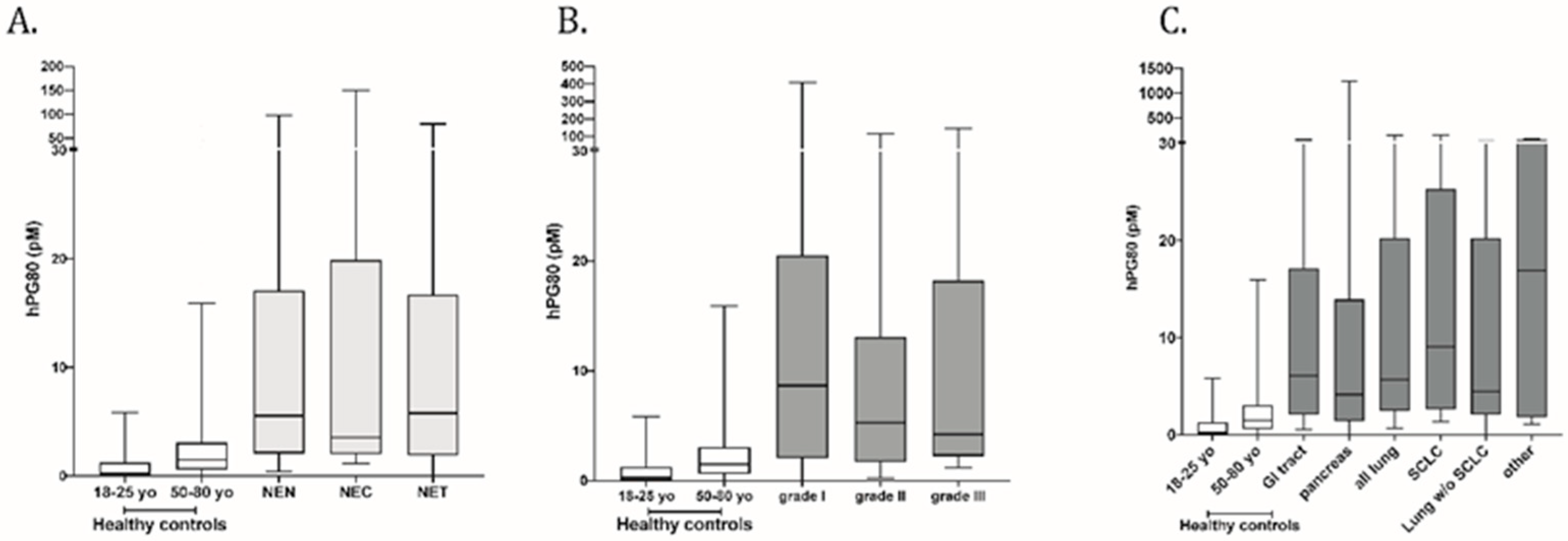
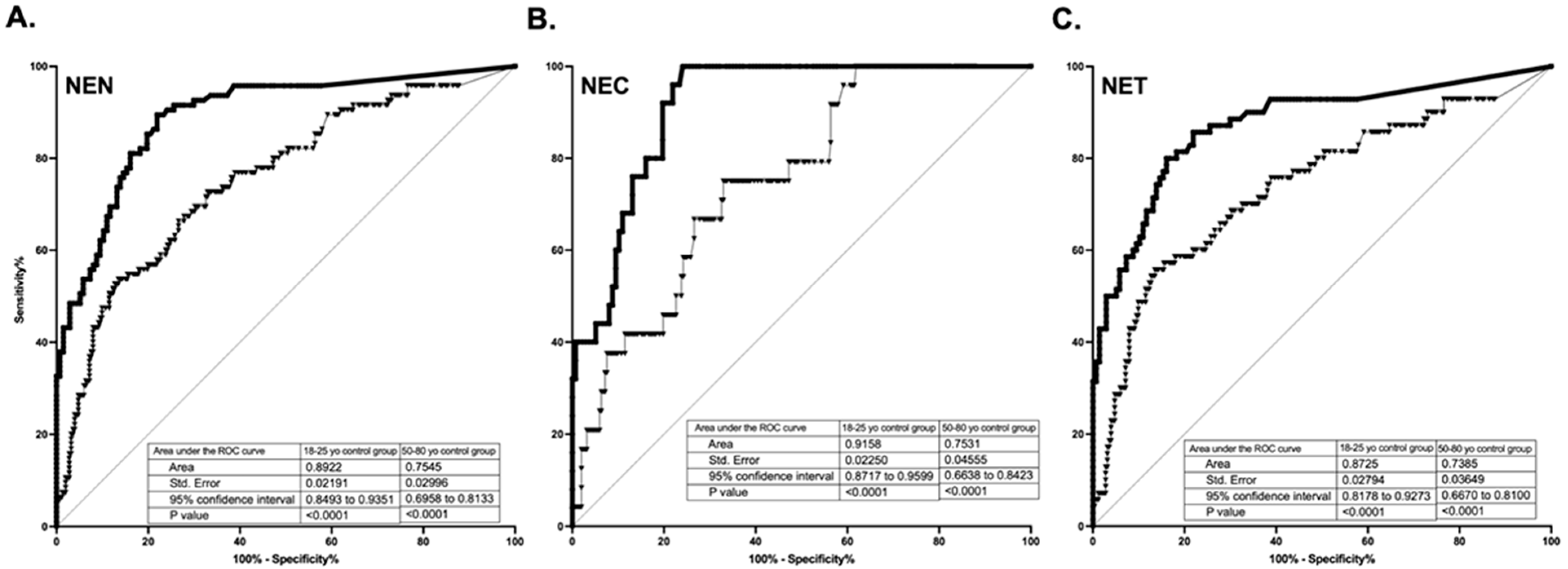
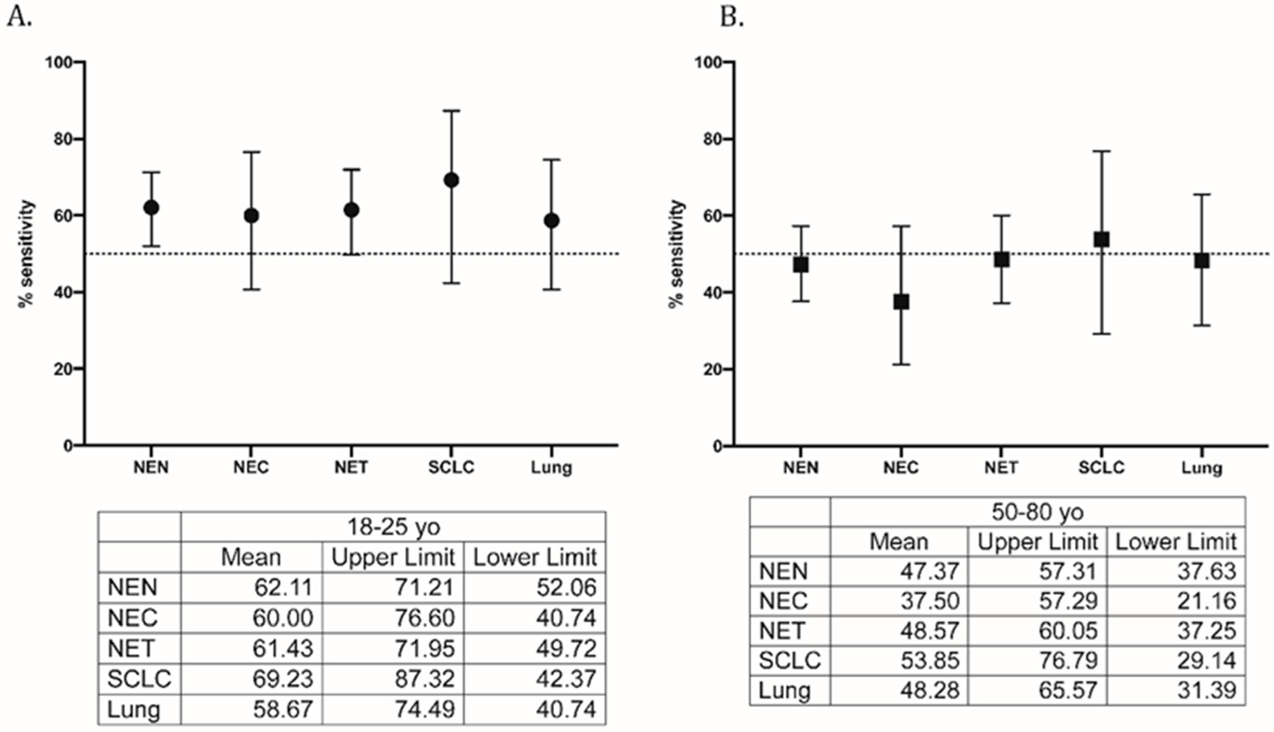
3.1. Diagnostic Performance of hPG80 in the Various Cohorts of Cancer Patients

The demographic characteristics of the neuroendocrine neoplasm (NEN) patients and control cohorts are shown in Table 1.
The median age of NEN patients was 61 years (37–86 y). Among the 95 NENs, 25 patients (26.3%) had high-grade neuroendocrine carcinoma (NEC) and 70 patients (73.7%) were diagnosed with well-differentiated neuroendocrine tumors (NET). The median hPG80 in NENs patients was 5.54 pM (IQR 2.07–17.11 pM) as compared to 1.5 pM (IQR 0.60–3.09 pM) for patients in the 50–80-year-old control group and 0.29 pM (IQR 0.00–1.27 pM) for patients in the 18–25-year-old cohort (p < 0.0001, two-tailed Mann–Whitney U-test). A subgroup analysis of NENs revealed a median hPG80 of 3.54 pM (IQR 2.02–19.91 pM) in neuroendocrine carcinoma (NEC n = 25) and 5.8 pM (IQR 1.91–16.74 pM) in neuroendocrine tumor (NET n = 70). Interestingly, the small-cell lung cancer sub-cohort (n = 13) also showed significant elevation of hPG80 with a median at 9.09 pM (IQR 2.66–25.33 pM). All the above-mentioned differences were statistically significant as compared to healthy controls (Figure 1). Table 2 shows the levels of plasma hPG80 in the various study subgroups.

3.2. Diagnostic Performance of hPG80 in Each Sub-Cohort of Cancer Type

As shown in Figure 2, diagnostic accuracy, estimated by the ROC AUCs, is 0.89 for all NENs, 0.87 for NETs, and 0.92 for NECs when compared to the young 18–25 y control group; for the older 50–80 y cohort, the values were 0.75 for all NENs, 0.74 for NETs, and 0.75 for NECs.
As shown in Figure 3, diagnostic performances of hPG80 were tested using 90% specificity (CI: 89.8% to 93.4% for the 18–25-year-old and 84.4% to 93.3% for the 50–80-year-old control groups) for all sub-cohorts. The sensitivity ranged from 58.67% (CI: 40.74% to 74.49%) for lung other than SCLC to 69.23% (CI: 42.37% to 87.32%) for SCLC, when compared to the 18–25 y control group. The sensitivity ranged from 37.50% (CI: 21.16% to 57.29%) for NEC to 53.85% (CI: 29.14% to 76.79%) for SCLC, when compared to the 50–80 y control group.

4. Discussion

Neuroendocrine neoplasms represent a group of diseases with a common neuroendocrine lineage however each subgroup maintains its unique morphology, molecular biology, and phenotype. WHO now clearly distinguishes poorly differentiated neuroendocrine carcinoma aka NEC from well differentiated neuroendocrine tumor, also known as NET. NEC is characterized by an aggressive clinical course, is distinctively poorly differentiated, displays large cell or small cell morphology, and has a molecular profile indicative of mutation in TP53 and loss of RB1. In contrast, the well differentiated NETs are relatively indolent and can be differentiated into grade 1, 2, and 3 based on Ki 67 index. It is critical to distinguish between NET and NEC as both can have a variable clinical course and management paradigms [17]. However, a common unifying factor for both NET and NEC is lack of reliable blood-based diagnostic biomarker. hPG80 (human circulating progastrin) is a pan tumor biomarker that has been found to be over-expressed in multiple malignancies. hPG80 synthesis is product of overexpression of GAST gene in cancer cells and human circulating progastrin can now be measured accurately with help of DxPG80 Enzyme-Linked Immunosorbent Assay (ELISA) based test [8,16].
In this study, we investigated the diagnostic value of plasma hPG80 in patients with both low- and high-grade NENs. Our data revealed that plasma hPG80 levels in 95 patients with NENs was significantly higher compared to healthy blood donors. NENs are a heterogenous group of diseases that can arise from any location in the body [18]. Because of its broad expression in all types of NENs, plasma hPG80 could represent a novel biomarker to diagnose NENs. In fact, except chromogranin A (CgA), which is secreted by most NENs, currently-used biomarkers (neuron-specific enolase, pancreatic peptide and 5-hydroxyindoleacetic acid) have restricted expression, thus limiting their diagnostic value [19]. We showed that hPG80 diagnostic sensitivity ranged from 58.67% to 69.23% with a 90% specificity in NENs. Only few biomarkers including CgA are currently used in the diagnosis of patients with NENs [20]. Although these biomarkers are considered useful to aid diagnosis, they display limited sensitivity and specificity. Indeed, the sensitivity and specificity of CgA ranges between 43 and 100%, and below 50%, respectively [3]. In addition, elevated CgA levels are also found in many other conditions such as renal and hepatic failure, inflammatory diseases, or following the use of proton-pump inhibitors [21]. Another limitation to the use of CgA for NENs diagnosis comes from the absence of direct involvement in the mechanisms that underlie tumorigenic process including cell proliferation, migration, invasion, and metastasis [22]. By contrast, numerous studies have demonstrated that hPG80 contribute to tumorigenesis and tumor development [9,11,12,13]. Plasma hPG80 levels correlate with tumor burden but also with tumor activity. Indeed, hPG80 level significantly decreases upon surgery in peritoneal carinomatosis and upon remission in hepatocellular cancer [8]. In addition, in hepatocellular cancer, the hPG80 level correlates with response to treatment and disease progression [8]. Last but not least, we have recently shown that hPG80 could be used for prognosticating survival in metastatic renal cell carcinoma [7]. In line with these data and to gain further insight into its potential utility as a biomarker in NENs, the role of hPG80 in disease monitoring is being studied in an NCI (National Cancer Institute) sponsored multi center NET clinical trial (ETCTN 10450). Plasma hPG80 will be serially collected at baseline, at each radiographic (CT/MRI) assessment, and at the time of progression.
Recently, the NETest, a blood-derived multianalyte test that measures the expression of 51 circulating mRNA in blood, has been shown to outperform CgA in the diagnosis of NENs [23]. NETest diagnostic accuracy, sensitivity, and specificity, were 97, 99, and 95%, respectively [23]. However widespread use of this biomarker may be limited [24].
Due to the rarity of NECs, most of the molecular studies have focused on NETs. They demonstrated that signaling pathways such as PI3K-Akt, Ras/Raf/MEK/ERK, Notch, and Wnt/β-catenin signaling contribute to NET pathogenesis [10]. The Wnt-signaling pathway is essential to regulate cell proliferation, migration during embryogenesis, and tumorigenesis in various cancers [25]. Gene mutations in well-known components of the Wnt-signaling pathways, such as β-catenin and APC, and repression of Wnt inhibitor genes by DNA methylation or histone modification of their promoters (SFRP-1, Axin-2, DKK-1, DKK-3, and WIF-1) are frequently observed in NET cell lines and human NET tumor samples [10]. In addition, analysis of somatic mutations across 21 types of NETs revealed that the gene MEN1, encoding a negative regulator of β-catenin, was the most commonly mutated gene in NETs (8 out of 21 NET types) [26,27]. Remarkably the gene GAST, encoding progastrin, is a direct target of the Wnt oncogenic pathway [28]. Therefore, we can hypothesize that abnormal expression of the Wnt signaling pathway in NENs could be linked to the overexpression of hPG80 in these tumors.
Our study presents some limitations starting with the retrospective nature of the study and the low patient number, especially in the different subgroups analyzed. Second, most of the patients (84%) have advanced disease (stage IV), not allowing for evaluation of the diagnostic value of hPG80 in early-stage patients. Lastly, the current study does not account for potential confounders like use of proton-pump inhibitors, or fasting vs. post-prandial state. We acknowledge that PPI usage could induce gastrin hyper secretion (hypergastrinemia); however, to our knowledge, there is no publication showing an increase in hPG80 during PPI usage. Furthermore, as described in Cappellini [16], we would like to mention that the kit (DxPG80) that was used to measure hPG80 (human circulating progastrin) does not recognize any other forms of gastrin peptides, avoiding any risk of false positivity due to PPI usage and hypergastrinemia. Nonetheless, we are currently setting up a clinical study to answer this very question. Finally, a large prospective study, including more cases with early-stage disease, should strengthens the significance of using hPG80 as a new diagnostic biomarker in NENs.

5. Conclusions

This first-ever study of plasma hPG80 in NENs confirms that hPG80 is elevated in both low- and high-grade NENs, and suggests that hPG80 could be considered as a potential diagnostic blood-based biomarker for NENs diagnosis.

Author Contributions

A.C.: conceptualization, methodology, investigation, data curation, data analysis, original draft preparation, writing and review; A.P.: investigation, methodology, data curation, data analysis, writing and review; J.K.: investigation, data curation, data analysis, writing and review; S.A.: investigation, data curation, data analysis, writing and review; L.P.: investigation, data curation, data analysis, writing and review; Y.M.: investigation, data curation, data analysis, writing and review; B.V.: investigation, data curation, data analysis, writing and review; M.S.: investigation, data curation, data analysis, writing and review; B.M.E.: investigation, data curation, data analysis, writing and review; D.J.: investigation, data curation, data analysis, writing and review; L.A.: investigation, data curation, data analysis, writing and review. All authors read and approved the final manuscript.

Funding

This research was supported by the BPTP Shared Resource of the University of Kentucky Markey Cancer Center (P30 CA177558) and ECS Progastrin.

Institutional Review Board Statement

The study was conducted according to the guidelines of the Declaration of Helsinki, and the protocol was approved by the Markey Cancer Center Institutional Review Board (IRB) Number: 50940, approved 4/21/2020.

Informed Consent Statement

Informed consent was not required.

Data Availability Statement

All data generated or analyzed during this study are included in this published article. Individual deidentified patient data can be shared upon request and IRB approval.

Acknowledgments

UK Markey Cancer Center’s Research Communications Office assisted with preparation of this manuscript.

Conflicts of Interest

The employees of ECS Progastrin are A.P., Y.M., B.V. and D.J. Those with no competing interest are J.K., S.A., L.P., M.S., B.M.E. and L.A.

References

  1. Chauhan, A.; Kohn, E.; Del Rivero, J. Neuroendocrine Tumors-Less Well Known, Often Misunderstood, and Rapidly Growing in Incidence. JAMA Oncol. 2020, 6, 21–22. [Google Scholar] [CrossRef] [PubMed]
  2. Dasari, A.; Shen, C.; Halperin, D.; Zhao, B.; Zhou, S.; Xu, Y.; Shih, T.; Yao, J.C. Trends in the Incidence, Prevalence, and Survival Outcomes in Patients with Neuroendocrine Tumors in the United States. JAMA Oncol. 2017, 3, 1335–1342. [Google Scholar] [CrossRef] [PubMed]
  3. Oberg, K.; Modlin, I.M.; De Herder, W.; Pavel, M.; Klimstra, D.; Frilling, A.; Metz, D.C.; Heaney, A.; Kwekkeboom, D.; Strosberg, J.; et al. Consensus on biomarkers for neuroendocrine tumour disease. Lancet Oncol. 2015, 16, e435–e446. [Google Scholar] [CrossRef] [Green Version]
  4. Rehfeld, J.F.; Zhu, X.; Norrbom, C.; Bundgaard, J.R.; Johnsen, A.H.; Nielsen, J.E.; Vikesaa, J.; Stein, J.; Dey, A.; Steiner, D.F.; et al. Prohormone convertases 1/3 and 2 together orchestrate the site-specific cleavages of progastrin to release gastrin-34 and gastrin-17. Biochem. J. 2008, 415, 35–43. [Google Scholar] [CrossRef] [Green Version]
  5. Varro, A.; Voronina, S.; Dockray, G.J. Pathways of processing of the gastrin precursor in rat antral mucosa. J. Clin. Investig. 1995, 95, 1642–1649. [Google Scholar] [CrossRef] [PubMed]
  6. Siddheshwar, R.K.; Gray, J.C.; Kelly, S.B. Plasma levels of progastrin but not amidated gastrin or glycine extended gastrin are elevated in patients with colorectal carcinoma. Gut 2001, 48, 47–52. [Google Scholar] [CrossRef]
  7. Kohli, M.; Tan, W.; Vire, B.; Liaud, P.; Blairvacq, M.; Berthier, F.; Rouison, D.; Garnier, G.; Payen, L.; Cousin, T.; et al. Prognostic Value of Plasma hPG80 (Circulating Progastrin) in Metastatic Renal Cell Carcinoma. Cancers 2021, 13, 375. [Google Scholar] [CrossRef] [PubMed]
  8. You, B.; Mercier, F.; Assenat, E.; Langlois-Jacques, C.; Glehen, O.; Soule, J.; Payen, L.; Kepenekian, V.; Dupuy, M.; Belouin, F.; et al. The oncogenic and druggable hPG80 (Progastrin) is overexpressed in multiple cancers and detected in the blood of patients. EBioMedicine 2019, 51, 102574. [Google Scholar] [CrossRef] [PubMed] [Green Version]
  9. Prieur, A.; Cappellini, M.; Habif, G.; Lefranc, M.P.; Mazard, T.; Morency, E.; Pascussi, J.M.; Flaceliere, M.; Cahuzac, N.; Vire, B.; et al. Targeting the Wnt Pathway and Cancer Stem Cells with Anti-progastrin Humanized Antibodies as a Potential Treatment for K-RAS-Mutated Colorectal Cancer. Clin. Cancer Res. 2017, 23, 5267–5280. [Google Scholar] [CrossRef] [Green Version]
  10. Kim, J.T.; Li, J.; Jang, E.R.; Gulhati, P.; Rychahou, P.G.; Napier, D.L.; Wang, C.; Weiss, H.L.; Lee, E.Y.; Anthony, L.; et al. Deregulation of Wnt/beta-catenin signaling through genetic or epigenetic alterations in human neuroendocrine tumors. Carcinogenesis 2013, 34, 953–961. [Google Scholar] [CrossRef] [Green Version]
  11. Giraud, J.; Failla, L.M.; Pascussi, J.M.; Lagerqvist, E.L.; Ollier, J.; Finetti, P.; Bertucci, F.; Ya, C.; Gasmi, I.; Bourgaux, J.F.; et al. Autocrine Secretion of Progastrin Promotes the Survival and Self-Renewal of Colon Cancer Stem-like Cells. Cancer Res. 2016, 76, 3618–3628. [Google Scholar] [CrossRef] [PubMed] [Green Version]
  12. Hollande, F.; Lee, D.J.; Choquet, A.; Roche, S.; Baldwin, G.S. Adherens junctions and tight junctions are regulated via different pathways by progastrin in epithelial cells. J. Cell Sci. 2003, 116, 1187–1197. [Google Scholar] [CrossRef] [PubMed] [Green Version]
  13. Pannequin, J.; Delaunay, N.; Buchert, M.; Surrel, F.; Bourgaux, J.F.; Ryan, J.; Boireau, S.; Coelho, J.; Pélegrin, A.; Singh, P.; et al. β-Catenin/Tcf-4 Inhibition after Progastrin Targeting Reduces Growth and Drives Differentiation of Intestinal Tumors. Gastroenterology 2007, 133, 1554–1568. [Google Scholar] [CrossRef]
  14. Singh, P.; Owlia, A.; Varro, A.; Dai, B.; Rajaraman, S.; Wood, T. Gastrin gene expression is required for the proliferation and tumorigenicity of human colon cancer cells. Cancer Res. 1996, 56, 4111–4115. [Google Scholar]
  15. White, M.C.; Holman, D.M.; Boehm, J.E.; Peipins, L.A.; Grossman, M.; Henley, S.J. Age and cancer risk: A potentially modifiable relationship. Am. J. Prev. Med. 2014, 46, S7–S15. [Google Scholar] [CrossRef] [PubMed] [Green Version]
  16. Cappellini, M.; Flaceliere, M.; Saywell, V.; Soule, J.; Blanc, E.; Belouin, F.; Ortiz, E.; Canterel-Thouennon, L.; Poupeau, S.; Tigrett, S.; et al. A novel method to detect hPG80 (human circulating progastrin) in the blood. Anal. Methods 2021, 13, 4468–4477. [Google Scholar] [CrossRef] [PubMed]
  17. Rindi, G.; Klimstra, D.S.; Abedi-Ardekani, B.; Asa, S.L.; Bosman, F.T.; Brambilla, E.; Busam, K.J.; de Krijger, R.R.; Dietel, M.; El-Naggar, A.K.; et al. A common classification framework for neuroendocrine neoplasms: An International Agency for Research on Cancer (IARC) and World Health Organization (WHO) expert consensus proposal. Mod. Pathol. 2018, 31, 1770–1786. [Google Scholar] [CrossRef] [PubMed]
  18. Oronsky, B.; Ma, P.C.; Morgensztern, D.; Carter, C.A. Nothing But NET: A Review of Neuroendocrine Tumors and Carcinomas. Neoplasia 2017, 19, 991–1002. [Google Scholar] [CrossRef]
  19. Herrera-Martinez, A.D.; Hofland, L.J.; Galvez Moreno, M.A.; Castano, J.P.; de Herder, W.W.; Feelders, R.A. Neuroendocrine neoplasms: Current and potential diagnostic, predictive and prognostic markers. Endocr. Relat. Cancer 2019, 26, R157–R179. [Google Scholar] [CrossRef] [Green Version]
  20. Ahmed, M. Gastrointestinal neuroendocrine tumors in 2020. World J. Gastrointest. Oncol. 2020, 12, 791–807. [Google Scholar] [CrossRef]
  21. Modlin, I.M.; Gustafsson, B.I.; Moss, S.F.; Pavel, M.; Tsolakis, A.V.; Kidd, M. Chromogranin A—Biological function and clinical utility in neuro endocrine tumor disease. Ann. Surg. Oncol. 2010, 17, 2427–2443. [Google Scholar] [CrossRef] [PubMed]
  22. Walenkamp, A.; Crespo, G.; Fierro Maya, F.; Fossmark, R.; Igaz, P.; Rinke, A.; Tamagno, G.; Vitale, G.; Oberg, K.; Meyer, T. Hallmarks of gastrointestinal neuroendocrine tumours: Implications for treatment. Endocr. Relat. Cancer 2014, 21, R445–R460. [Google Scholar] [CrossRef] [PubMed]
  23. Malczewska, A.; Kos-Kudla, B.; Kidd, M.; Drozdov, I.; Bodei, L.; Matar, S.; Oberg, K.; Modlin, I.M. The clinical applications of a multigene liquid biopsy (NETest) in neuroendocrine tumors. Adv. Med. Sci. 2020, 65, 18–29. [Google Scholar] [CrossRef] [PubMed]
  24. Ma, Z.Y.; Gong, Y.F.; Zhuang, H.K.; Zhou, Z.X.; Huang, S.Z.; Zou, Y.P.; Huang, B.W.; Sun, Z.H.; Zhang, C.Z.; Tang, Y.Q.; et al. Pancreatic neuroendocrine tumors: A review of serum biomarkers, staging, and management. World J. Gastroenterol. 2020, 26, 2305–2322. [Google Scholar] [CrossRef] [PubMed]
  25. Nusse, R.; Clevers, H. Wnt/beta-Catenin Signaling, Disease, and Emerging Therapeutic Modalities. Cell 2017, 169, 985–999. [Google Scholar] [CrossRef] [PubMed]
  26. Jiang, X.; Cao, Y.; Li, F.; Su, Y.; Li, Y.; Peng, Y.; Cheng, Y.; Zhang, C.; Wang, W.; Ning, G. Targeting beta-catenin signaling for therapeutic intervention in MEN1-deficient pancreatic neuroendocrine tumours. Nat. Commun. 2014, 5, 5809. [Google Scholar] [CrossRef] [Green Version]
  27. Cao, Y.; Zhou, W.; Li, L.; Wang, J.; Gao, Z.; Jiang, Y.; Jiang, X.; Shan, A.; Bailey, M.H.; Huang, K.L.; et al. Pan-cancer analysis of somatic mutations across 21 neuroendocrine tumor types. Cell Res. 2018, 28, 601–604. [Google Scholar] [CrossRef] [Green Version]
  28. Koh, T.J.; Chen, D. Gastrin as a growth factor in the gastrointestinal tract. Regul. Pept. 2000, 93, 37–44. [Google Scholar] [CrossRef]
Figure 1. Diagnostic performance of hPG80 in (A) NENs, NEC and NET patient cohorts, (B) by tumor grade, (C) by tumor site as compared to the 18–25-year-old and 50–80-year-old.
Cancers 14 00863 g001
Figure 2. Diagnostic accuracy, estimated by the Receiver Operator Characteristic (ROC) Area Under the Curve (AUC)s, is 0.89 for all NENs (A), 0.92 for NECs (B), and 0.87 for NETs (C) when compared to the young 18–25 y control group (square); for the older 50–80 y cohort, the values were 0.75 for all NENs (A), 0.75 for NECs (B), and 0.74 for NETs (C) (triangles).
Cancers 14 00863 g002
Figure 3. Sensitivity of hPG80 in the all-patient (neuroendocrine neoplasm) cohorts with a specificity set at 90% as compared to (A) 18–25 y control group and (B) 50–80 y control group.
Cancers 14 00863 g003
Table 1. Clinical and pathological characteristics for NEN, NEC, NET patients, and control cohorts.
NENNECNETControl Cohorts
18–25 Years Old50–80 Years Old
N (%)N (%)N (%)N (%)N (%)
n = 95n = 25n = 70n = 137n = 252
Age (years)Median (range)61 (37–86)61 (37–78)62 (37–86)21 (18–25)55 (50–80)
GenderMale38 (40%)10 (40%)28 (40%)79 (57.7%)99 (39.3%)
Female57 (60%)15 (60%)42 (60%)58 (42.3%)153 (60.7%)
hPG80Median (IQR), pM5.54 (0.00–1241)3.54 (1.13–154.1)5.8 (0.00–1241)<LoD<LoQ
Mean (SD) pM28.24 (128.8)20.7 (39.96)30.55 (148.5)<LoD3.82 (0.55)
StageI to III14 (14.8%)2 (8%)12 (17.2%)NA
IV80 (84.2%)23 (92%)57 (81.4%)
Unknown1 (1.0%)0 (0%)1 (1.4%)
Grade133033
(34.7%)(0%)(47.2%)
228028
(29.5%)(0%)(40%)
330255
(31.6%)(100%)(7.1%)
Unknown404
(4.2%)(0%)(5.7%)
Primary SiteGI tract46442
(48.4%)(16%)(60%)
Pancreas15213
(15.8%)(8%)(18.6%)
SCLC13130
(13.7%)(52%)(0%)
Lung w/o SCLC17413
(17.9%)(16%)(18.6%)
Other422
(4.2%)(8%)(2.8%)
NA: not applicable; LoD: Limit of Detection; LoQ: Limit of Quantification.
Table 2. hPG80 (pM) in plasma from neuroendocrine neoplasm patients depending on the grade and the primary site. Groups are not significantly different from each other.
GRADE
Grade IGrade IIGrade III
hPG80Median (range), pM8.66 (0.00–1241)5.3 (0.00–142.1)4.22 (1.13–154.1)
Mean (SD), pM48.71 (214.4)16.37 (32.1)19.18 (36.77)
PRIMARY SITE
GI TractPancreasSCLCLung not SCLCOther
hPG80Median (range), pM6.06 (0.00–142.1)4.14 (0.00–1241)9.09 (1.35–124.1)4.51 (0.00–34.85)16.89 (1.13–86.87)
Mean (SD), pM13.88 (24.02)88.9 (318.9)30.36 (52.78)11.37 (11.35)30.45 (39.78)
hPG80: circulating progastrin; IQR: interquartile range; pM: picomolar; SD: standard deviation; GI: gastrointestinal; SCLC: small-cell lung cancer.
Publisher’s Note: MDPI stays neutral with regard to jurisdictional claims in published maps and institutional affiliations.

