Article

Effects of Light-Emitting Diodes on the Accumulation of Phenolic Compounds and Glucosinolates in Brassica juncea Sprouts

1
Department of Crop Science, College of Agriculture and Life Sciences, Chungnam National University, 99 Daehak-ro, Yuseong-gu, Daejeon 34134, Korea
2
Division of Life Sciences and Bio-Resource and Environmental Center, College of Life Sciences and Bioengineering, Incheon National University, Incheon 406-772, Korea
3
Department of Samrt Agriculture Systems, College of Agriculture and Life Sciences, Chungnam National University, 99 Daehak-ro, Yuseong-gu, Daejeon 34134, Korea
*
Authors to whom correspondence should be addressed.
These authors contributed equally to this work.
Horticulturae 2020, 6(4), 77; https://doi.org/10.3390/horticulturae6040077
Submission received: 10 September 2020 / Revised: 5 November 2020 / Accepted: 6 November 2020 / Published: 9 November 2020
(This article belongs to the Special Issue Urban Horticulture - New Trends and Technologies)

Abstract

:
Recent improvements in light-emitting diode (LED) technology afford an excellent opportunity to investigate the relationship between different light sources and plant metabolites. Accordingly, the goal of the present study was to determine the effect of different LED (white, blue, and red) treatments on the contents of glucosinolates (glucoiberin, gluconapin, sinigrin, gluconasturtiin, 4-methoxyglucobrassicin, 4-hydroxyglucobrassicin, glucobrassicin, and neoglucobrassicin) and phenolic compounds (4-hydroxybenzonate, catechin, chlorogenic acid, caffeate, gallate, sinapate, and quercetin) in Brassica juncea sprouts. The sprouts were grown in a growth chamber at 25 °C under irradiation with white, blue, or red LED with a flux rate of 90 μmol·m−2·s−1 and a long-day photoperiod (16 h light/8 h dark cycle). Marked differences in desulfoglucosinolate contents were observed in response to treatment with different LEDs and different treatment durations. In addition, the highest total desulfoglucosinolate content was observed in response to white LED light treatment, followed by treatment with red LED light, and then blue LED light. Among the individual desulfoglucosinolates identified in the sprouts, sinigrin exhibited the highest content, which was observed after three weeks of white LED light treatment. The highest total phenolic contents were recorded after one week of white and blue LED light treatment, whereas blue LED irradiation increased the production of most of the phenolic compounds identified, including 4-hydroxybenzonate, gallate, sinapate, caffeate, quercetin, and chlorogenic acid. The production of phenolics decreased gradually with increasing duration of LED light treatment, whereas anthocyanin accumulation showed a progressive increase during the treatment. These findings indicate that white LED light is appropriate for glucosinolate accumulation, whereas blue LED light is effective in increasing the production of phenolic compounds in B. juncea sprouts.

1. Introduction

Brassica juncea is an annually growing perennial herb, which belongs to the Brassicaceae family and is known as mustard green, Indian mustard, oriental mustard, leaf mustard, or Chinese mustard. This plant can grow to over 1 m in height, has erect, patent branches and is widely distributed throughout Africa, Bangladesh, China, India, Japan, Korea, and Pakistan. B. juncea has been one of the most economically valuable plants in India for centuries, owing to its medicinal and nutritive properties [1]. Moreover, in Korea, it is used as food, both alone and as the main ingredient in kimchi, a traditional fermented vegetable product. Kimchi, containing leaves of B. juncea as an ingredient, has recently drawn attention as a functional food for health maintenance and disease control [2]. The essential oil of B. juncea seeds has been used in cosmetics [3], while the plant is used in the production of petroleum diesel [4]. In addition, B. juncea contains various bioactive compounds, including glucosinolates [3], isothiocyanates [4], phenolic compounds [5,6], fatty acids [7], kaempferol glycosides [8], and various flavonoids [9].
Glucosinolates are plant-derived nitrogen- and sulfur-containing phytochemicals common in members of the order Capparales, including the Brassicaceae. Accordingly, glucosinolates are also widely found in agriculturally valuable crop species (e.g., Brassica vegetables), as well as in the model plant Arabidopsis thaliana. These compounds have a common core structure containing a β-D-thioglucose group, a sulfonated aldoxime moiety, and a variable amino acid-derived side chain. About 200 types of glucosinolates are known to occur naturally in plants, where they are thought to function as natural pesticides. In addition, glucosinolate derivatives contribute to the various aromas and flavors of cruciferous vegetables and condiments and perform important roles in various biological processes, including plant defenses, auxin homeostasis, and cancer prevention in humans [10,11,12]. Previous studies that have examined the pharmacological activity of B. juncea have found that it possesses fungicidal [13], antiatherogenic [13], antidiabetic [14], antioxidant/peroxynitrite-scavenging [8,13], antimicrobial [15], and antitumor [16,17] activities; moreover, B. juncea has been shown to exert beneficial effects against a variety of metabolic disorders [18].
Phenolics are the most abundant phytochemicals, are ubiquitously found in most plants, are responsible for plant defenses against various abiotic/biotic stresses, and contribute to the development of plant color [19,20,21,22]. Due to their wide availability in plant-based foods [4], plant phenolics also form an integral part of the human diet. Moreover, owing to their biological activities, which include antioxidant [23], anti-inflammatory [24], and anticarcinogenic [25] activities, plant-based foods rich in phenolic compounds are recommended for the enhancement of human health.
Light quality, referring to the wavelength or color, significantly influences plant growth. Red and blue lights are most effective for plant growth. Blue light is primarily involved in leaf growth and vegetative growth. Red light combined with blue light leads to flowering in plants. Cool white lights, which contain mostly blue light and little red light, are utilized to encourage leafy growth [26]. Green light is slightly less effective than red and blue light, since it is mainly reflected by plants [27]; however, it promotes early stem elongation, antagonizing growth inhibition [28]. Additionally, light-emitting diodes are more efficient than fluorescent light for reproductive and vegetative growth of non-heading Chinese cabbage (Brassica rapa subsp. chinensis). In particular, blue LED light benefits vegetative growth, whereas red LED light supports reproductive growth [29]. Furthermore, light conditions, including light period, quality, and intensity, can alter the nature and concentrations of therapeutic compounds found in plants. For instance, light irradiation has been shown to significantly influence the production of phytochemicals [30,31], while irradiance levels are reported to influence the production and concentrations of both carotenoid pigments and glucosinolates [31,32]. Illumination by LEDs has been shown to alter the composition of phytochemicals found in Brassica plant species. Previously, Li et al. [29] reported that blue LED irradiation increased the production of chlorophylls, carotenoids, and vitamin C. Similarly, blue and red LED lights produced significantly higher levels of glucosinolates in kale (Brassica oleracea var. acephala) and Chinese cabbage, respectively [33]. Samuolienė et al. [34] reported that LED irradiation affected the growth and nutritional quality of microgreen of Kohlrabi, mustard, red pakchoi, and tatsoi and that the effect of different light quality and intensity on metabolite production was dependent on plant species.
To our knowledge, no studies have evaluated the influence of different LED wavelengths on glucosinolate and phenolic contents in B. juncea sprouts. Although our previous study reported the effect of LED lights on the production of phenolic compounds and glucosinolates in Brassica napus sprouts [35], it is necessary to investigate optimal light conditions for enhancing the growth and production of bioactive compounds in the B. juncea sprouts, since phytochemical production depends on plant species. Thus, this study is aimed to investigate the effect of three LED treatments on the accumulation of different types of desulfoglucosinolates (DS-GSLs) and phenolic compounds in B. juncea sprouts.

2. Materials and Methods

2.1. Plant Materials and Growth Conditions

B. juncea seeds were purchased from Asia Seed Co., Ltd. (Seoul, Korea). To establish sprouts, 100 seeds were immersed in sterilized water for 24 h and then placed in a plastic pot containing vermiculite. The sprouts were grown in a growth chamber at 25 °C under irradiation with white (450–660 nm), blue (450 nm), or red (660 nm) LED lighting with a flux rate of 90 μmol·m−2·s−1 and a long-day photoperiod (16 h light/8 h dark cycle). The blue and red LED treatment were monochromatic. Specifically, the PARUS LED light (PGL-PFL series, PARUS LED Co., Cheoan, Korea) consisted of white (R/B = 6/12), red, and blue components (Figure S1). The sprouts were harvested after one, two, and three weeks of LED light treatment. The sprouts were harvested with liquid nitrogen and then freeze-dried for further researchers. The experiments were repeated three times, and mixtures of sprouts from the three independent replicates were used for high-performance liquid chromatography (HPLC) analysis of glucosinolates and phenolic compounds.

2.2. Desulfoglucosinolate Extraction and HPLC Analysis

Desulfoglucosinolates were extracted using previously reported procedures [36,37], with slight modifications. Crude GSLs were extracted from 0.1 g of dried sample in 1.5 mL of boiling 70% (v/v) ethanol at 70 °C for 5 min in a water bath and then centrifuged at 4 °C at 12,000 rpm for 10 min. The supernatants were collected into 5 mL test tubes. The residues were re-extracted twice more in the same manner, and the collected supernatants were loaded onto a mini-column filled with DEAE-Sephadex A-25 and desulfated by the addition of 75 μL of an aryl sulfatase solution. The resulting DS-GSLs were then eluted into 2 mL tubes with 0.5 mL HPLC-grade water. The elution was performed three times (Figure S2). The HPLC analysis conditions, system, and gradient program were according to our previous study [36]. The individual GSLs were identified by comparing their HPLC retention times to those in our database and quantified according to their HPLC areas and response factors; sinigrin (0.1 mg/mL), subjected to the same extraction process of the B. juncea samples, was used as an external standard [37].

2.3. HPLC Analysis of Phenolic Compounds

Phenolics were analyzed using our previously reported method [38]. Phenolic compounds were extracted from 0.1 g of dried sprout powder with 1.5 mL of aqueous methanol (80% v/v). After sonication for 1 h at 25 °C, the crude extract was centrifuged at 10,000 rpm for 10 min, and the supernatant was transferred to a fresh tube. The remaining sludge was re-extracted twice more in this manner. The collected supernatant was filtered via a 0.45 μm filter into a vial. HPLC analysis conditions, system, and gradient program were according to our previous study [38]. Phenolic compounds were identified based on retention times and spiking tests and quantified with reference to corresponding calibration curves (Figure S2).

2.4. Determination of Total Anthocyanin Contents

Total anthocyanin contents were evaluated using the pH differential method [39]. A 1 mL aliquot of the extract was mixed with 4 mL of each of two buffers (CH3COONa buffer [0.4 M, pH 4.5] and KCl buffer [0.025 M, pH 1.0]), followed by incubation at 28 °C for 15 min. Absorbance was determined at 510 nm and 700 nm, using distilled water as a blank. Calculation of total anthocyanin content was performed using an equation reported in a previous study [39]. The result was converted to micrograms of cyanidin-3-glucoside equivalents (CGE) per gram dry weight (mg CGE/g dry weight).

2.5. Statistical Analysis

Analysis of variance with Duncan’s multiple range test at p < 0.05 was used for data analysis performed with SAS software version 9.2 (SAS Institute Inc., Cary, NC, USA). Two-way ANOVA interaction plots were generated from SPSS 24.0 (IBM, Chicago, IL, USA). Principal component analysis (PCA) for 18 metabolites detected in sprout samples was performed using MetaboAnalyst 4.0 (http://www.metaboanalyst.ca/) with auto-scaling.

3. Results

3.1. Sprout Dry Weight and Phenotype Change

Differential LED irradiation had a considerable influence on sprout dry weight; red and white LED light-irradiated sprouts presented higher dry weight values than those of sprouts irradiated with blue LED light (Table 1). Furthermore, the seedlings exhibited deeper purple colors in their cotyledons and hypocotyls, depending on the duration of exposure to LED irradiation. Interestingly, several sprouts exposed to blue LED light formed leaves (Figure 1 and Figure S3).

3.2. Glucosinolate Contents

Our analyses revealed that the B. juncea sprouts contained eight different DS-GSLs (4-hydroxyglucobrassicin, 4-methoxyglucobrassicin, glucoiberin, gluconapin, glucobrassicin, gluconasturtiin, sinigrin, and neoglucobrassicin), whose production was markedly enhanced with LED light treatment (Figure 2 and Figure S4). Among the eight DS-GSLs identified, sinigrin presented the greatest concentration. The highest glucoiberin content was observed after two weeks of blue and white LED light treatment, and the level of glucoiberin decreased after two weeks under all LED treatments. Sinigrin accumulation in the sprouts increased with increasing duration of LED light treatment, with the highest level (94.63% of the total DS-GSLs) observed after three weeks of treatment with white LED light. The sinigrin content increased in both white and red LED light-treated sprouts after three weeks and decreased in plants treated with blue LED irradiation. The highest gluconapin content was observed in white LED light-treated sprouts, followed by that of sprouts treated with blue and red LED light. The gluconapin content increased and then decreased with white LED light treatment for one, two, or three weeks. Furthermore, after three weeks, the gluconapin content had decreased under all the LED treatments. The highest 4-hydroxyglucobrassicin content (6.22% of the total DS-GSLs) was observed after one week of red LED light treatment, followed by those after one week of white and blue LED treatment. The 4-hydroxyglucobrassicin content decreased with red LED treatment after two and three weeks. The highest 4-methoxyglucobrassicin content was observed after two weeks of red LED light treatment, followed by those after one week of white and blue LED treatment. However, 4-methoxyglucobrassicin content decreased after two weeks of red LED light treatment. The highest gluconasturtiin content (4.71% of the total DS-GSLs) was recorded after two weeks of blue LED light treatment, followed by those after two weeks of red and white LED irradiation treatment. However, the gluconasturtiin content decreased after three weeks under blue LED lighting. The total glucosinolate content of B. juncea sprouts was differentially influenced by the different LED treatments and their duration; the highest level was achieved after three weeks under white LED illumination, and this level was higher than those observed in plants grown under red and blue LED lighting for three weeks (Figure 2). Overall, the glucosinolate concentration increased with increasing duration of LED treatment.

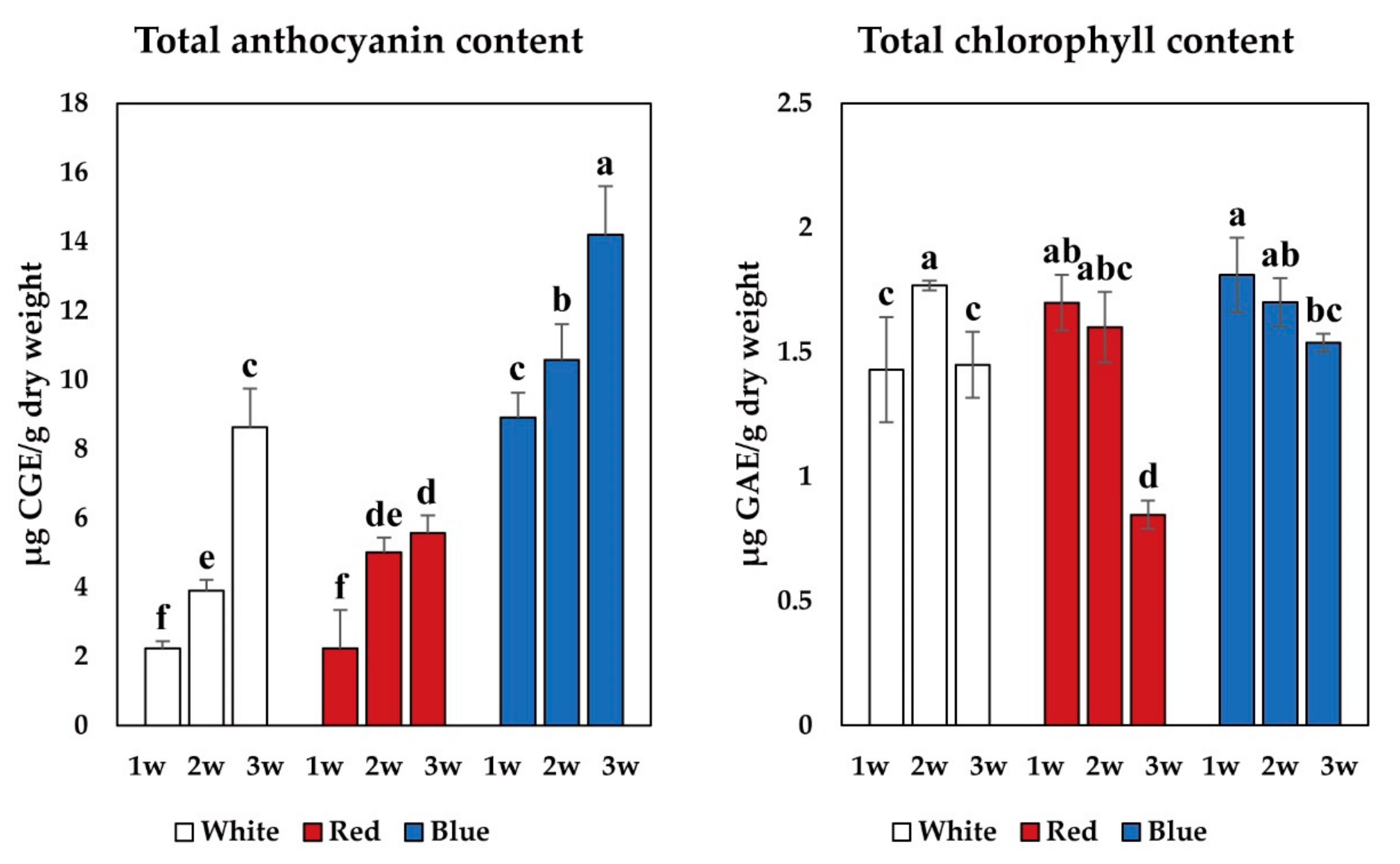
3.3. Total Anthocyanin, Total Chlorophyll, and Phenolic Contents

The total anthocyanin content in the B. juncea seedlings varied according to the duration of LED irradiation, ranging from 2.23 to 14.19 μg CGE/g dry weight (Figure 3). Anthocyanin accumulation showed a gradual increase with increasing duration of LED light treatment. The highest total anthocyanin content in the seedlings was detected after three weeks under blue LED light treatment (14.19 ± 1.41 μg CGE/g dry weight), followed by that after three weeks under red LED light treatment (8.63 ± 1.12 μg CGE/g dry weight). In contrast, chlorophyll accumulation showed a gradual decrease with increasing duration of LED light treatment, except after two weeks of white LED light treatment (Figure 3). Seven different phenolics were identified by HPLC in the B. juncea seedlings (4-hydroxybenzonate, gallate, catechin, chlorogenic acid, caffeate, quercetin, and sinapate), and the production of gallate, caffeate, and quercetin presented a gradual decline with LED light treatment (Figure 4 and Figure S5). The highest total phenolic contents were observed after one week of white and blue LED treatment. Caffeate and quercetin presented the greatest concentrations among the seven phenolics identified in B. juncea seedlings. However, caffeate and quercetin accumulation also showed a gradual decrease with increasing duration of LED light treatment. The highest levels of gallate, quercetin, and caffeate were observed after one week of white and blue LED light treatment. In contrast, the highest levels of catechin and sinapate were observed after one week of white LED light treatment and two weeks of blue LED light treatment, respectively. Interestingly, 4-hydroxybenzonate was only identified in blue LED light-treated seedlings.
The quantitation data of the 18 secondary metabolites were then subjected to PCA to investigate the differences in metabolite profiles among B. juncea sprouts grown under LED illumination of varying duration (Figure 5). Two principal components of the score plot accounted for 36.3% and 19.5 % of the total variance and resolved the separation of sprouts after one week of white, red, and blue LED light treatment from the others. The most important metabolites of component 1 in the loading plot were sinigrin, gluconapin, gluconasturtiin, catechin, and neoglucobrassicin, for which the eigenvector values were 0.36126, 0.30519, 0.24009, 0.12227, and 0.051137, respectively, and caffeate, gallate, rutin, quercetin, and 4-methoxyglucobrassicin, for which the eigenvector values were −0.38205, −0.36997, −0.34531, −0.33434, and −0.21886, respectively. The significant metabolites of component 2 were chlorogenic acid and neoglucobrassicin, for which the eigenvector values were 0.15844 and −0.46276, respectively.

4. Discussion

Plant development, morphogenesis, and growth are highly influenced by light quality. In this study, the sprouts irradiated with blue LEDs revealed the highest height; several of these sprouts formed leaves. This result is agreement with previous studies reporting that blue light can promote elongation growth of arugula and mustard sprouts as a shade-avoidance response [40], and blue LED light supports vegetative growth [29].
Brassica vegetables are a good source of glucosinolates and phenolic compounds. In this study, eight glucosinolates and seven phenolics were detected and quantified in B. juncea seedlings irradiated with different LED lighting. Furthermore, total anthocyanin contents were also determined in the seedlings. These findings are in agreement with those of previous studies that reported the identification of glucoiberin, sinigrin, gluconapin, 4-hydroxyglucobrassicin, 4-methoxyglucobrassicin, neoglucobrassicin, gluconasturtiin, and glucobrassicin in three stem mustard plants [41] and mustard hairy roots [42]. Furthermore, gallate, 4-hydroxybenzonate, caffeate, chlorogenic acid, and sinapate have been identified in potherb mustard [43] and mustard plants [44], while quercetin and catechin have been detected in mustard seeds [45].
In this study, we found that the levels of most glucosinolates increased in mustard sprouts exposed to different LED lighting, although only minor changes were observed. In addition, the DS-GSL levels in mature kale plants may differ from those of young seedlings, because the composition of glucosinolates has been shown to change throughout the development of various Brassica crops [46] as well as of Arabidopsis [47]. In the current study, all LED treatments affected the levels of the eight DS-GSLs identified in B. juncea sprouts. However, the levels of most of the DS-GSLs decreased after two weeks of LED treatment, regardless of the treatment used. Previous studies reported that the effect of light quality on glucosinolate accumulation varies with light intensity and spectrum, light source combinations, and plant species. For example, Moon et al. [48] described that sprouts of B. rapa ssp. Pekinensis var. BP79 grown under fluorescent + blue light contained higher levels of glucosinolates than sprouts grown under fluorescent light, whereas sprouts of B. rapa ssp. Pekinensis var. Tsao Huang Pa grown under fluorescent light had a little higher levels of glucosinolates. Park et al. [35] showed that the total glucosinolate contents in sprouts of B. napus grown under white, blue, and red LEDs were not statically different. Similarly, Tan et al. [40] reported that total glucosinolate contents in sprouts of B. rapa subsp. chinensis var. parachinensis grown under 80 μmol m2 s −1 white light, 80 μmol m−2 s−1 red + blue light, 160 μmol m−2 s−1 white light, and 160 μmol m−2 s−1 red + blue light did not significantly differ, and glucosinolate production presented a gradual decline from one-leafed seedlings to 30-day-old plants. These findings highlight that further studies are required to identify the specific intensity and duration of lighting that can be used to increase the production of essential DS-GSL in B. juncea sprouts.
The results of our phenolic compound analyses showed that exposure to blue LED light enhanced the production of most phenolic compounds, including gallate, quercetin, caffeate, sinapate, 4-hydroxybenzonate, and chlorogenic acid, in B. juncea sprouts. These findings are consistent with those of previous studies. For instance, Park et al. [35] demonstrated that irradiation with blue LED light for 14 days increased the production of most phenolics, including caffeate, (−)-epicatechin, and (+)-catechin, in B. napus sprouts. Similarly, blue LED irradiation has been reported to enhance the production of phenolic compounds in the sprouts of B. rapa ssp. Pekinensis after 12 days of treatment [44]. Additionally, blue LED irradiation led to the upregulation of phenylpropanoid/flavonoid biosynthesis in the sprouts of Fagopyrum tataricum L. [49] and resulted in the increased production of phenolics in Vigna unguiculata L. Walp. sprouts [50]. Szopa et al. [51] reported that blue LED light increased the accumulation of phenolic acids both in shoot cultures of Aronia prunifolia, Aronia arbutifolia, and Aronia melanocarpa and in callus cultures of Peucedanum japonicum Thunb. [52]. Additionally, the results of this study revealed that the production of phenolics gradually decreased with increasing duration of LED light treatment. This agrees with a previous study reporting that LED treatment progressively reduced phenylpropanoid/flavonoid biosynthesis in the sprouts of Triticum aestivum L. in a manner dependent on the duration of LED treatment [53].
Interestingly, 4-hydroxybenzonate was detected only in sprouts grown under blue LED treatment. It is carefully suggested that blue LED light affected the production of 4-hydroxybenzonate, since many previous studies reported that blue light boosts the production of phenolic compounds [54,55,56]. However, numerous factors might influence the production of phenolic compounds. Therefore, further studies are required. We also determined the total anthocyanin content in B. juncea seedlings. In contrast to the phenolic content, total anthocyanin content gradually increased with increasing duration of LED light treatment. Because the biosynthetic pathways of these metabolites share common intermediates, we tentatively suggest that a gradual increase in anthocyanin content induces a progressive decrease in phenolic compounds’ concentrations in B. juncea sprouts.
Narrow-bandwidth LED lighting can be used to directly influence the color, size, and secondary metabolite levels of most commercially valuable fruits and vegetables; consequently, an increasing number of studies have evaluated the use of LED irradiation as a means of improving the quality of various food products. Blue LED irradiation has been reported to increase the production of phenolics in the sprouts of F. tataricum L. [49] and Pisum sativum L. [57], while red LED light has been reported to increase the production of phenolics in Myrtus communis L. in vitro [58]. Additionally, the production of phenolics was reportedly not affected by blue, red, or blue + red LED illumination in F. tataricum L. sprouts [59]. A different study showed that natural light yielded greater chlorophyll and total carotenoid contents in V. unguiculata seedlings than irradiation with blue, yellow, and red LED light [60]. Azad et al. [61] reported that soybean sprouts grown under green LED treatment presented higher isoflavone contents and higher phenolic contents than sprouts grown under florescent light, and far-infrared irradiation (FIR) increased the total phenolic content and total isoflavones content in soybean sprouts. Additionally, UV-B irradiation at low dosages induced anthocyanin production in radish sprouts [62], and UV-C irradiation at relatively high dosages significantly increased total phenolic content, total flavonoid content, and proanthocyanidins in lemon pomace dried powder [63].
The results of the present and previous studies suggest that the effect of different light sources and wavelengths on plant secondary metabolite accumulation may be depending on plant species, cell and tissue type, and organ. In the present study, white LED irradiation in B. juncea sprouts affected sinigrin production, while red LED light treatment altered the production of other DS-GSLs when compared with white and blue LED treatments. In contrast, blue LED irradiation was found to be beneficial for the production of phenolics.

Supplementary Materials

The following are available online at https://www.mdpi.com/2311-7524/6/4/77/s1. Figure S1. Brassica juncea sprouts grown under different light-emitting diode (LED) lights (blue, white, and red) for three weeks., Figure S2. Spectral distribution of the white LED, Figure S3. Chromatogram of the phenolic compounds (A) and the desulfo-glucosinolates (B) extracted from mustard sprouts grown under blue LED lights. 1, Gallic acid; 2, catechin; 3, chlorogenic acid; 4, 4-hydroxybenzoic acid; 5, caffeic acid; 6, sinapic acid; 7, quercetin; 8, glucoiberin; 9, sinigrin; 10, gluconapin; 11, 4-hydroxyglucobrassicin; 12, glucobrassicin; 13, 4-methoxyglucobrassicin; 14, gluconasturtiin; 15, neoglucobrassicin., Figure S4. Two-way ANOVA interaction plots showing changes in means of phenolic contents by developmental time and light sources., Figure S5. Two-way ANOVA interaction plots showing changes in means of glucosinoalte contents by developmental time and light sources.

Author Contributions

S.U.P. and J.K.K. designed the experiments and analyzed the data. C.H.P., Y.E.P., and H.J.Y., performed the experiments and analyzed the data. C.H.P. and Y.E.P. wrote the manuscript. All authors have read and agreed to the published version of the manuscript.

Funding

This research received no external funding.

Acknowledgments

This work was supported by Golden Seed Project (213006051WTE11) funded by Ministry of Agriculture, Food and Rural Affairs (MAFRA), Ministry of Oceans and Fisheries (MOF), Rural Development Administration (RDA) and Korea Forest Service (KFS), Republic of Korea.

Conflicts of Interest

The authors declare no conflict of interest.

References

  1. Manohar, P.R.; Pushpan, R.; Rohini, S. Mustard and its uses in Ayurveda. Indian J. Tradit. Know. 2009, 8, 400–404. [Google Scholar]
  2. Kim, H.Y.; Yokozawa, T.; Cho, E.J.; Cheigh, H.S.; Choi, J.S.; Chung, H.Y. In vitro and in vivo antioxidant effects of mustard leaf (Brassica juncea). Phytother. Res. 2003, 17, 465–471. [Google Scholar] [PubMed]
  3. Yu, J.C.; Jiang, Z.-T.; Li, R.; Chan, S.M. Chemical composition of the essential oils of Brassica juncea (L.) Coss. grown in different regions, Hebei, Shaanxi and Shandong, of China. J. Food Drug Anal. 2003, 11, 22–26. [Google Scholar]
  4. Moser, B.R.; Evangelista, R.L.; Jham, G. Fuel properties of Brassica juncea oil methyl esters blended with ultra-low sulfur diesel fuel. Renew. Energy 2015, 78, 82–88. [Google Scholar] [CrossRef]
  5. Kumar, A.; D’Souza, S.S.; Tickoo, S.; Salimath, B.P.; Singh, H.B. Antiangiogenic and proapoptotic activities of allyl isothiocyanate inhibit ascites tumor growth in vivo. Integr. Cancer Ther. 2009, 8, 75–87. [Google Scholar] [CrossRef] [Green Version]
  6. Yoon, B.H.; Jung, J.W.; Lee, J.J.; Cho, Y.W.; Jang, C.G.; Jin, C.; Oh, T.H.; Ryu, J.H. Anxiolytic-like effects of sinapic acid in mice. Life Sci. 2007, 81, 234–240. [Google Scholar] [CrossRef] [PubMed]
  7. Joardar, A.; Das, S. Effect of fatty acids isolated from edible oils like mustard, linseed or coconut on astrocytes maturation. Cell. Mol. Neurobiol. 2007, 27, 973–983. [Google Scholar] [CrossRef]
  8. Jung, H.A.; Woo, J.J.; Jung, M.J.; Hwang, G.S.; Choi, J.S. Kaempferol glycosides with antioxidant activity from Brassica juncea. Arch. Pharm. Res. 2009, 32, 1379–1384. [Google Scholar] [CrossRef]
  9. Yokozawa, T.; Kim, H.Y.; Cho, E.J.; Choi, J.S.; Chung, H.Y. Antioxidant effects of isorhamnetin 3, 7-di-O-β-D-glucopyranoside isolated from mustard leaf (Brassica juncea) in rats with streptozotocin-induced diabetes. J. Agric. Food Chem. 2002, 50, 5490–5495. [Google Scholar] [CrossRef]
  10. Kliebenstein, D.J.; Kroymann, J.; Mitchell-Olds, T. The glucosinolate-myrosinase system in an ecological and evolutionary context. Curr. Opin. Plant Biol. 2005, 8, 264–271. [Google Scholar] [CrossRef]
  11. Mithen, R.F.; Dekker, M.; Verkerk, R.; Rabot, S.; Johnson, I.T. The nutritional significance, biosynthesis and bioavailability of glucosinolates in human foods. J. Sci. Food Agric. 2000, 80, 967–984. [Google Scholar] [CrossRef]
  12. Woodward, A.W.; Bartel, B. Auxin: Regulation, action, and interaction. Ann. Bot. 2005, 95, 707–735. [Google Scholar] [CrossRef] [PubMed] [Green Version]
  13. Lee, M.A.; Choi, J.H.; Choi, Y.S.; Han, D.J.; Kim, H.Y.; Shim, S.Y.; Chung, H.K.; Kim, C.J. The antioxidative properties of mustard leaf (Brassica juncea) kimchi extracts on refrigerated raw ground pork meat against lipid oxidation. Meat Sci. 2010, 84, 498–504. [Google Scholar] [CrossRef]
  14. Grover, J.K.; Yadav, S.; Vats, V. Hypoglycemic and antihyperglycemic effect of Brassica juncea diet and their effect on hepatic glycogen content and the key enzymes of carbohydrate metabolism. Mol. Cell. Biochem. 2002, 241, 95–101. [Google Scholar] [CrossRef]
  15. Ye, X.; Ng, T.B. Isolation and characterization of juncin, an antifungal protein from seeds of Japanese Takana (Brassica juncea var. integrifolia). J. Agric. Food Chem. 2009, 57, 4366–4371. [Google Scholar] [CrossRef]
  16. Monsalve, R.I.; de la Pena, M.A.G.; Menendez-Arias, L.; Lopez-Otin, C.; Villalba, M.; Rodriguez, R. Characterization of a new oriental-mustard (Brassica juncea) allergen, Bra j IE: Detection of an allergenic epitope. Biochem. J. 1993, 293, 625–632. [Google Scholar] [CrossRef] [Green Version]
  17. Thejass, P.; Kuttan, G. Allyl isothiocyanate (AITC) and phenyl isothiocyanate (PITC) inhibit tumour-specific angiogenesis by downregulating nitric oxide (NO) and tumour necrosis factor-α (TNF-α) production. Nitric Oxide 2007, 16, 247–257. [Google Scholar] [CrossRef]
  18. Yadav, S.P.; Vats, V.; Ammini, A.C.; Grover, J.K. Brassica juncea (Rai) significantly prevented the development of insulin resistance in rats fed fructose-enriched diet. J. Ethnopharmacol. 2004, 93, 113–116. [Google Scholar] [CrossRef]
  19. Akula, R.; Ravishankar, G.A. Influence of abiotic stress signals on secondary metabolites in plants. Plant Signal. Behav. 2011, 6, 1720–1731. [Google Scholar] [CrossRef] [PubMed]
  20. Bennett, R.N.; Wallsgrove, R.M. Secondary Metabolites in plant defense-mechanisms. New Phytol. 1994, 127, 617–633. [Google Scholar] [CrossRef]
  21. Dixon, R.A.; Paiva, N.L. Stress-induced phenylpropanoid metabolism. Plant Cell 1995, 7, 1085–1097. [Google Scholar] [CrossRef]
  22. Dai, J.; Mumper, R.J. Plant phenolics: Extraction, analysis and their antioxidant and anticancer properties. Molecules 2010, 15, 7313–7352. [Google Scholar] [CrossRef]
  23. Park, C.H.; Yeo, H.J.; Baskar, T.B.; Park, Y.E.; Park, J.S.; Lee, S.Y.; Park, S.U. In vitro antioxidant and antimicrobial properties of flower, leaf, and stem extracts of Korean mint. Antioxidants 2019, 8, 75. [Google Scholar] [CrossRef] [Green Version]
  24. Park, C.H.; Park, S.Y.; Lee, S.Y.; Kim, J.K.; Park, S.U. Analysis of metabolites in white flowers of Magnolia Denudata Desr. and violet flowers of Magnolia Liliiflora Desr. Molecules 2018, 23, 1558. [Google Scholar] [CrossRef] [Green Version]
  25. Ferrazzano, G.F.; Amato, I.; Ingenito, A.; de Natale, A.; Pollio, A. Anti-cariogenic effects of polyphenols from plant stimulant beverages (cocoa, coffee, tea). Fitoterapia 2009, 80, 255–262. [Google Scholar] [CrossRef]
  26. Parker, R.O. Introduction to Plant Science; Taylor & Francis: Abingdon-On-Thames, UK, 2004. [Google Scholar]
  27. Johkan, M.; Shoji, K.; Goto, F.; Hahida, S.N.; Yoshihara, T. Effect of green light wavelength and intensity on photomorphogenesis and photosynthesis in Lactuca sativa. Environ. Exp. Bot. 2012, 75, 128–133. [Google Scholar] [CrossRef]
  28. Folta, K.M. Green light stimulates early stem elongation, antagonizing light-mediated growth inhibition. Plant Physiol. 2004, 135, 1407–1416. [Google Scholar] [CrossRef] [Green Version]
  29. Li, H.; Tang, C.; Xu, Z.; Liu, X.; Han, X. Effects of different light sources on the growth of non-heading Chinese cabbage (Brassica campestris L.). J. Agric. Sci. 2012, 4, 262. [Google Scholar] [CrossRef] [Green Version]
  30. Kopsell, D.A.; Pantanizopoulos, N.I.; Sams, C.E.; Kopsell, D.E. Shoot tissue pigment levels increase in ‘Florida Broadleaf’mustard (Brassica juncea L.) microgreens following high light treatment. Sci. Hortic. 2012, 140, 96–99. [Google Scholar] [CrossRef]
  31. Lefsrud, M.G.; Kopsell, D.A.; Kopsell, D.E.; Curran-Celentano, J. Irradiance levels affect growth parameters and carotenoid pigments in kale and spinach grown in a controlled environment. Physiol. Plant. 2006, 127, 624–631. [Google Scholar] [CrossRef]
  32. Charron, C.S.; Sams, C.E. Glucosinolate content and myrosinase activity in rapid-cycling Brassica oleracea grown in a controlled environment. J. Am. Soc. Hortic. Sci. 2004, 129, 321–330. [Google Scholar] [CrossRef] [Green Version]
  33. Lee, M.K.; Arasu, M.V.; Park, S.; Byeon, D.H.; Chung, S.-O.; Park, S.U. LED lights enhance metabolites and antioxidants in chinese cabbage and kale. Braz. Arch. Biol. Technol. 2016, 59. [Google Scholar] [CrossRef] [Green Version]
  34. Samuolienė, G.; Brazaitytė, A.; Jankauskienė, J.; Viršilė, A.; Sirtautas, R.; Novičkovas, A.; Sakalauskienė, S.; Sakalauskaitė, J.; Duchovskis, P. LED irradiance level affects growth and nutritional quality of Brassica microgreens. Open Life Sci. 2013, 8, 1241–1249. [Google Scholar] [CrossRef]
  35. Park, C.H.; Kim, N.S.; Park, J.S.; Lee, S.Y.; Lee, J.W.; Park, S.U. Effects of light-emitting diodes on the accumulation of glucosinolates and phenolic compounds in sprouting canola (Brassica napus L.). Foods 2019, 8, 76. [Google Scholar] [CrossRef] [Green Version]
  36. Kim, S.J.; Kawaharada, C.; Jin, S.; Hashimoto, M.; Ishii, G.; Yamauchi, H. Structural elucidation of 4-(cystein-S-yl)butyl glucosinolate from the leaves of Eruca sativa. Biosci. Biotechnol. Biochem. 2007, 71, 114–121. [Google Scholar] [CrossRef] [Green Version]
  37. International Standards Organization (ISO). Part 1: Method Using High Performance Liquid Chromatography. In Rapeseed: Determination of Glucosinolates Content; (ISO 9167-1); International Standards Organization (ISO): Geneva, Switzerland, 1992; pp. 1–9. [Google Scholar]
  38. Park, C.H.; Bong, S.J.; Lim, C.J.; Kim, J.K.; Park, S.U. Transcriptome analysis and metabolic profiling of green and red mizuna (Brassica rapa L. var. japonica). Foods 2020, 9, 1079. [Google Scholar] [CrossRef]
  39. Park, C.H.; Yeo, H.J.; Baskar, T.B.; Kim, J.K.; Park, S.U. Metabolic profiling and chemical-based antioxidant assays of green and red lettuce (Lactuca sativa). Nat. Prod. Commun. 2018, 13, 315–322. [Google Scholar] [CrossRef] [Green Version]
  40. Tan, W.K.; Goenadie, V.; Lee, H.W.; Liang, X.; Loh, C.S.; Ong, C.N.; Tan, H.T.W. Growth and glucosinolate profiles of a common Asian green leafy vegetable, Brassica rapa subsp. chinensis var. parachinensis (choy sum), under LED lighting. Sci. Hortic. 2020, 261, 108922. [Google Scholar] [CrossRef]
  41. Sun, B.; Tian, Y.X.; Chen, Q.; Zhang, Y.; Luo, Y.; Wang, Y.; Li, M.Y.; Gong, R.G.; Wang, X.R.; Zhang, F.; et al. Variations in the glucosinolates of the individual edible parts of three stem mustards (Brassica juncea). R. Soc. Open Sci. 2019, 6, 182054. [Google Scholar] [CrossRef] [PubMed] [Green Version]
  42. Cuong, D.M.; Kim, J.K.; Bong, S.J.; Baek, S.A.; Jeon, J.; Park, J.S.; Park, S.U. Comparative analysis of glucosinolates and metabolite profiling of green and red mustard (Brassica juncea) hairy roots. 3 Biotech 2018, 8, 382. [Google Scholar] [CrossRef]
  43. Fang, Z.; Hu, Y.; Liu, D.; Chen, J.; Ye, X. Changes of phenolic acids and antioxidant activities during potherb mustard (Brassica juncea, Coss.) pickling. Food Chem. 2008, 108, 811–817. [Google Scholar] [CrossRef]
  44. Irtelli, B.; Navari-Izzo, F. Influence of sodium nitrilotriacetate (NTA) and citric acid on phenolic and organic acids in Brassica juncea grown in excess of cadmium. Chemosphere 2006, 65, 1348–1354. [Google Scholar] [CrossRef]
  45. Parikh, H.; Khanna, A. Pharmacognosy and phytochemical analysis of Brassica juncea seeds. Pharmacogn. J. 2014, 6, 47–54. [Google Scholar] [CrossRef] [Green Version]
  46. Kim, Y.J.; Kim, Y.B.; Li, X.; Choi, S.R.; Park, S.; Park, J.S.; Lim, Y.P.; Park, S.U. Accumulation of Phenylpropanoids by white, blue, and red light irradiation and their organ-specific distribution in chinese cabbage (Brassica rapa ssp. pekinensis). J. Agric. Food Chem. 2015, 63, 6772–6778. [Google Scholar] [CrossRef] [PubMed]
  47. Brown, P.D.; Tokuhisa, J.G.; Reichelt, M.; Gershenzon, J. Variation of glucosinolate accumulation among different organs and developmental stages of Arabidopsis thaliana. Phytochemistry 2003, 62, 471–481. [Google Scholar] [CrossRef]
  48. Moon, J.; Jeong, M.J.; Lee, S.I.; Lee, J.G.; Hwang, H.; Yu, J.; Kim, Y.-R.; Park, S.W.; Kim, J.A. Effect of LED mixed light conditions on the glucosinolate pathway in Brassica rapa. J. Plant Biotechnol. 2015, 42, 245–256. [Google Scholar] [CrossRef] [Green Version]
  49. Thwe, A.A.; Kim, Y.B.; Li, X.; Seo, J.M.; Kim, S.J.; Suzuki, T.; Chung, S.O.; Park, S.U. Effects of light-emitting diodes on expression of phenylpropanoid biosynthetic genes and accumulation of phenylpropanoids in Fagopyrum tataricum sprouts. J. Agric. Food Chem. 2014, 62, 4839–4845. [Google Scholar] [CrossRef]
  50. Yeo, H.J.; Park, C.H.; Lee, K.B.; Kim, J.K.; Park, J.S.; Lee, J.W.; Park, S.U. Metabolic analysis of vigna unguiculata sprouts exposed to different light-emitting diodes. Nat. Prod. Commun. 2018, 13, 1349–1354. [Google Scholar] [CrossRef] [Green Version]
  51. Szopa, A.; Starzec, A.; Ekiert, H. The importance of monochromatic lights in the production of phenolic acids and flavonoids in shoot cultures of Aronia melanocarpa, Aronia arbutifolia and Aronia × prunifolia. J. Photochem. Photobiol. B 2018, 179, 91–97. [Google Scholar] [CrossRef] [PubMed]
  52. Chen, C.C.; Agrawal, D.C.; Lee, M.R.; Lee, R.J.; Kuo, C.L.; Wu, C.R.; Tsay, H.S.; Chang, H.C. Influence of LED light spectra on in vitro somatic embryogenesis and LC-MS analysis of chlorogenic acid and rutin in Peucedanum japonicum Thunb.: A medicinal herb. Bot. Stud. 2016, 57, 9. [Google Scholar] [CrossRef] [Green Version]
  53. Cuong, D.; Ha, T.W.; Park, C.H.; Kim, N.S.; Yeo, H.J.; Chun, S.W.; Kim, C.; Park, S.U. Effects of LED lights on expression of genes involved in phenylpropanoid biosynthesis and accumulation of phenylpropanoids in wheat sprout. Agronomy 2019, 9, 307. [Google Scholar] [CrossRef] [Green Version]
  54. Taulavuori, K.; Julkunen-Tiitto, R.; Hyöky, V.; Taulavuori, E. Blue mood for superfood. Nat. Prod. Commun. 2013, 8. [Google Scholar] [CrossRef] [Green Version]
  55. Nascimento, L.B.; Leal-Costa, M.V.; Coutinho, M.A.; Moreira, N.d.S.; Lage, C.L.; Barbi, N.d.S.; Costa, S.S.; Tavares, E.S. Increased antioxidant activity and changes in phenolic profile of Kalanchoe pinnata (Lamarck) Persoon (Crassulaceae) specimens grown under supplemental blue light. Photochem. Photobiol. 2013, 89, 391–399. [Google Scholar] [CrossRef]
  56. Taulavuori, K.; Pyysalo, A.; Taulavuori, E.; Julkunen-Tiitto, R. Responses of phenolic acid and flavonoid synthesis to blue and blue-violet light depends on plant species. Environ. Exp. Bot. 2018, 150, 183–187. [Google Scholar] [CrossRef] [Green Version]
  57. Liu, H.K.; Chen, Y.Y.; Hu, T.T.; Zhang, S.J.; Zhang, Y.H.; Zhao, T.Y.; Yu, H.E.; Kang, Y.F. The influence of light-emitting diodes on the phenolic compounds and antioxidant activities in pea sprouts. J. Funct. Foods 2016, 25, 459–465. [Google Scholar] [CrossRef]
  58. Cioc, M.; Szewczyk, A.; Zupnik, M.; Kalisz, A.; Pawlowska, B. LED lighting affects plant growth, morphogenesis and phytochemical contents of Myrtus communis L. in vitro. Plant Cell Tissue Organ Cult. 2018, 132, 433–447. [Google Scholar] [CrossRef] [Green Version]
  59. Lee, S.W.; Seo, J.M.; Lee, M.K.; Chun, J.H.; Antonisamy, P.; Arasu, M.V.; Suzuki, T.; Al-Dhabi, N.A.; Kim, S.J. Influence of different LED lamps on the production of phenolic compounds in common and Tartary buckwheat sprouts. Ind. Crops Prod. 2014, 54, 320–326. [Google Scholar] [CrossRef]
  60. Lal, N.; Sachan, P. Effect of different visible light wavelengths on seed germination and photosynthetic pigment contents in Vigna unguiculata (L.) Walp. Indian J. Biol. 2017, 4, 132–136. [Google Scholar]
  61. Azad, M.; Kim, W.; Park, C.; Cho, D. Effect of artificial LED light and far infrared irradiation on phenolic compound, isoflavones and antioxidant capacity in soybean (Glycine max L.) sprout. Foods 2018, 7, 174. [Google Scholar] [CrossRef] [Green Version]
  62. Su, N.; Lu, Y.; Wu, Q.; Liu, Y.; Xia, Y.; Xia, K.; Cui, J. UV-B-induced anthocyanin accumulation in hypocotyls of radish sprouts continues in the dark after irradiation. J. Sci. Food Agric. 2016, 96, 886–892. [Google Scholar] [CrossRef]
  63. Papoutsis, K.; Vuong, Q.; Pristijono, P.; Golding, J.; Bowyer, M.; Scarlett, C.; Stathopoulos, C. Enhancing the total phenolic content and antioxidants of lemon pomace aqueous extracts by applying UV-C irradiation to the dried powder. Foods 2016, 5, 55. [Google Scholar] [CrossRef] [PubMed] [Green Version]
Figure 1. B. juncea sprouts grown under different LED lights (blue, white, and red) for three weeks. (a) Sprouts grown under white LED light, (b) red LED light, and (c) blue LED light.
Horticulturae 06 00077 g001
Figure 2. Glucosinolate contents of B. juncea sprouts grown under LED illumination of varying duration. Different letters indicate a significant difference among means, applying Duncan’s multiple range test (p < 0.05), and bars represent standard deviation of the mean. 1w, 2w, 3w indicate 1 week, 2 weeks, and 3 weeks, respectively.
Horticulturae 06 00077 g002
Figure 3. Total anthocyanin content and total chlorophyll content of B. juncea sprouts grown under LED illumination of varying duration. Different letters indicate a significant difference among means, applying Duncan’s multiple range test (p < 0.05), and bars represent standard deviation of the mean.
Horticulturae 06 00077 g003
Figure 4. Phenolic contents of B. juncea sprouts grown under LED illumination of varying duration. Different letters indicate a significant difference among means, applying Duncan’s multiple range test (p < 0.05), and bars represent standard deviation of the mean.
Horticulturae 06 00077 g004
Figure 5. Score plots (A) and loading plots (B) of the principal component analysis (PCA) results obtained for 18 secondary metabolites from B. juncea sprouts grown under LED illumination of varying duration.
Horticulturae 06 00077 g005
Table 1. Dry weight of Brassica juncea sprouts grown under varying durations of light-emitting diode (LED) illumination.
LED ColorDry Weight (mg)
1 Week2 Weeks3 Weeks
White0.22 ± 0.02 b Z0.56 ± 0.04 a 0.90 ± 0.08 a
Blue0.21 ± 0.01 b0.39 ± 0.07 b0.64 ± 0.03 b
Red0.28 ± 0.03 a0.63 ± 0.05 a1.00 ± 0.05 a
Z Different letters in the same row indicate a significant difference between means, applying Duncan’s multiple range test (p < 0.05).
Publisher’s Note: MDPI stays neutral with regard to jurisdictional claims in published maps and institutional affiliations.

