Phytochemical Profile and Biological Activities of Tendrils and Leaves Extracts from a Variety of Vitis vinifera L.

 , ,
, , 
 ,
, 
 , and
, and _Cherfan.png)
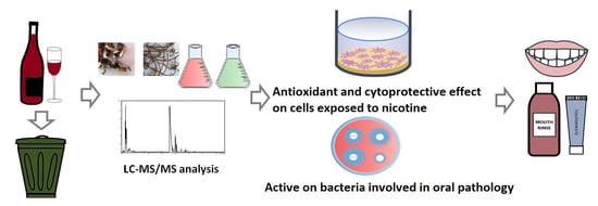
Abstract
1. Introduction
2. Materials and Methods
2.1. Preparation of Leaves and Tendrils Extract
2.1.1. Plant Material
2.1.2. Preparation of Extracts
2.2. Quantitative Determination of Bioactive Compounds
2.2.1. Chemicals
2.2.2. Determination of Total Flavonoid and Caffeic Acid Derivatives Content
2.2.3. Determination of Total Phenolic Content
2.2.4. Phytochemical Analysis by LC-MS/MS
2.3. Antioxidant Activity Evaluation
2.3.1. DPPH Radical Scavenging Activity
2.3.2. Ferric-Reducing Antioxidant Power (FRAP) Assay
2.4. Biological Activities on Cell Lines
2.4.1. Cell Culture
2.4.2. Cytocompatibility and Cytoprotective Effect against Nicotine-Induced Cytotoxicity
2.4.3. Antioxidant Activity on Cell Cultures
2.4.4. Anti-inflammatory Potential
2.5. Antimicrobial Potential of the Extracts
2.5.1. Test Microorganisms
2.5.2. Culture Media
2.5.3. Method
2.6. Statistical Analysis
3. Results and Discussion
3.1. Total Phenolic Contents and Antioxidant Activity
3.2. Chromatographic Analysis of V. vinifera Fn Leaves and Tendrils Extracts
3.3. Cytocompatibility and Cytoprotective Effect of LE and TE against Nicotine-Induced Cytotoxicity
3.4. Antioxidant Potential
3.5. Anti-Inflammatory Potential
3.6. Antimicrobial Activity
4. Conclusions
Author Contributions
Funding
Acknowledgments
Conflicts of Interest
References
- Jackson, R.S. Wine and Health-Principles and Applications. In Food Science and Technology; Academic Press: San Diego, CA, USA, 2008; pp. 1–13. [Google Scholar]
- Torregrosa, L.; Vialet, S.; Adivèze, A.; Iocco-Corena, P.; Thomas, M.R. Grapevine (Vitis vinifera L.). In Agrobacterium Protocols, 3nd ed.; Wang, K., Ed.; Springer: New York, NY, USA, 2007; Volume 2, pp. 177–181. [Google Scholar]
- Fontana, A.; Antoniolli, A.; Bottini, R. Extraction, Characterization and Utilization of Bioactive Compounds from Wine Industry Waste. In Utilisation of Bioactive Compounds from Agricultural and Food Waste; Vuong, Q.V., Ed.; CRC Press: Boca Raton, FL, USA, 2017; pp. 213–229. [Google Scholar]
- Llobera, A.; Cañellas, J. Antioxidant activity and dietary fibre of Prensal Blanc white grape (Vitis vinifera) by-products. Int. J. Food Sci. Technol. 2008, 43, 1953–1959. [Google Scholar] [CrossRef]
- European Parliament European Parliament Resolution of 19 January 2012 on How to Avoid Food Wastage: Strategies for a More Efficient Food Chain in the EU (2011/2175(INI)). Available online: https://op.europa.eu/en/publication-detail/-/publication/b2a48b16-fe76-11e2-a352-01aa75ed71a1 (accessed on 3 April 2020).
- Mateo, J.J.; Maicas, S. Valorization of winery and oil mill wastes by microbial technologies. Food Res. Int. 2015, 73, 13–25. [Google Scholar] [CrossRef]
- Fraternale, D.; Rudov, A.; Prattichizzo, F.; Olivieri, F.; Ricci, D.; Giacomini, E.; Carloni, S.; Azzolini, C.; Gordillo, B.; Jara-Palacios, M.J. Chemical composition and “in vitro” anti-inflammatory activity of Vitis vinifera L.(var. Sangiovese) tendrils extract. J. Funct. Foods 2016, 20, 291–302. [Google Scholar] [CrossRef]
- Desjardins, J.; Grenier, D. Neutralizing effect of green tea epigallocatechin-3-gallate on nicotine-induced toxicity and chemokine (C-C motif) ligand 5 secretion in human oral epithelial cells and fibroblasts. J. Investig. Clin. Dent. 2012, 3, 189–197. [Google Scholar] [CrossRef]
- Warnakulasuriya, S.N.; Rupasinghe, H.P.V. Novel long chain fatty acid derivatives of quercetin-3-O-glucoside reduce cytotoxicity induced by cigarette smoke toxicants in human fetal lung fibroblasts. Eur. J. Pharmacol. 2016, 781, 128–138. [Google Scholar] [CrossRef]
- Li, L.; Sun, W.; Wu, T.; Lu, R.; Shi, B. Caffeic acid phenethyl ester attenuates lipopolysaccharide-stimulated proinflammatory responses in human gingival fibroblasts via NF-κB and PI3K/Akt signaling pathway. Eur. J. Pharmacol. 2017, 794, 61–68. [Google Scholar] [CrossRef]
- Chang, Y.C.; Huang, F.M.; Tai, K.W.; Yang, L.C.; Chou, M.Y. Mechanisms of cytotoxicity of nicotine in human periodontal ligament fibroblast cultures in vitro. J. Periodontal Res. 2002, 37, 279–285. [Google Scholar] [CrossRef]
- Oniga, I.; Vlase, L.; Toiu, A.; Benedec, D.; Duda, M. Evaluation of phenolic acid derivatives and essential oil content in some Melissa officinalis L. varieties. Farmacia 2010, 58, 764–769. [Google Scholar]
- Moldovan, M.; Sonia, I.; Bischin Pușcaș, C.; Silaghi-Dumitrescu, R.; Hanganu, D.; Bogdan, C.; Vlase, L.; Oniga, I.; Benedec, D. A Design of Experiments strategy to enhance the recovery of polyphenolic compounds from Vitis vinifera by-products through heat reflux extraction. Biomolecules 2019, 9, 529. [Google Scholar] [CrossRef]
- Romanian Pharmacopoeia Commission National Medicines Agency. Romanian Pharmacopoeia, 10th ed.; Medical Publishing House: Bucharest, Romania, 1993; p. 335. [Google Scholar]
- Mureşan, M.; Benedec, D.; Vlase, L.; Oprean, R.; Toiu, A.; Oniga, I. Screening of polyphenolic compounds, antioxidant and antimicrobial properties of Tanacetum vulgare from Transylvania. Stud. Univ. Babes-Bolyai Chem. 2015, 60, 127–138. [Google Scholar]
- Benedec, D.; Hanganu, D.; Lorena, F.; Oniga, I.; Brindusa, T.; Olah, N.-K.; Gheldiu, A.-M.; Raita, O.; Vlase, L. Chemical, antioxidant and antibacterial studies of Romanian. Heracleum Phondylium. Farmacia 2017, 65, 252–256. [Google Scholar]
- European Pharmacopoeia Comission. European Pharmacopoiea, 5th ed.; Council of Europe: Strasbourg, France, 2005; p. 221. [Google Scholar]
- Singleton, V.L.; Orthofer, R.; Lamuela-Raventós, R.M. Analysis of total phenols and other oxidation substrates and antioxidants by means of folin-ciocalteu reagent. Methods Enzymol. 1999, 299, 152–178. [Google Scholar]
- Rusu, M.; Gheldiu, A.-M.; Mocan, A.; Moldovan, C.; Popa, D.-S.; Tomuta, I.; Vlase, L. Process Optimization for Improved Phenolic Compounds Recovery from Walnut (Juglans regia L.) Septum: Phytochemical Profile and Biological Activities. Molecules 2018, 23, 2814. [Google Scholar] [CrossRef] [PubMed]
- Balík, J.; Kyseláková, M.; Vrchotová, N.; Tříska, J.; Kumšta, M.; Veverka, J.; Híc, P.; Totušek, J.; Lefnerová, D. Relations between Polyphenols Content and Antioxidant Activity in Vine Grapes and Leaves. Czech J. Food Sci. 2008, 26, 25–32. [Google Scholar] [CrossRef]
- Thaipong, K.; Boonprakob, U.; Crosby, K.; Cisneros-Zevallos, L.; Byrne, D. Comparision of ABTS, DPPH, FRAP, and ORAC assays for estimating antioxidant activity from guava fruit extracts. J. Food Compos. Anal. 2006, 19, 669–675. [Google Scholar] [CrossRef]
- Benzie, I.F.F.; Strain, J.J. The ferric reducing ability of plasma (FRAP) as a measure of ‘antioxidant power’: The FRAP assay. Anal. Biochem. 1996, 239, 70–76. [Google Scholar] [CrossRef]
- Olah, N.-K.; Osser, G.; Ramona Flavia, C.; Furtuna, F.; Benedec, D.; Lorena, F.; Raita, O.; Hanganu, D. The study of polyphenolic compounds profile of some Rosmarinus officinalis L. extracts. Pak. J. Pharm. Sci. 2016, 29, 2355–2361. [Google Scholar]
- Pop, A.; Drugan, T.; Gutleb, A.; Lupu, D.; Cherfan, J.; Loghin, F.; Kiss, B. Estrogenic and anti-estrogenic activity of butylparaben, butylated hydroxyanisole, butylated hydroxytoluene and propyl gallate and their binary mixtures on two estrogen responsive cell lines (T47D-Kbluc, MCF-7). J. Appl. Toxicol. 2018, 38, 1–14. [Google Scholar] [CrossRef]
- Rusu, M.E.; Fizeșan, I.; Pop, A.; Gheldiu, A.M.; Mocan, A.; Crișan, G.; Vlase, L.; Loghin, F.; Popa, D.S.; Tomuta, I. Enhanced recovery of antioxidant compounds from hazelnut (Corylus avellana L.) involucre based on extraction optimization: Phytochemical profile and biological activities. Antioxidants 2019, 8, 460. [Google Scholar] [CrossRef]
- Carpa, R.; Dragan-Bularda, M.; Muntean, V. Microbiologie Generala-Lucrari Practice; Presa Universitara Clujeana: Cluj-Napoca, Romania, 2014; pp. 56–82. [Google Scholar]
- Atlas, R.M. Handbook of Microbiological Media; CRC Press: Washington, DC, USA, 2010. [Google Scholar]
- Katalinic, V.; Mozina, S.S.; Generalic, I.; Skroza, D.; Ljubenkov, I.; Klancnik, A. Phenolic profile, antioxidant capacity, and antimicrobial activity of leaf extracts from six Vitis vinifera L. varieties. Int. J. Food Prop. 2013, 16, 45–60. [Google Scholar] [CrossRef]
- Šuković, D.; Knežević, B.; Gašić, U.; Sredojević, M.; Ćirić, I.; Todic, S.; Mutic, J.; Tesic, Z. Phenolic profiles of leaves, grapes and wine of grapevine variety Vranac (Vitis vinifera L.) from Montenegro. Foods 2020, 9, 138. [Google Scholar]
- Ferhi, S.; Santaniello, S.; Zerizer, S.; Cruciani, S.; Fadda, A.; Sanna, D.; Dore, A.; Maioli, M.; D’hallewin, G. Total phenols from grape leaves counteract cell proliferation and modulate apoptosis-related gene expression in MCF-7 and HepG2 human cancer cell lines. Molecules 2019, 24, 612. [Google Scholar] [CrossRef] [PubMed]
- Akyurt, B.; Başyiğit, B.; Çam, M. Phenolic compounds content, antioxidant and antidiabetic potentials of seven edible leaves. GIDA 2018, 43, 876–885. [Google Scholar]
- Kuru, İ.; Orcan, P.; Akbaş, F.; Işıkalan, Ç.; Namli, S. The evaluation of total phenolic, flavonoid, sugar contents and antioxidant activity of Tayfi Grape in Turkey. Batman Univ. J. Life Sci. 2017, 7, 13–22. [Google Scholar]
- Fernandes, F.; Ramalhosa, E.; Pires, P.; Verdial, J.; Valentão, P.; Andrade, P.; Bento, A.; Pereira, J.A. Vitis vinifera leaves towards bioactivity. Ind. Crops Prod. 2013, 43, 434–440. [Google Scholar] [CrossRef]
- Doshi, P.; Adsule, P.; Banerjee, K. Phenolic composition and antioxidant activity in grapevine parts and berries (Vitis vinifera L.) cv. Kishmish Chornyi (Sharad Seedless) during maturation. Int. J. Food Sci. Technol. 2006, 41, 1–9. [Google Scholar] [CrossRef]
- Bodó, A.; Csepregi, K.; Szata, B.É.; Nagy, D.U.; Jakab, G.; Kocsis, M. Bioactivity of Leaves, Skins and Seeds of Berry Color Variant Grapevines (Vitis vinifera L.). Res. Rev. J. Pharmacogn. Phytochem. 2017, 5, 16–22. [Google Scholar]
- Pintać, D.; Četojević-Simin, D.; Berežni, S.; Orčić, D.; Mimica-Dukić, N.; Lesjak, M. Investigation of the chemical composition and biological activity of edible grapevine (Vitis vinifera L.) leaf varieties. Food Chem. 2019, 286, 686–695. [Google Scholar] [CrossRef]
- Dawbaa, S.; Aybastıer, Ö.; Demir, C. Ultrasensitive determination of DNA oxidation products by gas chromatography–tandem mass spectrometry and the role of antioxidants in the prevention of oxidative damage. J. Chromatogr. B Anal. Technol. Biomed. Life Sci. 2017, 1051, 84–91. [Google Scholar] [CrossRef]
- Fraternale, D.; De Bellis, R.; Calcabrini, C.; Potenza, L.; Cucchiarini, L.; Mancini, U.; Dachà, M.; Ricci, D. Aqueous extract from Vitis vinifera tendrils is able to enrich keratinocyte antioxidant defences. Nat. Prod. Commun. 2011, 6, 1315–1319. [Google Scholar] [CrossRef]
- Orfali, G.D.C.; Duarte, A.; Bonadio, V.; Martinez, N.; Araújo, M.; Priviero, F.; Carvalho, P.; Priolli, D. Review of anticancer mechanisms of isoquercitin. World J. Clin. Oncol. 2016, 7, 189–199. [Google Scholar] [CrossRef] [PubMed]
- Karak, P. Biological activities of flavonoids: An overview. Int. J. Pharm. Sci. Res. 2019, 10, 1567–1574. [Google Scholar]
- Nirmala, J.G.; Celsia, S.E.; Swaminathan, A.; Narendhirakannan, R.T.; Chatterjee, S. Cytotoxicity and apoptotic cell death induced by Vitis vinifera peel and seed extracts in A431 skin cancer cells. Cytotechnology 2018, 70, 537–554. [Google Scholar] [CrossRef] [PubMed]
- Anastasiadi, M.; Pratsinis, H.; Kletsas, D.; Skaltsounis, A.-L.; Haroutounian, S.A. Bioactive non-coloured polyphenols content of grapes, wines and vinification by-products: Evaluation of the antioxidant activities of their extracts. Food Res. Int. 2010, 43, 805–813. [Google Scholar] [CrossRef]
- Giovannelli, L.; Innocenti, M.; Santamaria, A.R.; Bigagli, E.; Pasqua, G.; Mulinacci, N. Antitumoural activity of viniferin-enriched extracts from Vitis vinifera L. cell cultures. Nat. Prod. Res. 2014, 28, 2006–2016. [Google Scholar] [CrossRef]
- Haber, J.; Wattles, J.; Crowley, M.; Mandell, R.; Joshipura, K.; Kent, R.L. Evidence for cigarette smoking as a major risk factor for periodontitis. J. Periodontol. 1993, 64, 16–23. [Google Scholar] [CrossRef]
- McGuire, J.R.; McQuade, M.J.; Rossmann, J.A.; Garnick, J.J.; Sutherland, D.E.; Scheidt, M.J.; Van Dyke, T.E. Cotinine in saliva and gingival crevicular fluid of smokers with periodontal disease. J. Periodontol. 1989, 60, 176–181. [Google Scholar] [CrossRef]
- Katono, T.; Kawato, T.; Tanabe, N.; Tanaka, H.; Suzuki, N.; Kitami, S.; Morita, T.; Motohashi, M.; Maeno, M. Effects of nicotine and lipopolysaccharide on the expression of matrix metalloproteinases, plasminogen activators, and their inhibitors in human osteoblasts. Arch. Oral Biol. 2009, 54, 146–155. [Google Scholar] [CrossRef]
- Tanaka, H.; Tanabe, N.; Shoji, M.; Suzuki, N.; Katono, T.; Sato, S.; Motohashi, M.; Maeno, M. Nicotine and lipopolysaccharide stimulate the formation of osteoclast-like cells by increasing macrophage colony-stimulating factor and prostaglandin E2 production by osteoblasts. Life Sci. 2006, 78, 1733–1740. [Google Scholar] [CrossRef]
- Almasri, A.; Wisithphrom, K.; Windsor, L.J.; Olson, B. Nicotine and lipopolysaccharide affect cytokine expression from gingival fibroblasts. J. Periodontol. 2007, 78, 533–541. [Google Scholar] [CrossRef]
- Palaska, I.; Papathanasiou, E.; Theoharides, T.C. Use of polyphenols in periodontal inflammation. Eur. J. Pharmacol. 2013, 720, 77–83. [Google Scholar] [CrossRef] [PubMed]
- Forester, S.C.; Lambert, J.D. The role of antioxidant versus pro-oxidant effects of green tea polyphenols in cancer prevention. Mol. Nutr. Food Res. 2011, 55, 844–854. [Google Scholar] [CrossRef] [PubMed]
- Paunović, M.G.; Ognjanović, B.I.; Matić, M.M.; Štajn, A.Š.; Saičić, Z.S. Protective effects of quercetin and vitamin C against nicotine-induced toxicity in the blood of Wistar rats. Arch. Ind. Hyg. Toxicol. 2016, 67, 304–310. [Google Scholar] [CrossRef] [PubMed]
- Balakrishnan, A.; Menon, V.P. Antioxidant properties of hesperidin in nicotine-induced lung toxicity. Fundam. Clin. Pharmacol. 2007, 21, 535–546. [Google Scholar] [CrossRef] [PubMed]
- Yarahmadi, A.; Zal, F.; Bolouki, A. Protective effects of quercetin on nicotine induced oxidative stress in ‘HepG2 cells’. Toxicol. Mech. Methods 2017, 27, 609–614. [Google Scholar] [CrossRef] [PubMed]
- Anastasiadi, M.; Pratsinis, H.; Kletsas, D.; Skaltsounis, A.L.; Haroutounian, S.A. Grape stem extracts: Polyphenolic content and assessment of their in vitro antioxidant properties. LWT Food Sci. Technol. 2012, 48, 316–322. [Google Scholar] [CrossRef]
- Goutzourelas, N.; Stagos, D.; Spanidis, Y.; Liosi, M.; Apostolou, A.; Priftis, A.; Haroutounian, S.; Spandidos, D.A.; Tsatsakis, A.M.; Kouretas, D. Polyphenolic composition of grape stem extracts affects antioxidant activity in endothelial and muscle cells. Mol. Med. Rep. 2015, 12, 5846–5856. [Google Scholar] [CrossRef] [PubMed]
- Pervin, M.; Hasnat, M.; Lee, Y.M.; Kim, D.H.; Jo, J.E.; Lim, B.O. Antioxidant activity and acetylcholinesterase inhibition of grape skin anthocyanin (GSA). Molecules 2014, 19, 9403–9418. [Google Scholar] [CrossRef] [PubMed]
- Marabini, L.; Melzi, G.; Lolli, F.; Dell’Agli, M.; Piazza, S.; Sangiovanni, E.; Marinovich, M. Effects of Vitis vinifera L. leaves extract on UV radiation damage in human keratinocytes (HaCaT). J. Photochem. Photobiol. B Biol. 2020, 204, 111810. [Google Scholar] [CrossRef] [PubMed]
- Li, H.; Wang, X.; Li, P.; Li, Y.; Wang, H. Comparative study of antioxidant activity of grape (Vitis vinifera) seed powder assessed by different methods. J. Food Drug Anal. 2008, 16, 1–7. [Google Scholar]
- Grace Nirmala, J.; Narendhirakannan, R.T. In vitro antioxidant and antimicrobial activities of grapes (Vitis vinifera L.) seed and skin extracts - Muscat variety. Int. J. Pharm. Pharm. Sci. 2011, 3, 242–249. [Google Scholar]
- Lakshmi, B.V.S.; Sudhakar, M.; Aparna, M. Protective potential of Black grapes against lead induced oxidative stress in rats. Environ. Toxicol. Pharmacol. 2013, 35, 361–368. [Google Scholar] [CrossRef] [PubMed]
- Mukherjee, S.; Das, S.K.; Vasudevan, D.M. Dietary grapes (Vitis vinifera) feeding attenuates ethanol-induced oxidative stress in blood and modulates immune functions in mice. Indian J. Biochem. Biophys. 2012, 49, 379–385. [Google Scholar] [PubMed]
- Pirinççioğlu, M.; Kızıl, G.; Kızıl, M.; Özdemir, G.; Kanay, Z.; Ketani, M.A. Protective effect of Öküzgözü (Vitis vinifera L. cv.) grape juice against carbon tetrachloride induced oxidative stress in rats. Food Funct. 2012, 3, 668–673. [Google Scholar] [CrossRef] [PubMed]
- Saada, H.N.; Said, U.Z.; Meky, N.H.; Azime, A.S.A. El Grape seed extract Vitis vinifera protects against radiation-induced oxidative damage and metabolic disorders in rats. Phyther. Res. 2009, 23, 434–438. [Google Scholar] [CrossRef]
- Saadaoui, N.; Weslati, A.; Barkaoui, T.; Khemiri, I.; Gadacha, W.; Souli, A.; Mokni, M.; Harbi, M.; Ben-Attia, M. Gastroprotective effect of leaf extract of two varieties grapevine (Vitis vinifera L.) native wild and cultivar grown in North of Tunisia against the oxidative stress induced by ethanol in rats. Biomarkers 2020, 25, 48–61. [Google Scholar] [CrossRef]
- Lafay, S.; Jan, C.; Nardon, K.; Lemaire, B.; Ibarra, A.; Roller, M.; Houvenaeghel, M.; Juhel, C.; Cara, L. Grape extract improves antioxidant status and physical performance in elite male athletes. J. Sport. Sci. Med. 2009, 8, 468–480. [Google Scholar]
- Sangiovanni, E.; Di Lorenzo, C.; Colombo, E.; Colombo, F.; Fumagalli, M.; Frigerio, G.; Restani, P.; Dell’Agli, M. The effect of in vitro gastrointestinal digestion on the anti-inflammatory activity of Vitis vinifera L. leaves. Food Funct. 2015, 6, 2453–2463. [Google Scholar] [CrossRef]
- Oeckinghaus, A.; Hayden, M.S.; Ghosh, S. Crosstalk in NF-κB signaling pathways. Nat. Immunol. 2011, 12, 695–708. [Google Scholar] [CrossRef]
- Shi, J.; Yu, J.; Pohorly, J.E.; Kakuda, Y. Polyphenolics in grape seeds—Biochemistry and functionality. J. Med. Food 2003, 6, 291–299. [Google Scholar] [CrossRef]
- Millan-Linares, M.C.; Bermudez, B.; Martin, M.E.; Muñoz, E.; Abia, R.; Millan, F.; Muriana, F.J.G.; Montserrat-de la Paz, S. Unsaponifiable fraction isolated from grape (Vitis vinifera L.) seed oil attenuates oxidative and inflammatory responses in human primary monocytes. Food Funct. 2018, 9, 2517–2523. [Google Scholar] [CrossRef] [PubMed]
- Singh, J.; Singh, A.K.; Singh, A. Analgesic and anti-inflammatory activity of methanolic extract of Vitis vinifera leaves. Pharmacologyonline 2009, 3, 496–504. [Google Scholar]
- Filip, A.; Clichici, S.; Daicoviciu, D.; Catoi, C.; Bolfa, P.; Postescu, I.D.; Gal, A.; Baldea, I.; Gherman, C.; Muresan, A. Chemopreventive effects of Calluna vulgaris and Vitis vinifera extracts on UVB-induced skin damage in SKH-1 hairless mice. J. Physiol. Pharmacol. 2011, 62, 385–392. [Google Scholar] [PubMed]
- Middleton, E.; Kandaswami, C.; Theoharides, T.C. The effects of plant flavonoids on mammalian cells: Implications for inflammation, heart disease, and cancer. Pharmacol. Rev. 2000, 52, 673–751. [Google Scholar] [PubMed]
- Mitjavila, M.T.; Moreno, J.J. The effects of polyphenols on oxidative stress and the arachidonic acid cascade. Implications for the prevention/treatment of high prevalence diseases. Biochem. Pharmacol. 2012, 84, 1113–1122. [Google Scholar] [CrossRef] [PubMed]
- Bunte, K.; Hensel, A.; Beikler, T. Polyphenols in the prevention and treatment of periodontal disease: A systematic review of in vivo, ex vivo and in vitro studies. Fitoterapia 2019, 132, 30–39. [Google Scholar] [CrossRef]
- Ahmed, S.M.U.; Luo, L.; Namani, A.; Wang, X.J.; Tang, X. Nrf2 signaling pathway: Pivotal roles in inflammation. Biochim. Biophys. Acta Mol. Basis Dis. 2017, 1863, 585–597. [Google Scholar] [CrossRef]
- Zhang, H.; Tsao, R. Dietary polyphenols, oxidative stress and antioxidant and anti-inflammatory effects. Curr. Opin. Food Sci. 2016, 8, 33–42. [Google Scholar] [CrossRef]
- Oral Health. Available online: https://www.who.int/news-room/fact-sheets/detail/oral-health (accessed on 16 March 2020).
- Wu, C.D. Grape Products and Oral Health. J. Nutr. 2009, 139, 1818S–1823S. [Google Scholar] [CrossRef]
- Metwalli, K.H.; Khan, S.A.; Krom, B.P.; Jabra-Rizk, M.A. Streptococcus mutans, Candida albicans, and the human mouth: A sticky situation. PLoS Pathog. 2013, 9, e1003616. [Google Scholar] [CrossRef]
- Chinsembu, K.C. Plants and other natural products used in the management of oral infections and improvement of oral health. Acta Trop. 2016, 154, 6–18. [Google Scholar] [CrossRef] [PubMed]
- Daniluk, T.; Fiedoruk, K.; Sciepuk, M.; Zaremba, M.L.; Rozkiewicz, D.; Cylwik-Rokicka, D.; Tokajuk, G.; Kedra, B.A.; Anielska, I.; Stokowska, W.; et al. Aerobic bacteria in the oral cavity of patients with removable dentures. Adv. Med. Sci. 2006, 51, 86–90. [Google Scholar] [PubMed]
- Lu, M.; Xuan, S.; Wang, Z. Oral microbiota: A new view of body health. Food Sci. Hum. Wellness 2019, 8, 8–15. [Google Scholar] [CrossRef]
- Furiga, A.; Lonvaud-Funel, A.; Badet, C. In vitro study of antioxidant capacity and antibacterial activity on oral anaerobes of a grape seed extract. Food Chem. 2009, 113, 1037–1040. [Google Scholar] [CrossRef]
- Yim, N.H.; Ha, D.T.; Trung, T.N.; Kim, J.P.; Lee, S.M.; Na, M.K.; Jung, H.J.; Kim, H.S.; Kim, Y.H.; Bae, K.H. The antimicrobial activity of compounds from the leaf and stem of Vitis amurensis against two oral pathogens. Bioorganic Med. Chem. Lett. 2010, 20, 1165–1168. [Google Scholar] [CrossRef]
- Cecchin, D.; Farina, A.P.; Souza, M.A.; Albarello, L.L.; Schneider, A.P.; Vidal, C.M.P.; Bedran-Russo, A.K. Evaluation of antimicrobial effectiveness and dentine mechanical properties after use of chemical and natural auxiliary irrigants. J. Dent. 2015, 43, 695–702. [Google Scholar] [CrossRef]
- Abed, A.; Harb, J.; Khasib, S.; Saad, B. In vitro assessment of cytotoxic, antioxidant and antimicrobial activities of leaves from two grape varieties collected from arid and temperate regions in Palestine. QSci. Connect 2015, 2015, 1–9. [Google Scholar]
- Miklasińska, M.; Kępa, M.; Wojtyczka, R.; Idzik, D.; Zdebik, A.; Orlewska, K.; Wąsik, T. Antibacterial activity of protocatechuic acid ethyl ester on Staphylococcus aureus clinical strains alone and in combination with antistaphylococcal drugs. Molecules 2015, 20, 13536–13549. [Google Scholar] [CrossRef]
- Miklasińska, M.; Kȩpa, M.; Wojtyczka, R.D.; Idzik, D.; Dziedzic, A.; Wąsik, T.J. Catechin hydrate augments the antibacterial action of selected antibiotics against Staphylococcus aureus clinical strains. Molecules 2016, 21, 244. [Google Scholar] [CrossRef]
- Gutiérrez-Venegas, G.; Gómez-Mora, J.A.; Meraz-Rodríguez, M.A.; Flores-Sánchez, M.A.; Ortiz-Miranda, L.F. Effect of flavonoids on antimicrobial activity of microorganisms present in dental plaque. Heliyon 2019, 5, e03013. [Google Scholar] [CrossRef]
- Smeriglio, A.; Denaro, M.; De Francesco, C.; Cornara, L.; Barreca, D.; Bellocco, E.; Ginestra, G.; Mandalari, G.; Trombetta, D. Feijoa fruit peel: Micro-morphological features, evaluation of phytochemical profile, and biological properties of its essential oil. Antioxidants 2019, 8, 320. [Google Scholar] [CrossRef] [PubMed]


| Samples | TFC (mg RE/g) | Caffeic Acid Derivatives (mg CAE/g) | TPC (mg GAE/g) | IC50 (DPPH) (mg/mL) | FRAP (µmol Te/mL) |
|---|---|---|---|---|---|
| TE | 14.21 ± 0.20 a | 4.14 ± 0.07 a | 35.65 ± 0.33 a | 0.155 ± 0.04 b,c | 10.60 ± 0.39 a |
| LE | 16.75 ± 0.12 | 6.39 ± 1.11 | 28.62 ± 0.24 | 0.248 ± 0.01 a | 6.29 ± 0.20 |
| Trolox | - | - | - | 0.011 ± 0.00 | - |
| Polyphenolic Compounds | m/z Value | tR ± SD (min) | LE (µg/g) | TE (µg/g) |
|---|---|---|---|---|
| Gallic acid | 169 | 1.50 ± 0.01 | 5.50 ± 0.05 | 6.90 ± 0.11 a |
| Protocatechuic acid | 153 | 2.80 ± 0.01 | 1.71 ± 0.02 | 7.92 ± 0.08 a |
| Caftaric acid | 311 | 3.54 ± 0.05 | < 0.02 | <0.02 |
| Catechin | 289 | 6.00 ± 0.03 | 23.31 ± 0.36 | 51.72 ± 1.06 a |
| Epicatechin | 289 | 9.00 ± 0.01 | 7.10 ± 0.09 | 2.60 ± 0.09 a |
| Hyperoside | 463 | 18.60 ± 0.12 | 147.09 ± 1.79 | 127.39 ± 2.61 a |
| Isoquercitrin | 463 | 19.60 ± 0.10 | 903.49 ± 6.51 | 541.34 ± 6.44 a |
| Rutin | 609 | 20.20 ± 0.15 | 385.63 ± 3.36 | 617.21 ± 6.79 a |
| Quercitrin | 447 | 23.64 ± 0.13 | 188.74 ± 2.26 | 100.87 ± 2.13 a |
| Quercetin | 301 | 26.80 ± 0.15 | 10.54 ± 0.24 | 8.89 ± 0.11 a |
| Bacterial/Fungal Strain | Inhibition Diameter for the Tested Samples (mm) ± SD | ||
|---|---|---|---|
| C | 1 | 2 | |
| Porphyromonas gingivalis ATCC 33277 | 0 ± 0.00 | 10 ± 0.00 | 13 ± 1.41 |
| Enterococcus faecalis ATCC 29212 | 0 ± 0.00 | 10 ± 0.00 | 12 ± 1.41 |
| Streptococcus mutans ATCC 25175 | 0 ± 0.00 | 10 ± 0.00 | 11.5 ± 0.71 |
| Staphylococcus aureus ATCC 25923 | 0 ± 0.00 | 14.5 ± 0.71 | 12 ± 1.41 |
| Escherichia coli ATCC 25922 | 0 ± 0.00 | 10 ± 1.41 | 11.5 ± 0.71 |
| Klebsiella sp. | 0 ± 0.00 | 0 ± 0.00 | 0 ± 0.00 |
| Candida albicans ATCC 10231 | 0 ± 0.00 | 0 ± 0.00 | 0 ± 0.00 |
© 2020 by the authors. Licensee MDPI, Basel, Switzerland. This article is an open access article distributed under the terms and conditions of the Creative Commons Attribution (CC BY) license (http://creativecommons.org/licenses/by/4.0/).
Share and Cite
Moldovan, M.L.; Carpa, R.; Fizeșan, I.; Vlase, L.; Bogdan, C.; Iurian, S.M.; Benedec, D.; Pop, A. Phytochemical Profile and Biological Activities of Tendrils and Leaves Extracts from a Variety of Vitis vinifera L. Antioxidants 2020, 9, 373. https://doi.org/10.3390/antiox9050373
Moldovan ML, Carpa R, Fizeșan I, Vlase L, Bogdan C, Iurian SM, Benedec D, Pop A. Phytochemical Profile and Biological Activities of Tendrils and Leaves Extracts from a Variety of Vitis vinifera L. Antioxidants. 2020; 9(5):373. https://doi.org/10.3390/antiox9050373
Chicago/Turabian StyleMoldovan, Mirela L., Rahela Carpa, Ionel Fizeșan, Laurian Vlase, Cătălina Bogdan, Sonia M. Iurian, Daniela Benedec, and Anca Pop. 2020. "Phytochemical Profile and Biological Activities of Tendrils and Leaves Extracts from a Variety of Vitis vinifera L." Antioxidants 9, no. 5: 373. https://doi.org/10.3390/antiox9050373
APA StyleMoldovan, M. L., Carpa, R., Fizeșan, I., Vlase, L., Bogdan, C., Iurian, S. M., Benedec, D., & Pop, A. (2020). Phytochemical Profile and Biological Activities of Tendrils and Leaves Extracts from a Variety of Vitis vinifera L. Antioxidants, 9(5), 373. https://doi.org/10.3390/antiox9050373