Next Article in Journal
A Novel Risk Assessment and Analysis Method for Correlation in a Complex System Based on Multi-Dimensional Theory
Next Article in Special Issue
Special Feature: Permanent and Long-Term Biodegradable Biomaterials
Previous Article in Journal
Physicochemical and Microstructural Properties of Red Muds under Acidic and Alkaline Conditions
Previous Article in Special Issue
Assessment of Chitosan-Rue (Ruta graveolens L.) Essential Oil-Based Coatings on Refrigerated Cape Gooseberry (Physalis peruviana L.) Quality
 
 
Retraction published on 25 August 2020, see Appl. Sci. 2020, 10(17), 5865.
Article

Experimental Study on the Influence of Apigenin K and Melatonin in Socket Preservation as Bone Stimulators: An Experimental Study in Beagle Dogs

by
José Luis Calvo-Guirado
1,*,
Manuel Fernández-Domínguez
2,
Juan Manuel Aragoneses
3,
Enrique Fernández-Bodereau
4,
Miguel Angel Garcés-Villalá
5,
Felix De Carlos-Villafranca
6,
Manuel Cabello-Colás
7,
Rocío Jiménez-López
7,
Carlos Pérez Albacete-Martínez
1 and
Rafael Arcesio Delgado-Ruiz
8
1
Department of Oral and Implant Surgery, Faculty of Health Sciences, Universidad Católica San Antonio de Murcia (UCAM), 30170 Murcia, Spain
2
Department of Oral Surgery and Implantology and Director of Research in Dentistry in the Doctoral, Program of Translational Medicine, CEU San Pablo University, 28223 Madrid, Spain
3
Department of Dental Research, Universidad Federico Henríquez y Carvajal (UFHEC), Santo Domingo 10107, Dominican Republic
4
Carrera Especialización en Prótesis Fija, Removible e Implantología, Facultad de Odontología, Universidad Nacional de Córdoba, Córdoba X5000HUA, Argentine
5
Department of Implant and Biomaterial Research, Fundación Corazón de Jesús, Av. Rawson 1373 sur, San Juan 5400, Argentina
6
Department of Orthodontics, Faculty of Medicine, University of Oviedo, 33006 Asturias, Spain
7
Pre-graduate Student, Faculty of Health Sciences, Universidad Católica San Antonio de Murcia (UCAM), 30170 Murcia, Spain
8
Department of Prosthodontics and Digital Technology, Stony Brook University, School of Dental Medicine, Stony Brook, NY 11794, USA
*
Author to whom correspondence should be addressed.
Appl. Sci. 2020, 10(9), 3006; https://doi.org/10.3390/app10093006
Submission received: 28 March 2020 / Revised: 21 April 2020 / Accepted: 22 April 2020 / Published: 25 April 2020
(This article belongs to the Special Issue Permanent and Long-Term Biodegradable Biomaterials)

Abstract

:

Featured Application

The study promotes the use of two different natural substances, apigenin K and melatonin, like bone stimulators for bone regeneration.

Abstract

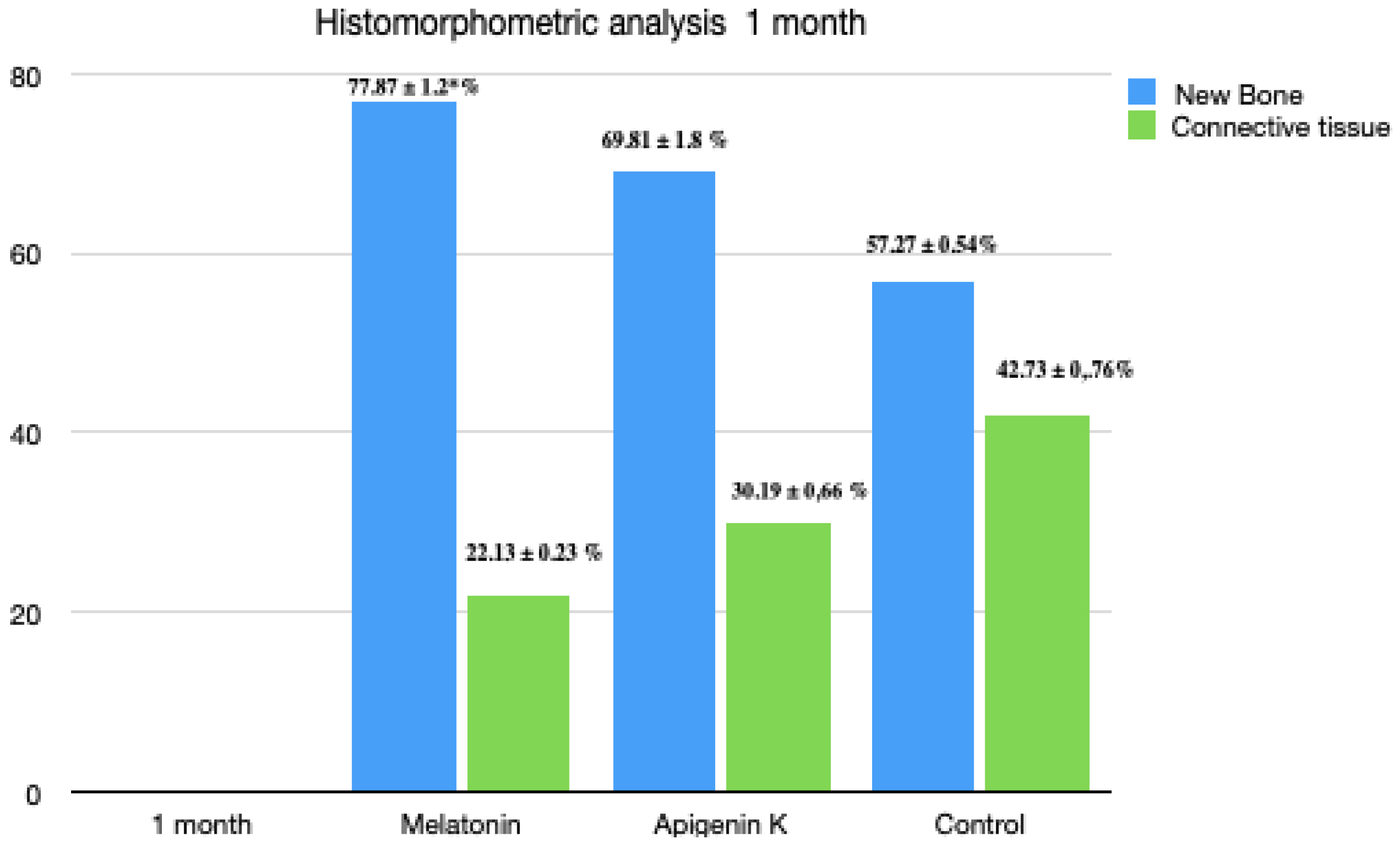
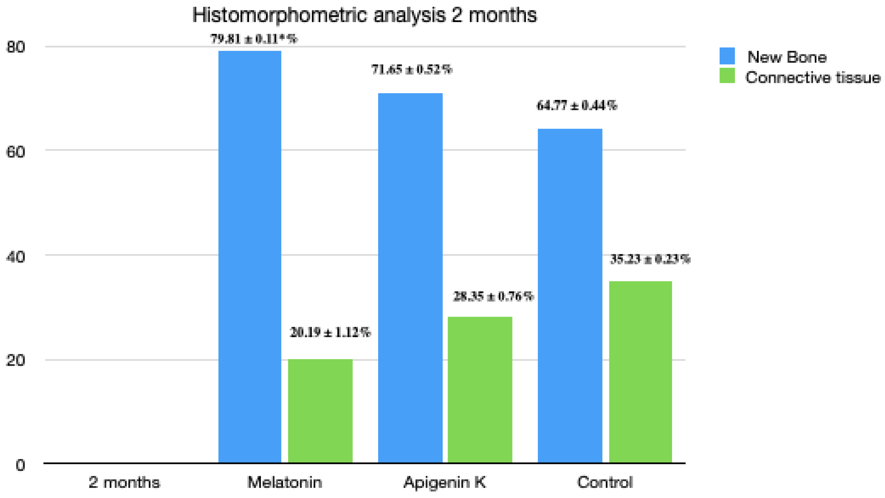
The aim is to evaluate whether apigenin K and melatonin M5250 were able to stimulate bone formation after tooth extraction at one, two, and three months follow-up. Six male beagle dogs were used. Apigenin K and melatonin M5250 immersed in hemostatic collagen sponges were placed in the third and fourth premolar and the first molar extracted sockets; the second premolar was used as control. At one, two, and three months, bone core biopsies were performed, and picrosirius–hematoxylin was used for the staining process. In the first month, a higher amount of calcified bone tissue was observed in the melatonin (77.87% ± 1.2%) and apigenin K (69.81% ± 1.8%) groups than the control group (57.27% ± 0.54%), with apparent discrepancies in values between the three groups (p < 0.04). In the second month, there was a considerable improvement in the results in the areas with melatonin (79.81% ± 0.11%) than in those of apigenin K (71.65% ± 0.52%) and control (64.77% ± 0.44%) (p < 0.04). In the third month, the number of mature bone was similar to all the groups. The creation of new bone was significant in the melatonin group (82.78% ± 0.87%), followed by the apigenin K group (78.76% ± 0.43%) and the control group (57.27% ± 0.11%). From this experimental study in dogs, it can be concluded that melatonin and apigenin K can accelerate the process of mineralization of the bone matrix, and thus the creation of laminae in the early stages of healing (1 month). Less reabsorption of the post-extraction sockets can be expected with the topical application of melatonin and apigenin K. It seems that the stimulatory effects of bone healing induced by the topical application of melatonin and apigenin K are defect-size-dependent, being more evident in small defects compared to larger defects.

1. Introduction

Tooth extraction results in changes of the alveolar bone dimensions; these changes occur over time in vertical and horizontal directions in different proportions [1,2]. Studies have shown that the bone contours change from the first 60 to 90 days after extraction [1,2] and that the bone loss is higher vertically than horizontally [1,2,3]. It has been noted that horizontal bone resorption can reach up to 57%, and the amount of lingual bone reduction could amount up to 30% [4]. To compensate for these dimensional changes, various biomaterials are used for bone regeneration; autogenous bone, non-protein type of materials close to human bone, natural and synthetic biomaterials, and even ground teeth can be used as bone grafts [5,6,7,8,9]. Other approaches, like the application of natural substances (apigenin and melatonin), can stimulate bone formation in areas with bone defects [10,11,12]. In several articles, it has been stated that melatonin is an essential mediator in the ability to stimulate and form new bone [13,14,15]. It has been observed that melatonin puts on the production of type I collagen [16]. Melatonin can also inhibit the bone reabsorption by regulating the formation and activation of osteoclasts mediated by RANK-L (nuclear factor receptor activator of the kappa-B ligand) [17]. Therefore, melatonin could be considered for treatments where an enhanced formation of bone is required [18,19,20,21,22]. The oral cavity is affected by a number of conditions such as mucositis, tooth decay, periodontitis, and cancers, which affect the oral cavity normally. However, the use of melatonin has been observed to enhance implant osseointegration, bone regeneration, and can also help in the management of periodontitis [23,24,25,26,27,28,29,30]. Today, there are many natural products that play a significant role in medicine, and many essential substances from plants, such as flavonoids, help as medicine for cancer [31]. Flavonoids, found in vegetables, fruits, and plants, are natural compounds with health benefits such as creating new bone and reducing its resorption [32,33,34,35]. Among the flavonoids, apigenin compounds seem to stimulate the proliferation of osteoblasts, thus preventing osteoporosis based on their antioxidant activity [32,33,34,35]. According to some animal studies, other flavonoids can reduce bone resorption in ovariectomized rats. Osteoclastogenesis has also been shown to be reduced by controlling the factor-kappa β and a protein activator (AP-1) in humans [36]. It has been demonstrated that naringenin interferes in osteoclastogenic processes by inhibiting the osteoclasts from differentiating and, thus, reduces bone resorption [36,37]. Apart from bone resorption by osteoclasts, apigenin induces cellular apoptosis of osteoclasts, and other beneficial effects reported in cancer, Alzheimer’s disease, amnesia, depression, insomnia, and diabetes [38,39,40]. It possesses remarkable anti-inflammatory, antioxidant, and estrogenic properties [41]. In addition, apigenin has antimicrobial activity, and resveratrol helps apigenin to bypass hepatic metabolism and maintain apigenin’s anti-inflammatory activities in the body [42,43]. The aim of our study is to evaluate the capacity of apigenin K and melatonin to stimulate the bone regeneration process after teeth extraction at 1, 2, and 3 months follow-up.

2. Materials and Methods

The study used six Beagle dogs who were about one year old and weighed approximately 14 to 15 kg. The Animal Research Ethics Committee of the State University of Murcia, Spain, following the guidelines established by the European Union Council Directive of 1 February 2013/53/EEC, approved the protocol. The dogs were placed in quarantine and vaccinated against rabies, as well as given essential vitamins. Throughout the study, the dogs were kept in cages and provided with veterinary care, water, and nurture.
The experiment was a split-mouth study design (Figure 1).
The dogs were pre-anesthetized with 10% Zelazol at 0.15 mL/kg and acepromazine maleate 0.10–0.25 mg/kg (Calmo-Neosan; Pfizer, Madrid, Spain) and Domtor 30 mg/kg (Medetor 1 mg, Virbac, CPPharma Handelsgesellschaft GmbH, Burgdorf, Germany). Afterwards, the animals were taken to the operating room where an intravenous catheter (diameter 20 G) was placed in the cephalic vein, and propofol was infused at a rate of 0.4 mg/kg/min. Volatile anesthetics were used, and the dogs were given tracheal intubation to administer the anesthetic (isoflurane diluted in oxygen 2V%). In addition, the dogs were injected with local anesthesia Artinibsa (articaine 40 mg/mL + 0.01% epinephrine, Inibsa®, Barcelona, Spain) in the surgical sites. A veterinarian supervised the entire procedure.
Lower premolars (2P2, 3P3, and 4P4) and first molars (1M1) were extracted bilaterally under general anesthesia. The multi-radicular teeth were divided using a multilayer tungsten-carbide bur, and the roots were removed one by one, to reduce the damage that could be exerted to the buccal and lingual cortical areas (Figure 2a,b).
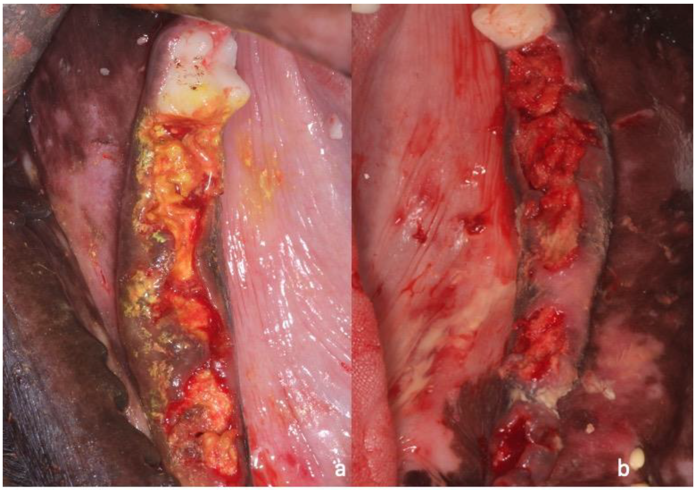
A collagen sponge soaked in apigenin K (Nutrafur, Alcantarilla, Murcia, Spain) was introduced into the lower right premolars (3P3 and 4P4) and the right first molars (1M1); empty sockets at the right side were done in second premolars sockets 2P2, Meanwhile, at the left side at the same premolar and molar sites, a collagen sponge containing melatonin M5250 (Sigma Aldrich, Merck Life Science SL, Madrid, Spain) was inserted. Second premolar sockets were used as controls (Figure 3a,b and Figure 4a,b). Resorbable sutures S90 (Lorca Marin, Lorca, Murcia, Spain) were used to close the wound.
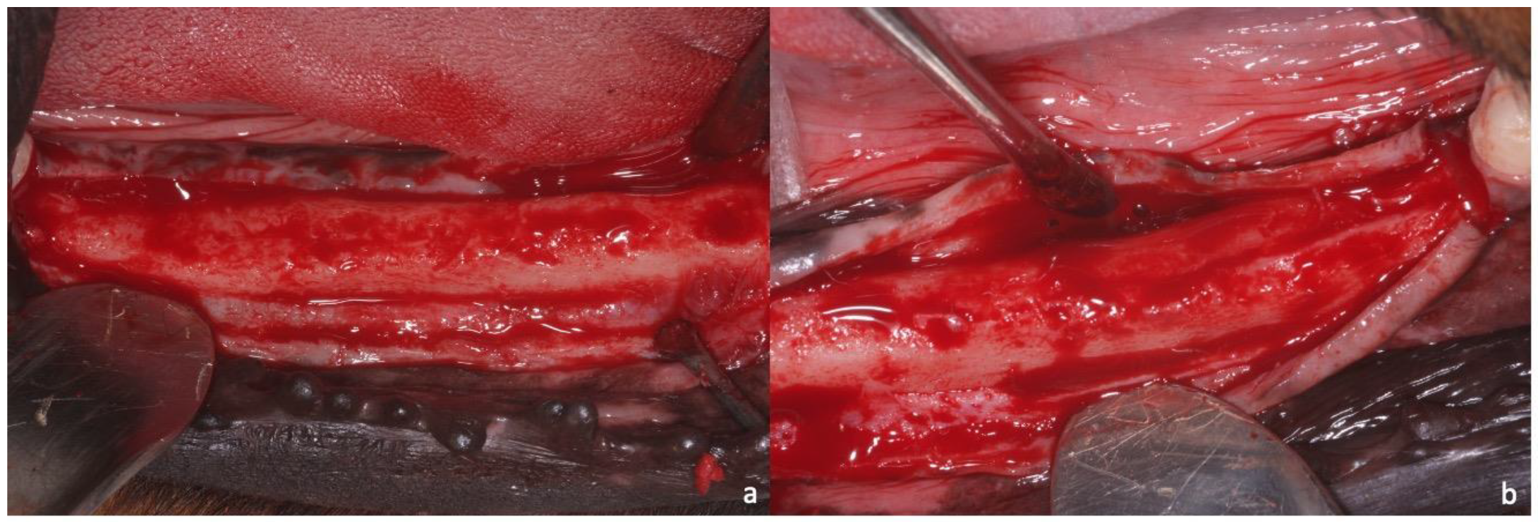
No incisors or canines were removed so that the animals could chew with the remaining teeth. The entire process was carried out under the supervision of the University’s Animal Research Department veterinarian. During the whole surgical procedure, glycoside saline (200 cm3) was used for the hydration of the venous line, so that the dogs would have a better post-operative period. Once the surgery was finished, the animals received the following anti-inflammatory, analgesic, and antibiotic medication: anti-inflammatory Voren® 2 mL intramuscularly (Boehringer Ingelheim España, S.A. Sant Cugat del Vallès, Barcelona) and antibiotic Bivamox® 2 mL intramuscularly (Boehringer Ingelheim España, S.A. Sant Cugat del Vallès, Barcelona) for one week. The dogs were re-evaluated to check their post-extraction wounds and general health. After the surgery, the dogs were fed ad-libitum for 15 days with a soft diet and water. Oral cleaning of the wound and adjacent teeth was done with gauze soaked in SEA 4 (Blue Sea Laboratories, Alicante, Spain). After 1, 2, and 3 months, the dogs were anesthetized with Artinibsa (articaine 40 mg/mL + 0.01% epinephrine, Inibsa®, Barcelona, Spain) into the buccal and lingual areas. After that, a full-thickness crestal flap was raised to obtain a biopsy (Figure 5a,b). For the biopsy, a 2.75 mm diameter trephine bur was used in the control, melatonin, and apigenin K areas at 1, 2, and 3 months, respectively (Figure 6a,b)

2.1. Sample Preparation, Histology, and Histomorphometry

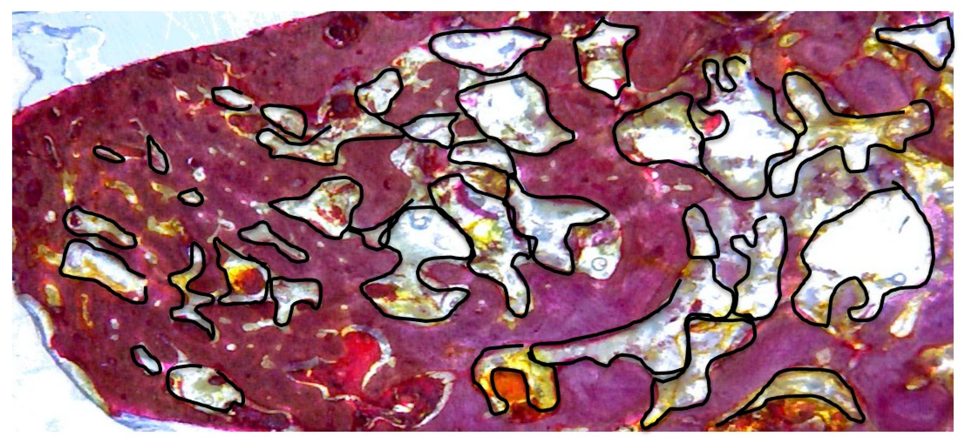
Biopsies cores were inserted into 10% buffered formalin for 10 days. Afterward, the samples were decalcified for 35 days using TBD-2 (Anatomical Pathology International, Runcorn, Cheshire, UK). The samples were then dehydrated in an ethanol solution and included in resin (Technovit 7200 VLC, Kulzer, Wehrheim, Germany). The samples were sectioned using an IsoMet 1000 (Buehler, Lake Bluff, IL, USA), and they were sawed, grounded, and polished to 10-micron thickness. Two slides were obtained per biopsy. Staining was completed with the picrosirius–hematoxylin technique. For histomorphometric analysis, images were magnified 20X, and 10 random areas were evaluated per field using an optical microscope (DP12; Olympus, Nagano, Japan). Microimage 4.0 software (Media Cybernetics, Silver Spring, MD, USA) was used for image analysis. For the histomorphometric analysis, the software ImageTool for Microsoft Windows™ (version 5.02, University of Texas) was used. The percentage of mature bone within the total bone area was determined in each core. Mature bone was characterized by images of totally mineralized bone with numbers of cells and large medullar cavities, differentiating this from mature bone characterized by images showing a predominance of new osteons made up of bone laminae organized concentrically around Haversian canals. The biopsy laboratory procedure was always the same and done by the same pathologist in the same laboratory. The difference between the areas without bone, divided by 100% of new bone, was determined as connective tissue (Figure 7).

2.2. Statistical Analysis

The results were recorded as mean ± standard deviation data. The Mann–Whitney test was applied with a significance level of 95% (p < 0.05). The Kolmogorov–Smirnov and Friedmann tests were applied for the non-parametric comparison within each time and group. SPSS 15.0 software was used for statistical analysis. The significance was established as p < 0.05.

3. Results

3.1. One Month Optical Microscopy

The biopsy of the control groups showed ideal new bone formation on one side of the defect, but it was utterly immature, with highly disorganized tissues and large medullar spaced areas (Figure 8a). In the biopsy of the test groups (melatonin and apigenin K), new bone was evident with more mineralization and more organized osteons (laminae arranged concentrically around Haversian channels). This much more organized bone was present in the central areas where melatonin and apigenin K were placed. The new bone had irregular disposition in some areas with high levels of cellularity and thicker trabecular structure in the middle and apical areas than the control group (Figure 8b,c).
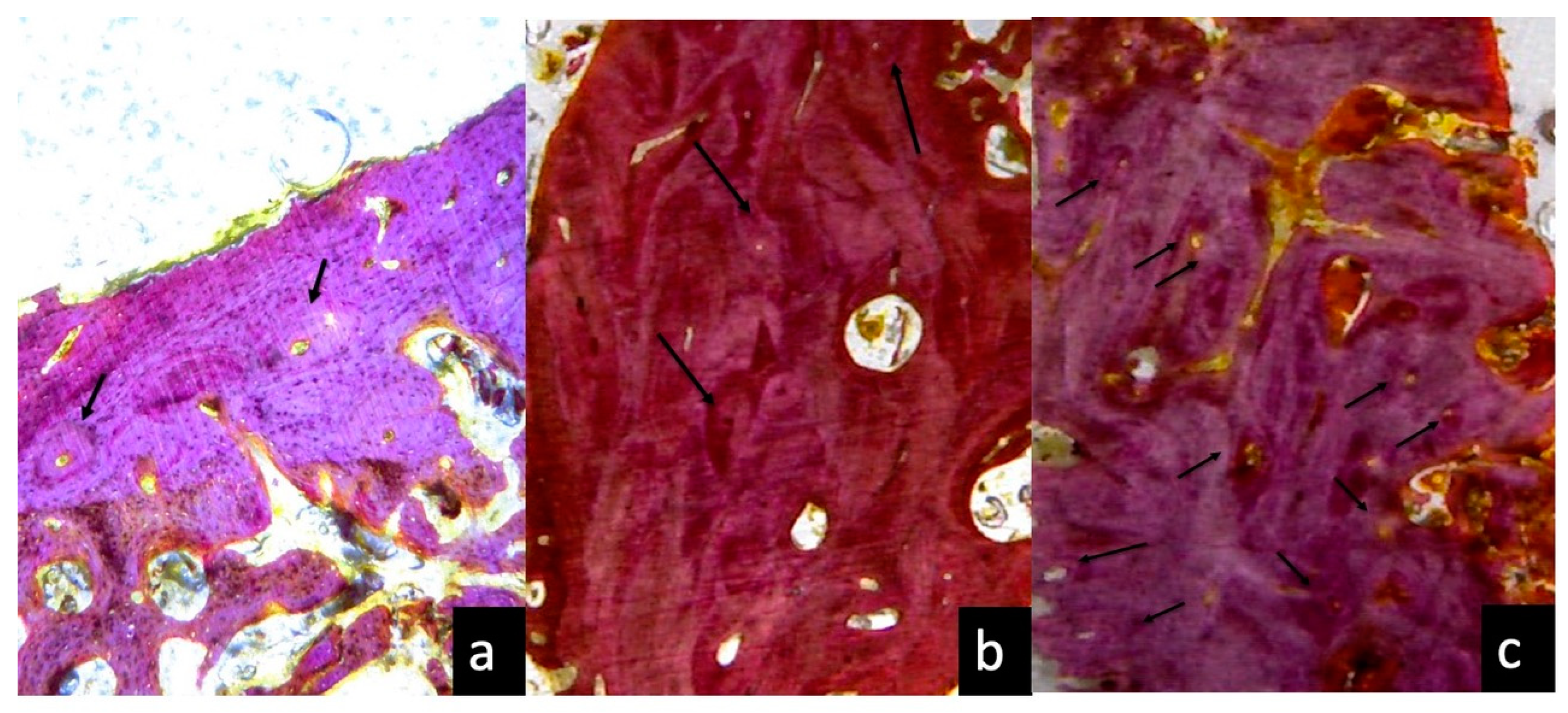
In Figure 9, we can observe at one month, the amount of osteons in melatonin (Figure 9c) and apigenin K groups (Figure 9b) is larger than the control group (Figure 9a).

3.2. One Month Histomorphometric Analysis

After one month of healing, some differences were observed between groups. Higher percentages of new bone (77.87% ± 1.2%) were measured in the melatonin-treated defects, followed by the apigenin K-treated defects (69.81% ± 1.8%) and the control group (57.27% ± 0.54%). Results showed significant differences between the groups (p < 0.05; Figure 10). Meanwhile, the new bone percentages were higher for the test groups (melatonin and apigenin K), and the connective tissue percentages were higher in the control group.

3.3. Two Months Optical Microscopy

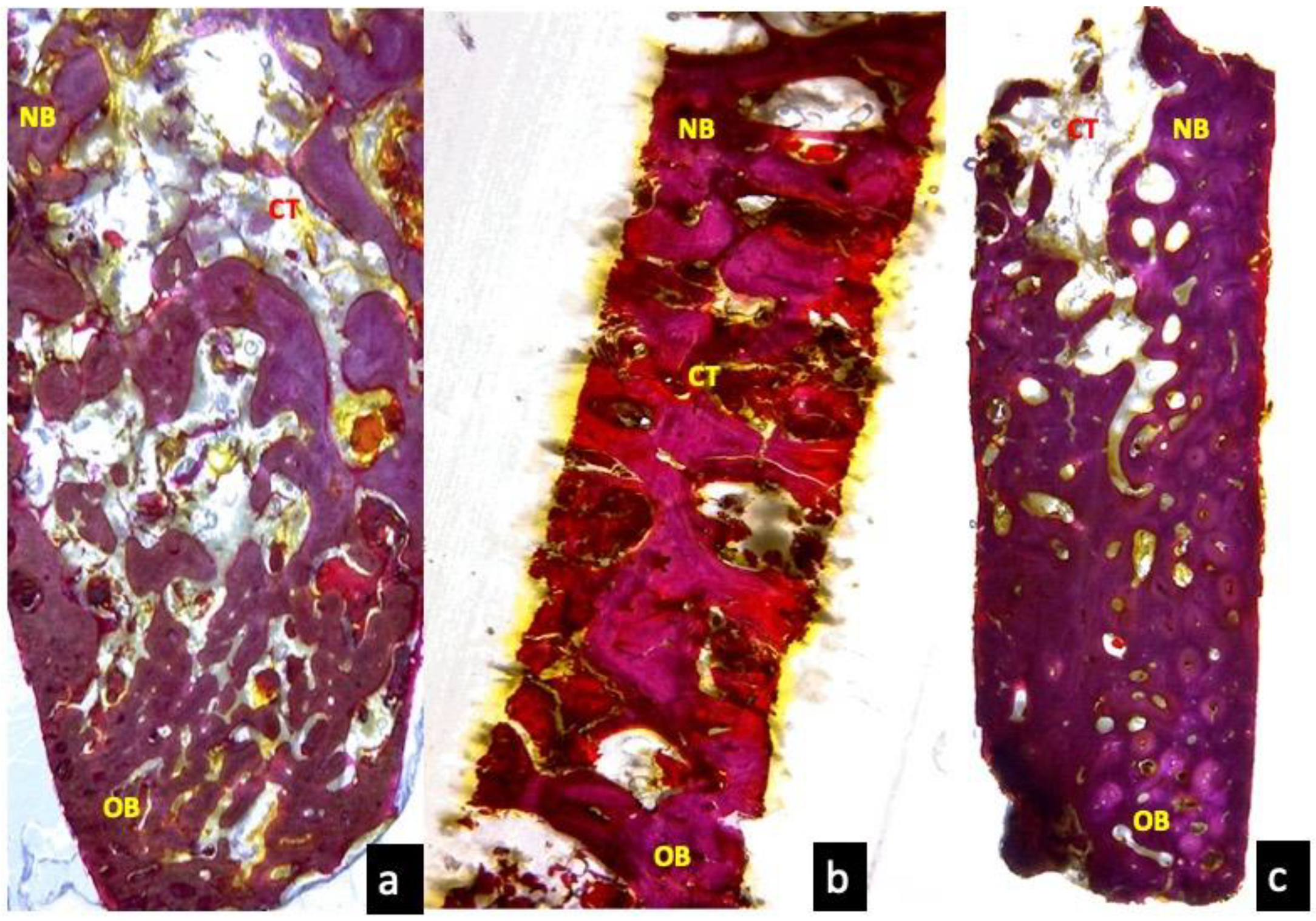
After two months of healing, the control group showed trabeculae bone organization in the middle of the defect compared to the first month. However, the bone was still disorganized in the entire defect (Figure 11a). In the test groups, a higher quantity of organized bone was observed in the middle and the apical area. In the melatonin group, the images showed more mature bone and more organized osteons, followed by these observed in the apigenin K and control groups (Figure 11b,c).

3.4. Two Months Histomorphometric Analysis

After two months, the bone percentages were 79.81% ± 0.11% for the melatonin treated defects, while in the apigenin-treated defects, the bone formation was 71.65% ± 0.52%, both higher compared to the control group (64.77% ± 0.44%). There were statistically significant differences among groups (p < 0.05; Figure 12).

3.5. Three Months Optical Microscopy

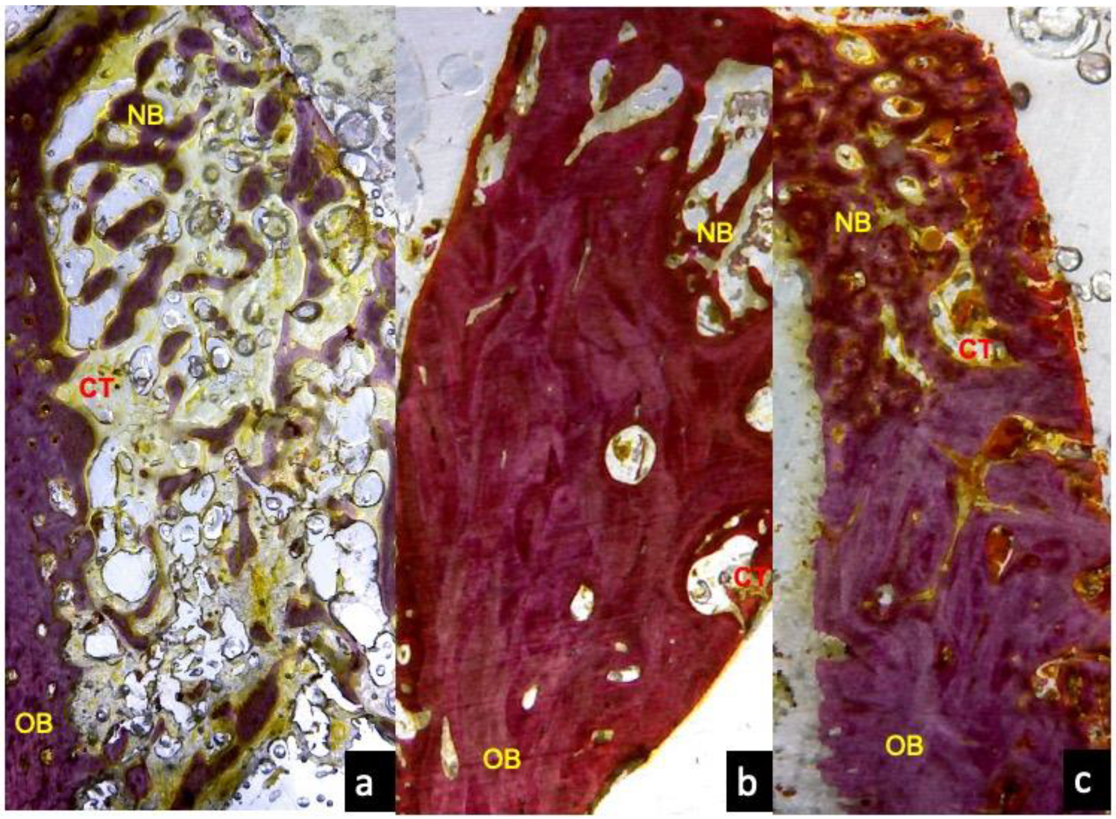
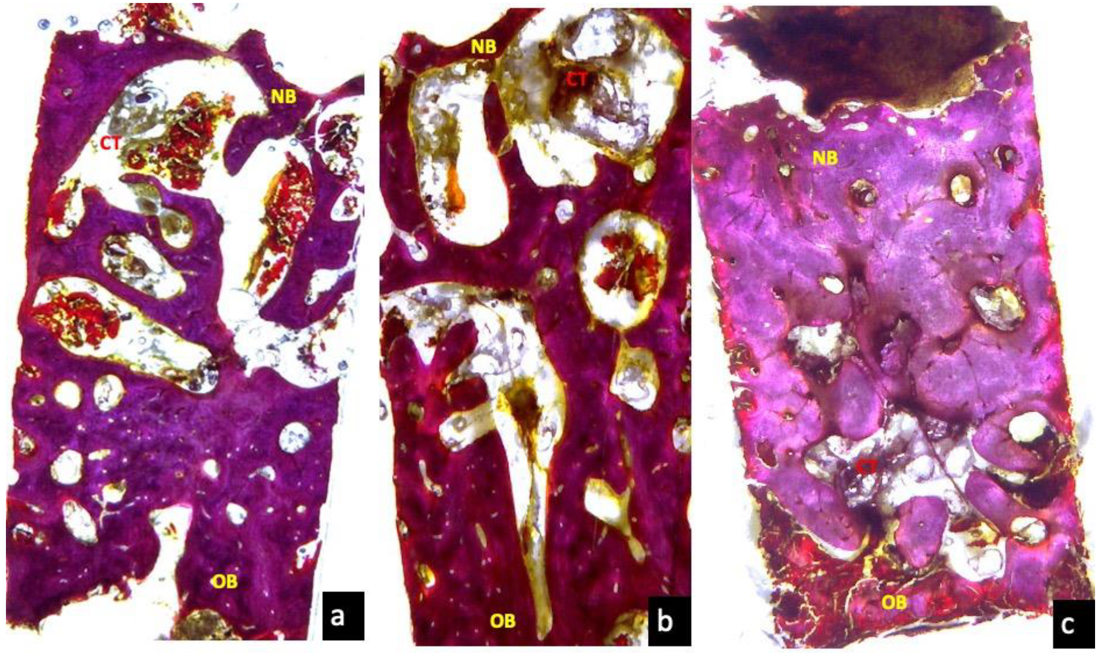
After three months, all groups showed increased mature bone, especially in the melatonin and apigenin K groups. The control group showed nice and stiff bone at the base of the bone core, but on top of it, the trabecular pattern was thin and the medullar spaces wider (Figure 13a). Apigenin K also showed newly formed trabecular bone and marrow spaces in the middle of the bone core (Figure 13b). Furthermore, they presented small areas of disorganized tissue on top of the core. Melatonin stimulated bone maturation, thicker trabeculae, and smaller medullar spaces compared to the other groups (Figure 13c)

3.6. Three Months Histomorphometric Analysis

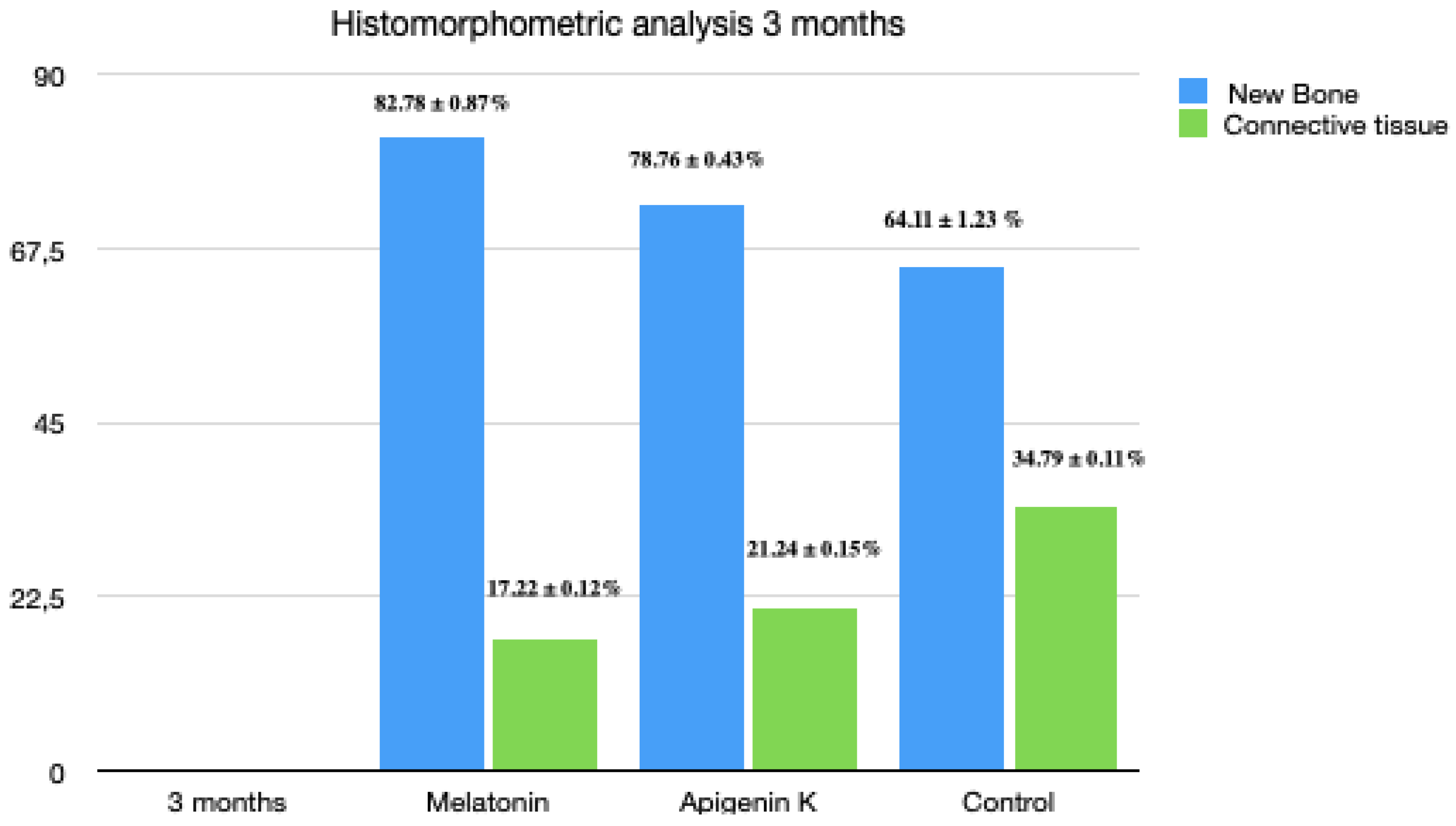
At this period, the new bone was 82.78% ± 0.87% in the melatonin-treated defects, while the apigenin-treated defects, the new bone was 78.76% ± 0.43%, and in the control group, the new bone was 64.11% ± 1.23% (p < 0.05; Figure 14). The behavior of apigenin K and melatonin was very similar at three months follow-up.

4. Discussion

The aim of this experimental study is to determine if apigenin K and melatonin M5250 delivered into post-extraction sockets could stimulate the socket healing at different time frames (1, 2, and 3 months). Our findings suggest that apigenin and melatonin effectively increased the new bone formation during the first month compared to control sites without the bone stimulation substances.
These findings on melatonin can be explained through their effect on the osteoblast, in which it was shown that melatonin stimulates their differentiation [44,45] and can regulate their function as well [18,19,20,21,46,47]. This is produced through a mechanism of acceleration in the mesenchymal stem cell differentiation to osteoblasts [44,45]. Another potential explanation for the stimulation of bone formation observed in the melatonin group was presented by Ramirez-Fernandez et al., who found that melatonin increased the angiogenesis during the first four weeks of bone healing, which explains the numerous endothelial buds and capillary blood vessels observed in our study at the first month in the melatonin group [48,49,50]. Furthermore, it has been shown that melatonin acts on the metabolism of calcium, which could explain the organized bone configuration observed during the second and third month in the melatonin group [51,52]. In addition, the more lamellar appearance in the melatonin group compared to the control group can be explained by the stimulation of the synthesis of type I collagen, and other proteins of the bone matrix [24,25], as well as increased osteocalcin and osteopontin synthesis [48] induced by melatonin. The findings for the apigenin group also showed that apigenin K stimulated more bone formation than controls during the first month, which has been explained based on the effects of the pre-osteoblast differentiation [53,54,55,56] and the reduction of bone loss in ovariectomized rats [57,58,59,60,61]. Besides, several studies have shown that apigenin increases the rate of osteoblastic differentiation [62] because it seems as though apigenin supports the binding of alpha/beta-type estrogen receptors (α, β) [63]. Apigenin increased the processes of differentiation and proliferation of osteoblasts, which can be explained by its effect on the β receptors present in osteoblasts [61,64,65,66].
The morphologic changes appreciated in the melatonin and apigenin K groups compared to the controls at two months (thicker cortical and more lamellar characteristics) are all the global effects of the bone stimulation expressed as an acceleration of the osteoblasts differentiation, reduced bone resorption, increased synthesis of collagen type I, and increased mineralization induced by these two substances [62,67]. It should also be noted that the collagen sponge could have supported the bone formation, but the lack of traces of the collagen sponge at one month indicated that the benefits observed in the bone healing were related to the stimulant substances (apigenin K and melatonin) and not to the collagen sponge.

5. Conclusions

Within the limitations of this experimental study in dogs, it can be concluded that melatonin and apigenin K can accelerate the process of mineralization of the bone matrix, and thus the creation of laminae in the early stages of healing (one month). Less reabsorption of the post-extraction sockets can be expected with the topical application of melatonin and apigenin K. It seems that the stimulatory effects of bone healing induced by the topical application of melatonin and apigenin K are defect-size-dependent, being more evident in small defects compared to larger defects.

Author Contributions

Conceptualization: J.L.C.-G., M.A.G.-V., C.P.A.-M. and F.D.C.-V.; methodology: J.L.C.-G., software, M.F.-D., F.D.C.-V., and R.A.D.-R.; validation, J.M.A., M.C.-C., R.A.D.-R., and S.A.G.; formal analysis, J.L.C.-G. and C.P.A.-M.; investigation, J.M.A. and M.C.-C.; resources, E.F.-B., F.D.C.-V., and R.A.D.-R.; data curation, M.F.-D. and C.P.A.-M.; writing—original draft preparation, J.L.C.-G. and M.C.-C.; writing—review and editing, J.L.C.-G., R.J.-L., and S.A.G.; visualization, E.F.-B., M.C.-C., and R.A.D.-R.; supervision, J.L.C.-G., J.M.A., and M.A.G.-V.; project administration, J.L.C.-G., M.A.G.-V., and R.J.-L.; funding acquisition, J.L.C.-G., E.F.-B., R.J.-L., and R.A.D.-R. All authors have read and agreed to the published version of the manuscript.

Funding

This research received no external funding.

Conflicts of Interest

The authors declare no conflict of interest.

References

  1. Araújo, M.G.; Sukekava, F.; Wennström, J.L.; Lindhe, J. Ridge alterations following implant placement in fresh extraction sockets: An experimental study in the dog. J. Clin. Periodontol. 2005, 32, 645–652. [Google Scholar] [CrossRef] [PubMed]
  2. Araújo, M.G.; Lindhe, J. Ridge alterations following tooth extraction with and without flap elevation: An experimental study in the dog. Clin. Oral Implant. Res. 2009, 20, 545–549. [Google Scholar] [CrossRef] [PubMed]
  3. Januário, A.L.; Duarte, W.R.; Barriviera, M.; Mesti, J.C.; Araújo, M.G.; Lindhe, J. Dimension of the facial bone wall in the anterior maxilla: A cone-beam computed tomography study. Clin. Oral Implant. Res. 2011, 22, 1168–1171. [Google Scholar] [CrossRef] [PubMed]
  4. Botticelli, D.; Berglundh, T.; Lindhe, J. Hard-tissue alterations following immediate implant placement in extraction sites. J. Clin. Periodontol. 2004, 31, 820–828. [Google Scholar] [CrossRef] [PubMed]
  5. Mastrogiacomo, M.; Muraglia, A.; Komlev, V.; Peyrin, F.; Rustichelli, F.; Crovace, A.; Cancedda, R. Tissue engineering of bone: Search for a better scaffold. Orthod. Craniofacial Res. 2005, 28, 277–284. [Google Scholar] [CrossRef] [PubMed]
  6. Mastrogiacomo, M.; Scaglione, S.; Martinetti, R.; Dolcini, L.; Beltrame, F.; Cancedda, R.; Quarto, R. Role of scaffold internal structure on in vivo bone formation in macroporous calcium phosphate bioceramics. Biomaterials 2006, 27, 3230–3237. [Google Scholar] [CrossRef]
  7. Misawa, M.; Lindhe, J.; Araújo, M.G. The alveolar process following single-tooth extraction: A study of maxillary incisor and premolar sites in man. Clin. Oral Implant. Res. 2016, 27, 884–889. [Google Scholar] [CrossRef]
  8. Calvo-Guirado, J.L.; Cegarra Del Pino, P.; Sapoznikov, L.; Delgado Ruiz, R.A.; Fernández-Domínguez, M.; Gehrke, S.A. A new procedure for processing extracted teeth for immediate grafting in post-extraction sockets. An experimental study in American Fox Hound dogs. Ann Anat. 2018, 217, 14–23, Erratum in: Ann Anat. 2018, 10, 218:213. [Google Scholar] [CrossRef]
  9. Maté Sánchez de Val, J.E.; Calvo-Guirado, J.L.; Gómez-Moreno, G.; Gehrke, S.; Mazón, P.; De Aza, P.N. Influence of hydroxyapatite granule size, porosity, and crystallinity on tissue reaction in vivo. Part B: A comparative study with biphasic synthetic biomaterials. Clin. Oral Implant. Res. 2018, 29, 1077–1084. [Google Scholar]
  10. Acuña-Castroviejo, D.; Reiter, R.J.; Menéndez-Peláez, A. Characterization of high-affinity melatonin binding sites in purified cell nuclei of rat liver. J. Pineal Res. 1994, 16, 100–112. [Google Scholar] [CrossRef]
  11. Hirose, T.; Smith, R.J.; Jetten, A.M. ROR gamma: The third member of ROR/RZR orphan receptor subfamily that is highly expressed in skeletal muscle. Biochem. Biophys. Res. Commun. 1994, 205, 1976–1983. [Google Scholar] [CrossRef] [PubMed] [Green Version]
  12. Carlberg, C.; Hooft van Huijsduijnen, R.; Staple, J.K.; DeLamarter, J.F.; Becker-André, M. RZRs, a new family of retinoid-related orphan receptors that function as both monomers and homodimers. Mol. Endocrinol. 1994, 8, 757–770. [Google Scholar] [PubMed] [Green Version]
  13. Cardinali, D.P.; Ladizesky, M.G.; Boggio, V.; Cutrera, R.A.A.; Mautalen, C. Melatonin effects bone: Experimental facts and clinical perspectives. J. Pineal Res. 2003, 34, 81–87. [Google Scholar] [CrossRef] [PubMed]
  14. Macías, M.; Escames, G.; Leon, J.; Coto, A.; Sbihi, Y.; Osuna, A.; Acuña-Castroviejo, D. Calreticulin–melatonin. An unexpected relationship. Eur. J. Biochem. 2003, 270, 832–840. [Google Scholar] [CrossRef] [PubMed] [Green Version]
  15. Suzuki, N.; Somei, M.; Seki, A.; Reiter, R.J.; Hattori, A. Novel bromomelatonin derivatives as potentially effective drugs to treat bone diseases. J. Pineal Res. 2008, 45, 229–234. [Google Scholar] [CrossRef] [Green Version]
  16. Roth, J.A.; Kim, B.G.; Lin, W.L.; Cho, M.I. Melatonin promotes osteoblast differentiation and bone formation. J. Biol. Chem. 1999, 274, 22041–22047. [Google Scholar] [CrossRef] [Green Version]
  17. Galano, A.; Reiter, R.J. Melatonin and its metabolites vs oxidative stress: From individual actions to collective protection. J Pineal Res. 2018, 65, e12514. [Google Scholar] [CrossRef] [Green Version]
  18. Calvo-Guirado, J.L.; Gómez-Moreno, G.; Barone, A.; Cutando, A.; Alcaraz-Baños, M.; Chivas, F.; López-Marí, L.; Guardia, J. Melatonin plus porcine bone on discrete calcium deposit implant surface stimulates osteointegration in dental implants. J. Pineal Res. 2009, 47, 164–172. [Google Scholar] [CrossRef]
  19. Calvo-Guirado, J.L.; Gómez-Moreno, G.; López-Marí, L.; Guardia, J.; Martínez-González, J.M.; Barone, A.; Tresguerres, I.F.; Paredes, S.D.; Fuentes-Breto, L. Actions of melatonin mixed with collagenized porcine bone versus porcine bone only on osteointegration of dental implants. J. Pineal Res. 2010, 48, 194–203. [Google Scholar] [CrossRef]
  20. Calvo-Guirado, J.L.; Ramírez-Fernández, M.P.; Gómez-Moreno, G.; Maté-Sánchez, J.E.; Delgado-Ruiz, R.; Guardia, J.; López-Marí, L.; Barone, A.; Ortiz-Ruiz, A.J.; Martínez-González, J.M.; et al. Melatonin stimulates the growth of new bone around implants in the tibia of rabbits. J. Pineal Res. 2010, 49, 356–363. [Google Scholar] [CrossRef]
  21. Tan, D.X.; Manchester, L.C.; Terron, M.P.; Flores, L.J.; Reiter, R.J. One molecule, many derives: A never ending interaction of melatonin with reactive oxygen and reactive nitrogen species? J. Pineal Res. 2007, 42, 28–42. [Google Scholar] [CrossRef] [PubMed]
  22. Tan, W.L.; Wong, T.L.T.; Wong, M.C.M.; Lang, N.P. A systematic review of post-extractional alveolar hard and soft tissue dimensional changes in humans. Clin. Oral Implant. Res. 2012, 23, 1–21. [Google Scholar] [CrossRef] [PubMed]
  23. Liu, J.; Huang, F.; He, H.W. Melatonin effects on hard tissues: Bone and tooth. Int. J. Mol. Sci. 2013, 14, 10063–10074. [Google Scholar] [CrossRef] [PubMed] [Green Version]
  24. Cutando, A.; Gómez-Moreno, G.; Arana, C.; Acuña-Castroviejo, D.; Reiter, R.J. Melatonin: Potential functions in the oral cavity. J. Periodontology 2007, 78, 1094–1102. [Google Scholar] [CrossRef]
  25. Cutando, A.; Aneiros-Fernández, J.; López-Valverde, A.; Arias-Santiago, S.; Aneiros-Cachaza, J.; Reiter, R.J. A new perspective in Oral health: Potential importance and actions of melatonin receptors MT1, MT2, MT3, and RZR/ROR in the oral cavity. Arch. Oral Biol. 2011, 56, 944–950. [Google Scholar] [CrossRef]
  26. Najeeb, S.; Khurshid, Z.; Zohaib, S.; Zafar, M.S. Therapeutic potential of melatonin in oral medicine and periodontology. Kaohsiung J. Med. Sci. 2016, 32, 391–396. [Google Scholar] [CrossRef]
  27. Abdel Moneim, A.E.; Guerra-Librero, A.; Florido, J.; Shen, Y.Q.; Fernández-Gil, B.; Acuña-Castroviejo, D.; Escames, G. Oral Mucositis: Melatonin Gel an Effective New Treatment. Int. J. Mol. Sci. 2017, 7, 18. [Google Scholar] [CrossRef]
  28. Guerrero-Gironés, J.; Alcaina-Lorente, A.; Ortiz-Ruiz, C.; Ortiz-Ruiz, E.; Pecci-Lloret, M.P.; Rodríguez-Lozano, F.J.; Martínez, C.M.; Ortiz-Ruiz, A.J. Melatonin as an Agent for Direct Pulp-Capping Treatment. Int. J. Env. Res. Public Health 2020, 6, 17. [Google Scholar] [CrossRef] [Green Version]
  29. Stanciu, A.E.; Zamfir-Chiru-Anton, A.; Stanciu, M.M.; Stoian, A.P.; Jinga, V.; Nitipir, C.; Bucur, A.; Pituru, T.S.; Arsene, A.L.; Dragoi, C.M.; et al. Clinical significance of serum melatonin in predicting the severity of oral squamous cell carcinoma. Oncol. Lett. 2020, 19, 1537–1543. [Google Scholar] [CrossRef] [Green Version]
  30. Permuy, M.; López-Peña, M.; González-Cantalapiedra, A.; Muñoz, F. Melatonin: A Review of Its Potential Functions and Effects on Dental Diseases. Int. J. Mol. Sci. 2017, 18, E865. [Google Scholar] [CrossRef] [Green Version]
  31. Lee, J.A.; Ha, S.K.; Cho, E.; Choi, I. Resveratrol as a Bioenhancer to Improve Anti-Inflammatory Activities of Apigenin. Nutrients 2015, 7, 9650–9661. [Google Scholar] [CrossRef] [PubMed] [Green Version]
  32. Nakamura, H.; Ukai, T.; Yoshimura, A.; Kozuka, Y.; Yoshioka, H.; Yoshinaga, Y.; Abe, Y.; Hara, Y. Green tea catechin inhibits lipopolysaccharide-induced bone resorption in vivo. J. Periodontal Res. 2010, 45, 23–30. [Google Scholar] [CrossRef]
  33. Barreca, D.; Mandalari, G.; Calderaro, A.; Smeriglio, A.; Trombetta, D.; Felice, M.R.; Gattuso, G. Citrus Flavones: An Update on Sources, Biological Functions, and Health Promoting Properties. Plants 2020, 9, E288. [Google Scholar] [CrossRef] [PubMed] [Green Version]
  34. Sánchez-Marzo, N.; Pérez-Sánchez, A.; Ruiz-Torres, V.; Martínez-Tébar, A.; Castillo, J.; Herranz-López, M.; Barrajón-Catalán, E. Antioxidant and Photoprotective Activity of Apigenin and its Potassium Salt Derivative in Human Keratinocytes and Absorption in Caco-2 Cell Monolayers. Int. J. Mol. Sci. 2019, 20, E2148. [Google Scholar] [CrossRef] [PubMed] [Green Version]
  35. Gutiérrez-Venegas, G.; Gómez-Mora, J.A.; Meraz-Rodríguez, M.A.; Flores-Sánchez, M.A.; Ortiz-Miranda, L.F. Effect of flavonoids on antimicrobial activity of microorganisms present in dental plaque. Heliyon 2019, 5, e03013. [Google Scholar] [CrossRef] [PubMed] [Green Version]
  36. La, V.D.; Tanabe, S.; Grenier, D. Naringenin inhibits human osteoclastogenesis and osteoclastic bone resorption. J. Periodontal Res. 2009, 44, 193–198. [Google Scholar] [CrossRef] [PubMed]
  37. Leyva-López, N.; Gutierrez-Grijalva, E.P.; Ambriz-Perez, D.L.; Heredia, J.B. Flavonoids as Cytokine Modulators: A Possible Therapy for Inflammation-Related Diseases. Int. J. Mol. Sci. 2016, 17, E921. [Google Scholar] [CrossRef] [PubMed]
  38. Salehi, B.; Venditti, A.; Sharifi-Rad, M.; Kręgiel, D.; Sharifi-Rad, J.; Durazzo, A.; Lucarini, M.; Santini, A.; Souto, E.B.; Novellino, E.; et al. The therapeutic potential of apigenin. Int. J. Mol. Sci. 2019, 20, 1305. [Google Scholar] [CrossRef] [Green Version]
  39. Hasnat, M.A.; Pervin, M.; Lim, J.H.; Lim, B.O. Apigenin Attenuates Melanoma Cell Migration by Inducing Anoikis through Integrin and Focal Adhesion Kinase Inhibition. Molecules 2015, 20, 21157–21166. [Google Scholar] [CrossRef]
  40. Park, J.A.; Ha, S.K.; Kang, T.H.; Oh, M.S.; Cho, M.H.; Lee, S.Y.; Park, J.H.; Kim, S.Y. Protective effect of apigenin on ovariectomy-induced bone loss in rats. Life Sci. 2008, 20, 1217–1223. [Google Scholar] [CrossRef]
  41. Hardeland, R. Melatonin and inflammation-Story of a double-edged blade. J Pineal Res. 2018, 65, e12525. [Google Scholar] [CrossRef] [PubMed] [Green Version]
  42. Benavente-García, O.; Castillo, J. Update on uses and properties of citrus flavonoids: New findings in anticancer, cardiovascular, and anti-inflammatory activity. J. Agric. Food Chem. 2008, 13, 6185–6205. [Google Scholar] [CrossRef] [PubMed]
  43. Lee, H.H.; Jung, J.; Moon, A.; Kang, H.; Cho, H. Antitumor and Anti-Invasive Effect of Apigenin on Human Breast Carcinoma through Suppression of IL-6 Expression. Int. J. Mol. Sci. 2019, 27, 20. [Google Scholar] [CrossRef] [PubMed] [Green Version]
  44. Strewler, G.J. Local and systemic control of the osteoblast. J. Clin. Investig. 2001, 107, 271–272. [Google Scholar] [CrossRef] [Green Version]
  45. Zhou, Y.; Wang, C.; Si, J.; Wang, B.; Zhang, D.; Ding, D.; Zhang, J.; Wang, H. Melatonin up-regulates bone marrow mesenchymal stem cells osteogenic action but suppresses their mediated osteoclastogenesis via MT2-inactivated NF-κB pathway. Br. J. Pharmacol. 2020, 177, 2106–2122. [Google Scholar] [CrossRef]
  46. Omori, Y.; Iezzi, G.; Perrotti, V.; Piattelli, A.; Ferri, M.; Nakajima, Y.; Botticelli, D. Influence of the Buccal Bone Crest Width on Peri-Implant Hard and Soft Tissues Dimensions: A Histomorphometric Study in Humans. Implant. Dent. 2018, 27, 415–423. [Google Scholar] [CrossRef]
  47. Maria, S.; Samsonraj, R.M.; Munmun, F.; Glas, J.; Silvestros, M.; Kotlarczyk, M.P.; Rylands, R.; Dudakovic, A.; van Wijnen, A.J.; Enderby, L.T.; et al. Biological effects of melatonin on osteoblast/osteoclast cocultures, bone, and quality of life: Implications of a role for MT2 melatonin receptors, MEK1/2, and MEK5 in melatonin-mediated osteoblastogenesis. J. Pineal Res. 2018, 64, e12465. [Google Scholar] [CrossRef]
  48. Martínez-Sanz, E.; Ossipov, D.A.; Hilborn, J.; Larsson, S.; Jonsson, K.B.; Varghese, O.P. Bone reservoir: Injectable hyaluronic acid hydrogel for minimal invasive bone augmentation. J. Control. Release 2011, 152, 230–240. [Google Scholar] [CrossRef]
  49. Ramírez-Fernández, M.P.; Calvo-Guirado, J.L.; de-Val, J.E.; Delgado-Ruiz, R.A.; Negri, B.; Pardo-Zamora, G.; Peñarrocha, D.; Barona, C.; Granero, J.M.; Alcaraz-Baños, M. Melatonin promotes angiogenesis during repair of bone defects: A radiological and histomorphometric study in rabbit tibiae. Clin. Oral Investig. 2013, 17, 147–158. [Google Scholar] [CrossRef]
  50. Gómez-Moreno, G.; Guardia, J.; Ferrera, M.J.; Cutando, A.; Reiter, R.J. Melatonin in diseases of the oral cavity. Oral Dis. 2010, 16, 242–247. [Google Scholar] [CrossRef]
  51. Witt-Enderby, P.A.; Radio, N.M.; Doctor, J.S.; Davis, V.L. Therapeutic treatments potentially mediated by melatonin receptors: Potential clinical uses in the prevention of osteoporosis, cancer and as an adjuvant therapy. J. Pineal Res. 2006, 41, 297–305. [Google Scholar] [CrossRef] [PubMed]
  52. Salomó-Coll, O.; de Maté-Sánchez, J.E.; Ramírez-Fernandez, M.P.; Hernández-Alfaro, F.; Gargallo-Albiol, J.; Calvo-Guirado, J.L. Osseoinductive elements around immediate implants for better osteointegration: A pilot study in foxhound dogs. Clin. Oral Implant. Res. 2018, 29, 1061–1069. [Google Scholar] [CrossRef] [PubMed]
  53. Guardia, J.; Gómez-Moreno, G.; Ferrera, M.J.; Cutando, A. Evaluation of effects of topic melatonin on implant surface at 5 and 8 weeks in Beagle dogs. Clin. Implant. Dent. Relat. Res. 2011, 13, 262–268. [Google Scholar] [CrossRef] [PubMed]
  54. Ito, K.; Minegishi, T.; Takayama, T.; Tamura, T.; Yamada, Y.; Sato, S. Effects of ipriflavone on augmented bone using a guided bone regeneration procedure. Clin. Oral Implant. Res. 2007, 18, 60–68. [Google Scholar] [CrossRef] [PubMed]
  55. Guskuma, M.H.; Hochuli-Vieira, E.; Pereira, F.P.; Rangel-Garcia Junior, I.; Okamoto, R.; Okamoto, T.; Magro-Filho, O. Bone regeneration in surgically created defects filled with autogenous bone: An epifluorescence microscopy analysis in rats. J. Appl. Oral Sci. 2010, 18, 346–353. [Google Scholar] [CrossRef] [PubMed]
  56. Popović, M.; Caballero-Bleda, M.; Benavente-García, O.; Castillo, J. The flavonoid apigenin delays forgetting of passive avoidance conditioning in rats. J. Psychopharmacol. 2014, 28, 498–501. [Google Scholar] [CrossRef]
  57. Ginwala, R.; Bhavsar, R.; Chigbu, D.I.; Jain, P.; Hhan, Z.K. Potential Role of Flavonoids in Treating Chronic Inflammatory Diseases with a Special Focus on the Anti-Inflammatory Activity of Apigenin. Antioxidants 2019, 8, E35. [Google Scholar] [CrossRef] [Green Version]
  58. Songlin, P.; Ge, Z.; Yixin, H.; Xinluan, W.; Pingchung, L.; Kwoksui, L.; Ling, Q. Epimedium-derived flavonoids promote osteoblastogenesis and suppress adipogenesis in bone marrow stromal cells while exerting an anabolic effect on osteoporotic bone. Bone 2009, 45, 534–544. [Google Scholar] [CrossRef]
  59. Maki, K.; Nishida, I.; Mitsutaka, K. The effect of oral ipriflavone on the rat mandible during growth. Eur. J. Orthod. 2005, 27, 27–31. [Google Scholar] [CrossRef]
  60. Ostrowska, Z.; Kos-Kudla, B.; Nowak, M.; Swietochowska, E.; Marek, B.; Gorski, J.; Kajdaniuk, D.; Wolkowska, K. The relationship between bone metabolism, melatonin and other hormones in sham-operated and pinealectomized rats. Endocr. Regul. 2003, 37, 211–224. [Google Scholar]
  61. Rha, C.S.; Jeong, H.W.; Park, S.; Lee, S.; Jung, Y.S.; Kim, D.O. Antioxidative, Anti-Inflammatory, and Anticancer Efects of Purified Flavonol Glycosides and Aglycones in Green Tea. Antioxidants 2019, 8, 278. [Google Scholar] [CrossRef] [PubMed] [Green Version]
  62. Siddiqui, J.A.; Swarnkar, G.; Sharan, K.; Chakravarti, B.; Sharma, G.; Rawat, P.; Kumar, M.; Khan, F.M.; Pierroz, D.; Maurya, R.; et al. 8,8″-Biapigeninyl stimulates osteoblast functions and inhibits osteoclast and adipocyte functions: Osteoprotective action of 8,8″-biapigeninyl in ovariectomized mice. Mol. Cell Endocrinol. 2010, 29, 256–267. [Google Scholar] [CrossRef] [PubMed]
  63. Bandyopadhyay, S.; Lion, J.M.; Mentaverri, R.; Ricupero, D.A.; Kamel, S.; Romero, J.R.; Chattopadhyay, N. Attenuation of osteoclastogenesis and osteoclast function by apigenin. Biochem. Pharmacol. 2006, 14, 184–197. [Google Scholar] [CrossRef] [PubMed]
  64. Choi, E.J.; Kim, G.H. Apigenin Induces Apoptosis through a Mitochondria/Caspase-Pathway in Human Breast Cancer MDA-MB-453 Cells. J. Clin. Biochem. Nutr. 2009, 44, 260–265. [Google Scholar] [CrossRef] [Green Version]
  65. Venturelli, S.; Burkard, M.; Biendl, M.; Lauer, U.M.; Frank, J.; Busch, C. Prenylated chalcones and flavonoids for the prevention and treatment of cancer. Nutrition 2016, 32, 1171–1178. [Google Scholar] [CrossRef]
  66. Mak, P.; Leung, Y.K.; Tang, W.Y.; Harwood, C.; Ho, S.M. Apigenin suppresses cancer cell growth through ERbeta. Neoplasia 2006, 8, 896–904. [Google Scholar] [CrossRef] [Green Version]
  67. Agati, G.; Azzarello, E.; Pollastri, S.; Tattini, M. Flavonoids as antioxidants in plants: Location and functional significance. Plant Sci. 2012, 196, 67–76. [Google Scholar] [CrossRef]
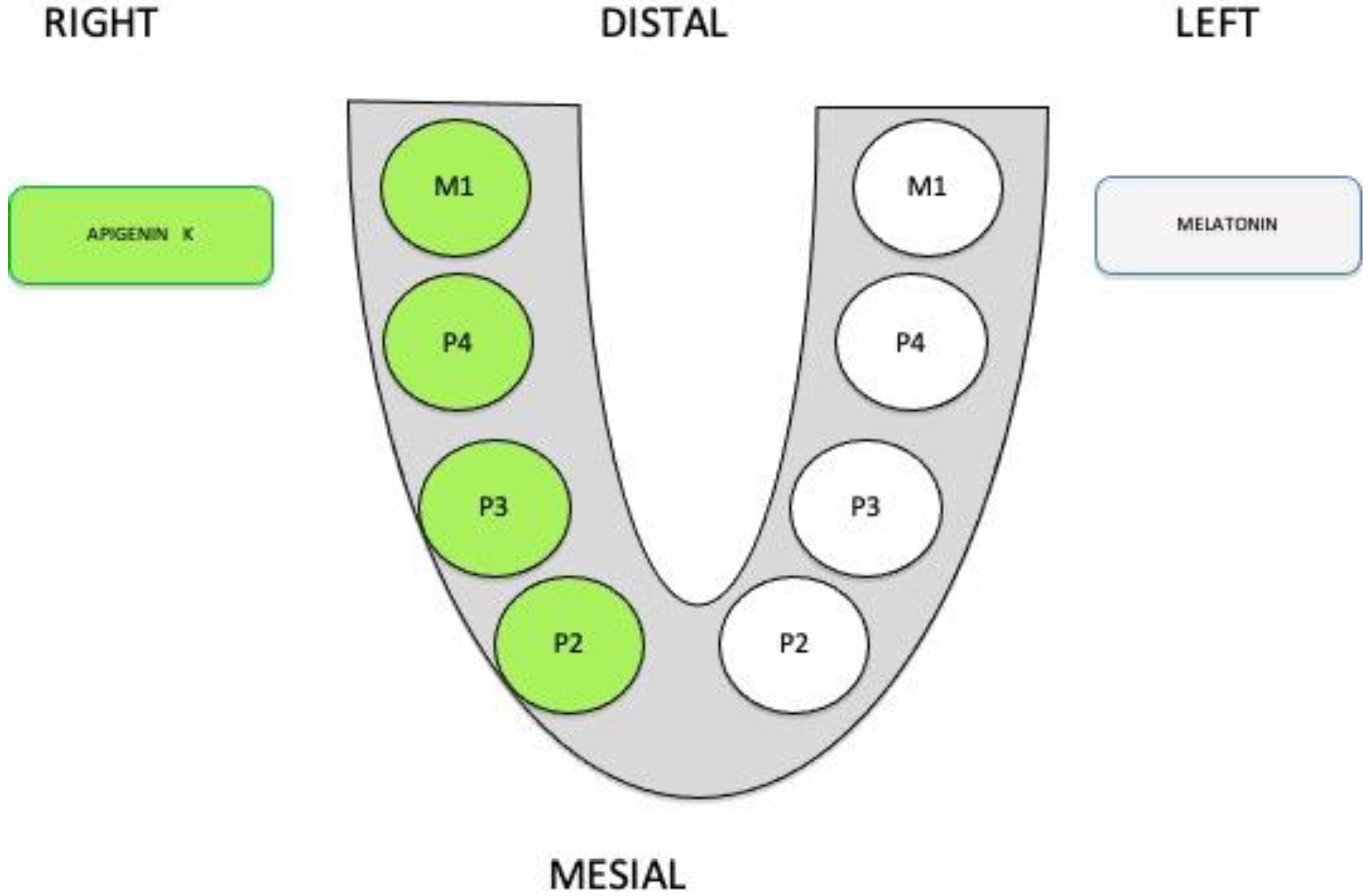
Figure 1. Apigenin K was placed on the right side and melatonin M5250 was placed on the opposite side of the dog’s mandible. P2 second premolar, P3 third premolar, P4 forth preomolar and M1 first molar.
Applsci 10 03006 g001
Figure 2. (a) Premolars and molar sectioned in buccal-lingual direction before extraction; (b) Empty alveolar sockets of mandibular P2, P3, P4, and M1 (premolars and first molar).
Applsci 10 03006 g002
Figure 3. (a) A collagen sponge soaked into yellow apigenin; (b) white collagen sponge soaked in melatonin.
Applsci 10 03006 g003
Figure 4. (a) Apigenin K placed in all sockets except Premolar 2; (b) melatonin sponges in place, leaving the P2 area empty as a control.
Applsci 10 03006 g004
Figure 5. (a): Right side healing bone after apigenin K use; (b): Left side of healing bone after melatonin placement.
Applsci 10 03006 g005
Figure 6. (a) The biopsies were taken with a trephine bur of 2.75 mm diameter after 3 months of healing; (b) 10-mm length of a healed bone sample.
Applsci 10 03006 g006
Figure 7. This image represents all the measurements of no bone formation (black landmarks) compared with total bone core (black landmark).
Applsci 10 03006 g007
Figure 8. (a) Biopsies of post-extraction sockets at 30 days at control group with less bone formation; (b) core sample of apigenin K, showing newly bone formation at the base of the core and less organization on top of it; (c) melatonin core after 30 days with the high bone formation with red dots that show an increase in vascularization. Staining made by picrosirius–hematoxylin. The images have been captured with the objective of 200X. NB: new bone; OB: old bone; CT: connective tissue.
Applsci 10 03006 g008
Figure 9. High magnification of control group (a), apigenin K group (b), and melatonin group (c) at the one-month follow-up. Great amount of osteons in the melatonin group were observed compared with apigenin K and control groups. The images have been captured with the objective of 500X. Black arrows represent the number of osteons formed.
Applsci 10 03006 g009
Figure 10. New bone and connective tissue at the one-month evaluation. The level of significance was set at p < 0.05. Non-parametric Man–Whitney test was applied.
Applsci 10 03006 g010
Figure 11. (a) Control group at 2 months with organized areas in the base area and disorganized areas on top of the core; (b) apigenin K bone core with an established well-organized bone; (c) melatonin group showed well-organized bone of the whole sample. Staining made by picrosirius–hematoxylin. The images have been captured with the objective of 200X. CT: connective tissue; OB: old bone; NB: new bone.
Applsci 10 03006 g011
Figure 12. Bone and connective tissue formation at 2 months of evaluation. The level of significance was set at p < 0.05. Non-parametric Man–Whitney test was applied.
Applsci 10 03006 g012
Figure 13. (a) Control group; (b) apigenin K group; (c) melatonin group. Staining made by picrosirius–hematoxylin. The images have been captured with the objective of 200X. OB: Old bone; NB: new bone; CT: Connective tissue.
Applsci 10 03006 g013
Figure 14. Connective tissue and new bone formation at the 3-month study time.
Applsci 10 03006 g014