Article

A Comparative Study of Virgin Coconut Oil, Coconut Oil and Palm Oil in Terms of Their Active Ingredients

by
Suryani Suryani
1,*,
Sariani Sariani
2,
Femi Earnestly
1,
Marganof Marganof
3,
Rahmawati Rahmawati
4,
Sevindrajuta Sevindrajuta
4,
Teuku Meurah Indra Mahlia
5 and
Ahmad Fudholi
6
1
Department of Chemistry, Faculty of Science and Technology, Muhammadiyah University of West Sumatera, Padang 25171, West Sumatera, Indonesia
2
Department of English, Politeknik Negeri Padang, Padang 2500, West Sumatera, Indonesia
3
Department of Forestry, Faculty of Foresttry, Muhammadiyah University of West Sumatera, Padang 25171, West Sumatera, Indonesia
4
Department of Agro Technology, Faculty of Agriculture, Muhammadiyah University of West Sumatera, Padang 25171, West Sumatera, Indonesia
5
School of Information, Systems and Modelling, Faculty of Engineering and Information Technology, University of Technology Sydney, Sydney NSW 2007, Australia
6
Solar Energy Research Institute, Universiti Kebangsaan Malaysia, 43600 UKM Bangi, Malaysia
*
Author to whom correspondence should be addressed.
Processes 2020, 8(4), 402; https://doi.org/10.3390/pr8040402
Submission received: 21 November 2019 / Revised: 14 March 2020 / Accepted: 19 March 2020 / Published: 30 March 2020
(This article belongs to the Section Environmental and Green Processes)

Abstract

:
This research aims to study the unique factors of virgin coconut oil (VCO) compared with coconut oil (i.e., coconut oil processed through heating the coconut milk and palm oil sold on the market). Its novelty is that it (VCO) contains lactic acid bacteria and bacteriocin. Lauric acid content was analyzed by the Chromatographic Gas method. Isolation of lactic acid bacteria (LAB) was conducted by the dilution method using MRSA + 0.5% CaCO3 media. Iodium number, peroxide, and %FFA were analyzed using a general method, and isolation bacteriocin by the deposition method using ammonium sulfate. In addition, macromolecular identification was conducted by 16S rRNA. VCO was distinguished by a higher content of lauric acid (C12:0) 41%–54.5% as compared with 0% coconut and 0, 1% palm oil, respectively. The VCO also contains LAB, namely Lactobacillus plantarum and Lactobacillus paracasei, and can inhibit the growth of pathogenic bacteria, such as Pseudomonas aeruginosa, Klebsiella, Staphylococcus aureus, S. epidermidis, Proteus, Escherichia coli, Listeria monocytogenes, Bacillus cereus, Salmonella typhosa and bacteriocin. Comparison with VCO is based on having a high content of lauric acid, 54%, and LAB content. The difference between VCO and coconut oil and palm oil is fatty acids. In VCO there are lauric acid and stearic acid, namely lauric acid VCO (A) 54.06%, VCO (B) 53.9% and VCO (C) 53.7%. The content of stearic acid VCO (A) is 12.03%, VCO (B) 12.01% and VCO (C) 11.9%. Coconut oil contains a little lauric acid, which is 2.81%, stearic acid 2.65% and palmitic acid 2.31%. Palm oil can be said to have very little lauric acid, namely in palm oil 1, 0.45%, and even in palm oil 2, 0%; in turn, palmitic acid palm oil 1 has 2.88% and palm oil 2 palmitic acid has 24.42%.

1. Introduction

Virgin coconut oil (VCO) can be made through several methods, such as by fermenting coconut milk [1,2,3] and by adding microbes (Lactobacillus fermentum and Lactobacillus plantarum) as starter cultures [4,5]. VCO can also be produced through centrifugation [6] and microwave processes [7], and by fermentation without the addition of microbes as a starter [8]. This oil is called virgin oil because it is made without any heating [9].
VCO has been used widely because it is believed to have benefits compared to coconut oil, made through a heating process, and palm oil. VCO is useful against microbes, bacteria and viruses [10], and is useful for helping one lose weight in terms of metabolism. VCO contains medium chain triglycerides [11,12], which is initially digested or processed in the body from carbohydrates that can cut back hunger [13]. Thus, it causes people to consume less carbohydrates, which eventually reduces body weight [14,15]. VCO also affects the healing after an ovariectomy [16] and can be used as an antioxidant [17]. VCO can also reduce blood pressure. In addition, VCO can also be used for skincare [18,19,20], as an external drug, such as wound medicine, and can function as a probiotic [4,21,22,23].
VCO and palm oil have different characteristics with different functions [24,25,26]. VCO gravitates more towards medicine, probiotics and cosmetics, whereas palm oil characteristics are quite suitable to be converted to diesel fuel. Other vegetable oils than palm oil have also been proven to be economical and can be converted to biodiesel production at a large scale [27,28,29]. The chemical composition of VCO has been studied in including the iodine number, the saponification number, the amount of free fatty acids (%FFA), viscosity and color. This chemical composition needs to be examined prior to consumption in order to follow Asian and Pacific Coconut Community (APCC) standardization, because VCO is originally made from fresh coconut milk [30].
VCO has been known for its high lauric acid content, which is between 46.36% and 48.42% [31] Processed from coconut milk, which is high in carbohydrates and proteins, the fermentation process of VCO results in lactic acid bacteria (LAB) [32,33,34,35]. LAB from VCO have been isolated and their antimicrobial ability also has been studied [36]. This atypical microbial ability exists because LAB contains bacteriocin, which can kill pathogenic bacteria. Further, bacteriocin from Lactobacillus plantarum [34,35,37,38] has been isolated as well. Thus, the aim of the research was comparison of the psychochemical parameters and contents of the fatty acids (lauric, palmitic and stearic) of coconut oil, palm oil and VCO as indicators of the characteristics of each oil. The antimicrobial ability of VCO was also analyzed.
The newest fact referring to VCO, which is yet to be acknowledged, is the presence of lactic acid bacteria (LAB) in the oil and blondo layers (VCO dregs). This LAB will be present when VCO is made through traditional fermentation processes or fermentation using the existing bacteria in the air [36].

2. Materials and Methods

2.1. Materials

The samples were three types of virgin coconut oils, with different methods of extraction/fermentation, coconut oil derived from heating coconut milk and consumer-grade palm oil.

2.1.1. Chemical Material

For lauric acid analysis, -hexane (p.a), CHCl3 and Aquadest reagent for sample preparation, namely saturated NaCl, Na2SO4 anhydrate, BF3, methanol and N2gas to stop the oxidation occurring, were used. The internal fatty acid standard was used. Whereas, for acid number analysis (%FFA), 95% alcohol (pa), blue bromothymol indicator, phenolphthalein indicator, HCl 0.5 N standard solution and KOH standard solution were used. KOH ethanolic, HCl 0.5 N standard solution was taken for saponification number analysis, and potassium iodide was used for peroxide number analysis. Meanwhile MRSA (deMann Rogosa Sharpe Agar) p.a + 0.5% CaCO3 media was used for isolation of lactic acid bacteria and in order to increase their amount using MRS media. The materials used for isolation bacteriocin were MRSB media (Merck), Lactobacillus plantarum M0, ammonium sulfate (NH4) 2SO4, phosphate buffer, DEAE cellulose, butanol, SDS stacking gel and markers.

2.1.2. Instruments

The instrument used where ordinary laboratory glassware, such as petri dishes, Erlenmeyer flasks, test tubes and beaker glass, all of them made by Pyrex. In addition, gas chromatography (GC) GC–MS Shimadzu QP 2010S (Shimadzu Corp, Kyoto, Japan) was set for lauric acid analysis and Autocklav Yamata SN 21 for sterilization and laminar flow as the working media for isolating the bacteria and for performing antimicrobial analysis.

2.2. The Standardization of the VCO

2.2.1. The Determination of pH

The pH of the sample is determined using the pH meter in accordance with the applicable general procedure.

2.2.2. The Analysis of Fatty Acid Sample Using GC–MS

Prior to injecting the sample into a GC–MS instrument, the oil sample was prepared by setting 50 g of VCO sample and adding 400 µL of NaOH Metanolic. This mixture was vortexed and heated at 50 °C for 10 min. After undergoing the cooling process, 1 mL CH3COOH, 1 mL distilled water and 1 mL n-hexane were added, respectively. Then this mixture was vortexed and cooled for several minutes where two layers were formed as the result.
About 1 µL at the top layer was taken as the sample to be injected and analyzed in a GC–MS, Shimadzu QP2010 which was equipped with capillary column of (30 m) × 0.25 mm ID; 0.25 ìm (interspersed by DB5MS. Japan), by injecting the sample into the capillary column. The carrier gas was helium, where the injector and detector temperatures were set at 280 °C. The injection was performed using the split mode (1:30). The column temperature was programmed to change from 50 °C to 280 °C at a rate of 5 °C per minute. Fatty acid ethyl esters were separated at the constant pressure (100 kPa), and the peak was identified through the comparison of mass spectra with mass spectral as the database (internal standard). The compound identification was with regard to the comparison of its mass spectrum with the NIST Mass Spectral Library 2008.

2.2.3. The Determination of Water Content

Porcelain dishes along with loose-fitting covers were cleaned and dried in the dryer oven at 105 °C for 1 h. Then they were taken using a pair of clamps and inserted into desiccators where the lid was detached. Soon after the dish has cooled, the lid was tightened and weighed. A 2 g sample was weighed into the dish and undergone another eight hours dry-heating in a hot-air oven at 105 °C, until reaching a constant weight. Another cooling process in the desiccators was conducted for 30 min before determining its water level.

2.2.4. The Determination of Acid Number

A total of 5 g of the sample was weighed into a 300 mL Erlenmeyer flask, and then 25 mL neutral alcohol was added; after that the flask was connected to an upright condenser, and boiled for 30 min. After cooling down, the sample was titrated with NaOH 0.1 M using a pp indicator. The volume of the NaOH titer was recorded.

2.2.5. The Determination of Iodine Number

A total of 0.5 g of oil was weighted in a covered Erlenmeyer flask, then 10 mL chloroform was added and shaken. Then 15 mL Wij’s solution was added and shaken slowly for another 30 min where the Erlenmeyer continued to be covered. The lid and inner wall of the Erlenmeyer were washed with 50 mL distilled water (initially heated and cooled). The next step was titration with 0.1 N Tio (Na2S2O3) until the color changed into light brown, and 2 mL of 1% starch was used as indicator. The titration was continuously conducted until the dark blue color disappeared.

2.2.6. The Isolation of Lactic Acid Bacteria

The oil sample was diluted utilizing saline solution with dilution up to 10−7. Then this sample was planted on the dish containing MRSA + 0.5% CaCO3 selective media, and incubated overnight at 37 °C. The growth was observed.

2.2.7. The Molecular Identification

Initially, the identification began with isolating genomic DNA of lactic acid bacteria, then, continued by amplifying 16S rRNA gen. In order to determine the size of its DNA, this gen was analyzed using electrophoresis gel and ended with sequencing.

2.2.8. Antimicrobial Analysis

The antimicrobial analysis was carried out using the agar-disk method, which has been modified using a disk made from filter paper. It was assumed that 1 unit of antimicrobial activity was defined as the 1 mm2 inhibited zone area or “halo” zone. It began with testing bacteria grown on NA media (Merck) of 1.5% agar concentration at 37 °C. Incubated overnight, 1 dose of testing bacteria was moved into test tubes containing 10 mL sterile distilled water. After that, the cell numbers were conformed to McFarland 0.5, which was estimated to be 106–107 CFU mL−1. Toothpicks wrapped with cotton and sterilized were dipped into testing bacteria (more or less 100 µL). After being left to dry, we took the filter paper that was sterilized and perforated it like a disk with a 80 mm diameter, dipped it into LAB isolate, and stuck it onto the solid media surface in petri dishes, which were smeared by testing bacteria. Samples were incubated for the period of 3 × 24 h and observed until the clear zone or “halo” zone was formed, indicating the occurrence of the growth inhibition of pathogenic bacteria by lactic acid bacteria. Finally, the diameter of the clear zone was measured.

3. Results and Discussion

3.1. Composistion and Properties of VCO, Coconut Oil and Palm Oil

From the research conducted on virgin coconut oil (VCO), coconut oil and palm oil, differences among the three were found. Virgin coconut oil contains lauric acid (53.70%–54.06%), stearic acid (2.65%–12.10%) and lactic acid bacteria (Lactobacillus plantarum and Lactobacillus paracasei). Coconut oil contains very little lauric acid (and stearic and palmitic acids), while palm oil contains only palmitic acid. Because of the presence of lactic acid bacteria containing bacteriocin, VCO is characterized as an antimicrobial, in contrast to coconut and palm oils.
As shown in Table 1 below, the lauric acid content of VCO is the highest with 54.06%. In contrast, coconut oil contains only 2.81% and palm oil none (0%). Table 1 shows that there are two types of fatty acid contents in VCO; 54.06% lauric acid and 12.06% stearic acid, and no (0%) palmitic acid. The absence of palmitic acid is because VCO is not made of palm oil. Palmitic acid is found in palm oil. The lauric acid content of VCO is considered high compared to what has been obtained through this research [12]. This is because VCO is processed without heating, or through fermentation. Thus, the fatty acid carbon bonds are not broken, in other words, the fatty acid is in the form of medium chain triglycerides, particularly lauric acid. On the other hand, coconut oil made from cooked coconut milk has a low lauric acid content, 2.81%, containing 2.65% saturated acids due to the production process involving heating. Conversely, palm oil has absolutely no lauric acid content, whereas its palmitic acid is high (2.28%) compared to VCO and coconut oil.
From Table 1 above it can be said that VCO has an acid number of 1.0165, which is higher than coconut milk (acid number of 0.39695) and palm oil (acid number of 0.39645). VCO water content is similar to palm oil, which is 0.11%, whereas coconut oil processed through heating has a lower water content of 0.10%. However, the water content of the three samples still meet the limit standard of cooking oil 2177 SNI 3741 2013. In other words, the smaller number of the three samples’ water content simplifies the heating process so the three samples can be used as biodiesels, in accordance with current guidelines.
The analysis result of %FFA contains 0.264 VCO, 0.281 coconut oil and 0.51 palm oil. It shows that VCO heating process is much better than other oil samples according to the study of the VCO saponification number being 348.003, whereas coconut milk has a saponification number of 269.6266 and palm oil 204.0045. The high saponification number reflects the number of the fatty acid molecules. The bigger the saponification number, the smaller the molecules, or consisting of smaller fatty acid molecules or shorter chains, and vice versa. Hence, the higher saponification number of the VCO is because it consists of medium chain triglyceride fatty acids [14,31,39,40,41]. The higher saponification number compared to palm and coconut oils mean that the saponification occurring in VCO is greater, even though still within tolerable limits.

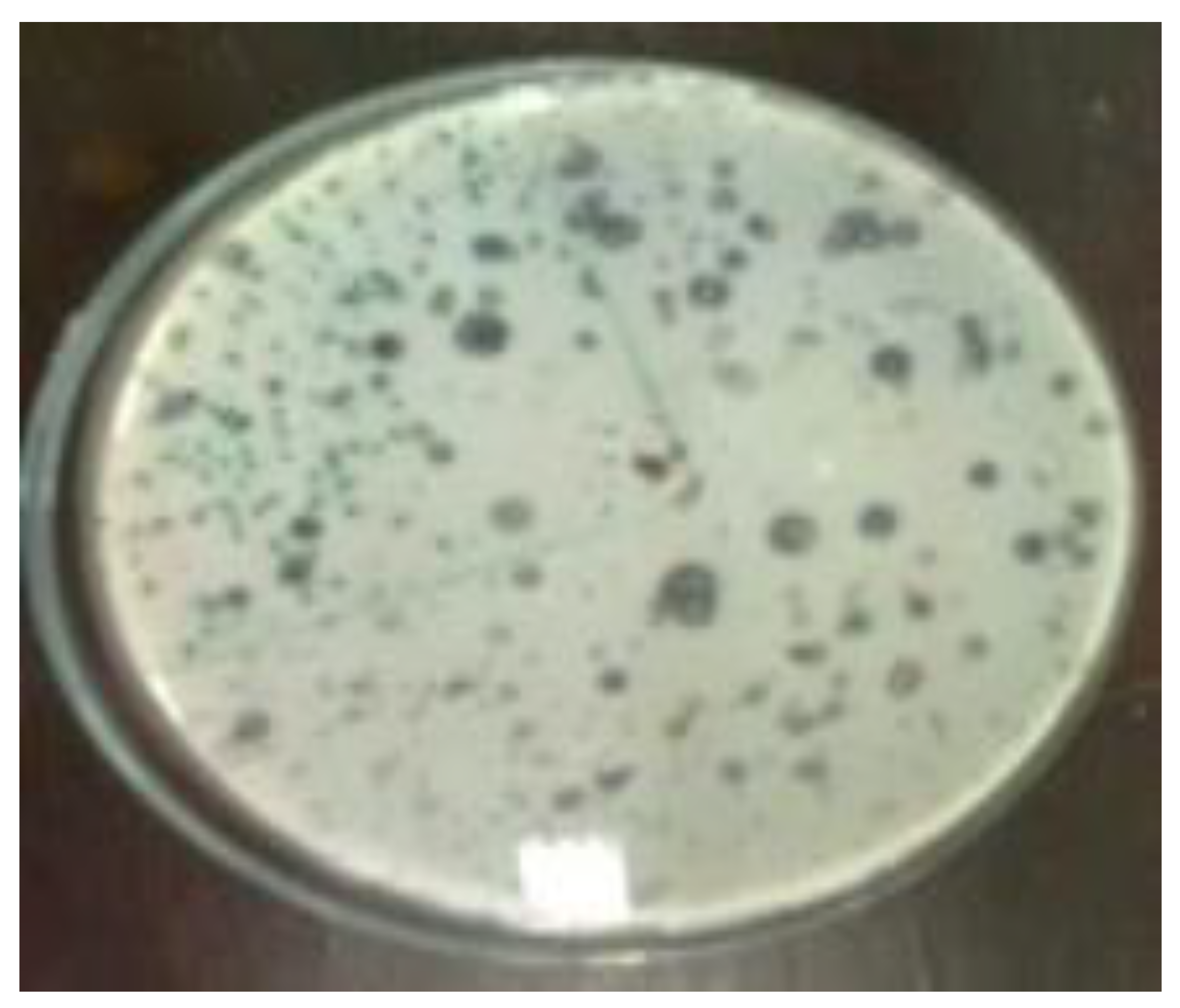
3.2. Result of Lactid Acid Bacteria Isolation

Figure 1 below presents the isolated result of lactic acid bacteria onto VCO, coconut oil and palm oil. It is shown that VCO contains lactic acid bacteria, as when it was being isolated using MRSA + CaCO3 selective media, the lactic acid bacteria colony that can grow in the “Halo” area is found. This area is a clear zone where it can produce lactic acid, neutralizing CaCO3. It is in line with, and as an addition to, the results in Figure 2 and Figure 3, pointing out the identification of the lactic acid bacteria using 16S rDNA, which turned out to be Lactobacillus plantarum and Lactobacillus paracasei as mentioned.
It can be seen, in Figure 1, there is a clear area; in the middle there is a white dot that is a colony of lactic acid bacteria present in the VCO oil. This is proof that VCO contains lactic acid bacteria [29,32].
Meanwhile, in Figure 2, is a petri dish filled with coconut oil and palm oil grown on the same media. No visible growth of lactic acid bacteria was observed. This proves that lactic acid bacteria do not exist in coconut oil and palm oil. This is in accordance with [8,35,42] but not so in [35], who only uses MRSA media without the addition of CaCO3. But growing colonies are not in the “Halo” area.

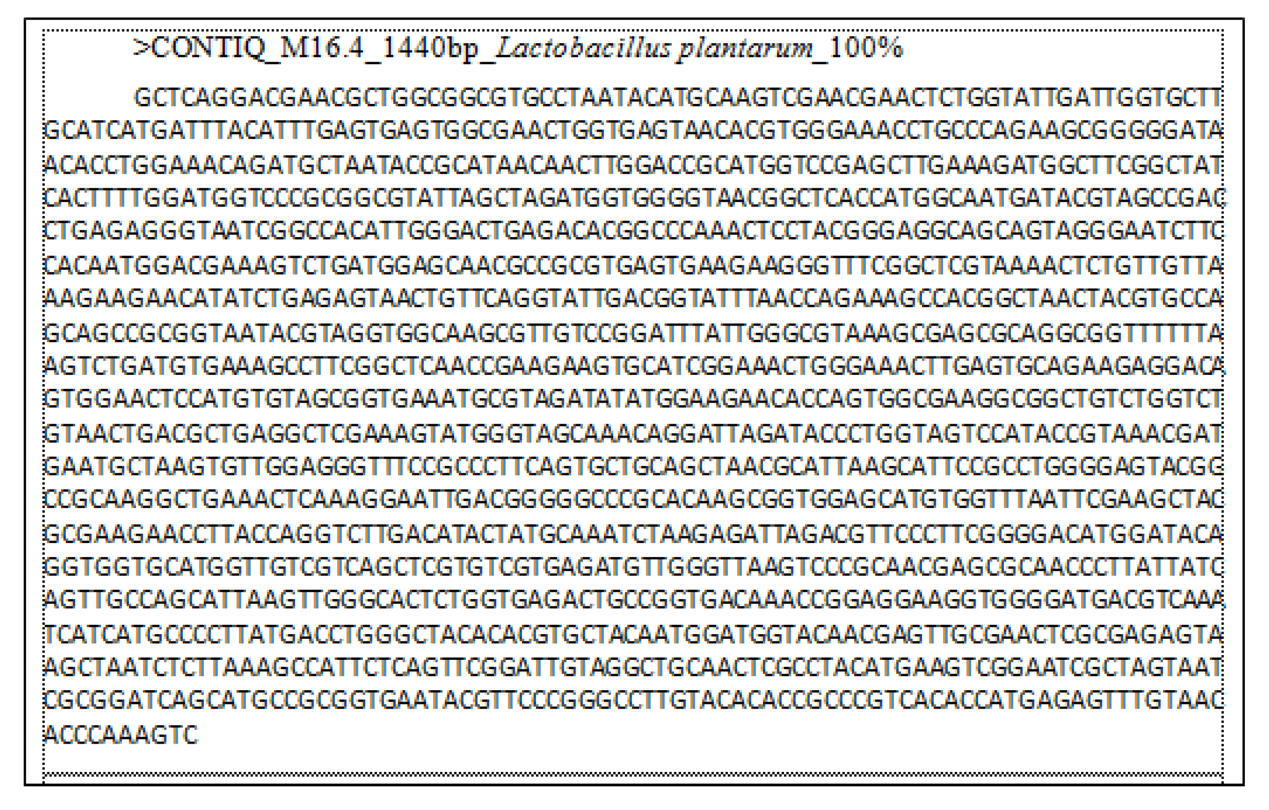
3.3. Molecular Identification of Lactid Acid Bacteria

The molecular identification of lactic acid bacteria produced Lactobacillus plantarum, as shown in the data below Figure 3 and Figure 4.

3.4. Result of Antimicrobial Analysis

The antimicrobial ability of VCO is also analyzed below. Antimicrobial analysis was performed with Lactobacillus paracasei and Lactobacillus plantarum lactic acid bacteria onto the following testing bacteria: Pseudomonas aeruginosa, Klebsiella, Staphylococcus aureus, Staphylococcus epidermidis, Proteus, from [31,35], Escherichia coli, Listeria monocytogenes, Bacillus cereus, and Salmonella typhosa.
As seen in Figure 5 below, clear zones show that the growth of pathogenic bacteria was inhibited by Lactobacillus plantarum and Lactobacillus paracasei.
As illustrated in Figure 5, VCO has the antimicrobial ability derived from two types of lactic acid bacteria against nine testing bacteria. It is seen that these lactic acid bacteria have a good ability to kill pathogenic bacteria, e.g., Listeria monocytogenes and E. coli, as stated in [8], where the antimicrobial ability of Lactobacillus plantarum is found to be most effective against Listeria monocytogenes, E.coli and Bacillus subtilis testing bacteria.
In Table 2 it can be seen that the lactic acid bacteria present in the VCO are able to inhibit the growth of pathogenic bacteria as Staphylococcus aureus, Pseudomonas aeruginosa, Klebsiella, Staphylococcus epidermidis, and Proteus, in accordance with [36].

4. Conclusions

Compared with coconut oil and palm oil, virgin coconut oil (VCO) has a higher content of lauric acid and lactic acid bacteria. Nevertheless, based on the content of water, free fatty acid and iodine number, VCO has other features in the field of health because it has lactic acid bacteria that can kill pathogens, characterizing it as an antimicrobial, in contrast to coconut and palm oil. The lauric acid content of the VCO is the highest (54.06%) compared to the lauric acid content of coconut oil (0.45%) and palm oil (0%). Having a high acid number, 1.10165, and high saponification number and iodine number demonstrate the characteristic of VCO, as it contains lauric acid with small molecules, which are medium-chain triglycerides (MCT). In the future, a more specialized assessment needs to be done, especially in the economic field to examine whether VCO is more appropriate as a fuel alternative or as an antimicrobial.

Author Contributions

Conceptualization: S.S. (Suryani Suryani) and S.S. (Sariani Sariani); resources: M.M., and S.S. (Suryani Suryani); methodology: S.S. (Suryani Suryani) and M.M.; software: S.S. (Suryani Suryani); validation: S.S. (Suryani Suryani), M.M.; formal analysis: S.S. (Suryani Suryani) and M.M.; writing—original draft preparation: S.S. (Sariani Sariani), T.M.I.M. and S.S. (Suryani Suryani); writing—review and editing: S.S. (Suryani Suryani), R.R., F.E., S.S. (Sariani Sariani) and A.F.; project administration: S.S. (Sevindrajuta Sevindrajuta) and R.R. All authors have read and agreed to the published version of the manuscript.

Funding

This research was funded by DRPM Kemenristekdikti, Indonesia year 2019, grant number: 232/SP2H/LT/DRPM/2019 dated DIPA 05 December 2018, SP DIPA-042.06.1.401516/2019, Directorate of Research and Community Service, Ministry of Research, Technology, and Higher Institution.

Acknowledgments

There was great supports received for the establishment of this research, therefore gratitude is given to: (1) Head of Basic Laboratory LLDIKTI, Area X. (2) Head of LPPM University of Muhammadiyah, West Sumatera. (3) Head of Agriculture Product Technology Laboratory, Andalas University. (4) Head of Biochemistry Laboratory, Medical Faculty, Andalas University. The benefactors do not have any role in this research design; data collection, analysis, or data interpretation; manuscript writing, or in any decision made in publishing the result.

Conflicts of Interest

The authors declare no conflict of interest.

References

  1. Kumalaningsih, S.; Padaga, M. The Utilization of Microorganisms Isolated From Fermented Coconut Milk For The Production of Virgin Coconut Oil. J. Basic Appl. Sci. Res. 2012, 2, 2286–2290. [Google Scholar]
  2. Satheesh, N.; Prasad, N.B.L. Optimization of Parameters for Fermentative Production of Virgin Coconut Oil by Lactobacillus fermentum NDRI 141. J. Food Sci. Eng. 2012, 2, 44–50. [Google Scholar] [CrossRef]
  3. Marina, A.M.; Rosli, W.I.W.; Neoh, S.L. Frying quality of virgin coconut oil as affected by Zea mays extract. Sains Malaysiana 2014, 43, 1311–1315. [Google Scholar]
  4. Prasad, N.; Satheesh, N. Production of virgin coconut oil by induced fermentation with Lactobacillus plantarum NDRI strain 184. Hrvatski časopis za Prehrambenu Tehnologiju, Biotehnologiju i Nutricionizam 2014, 9, 37–42. [Google Scholar]
  5. Redjeki, S.; Kurniati, E. The Kinetic Reacion of Virgin Coconut Oil (VCO) Fermentation in an Ideal Bioreact, or Tank in a Batch Process. J. Chem. Chem. Eng. 2013, 7, 159–163. [Google Scholar]
  6. Wong, Y.C.; Hartina, H. Virgin coconut oil production by centrifugation method. Orient. J. Chem. 2014, 30, 237–245. [Google Scholar] [CrossRef] [Green Version]
  7. Khatir, Z.; Agustina, R.; Hartuti, R.; Fahmi, S. Improving Fermented Virgin Coconut Oil Quality by Using Microwave Heating. In Earth and Enviromental Science; IOP: Bristol, UK, 2020. [Google Scholar]
  8. Suryani, A.D.; Dharma, A.; Manjang, Y.; Arief, S.; Munaf, E.; Nasir, N. Antimicrobial and antifungal activity of Lactic Acid Bacteria isolated from coconut milk fermentation. Res. J. Pharm. Biol. Chem. Sci. 2014, 5, 1587–1595. [Google Scholar]
  9. Bawalan, D.D. Processing Manual for Virgin Coconut Oil, its Products and By-products for Pacific Island Countries and Territories; Secretariat of the Pacific Community: Noumea, New Caledonia, 2011. [Google Scholar]
  10. Carandang, E.V. Health Benefits of Virgin Coconut Oil. Indian Coconut J. 2008, 9, 8. [Google Scholar]
  11. Hayatullina, Z.; Muhammad, N.; Mohamed, N.; Soelaiman, I.N. Virgin coconut oil supplementation prevents bone loss in osteoporosis rat model. Evid.-Based Complement. Altern. Med. 2012, 2012. [Google Scholar] [CrossRef] [Green Version]
  12. Nurul-Iman, B.S.; Kamisah, Y.; Jaarin, K.; Qodriyah, H.M.S. Virgin coconut oil prevents blood pressure elevation and improves endothelial functions in rats fed with repeatedly heated palm oil. Evid.-Based Complement. Altern. Med. 2013, 2013. [Google Scholar] [CrossRef] [Green Version]
  13. Dumancas, G.G. Health Benefits of Virgin Coconut Oil. In Vegetable Oil: Properties, Uses and Benefits; NOVA: Burleigh, Australia, 2016; pp. 161–194. [Google Scholar]
  14. An, H.M.; Park, S.Y.; Lee, D.K.; Kim, J.R.; Cha, M.K.; Lee, S.W.; Ha, N.J. Antiobesity and lipid-lowering effects of Bifidobacterium sin high fat diet-induced obese rats. Lipids Health Dis. 2011, 10, 116. [Google Scholar] [CrossRef] [PubMed] [Green Version]
  15. Allen, S.; Jordan, S.; Storey, M.; Thornton, C.A.; Gravenor, M.; Garaiova, I.; Plummer, S.F.; Wang, D.; Morgan, G. Dietary Supplementation with Lactobacilli and Bifidobacteria Is Well Tolerated and Not Associated with Adverse Events during Late Pregnancy and Early Infancy. J. Nutr. 2010, 140, 483–488. [Google Scholar] [CrossRef] [PubMed]
  16. Abujazia, M.A.; Muhammad, N.; Shuid, A.N.; Soelaiman, I.N. The effects of virgin coconut oil on bone oxidative status in ovariectomised rat. Evid.-Based Complement. Altern. Med. 2012, 2012. [Google Scholar] [CrossRef] [Green Version]
  17. Arlee, R.; Suanphairoch, S.; Pakdeechanuan, P. Differences in chemical components and antioxidant-related substances in virgin coconut oil from coconut hybrids and their parents. Int. Food Res. J. 2013, 20, 2103–2109. [Google Scholar]
  18. Pandiselvam, R.; Ramarathinam, M.; Plantation, C.; Beegum, S. Virgin Coconut Oil infused healthy cosmetics. Indian Coconut J. 2019, 13, 30–32. [Google Scholar]
  19. Kappally, S.; Shirwaikar, A.; Shirwaikar, A. Hygeia: Journal for drugs and medicines coconut oil—A review of potential applications. Hygeia J. Drugs Med. 2015, 7, 34–41. [Google Scholar]
  20. Varma, S.R.; Sivaprakasam, T.O.; Arumugam, I.; Dilip, N.; Raghuraman, M.; Pavan, K.; Rafiq, M.; Paramesh, R. In vitro anti-inflammatory and skin protective properties of Virgin coconut oil. J. Tradit. Complement. Med. 2019, 9, 5–14. [Google Scholar] [CrossRef]
  21. Law, K.S.; Azman, N.; Omar, E.A.; Musa, M.Y.; Yusoff, N.M.; Sulaiman, S.A.; Hussain, N.H.N. The effects of virgin coconut oil (VCO) as supplementation on quality of life (QOL) among breast cancer patients. Lipids Health Dis. 2014, 13, 139. [Google Scholar] [CrossRef] [Green Version]
  22. Krishna, A.G.G.; Raj, G.; Singh, B.A.; Kumar, P.K.P.; Chandrashekar, P. Coconut oil: Chemistry, production and its applications—A review. Indian Coconut J. 2010, 53, 15–27. [Google Scholar]
  23. Syukur, S.; Rajagukguk, H.; Syaputri, Y.; Iwahashi, H. Probiotic research in several products of virgin coconut oil from Padang, Indonesia. J. Phys. Conf. Ser. 2018, 1116. [Google Scholar] [CrossRef]
  24. Gesteiro, E.; Guijarro, L.; Sánchez-Muniz, F.J.; Vidal-Carou, M.D.C.; Troncoso, A.M.; Venanci, L.; Vinatea, V.J.; Quilez, J.; Anadón, A.; González-Gross, M. Palm Oil on the Edge. Nutrients 2019, 11, 2008. [Google Scholar] [CrossRef] [PubMed] [Green Version]
  25. Ong, H.C.; Masjuki, H.H.; Mahlia, T.M.I.; Silitonga, A.S.; Chong, W.T.; Yusaf, T. Engine performance and emissions using Jatropha curcas, Ceiba pentandra and Calophyllum inophyllum biodiesel in a CI diesel engine. Energy 2014, 69, 427–445. [Google Scholar] [CrossRef]
  26. Ong, H.C.; Milano, J.; Silitonga, A.S.; Hassan, M.H.; Shamsuddin, A.H.; Wang, C.T.; Mahlia, T.M.I.; Siswantoro, J.; Kusumo, F.; Sutrisno, J. Biodiesel production from Calophyllum inophyllum-Ceiba pentandra oil mixture: Optimization and characterization. J. Clean. Prod. 2019, 219, 183–198. [Google Scholar] [CrossRef]
  27. Silitonga, A.; Shamsuddin, A.; Mahlia, T.M.I.; Milano, J.; Kusumo, F.; Siswantoro, J.; Dharma, S.; Sebayang, A.; Masjuki, H.; Ong, H.C. Biodiesel synthesis from Ceiba pentandra oil by microwave irradiation-assisted transesterification: ELM modeling and optimization. Renew. Energy 2020, 146, 1278–1291. [Google Scholar] [CrossRef]
  28. Silitonga, A.S.; Masjuki, H.H.; Mahlia, T.M.I.; Ong, H.C.; Chong, W.T.; Boosroh, M.H. Overview properties of biodiesel diesel blends from edible and non-edible feedstock. Renew. Sustain. Energy Rev. 2013, 22, 346–360. [Google Scholar] [CrossRef]
  29. Mahlia, T.M.I.; Syazmi, Z.; Mofijur, M.; Abas, A.E.P.; Bilad, M.R.; Ong, H.C.; Silitonga, A.S. Patent landscape review on biodiesel production: Technology updates. Renew. Sustain. Energy Rev. 2020, 118. [Google Scholar] [CrossRef]
  30. Ghani, N.A.A.; Channip, A.A.; Hwa, P.C.H.; Ja’afar, F.; Yasin, H.M.; Usman, A. Physicochemical properties, antioxidant capacities, and metal contents of virgin coconut oil produced by wet and dry processes. Food Sci. Nutr. 2018, 6, 1298–1306. [Google Scholar] [CrossRef]
  31. Mansor, T.S.T.; Man, Y.B.C.; Shuhaimi, M.; Afiq, M.J.A.; Nurul, F.K.M.K. Physicochemical properties of virgin coconut oil extracted from different processing methods. Int. Food Res. J. 2012, 19, 837–845. [Google Scholar]
  32. Gupta, S.; Pandey, S. Isolation and characterization of bacteriocin producing bacteria from sweet lime juice. J. Pure Appl. Microbiol. 2018, 12, 953–960. [Google Scholar] [CrossRef]
  33. Khan, H. Production Characterization and Utilization of the Bacteriocin Produced by Enterococcus faecalis B9510. Appl. Chem. Biotechnol. 2013, 162, 1–186. [Google Scholar]
  34. Zhou, F.; Zhao, H.; Bai, F.; Dziugan, P.; Liu, Y.; Zhang, B. Purification and characterisation of the bacteriocin produced by Lactobacillus plantarum, isolated from Chinese pickle. Czech J. Food Sci. 2014, 32, 430–436. [Google Scholar] [CrossRef] [Green Version]
  35. Suryani, A.D. Isolation and Characterization of Bacteriocins Bacteria Lactobacillus Plantarum Strain NM178-5 from Fermentation Process with Contained on Coconut Milk. Transylv. Rev. 2016, 24, 614–628. [Google Scholar]
  36. Suryani, S.; Dharma, A.; Nasir, N. Isolation and identification of pathogenic bacteria secretion of chronic suppurative otitis media patients. Rasayan J. Chem. 2018, 11, 1139–1143. [Google Scholar] [CrossRef]
  37. Tolinački, M.; Kojić, M.; Lozo, J.; Terzić-Vidojević, A.; Topisirović, L.; Fira, D. Characterization of the bacteriocin-producing strain Lactobacillus paracasei subsp. Paracasei BGUB9. Arch. Biol. Sci. 2010, 62, 889–899. [Google Scholar] [CrossRef]
  38. Wang, Y.; Qin, Y.; Xie, Q.; Zhang, Y.; Hu, J.; Li, P. Purification and Characterization of Plantaricin LPL-1, a Novel Class IIa Bacteriocin Produced by Lactobacillus plantarum LPL-1 Isolated From Fermented Fish. Front. Microbiol. 2018, 9, 1–12. [Google Scholar] [CrossRef] [PubMed]
  39. Akinola, F.F.; Oguntibeju, O.O.; Adisa, A.W.; Owojuyigbe, O.S. Physico-chemical properties of palm oil from different palm oil local factories in Nigeria. J. Food Agric. Environ. 2010, 8, 264–269. [Google Scholar]
  40. Braga, J.D.; Lauzon, R.D.; Galvez, L.A. Physicochemical characterization of used coconut oil from vacuum frying of jackfruit (Artocarpus heterophyllus lam) pulp eviarc sweet variety as affected by frying cycle. Philipp. J. Sci. 2019, 148, 587–595. [Google Scholar]
  41. Marina, A.M.; Man, Y.B.C.; Amin, I. Virgin coconut oil: Emerging functional food oil. Trends Food Sci. Technol. 2009, 20, 481–487. [Google Scholar] [CrossRef]
  42. Kumar, V.; Kumari, A.; Angmo, K.; Bhalla, T.C. Isolation and characterization of lactic acid bacteria from traditional pickles of Himachal Pradesh, India. J. Food Sci. Technol. 2017, 54, 1945–1952. [Google Scholar]
Figure 1. Lactid acid bacteria isolate grown in MRS + 0.5% CaCO3 media.
Processes 08 00402 g001
Figure 2. Result of lactid acid bacteria isolation from coconut oil and from palm oil with no evidence of growth.
Processes 08 00402 g002
Figure 3. Lactid acid bacteria gene sequence from isolation of Lactobacillus plantarum.
Processes 08 00402 g003
Figure 4. The result of molecular identification of lactid acid bacteria showing Lactobacillus paracasei.
Processes 08 00402 g004
Figure 5. Results of the antimicrobial analysis of Lactobacillus plantarum lactid acid bacteria onto E. coli and S. aureus testing bacteria.
Processes 08 00402 g005
Table 1. The results: Composition and properties of virgin coconut oil, coconut oil and palm oil.
Type of the OilFatty Acids%Acid NumberSaponification Number%FFAIodine NumberpHWater Content %
VCO (A)Lauric Acid (C12:0)54.061.01348.000.265.326.500.11
Palmitic Acid-
Stearic Acid(C18:0)12.03
VCO (B)Lauric Acid (C12:0)53.901.03345.700.255.246.400.12
Palmitic Acid (C16:0)-
Stearic Acid (C18:0)12.01
VCO (C)Lauric Acid (C12:0)53.701.02346.640.265.256.500.11
Palmitic Acid (C16:0) -
Stearic Acid (C18:0)11.9
Coconut oilLauric Acid (C12:0)2.810.39269.620.287.026.900.11
Palmitic Acid (C16:0)2.31
Stearic Acid (C18:0)2.65
Palm Oil 1Lauric Acid (C12:0)0.450.39204.000.5151.006.600.09
Palmitic Acid (C16:0)2.88
Stearic Acid (C18:0)-
Palm Oil 2Lauric Acid (C12:0)-0.39203.020.7349.716.500.09
Palmitic Acid (C16:0)24.42
Stearic Acid (C18:0)-
Table 2. Antimicrobial activity analysis of LAB in the form of clear zone diameter (mm).
No.Testing BacteriaLactobacillus plantarumLactobacillus paracasei
1.Escherichia coli1616
2.Listeria monocytogenes1718
3.Bacillus substiliss1511
4.Salmonella typhy1211
5.Staphylococcus aureus1111
6.Pseudomonas aeruginosa1714
7.Klebsiella1312
8.Staphylococcus epidermidis1312
9.Proteus1413

