Abstract
Fibroblast growth factor receptors (FGFRs) are a family of receptor tyrosine kinases expressed on the cell membrane that play crucial roles in both developmental and adult cells. Dysregulation of FGFRs has been implicated in a wide variety of cancers, such as urothelial carcinoma, hepatocellular carcinoma, ovarian cancer and lung adenocarcinoma. Due to their functional importance, FGFRs have been considered as promising drug targets for the therapy of various cancers. Multiple small molecule inhibitors targeting this family of kinases have been developed, and some of them are in clinical trials. Furthermore, the pan-FGFR inhibitor erdafitinib (JNJ-42756493) has recently been approved by the U.S. Food and Drug Administration (FDA) for the treatment of metastatic or unresectable urothelial carcinoma (mUC). This review summarizes the structure of FGFR, especially its kinase domain, and the development of small molecule FGFR inhibitors.
1. Introduction
The human fibroblast growth factor receptor (FGFR) family consists of four members: FGFR1 to FGFR4. Despite being encoded by separate genes, the four members share high homology, with their sequence identity varying from 56% to 71% [1]. Similar to other receptor tyrosine kinases (RTKs), FGFRs are expressed on the cell membrane and can be stimulated and activated by extracellular signals. The native ligand of FGFRs is fibroblast growth factors (FGFs) [2,3,4]. The binding of FGFs drives the dimerization of FGFRs; subsequently, a transautophosphorylation event of the intracellular kinase domain is induced, followed by the activation of downstream transduction pathways [5,6]. Through triggering downstream signaling pathways, FGFRs participate in various vital physiological processes, such as proliferation, differentiation, cell migration and survival [7,8,9].
Aberrant expression of FGFRs has been shown in various kinds of solid tumors, and moreover, the aberrancy is considered an oncogenic signaling pathway [10,11,12]. It is believed that small molecules that competitively bind to the adenosine triphosphate (ATP) pocket of aberrant FGFRs while exhibiting little or no toxicity provide limitless prospects for the treatment of relevant tumors. The structure of FGFRs, especially the kinase domain, and the design of small molecular inhibitors have attracted intensive study in the past two decades. Multiple small molecule inhibitors have been developed, and some of them are currently being used in clinical trials, such as FGF401, which targets FGFR4 for the treatment of hepatocarcinoma (HCC) [13]; AZD4547, which targets FGFR1-3 for the treatment of a variety of tumors [14]. Moreover, erdafitinib (JNJ-42756493) [15] has been approved recently by U.S. Food and Drug Administration (FDA) for the treatment of mUC. More than 20 FGFR kinase/inhibitor complex structures have been determined to-date, and these structures have yielded extensive insights into the understanding of inactivation of FGFRs for related disease therapy.
2. Organization of FGFR
FGFRs share a canonical RTK architecture. From the N- to the C-terminus, all four FGFR members contain a large extracellular ligand-binding domain that comprises three immunoglobulin (Ig)-like subunits (D1, D2 and D3) followed by a single transmembrane helix and an intracellular tyrosine kinase domain [1,16] (Figure 1A). The linker region between D1 and D2 contains a highly conserved motif that is rich in aspartate acids, called the acid box [17]. The detailed function of those structural units will be further introduced below.
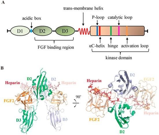
Figure 1.
Schematic diagram of FGFRs and the structure of the FGFR extracellular domain. (A) Organization of FGFRs. Important functional elements are highlighted. (B) Crystal structure of the FGF2:FGFR1:heparin ternary complex (PDB ID 1FQ9). The two copies of FGFR1 molecules are colored in green and light blue respectively. Heparin molecules are shown in red stick representation; FGF2 (colored in orange) and FGFR1 are shown in cartoon representation.
FGFs are the native ligand for this family of kinases. Through its extracellular domain, FGFR recognizes and is stimulated by specific FGFs. The FGF binding pocket is formed by the D2 and D3 subregions [18]. There have been contradicting views regarding the stoichiometry of the FGF/FGFR complex. Schlessinger, J. et al. solved the ternary complex structure of FGF2/FGFR1/heparin [19]. With the help of heparin, FGFR1 was dimerized after the binding of FGF2 to form the complex at a symmetric 2:2:2 stoichiometry ratio; both the FGF2 and heparin molecules simultaneously contacted the two FGFR1 monomers (Figure 1B). In the FGF1/FGFR2/heparin crystal structure solved by Pellegrini et al., the complex was assembled by asymmetric 2:2:1 stoichiometry [20]. By utilizing nuclear magnetic resonance, Saxena et al. studied the interactions of FGF1(FGF2)/FGFR4/HM (HM: heparin mimetics) complex, and their results supported the formation of the symmetric mode of FGF/FGFR dimerization in solution [21]. Interestingly, although all FGFs have a heparin sulfate binding site on their surface [22,23], endocrine FGFs such as FGF21 and FGF23 show a lower binding affinity to heparin sulfate [23] and require Klotho coreceptors instead to act as cofactors for FGFR activation [24,25,26].
In addition to acting as the ligand sensor, the extracellular domain also undertakes an autoinhibitory role, which relies on regulation by D1 and the acid box [27,28]. Several studies have proposed that the acid box could competitively bind to the heparin binding site of D2 to suppress heparin binding, while D1 forms intramolecular contacts with D2-D3, thus blocking the binding of FGFs [28,29,30]. Nevertheless, the mechanisms of autoinhibition need to be further clarified.
3. Structure of FGFR Kinase Domain
The intracellular tyrosine kinase domain is the most well studied region of the FGFR protein. This domain exhibits the canonical bilobed architecture of protein kinases [31,32,33,34,35]. The fold of the N-terminal small lobe (N-lobe, ~100 amino acid residues) consists of a five-stranded antiparallel β-sheet (β1–β5) and the αC-helix, an important regulatory element. The C-terminal large lobe (C-lobe, ~200 amino acid residues) predominately comprises seven a helices (αD, αE, αEF, αF-αI) (Figure 2A). The active site, which is responsible for ATP and substrate protein binding, is located in a clef between the two lobes (Figure 2B).
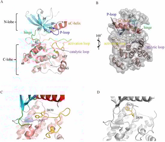
Figure 2.
Structure of the FGFR kinase domain. (A) Overall crystal structure of FGFR4 in cartoon representation. The five β-sheets of the N-lobe are labeled in cyan, and the helixes of the C-lobe are colored in salmon. The αC helix (red), P-loop (blue), catalytic loop (magenta), activation loop (bright orange) and hinge (green) are highlighted. (B) Surface presentation of FGFR4. The ATP binding pocket located between the N- and C-lobe is indicated by the dashed circle. (C) DFG-out conformation of the FGFR4 activation loop. The side chains of D630 and F631 are shown in stick representation. (D) DFG-in status of the FGFR4 activation loop. (A–C) were prepared from PDB ID 4UXQ; (D) was prepared from PDB ID 5JKG.
The C-lobe folds tightly with the αF-helix to form a hydrophobic core, around which the other secondary segments are packed. In addition to the primary helixes, the C-lobe contains a short helix located between the activation loop (A-loop) and the αF-helix named the αEF-helix, which is conserved among all FGFR members as well as other protein kinases [36]. Two short β-strands (β7 and β8) (Figure 2A) between the catalytic loop and activation loop (introduced below) interact with each other and are believed to participate in the regulation of FGFR activation [37]. In contrast to the C-lobe, the N-lobe folds in a more flexible manner, which benefits the binding and release of ATP/ADP and substrates.
There are several functionally important loops in both lobes. The loop between β8 and the αEF-helix is an activation loop (A-loop), which is essential for kinase activation [38,39,40]. The conformation of the highly conserved Asp-Phe-Gly motif (DFG-motif) in the A-loop is an indicator of kinase activity status [39,41]. Generally, the DFG-motif exists in two states: the active DFG-in and inactive DFG-out conformations [42,43] (Figure 2C,D). In the DFG-in state, the aspartate residue of the DFG-motif plays an essential role in ATP binding through the coordination of all three phosphate groups of ATP, either directly or via magnesium ions, while these interactions are sterically impossible when the motif is flipped into the DFG-out conformation. Phosphorylation is catalyzed by the conserved aspartate of the His-Arg-Asp (HRD) motif in the catalytic (αE–β7) loop [44]. The glycine rich P-loop (also called the nucleotide binding loop), located between the β1- and β2-strands, folds over to enclose ATP for phosphotransfer [45]. The so-called molecular brake located at the hinge region that connects the N- and C-lobes plays a critical role in the regulation of autoinhibition and activation [46].
The catalytic activity of the kinase domain is precisely controlled. There are two general conformations for all protein kinases, including those of the FGFR family. Activation typically involves changes in the orientation of the αC-helix in the small lobe and the activation loop in the C-lobe. During the catalytic cycle, the active kinase toggles between open and closed conformations. In the open form, the kinase binds MgATP and the protein substrate, while during catalysis, the kinase adopts the closed form. Once catalysis is completed, the MgADP and phosphorylated substrate are released, and the enzyme recovers to the open conformation, preparing for the next catalytic cycle [16,47].
4. Characteristics of FGFR/Inhibitor Interaction
As noted above, aberrantly expressed FGFRs have been implicated in various tumors. Therefore, extensive work has been performed on the development of FGFR inhibitors. The inhibitors that are in clinical trials or approved by the FDA for clinical use are summarized in Table 1, and the chemical structures of those inhibitors are shown in Figure 3.

Table 1.
FGFR inhibitors that are in clinical trials or approved by the FDA.
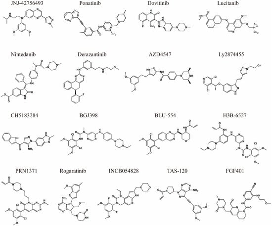
Figure 3.
Chemical structure of FGFR small molecule inhibitors.
FGFR inhibitors can generally be divided into two groups according to their binding behaviors, namely, type I and type II inhibitors [58,59]. Type I inhibitors bind FGFRs in the DFG-in enzymatic active conformation in an ATP-competitive manner, while the binding of type II requires the DFG-motif to be flipped to the DFG-out state [60,61]. The X-ray crystallographic structures of AZD4547 (PDB ID 4V05) [34] and PD173074 (PDB ID 2FGI) [62] bound to FGFR1 demonstrate that these two inhibitors are type I inhibitors. Taking the FGFR1/AZD4547 structure as an example, the AZD4547 occupying the ATP pocket of FGFR1 forms a hydrogen bond with the backbone nitrogen atom of the DFG aspartate (Asp641) and forms three hydrogen bonds with the hinge residues [34] (Figure 4A). In contrast, both the DFG-motifs of FGFR4 and FGFR1 are flipped out into an inactive conformation in the FGFR4/ponatinib (PDB ID 4UXQ) and FGFR1/ponatinib (PDB ID 4V04) structures [34]. In addition to the basal interactions, a hydrogen bond formed between ponatinib and the side chain of the strictly conserved glutamate from the αC-helix (Glu520 in FGFR4 and Glu531 in FGFR1) was also observed, which is characteristic of a type II inhibitor [61] (Figure 4B). Thus, these structures reveal ponatinib to be a type II inhibitor for FGFRs. As a consequence, the flip of the phenylalanine side chain breaks the regulatory spine and creates an additional induced-fit hydrophobic pocket that allows deeper binding of the inhibitor and provides better selectivity [34,43] as well as slower dissociation kinetics [34,63].
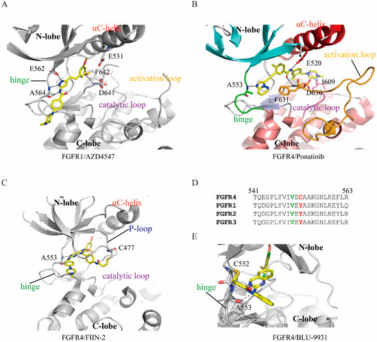
Figure 4.
FGFRs/inhibitor interaction features. All inhibitors are presented in yellow stick representation. (A) Structure of AZD4547 bound to FGFR1. The side chains of A564, E562 and D641, which directly form hydrogen bonds with the inhibitor and F642 of the DFG-motif, are shown. The hydrogen bonds are indicated by dashed lines. AZD4547 binds FGFR1 into DFG-in status, the side chain of F642 points out from the ATP pocket. (B) The structure of FGFR4 in a complex with ponatinib. The DFG-motif of FGFR4 flipped to an out conformation with F631 benzene ring flipped into the ATP-pocket and D630 point out from the pocket. (C) Covalent interaction of FIIN-2 and FGFR4. The side chains of A553 and C477, which interact with ponatinib, are shown in stick representation. Covalent bond formed between C477 and the acrylamide group of FIIN-2. (D) Sequence alignment of the FGFR hinge region. The C552 in FGR4 is replaced by a tyrosine in the other 3 members. The gatekeeper residues which locate at the kinases hinge region and play an essential role in determining pocket accessibility for inhibitors are highlighted in green. (E) Structure of BLU-9931 in complex with FGFR4. Unlike the pan-FGFR covalent inhibitors, BLU-9931 targets the unique C552 of FGFR4 to form covalent interactions.
The interaction between a small molecule inhibitor and protein kinase can be covalent (irreversible) or noncovalent (reversible) [64,65]. Typically, covalent inhibitors have a functional group known as the warhead, which can improve binding affinity and selectivity through covalent interaction with a certain residue of the target kinase [65,66]. Moreover, a well-designed warhead could provide better performance against drug resistance than reversible inhibitors [67,68]. The reported covalent reactive residues in protein kinases include cysteine [69], aspartic acid [70], lysine [71] and others [72]. For FGFRs, the conserved cysteine of the P-loop (C488 in FGFR1, C491 in FGFR2, C482 in FGFR3 and C477 in FGFR4) and the unique C552 in FGFR4 from the hinge region are the covalent binding sites. The FGFR4/FIIN-2 complex structure (PDB accession number 4QQC) is the first solved irreversible structure, where FIIN-2 formed a covalent bond through its reactive acrylamide group with the hydrosulfonyl side chain of FGFR4 C488 [73] (Figure 4C).
5. Current Status of Small Molecule FGFR Inhibitor Development
Designing specific small molecule inhibitors targeting protein kinases is challenging because the ATP binding pockets of the human kinome are similar [74,75]. Inhibitor research for FGFRs has gone through several stages. Initially, nonselective multiple-kinase inhibitors were developed to treat FGFR aberrations. Those nonselective inhibitors, including ponatinib [76], dovitinib [77] lucitanib [78] and nintedanib [79] (see Table 1 for details), were originally designed for other kinases and then proved to have potent inhibition activity toward FGFRs. For instance, the type II inhibitor ponatinib was originally developed to overcome the BCR-ABL T315I gatekeeper mutant and showed single-digit nanomolar binding strength to FGFR1-4 in later researches [76]. Although nonselective inhibitors might be clinically beneficial and achieve therapeutic success to some extent, the development of those inhibitors has been restricted due to the undesirable off-target toxicities [80,81].
To overcome the off-target effects of nonselective inhibitors, efforts have been made to develop FGFR-selective inhibitors (pan-FGFR inhibitors). In the earlier stage, multiple noncovalent pan-FGFR inhibitors were developed, including the well-known AZD4547 [14] and LY2874455 [48] (see Table 1 for details). Those pan-FGFR inhibitors are typically type I inhibitors. For example, AZD4547 is capable of potently inhibiting FGFR1-3 but shows negligible binding affinity to FGFR4. AZD4547 is currently in phase II clinical trials. However, preclinical data show that AZD4547 is not able to overcome the gatekeeper mutation V555M in FGFR3 [82]. Unlike AZD4547, the inhibitor LY2874455 shows inhibition efficacy against all 4 FGFRs, and the crystal structure as well as in vitro and vivo experiments confirmed that this inhibitor maintained equal inhibitory ability against the gatekeeper mutant V550M/V550L of FGFR4 [32].
Given that covalent inhibitors confer better binding kinetics and selectivity than noncovalent ones, developing irreversible inhibitors of FGFRs has attracted intensive pharmaceutical and academic attention in recent years. Since the first covalent inhibitor FIIN-1 [83], this field has achieved much progress. A number of FGFR covalent inhibitors have been developed, and some of those agents are already in clinical trials (see Table 1 for details). Moreover, several irreversible inhibitor/FGFR structures have been revealed by crystal structures, including FGFR4/BLU9931 (PDB ID: 4XCU) [84], FGFR4/FIIN-3 (PDB ID: 4R6V) [73], FGFR4/FIIN-2 (PDB ID: 4QQ5) [85], FGFR4/CGA159527 (PDB ID: 5NUD) [86], FGFR1(Y563C)/H3B-6527 (PDB ID: 5VND) [86], and FGFR1/TAS-120 (PDB ID: 6MZW) [54].
The kinase domains of FGFRs are highly homologous, with sequence identity varying from 74% to 77% [34]. Unexpectedly, most of the reported pan-FGFR reversible inhibitors tend to bind FGFR1-3 but exhibit greatly reduced potency toward FGFR4 [87]. The underlying mechanism is not quite clear. Tucker et al. proposed that the innate flexibility of the FGFR4 kinase domain might be responsible for the decrease in binding ability [34]. This feature, together with the unique C552 of FGFR4, which replaces a tyrosine in FGFR1-3, confers the opportunity to develop FGFR4-selective inhibitors [88,89] (Figure 4D). Indeed, H3B-6527 [56], BLU-9931 [84], BLU-554 [84] and FGF401 [90] were developed as FGFR4-selective covalent inhibitors that target C552 for irreversible binding (Figure 4E). Among these four molecules, FGF401 is the most interesting because the covalent bond it forms is reversible, which reduces the off-target effect and prolongs the residence time [91,92]. The crystal structure of FGF401/FGFR4 was recently reported by our laboratory (PDB ID 6JPJ) [57], and its potential utility is currently under intensive research.
In addition to the kinase domain, the ectodomains of FGFRs have also attracted intensive interests for drug discovery. Unlike the highly conserved kinase domain, the ectodomains of FGFRs are less conserved; targeting this domain may offer better isoform selectivity. The dominant strategy to target FGFR ectodomains is using monoclonal antibody/antibody-drug conjugate [93]. Several anti-FGFR monoclonal antibodies have been developed, and some of them are in clinical trials [94,95,96,97,98]. In addition, efforts have been made in the development of small molecule inhibitor targeting FGFR ectodomains. An inhibitor, SSR128129E, which allosterically binds to the ectodomain of FGFR in a non-FGF competitive manner, has been reported to inhibit FGF-induced signaling [99,100].
6. FGFR Gatekeeper Mutation and Drug Resistance
The long-term efficacy of kinase inhibitors in cancer treatment is often disturbed by acquired resistance. One common mechanism of resistance is generated by mutating the so-called gatekeeper residue of the kinase domain [101]. The gatekeeper mutation has been reported in various kinases, such as Bcr-Abl (T315I) [102], EGFR (T790M) [68], PDGFR (T674I) [103], FGFR1 (V561M) [104] and FGFR2 (V565I) [105]. The gatekeeper residue lies at the beginning of the hinge region and controls the accessibility of the hydrophobic pocket. Most protein kinases harbor a threonine that plays a major role in the interaction with the inhibitor by forming a critical hydrogen bond via its side chain hydroxyl oxygen. The mutation of this residue to a bulky hydrophobic amino acid, either Met or Ile, breaks the hydrogen interaction and introduces steric hindrance for inhibitor binding [76,106].
The drug resistance of FGFR gatekeeper mutations has been extensively verified, both in vitro and in vivo. For instance, the V564M mutation of FGFR2 confers the ability to resist dovitinib and BGJ398 [107]. Furthermore, an array of FGFR gatekeeper mutations have been identified in clinical samples. For example, the FGFR4 V550M mutation was detected in 13% of neuroendocrine breast carcinomas [108]. In FGFRs, the gatekeeper residue is a valine (Figure 4D); as a consequence, its side chain cannot form hydrogen interactions with inhibitors (see above context), and the resistance thus arises mainly through the introduction of steric hindrance. Several FGFR inhibitors have been shown to have the ability to overcome FGFR gatekeeper mutations. For example, Ly2874455 has almost equal binding affinity to wild-type FGFR4, FGFR4 (V550M), FGFR4 (V550L) FGFR1 (V561M), FGFR2 (V564F) and FGFR3 (V555M) [32]; FGF401 has similar affinity to wild-type FGFR4, FGFR4 (V550M) and FGFR4 (V550L) [57]; FIIN-2 shows a binding affinity loss of ~10-fold for FGFR4 (V550L) compared with the wild-type kinase [85].
7. Conclusions
Increasing evidence indicates that aberrant Fibroblast growth factor receptors (FGFR)signaling plays a crucial role in tumorigenesis and progression. Now, small molecule inhibitors targeting FGFRs offer a novel and effective strategy for the therapy of cancers caused by FGFR aberrations. Efforts and progress have been made in the field of FGFR inhibitor development. Some small molecules show promising antitumor activity and are evaluated in clinical trials. Recently, the pan-FGFR inhibitor erdafitinib has been approved by the Food and Drug Administration for the treatment of mUC, making it the first approved FGFR-targeted drug. However, there are still challenges in the field of FGFR inhibitor development, such as the need for more potent and selective FGFR inhibitors, and inhibitors with the ability to overcome gatekeeper mutations. Most FGFR inhibitors currently under evaluation are typical type I inhibitors that occupy only the ATP binding pocket. Development of type II FGFR inhibitors, which could be inserted deeper into the pocket, could confer better potency and selectivity. In addition, the development of covalent irreversible or covalent reversible FGFR inhibitors might be another strategy to improve safety and efficacy for cancer treatment.
Author Contributions
All authors were involved in the design of the review, and in writing or revising the manuscript. All authors approved the submitted version.
Funding
This research was funded by National Natural Science Foundation of China (81372904 and 81570537 to Y.C.).
Conflicts of Interest
The authors declare no conflict of interest.
References
- Itoh, N.; Ornitz, D.M. Evolution of the Fgf and Fgfr gene families. Trends Genet. 2004, 20, 563–569. [Google Scholar] [CrossRef] [PubMed]
- Schlessinger, J. Cell signaling by receptor tyrosine kinases. Cell 2000, 103, 211–225. [Google Scholar] [CrossRef]
- Weiner, H.L.; Zagzag, D. Growth factor receptor tyrosine kinases: Cell adhesion kinase family suggests a novel signaling mechanism in cancer. Cancer Investig. 2000, 18, 544–554. [Google Scholar] [CrossRef]
- Lemmon, M.A.; Schlessinger, J. Cell signaling by receptor tyrosine kinases. Cell 2010, 141, 1117–1134. [Google Scholar] [CrossRef] [PubMed]
- Schlessinger, J. Cell signaling by receptor tyrosine kinases: From basic concepts to clinical applications. Eur. J. Cancer Suppl. 2006, 4, 3–26. [Google Scholar] [CrossRef][Green Version]
- Schlessinger, J. Receptor Tyrosine Kinases: Legacy of the First Two Decades. Cold Spring Harb. Perspect. Biol. 2014. [Google Scholar] [CrossRef] [PubMed]
- Ornitz, D.M.; Itoh, N. The Fibroblast Growth Factor signaling pathway. Wiley Interdiscip. Rev. Dev. Biol. 2015, 4, 215–266. [Google Scholar] [CrossRef] [PubMed]
- Andrew, B.; Moosa, M. The FGF family: Biology, pathophysiology and therapy. Nat. Rev. Drug Discov. 2009, 8, 235–253. [Google Scholar]
- Karel, D.; Enrique, A. FGF signalling: Diverse roles during early vertebrate embryogenesis. Development 2010, 137, 3731–3742. [Google Scholar]
- Gowardhan, B.; Douglas, D.A.; Mathers, M.E.; McKie, A.B.; McCracken, S.R.C.; Robson, C.N.; Leung, H.Y. Evaluation of the fibroblast growth factor system as a potential target for therapy in human prostate cancer. Br. J. Cancer 2005, 92, 320–327. [Google Scholar] [CrossRef]
- Brooks, A.N.; Kilgour, E.; Smith, P.D. Molecular pathways: Fibroblast growth factor signaling: A new therapeutic opportunity in cancer. Clin. Cancer Res. 2012, 18, 1855–1862. [Google Scholar] [CrossRef] [PubMed]
- Turner, N.; Grose, R. Fibroblast growth factor signalling: From development to cancer. Nat. Rev. Cancer 2010, 10, 116–129. [Google Scholar] [CrossRef] [PubMed]
- Porta, D.G.; Weiss, A.; Fairhurst, R.A.; Wartmann, M.; Stamm, C.; Reimann, F.; Buhles, A.; Kinyamu-Akunda, J.; Sterker, D.; Murakami, M. Abstract 2098: NVP-FGF401, a first-in-class highly selective and potent FGFR4 inhibitor for the treatment of HCC. Cancer Res. 2017. [Google Scholar] [CrossRef]
- Gavine, P.R.; Mooney, L.; Kilgour, E.; Thomas, A.P.; Al-Kadhimi, K.; Beck, S.; Rooney, C.; Coleman, T.; Baker, D.; Mellor, M.J.; et al. AZD4547: An orally bioavailable, potent, and selective inhibitor of the fibroblast growth factor receptor tyrosine kinase family. Cancer Res. 2012, 72, 2045–2056. [Google Scholar] [CrossRef] [PubMed]
- Perera, T.P.S.; Jovcheva, E.; Mevellec, L.; Vialard, J.; De Lange, D.; Verhulst, T.; Paulussen, C.; Van De Ven, K.; King, P.; Freyne, E.; et al. Discovery and Pharmacological Characterization of JNJ-42756493 (Erdafitinib), a Functionally Selective Small-Molecule FGFR Family Inhibitor. Mol. Cancer Ther. 2017, 16, 1010–1020. [Google Scholar] [CrossRef]
- Farrell, B.; Breeze, A.L. Structure, activation and dysregulation of fibroblast growth factor receptor kinases: Perspectives for clinical targeting. Biochem. Soc. Trans. 2018, 46, 1753–1770. [Google Scholar] [CrossRef] [PubMed]
- Sanchez-Heras, E.; Howell, F.V.; Williams, G.; Doherty, P. The fibroblast growth factor receptor acid box is essential for interactions with N-cadherin and all of the major isoforms of neural cell adhesion molecule. J. Biol. Chem. 2006, 281, 35208–35216. [Google Scholar] [CrossRef]
- Wang, F.; Kan, M.; Xu, J.; Yan, G.; McKeehan, W.L. Ligand-specific structural domains in the fibroblast growth factor receptor. J. Biol. Chem. 1995, 270, 10222–10230. [Google Scholar] [CrossRef]
- Schlessinger, J.; Plotnikov, A.N.; Ibrahimi, O.A.; Eliseenkova, A.V.; Yeh, B.K.; Yayon, A.; Linhardt, R.J.; Mohammadi, M. Crystal structure of a ternary FGF-FGFR-heparin complex reveals a dual role for heparin in FGFR binding and dimerization. Mol. Cell 2000, 6, 743–750. [Google Scholar] [CrossRef]
- Pellegrini, L.; Burke, D.F.; von Delft, F.; Mulloy, B.; Blundell, T.L. Crystal structure of fibroblast growth factor receptor ectodomain bound to ligand and heparin. Nature 2000, 407, 1029–1034. [Google Scholar] [CrossRef]
- Saxena, K.; Schieborr, U.; Anderka, O.; Duchardt-Ferner, E.; Elshorst, B.; Gande, S.L.; Janzon, J.; Kudlinzki, D.; Sreeramulu, S.; Dreyer, M.K.; et al. Influence of heparin mimetics on assembly of the FGF.FGFR4 signaling complex. J. Biol. Chem. 2010, 285, 26628–26640. [Google Scholar] [CrossRef] [PubMed]
- Eriksson, A.E.; Cousens, L.S.; Weaver, L.H.; Matthews, B.W. Three-dimensional structure of human basic fibroblast growth factor. Proc. Natl. Acad. Sci. USA 1991, 88, 3441–3445. [Google Scholar] [CrossRef] [PubMed]
- Goetz, R.; Mohammadi, M. Exploring mechanisms of FGF signalling through the lens of structural biology. Nat. Rev. Mol. Cell Biol. 2013, 14, 166–180. [Google Scholar] [CrossRef] [PubMed]
- Razzaque, M.S. The FGF23-Klotho axis: Endocrine regulation of phosphate homeostasis. Nat. Rev. Endocrinol. 2009, 5, 611–619. [Google Scholar] [CrossRef] [PubMed]
- Chen, G.; Liu, Y.; Goetz, R.; Fu, L.; Jayaraman, S.; Hu, M.C.; Moe, O.W.; Liang, G.; Li, X.; Mohammadi, M. alpha-Klotho is a non-enzymatic molecular scaffold for FGF23 hormone signalling. Nature 2018, 553, 461–466. [Google Scholar] [CrossRef] [PubMed]
- Lee, S.; Choi, J.; Mohanty, J.; Sousa, L.P.; Tome, F.; Pardon, E.; Steyaert, J.; Lemmon, M.A.; Lax, I.; Schlessinger, J. Structures of beta-klotho reveal a ‘zip code’-like mechanism for endocrine FGF signalling. Nature 2018, 553, 501–505. [Google Scholar] [CrossRef] [PubMed]
- Mohammadi, M.; Olsen, S.K.; Ibrahimi, O.A. Structural basis for fibroblast growth factor receptor activation. Cytokine Growth Factor Rev. 2005, 16, 107–137. [Google Scholar] [CrossRef]
- Kalinina, J.; Dutta, K.; Ilghari, D.; Beenken, A.; Goetz, R.; Eliseenkova, A.V.; Cowburn, D.; Mohammadi, M. The alternatively spliced acid box region plays a key role in FGF receptor autoinhibition. Structure 2012, 20, 77–88. [Google Scholar] [CrossRef]
- Olsen, S.K.; Ibrahimi, O.A.; Raucci, A.; Zhang, F.; Eliseenkova, A.V.; Yayon, A.; Basilico, C.; Linhardt, R.J.; Schlessinger, J.; Mohammadi, M. Insights into the molecular basis for fibroblast growth factor receptor autoinhibition and ligand-binding promiscuity. Proc. Natl. Acad. Sci. USA 2004, 101, 935–940. [Google Scholar] [CrossRef]
- Wang, F.; Kan, M.; Yan, G.; Xu, J.; McKeehan, W.L. Alternately spliced NH2-terminal immunoglobulin-like Loop I in the ectodomain of the fibroblast growth factor (FGF) receptor 1 lowers affinity for both heparin and FGF-1. J. Biol. Chem. 1995, 270, 10231–10235. [Google Scholar] [CrossRef]
- Mohammadi, M.; Schlessinger, J.; Hubbard, S.R. Structure of the FGF receptor tyrosine kinase domain reveals a novel autoinhibitory mechanism. Cell 1996, 86, 577–587. [Google Scholar] [CrossRef]
- Wu, D.; Guo, M.; Min, X.; Dai, S.; Li, M.; Tan, S.; Li, G.; Chen, X.; Ma, Y.; Li, J.; et al. LY2874455 potently inhibits FGFR gatekeeper mutants and overcomes mutation-based resistance. Chem. Commun. (Camb.) 2018, 54, 12089–12092. [Google Scholar] [CrossRef] [PubMed]
- Wu, D.; Guo, M.; Philips, M.A.; Qu, L.; Jiang, L.; Li, J.; Chen, X.; Chen, Z.; Chen, L.; Chen, Y. Crystal Structure of the FGFR4/LY2874455 Complex Reveals Insights into the Pan-FGFR Selectivity of LY2874455. PLoS ONE 2016, 11, e0162491. [Google Scholar] [CrossRef] [PubMed]
- Tucker, J.A.; Klein, T.; Breed, J.; Breeze, A.L.; Overman, R.; Phillips, C.; Norman, R.A. Structural insights into FGFR kinase isoform selectivity: Diverse binding modes of AZD4547 and ponatinib in complex with FGFR1 and FGFR4. Structure 2014, 22, 1764–1774. [Google Scholar] [CrossRef] [PubMed]
- Ni, F.; Kung, A.; Duan, Y.; Shah, V.; Amador, C.D.; Guo, M.; Fan, X.; Chen, L.; Chen, Y.; McKenna, C.E.; et al. Remarkably Stereospecific Utilization of ATP alpha,beta-Halomethylene Analogues by Protein Kinases. J. Am. Chem. Soc. 2017, 139, 7701–7704. [Google Scholar] [CrossRef] [PubMed]
- Knighton, D.R.; Zheng, J.H.; Ten Eyck, L.F.; Ashford, V.A.; Xuong, N.H.; Taylor, S.S.; Sowadski, J.M. Crystal structure of the catalytic subunit of cyclic adenosine monophosphate-dependent protein kinase. Science 1991, 253, 407–414. [Google Scholar] [CrossRef] [PubMed]
- Yang, Y.; Ye, Q.; Jia, Z.; Cote, G.P. Characterization of the Catalytic and Nucleotide Binding Properties of the alpha-Kinase Domain of Dictyostelium Myosin-II Heavy Chain Kinase A. J. Biol. Chem. 2015, 290, 23935–23946. [Google Scholar] [CrossRef]
- Furdui, C.M.; Lew, E.D.; Schlessinger, J.; Anderson, K.S. Autophosphorylation of FGFR1 kinase is mediated by a sequential and precisely ordered reaction. Mol. Cell 2006, 21, 711–717. [Google Scholar] [CrossRef]
- Kornev, A.P.; Taylor, S.S.; Ten Eyck, L.F. A helix scaffold for the assembly of active protein kinases. Proc. Natl. Acad. Sci. USA 2008, 105, 14377–14382. [Google Scholar] [CrossRef]
- Duan, Y.; Chen, L.; Chen, Y.; Fan, X.G. c-Src binds to the cancer drug Ruxolitinib with an active conformation. PLoS ONE 2014, 9, e106225. [Google Scholar] [CrossRef]
- Hu, J.; Ahuja, L.G.; Meharena, H.S.; Kannan, N.; Kornev, A.P.; Taylor, S.S.; Shaw, A.S. Kinase regulation by hydrophobic spine assembly in cancer. Mol. Cell. Biol. 2015, 35, 264–276. [Google Scholar] [CrossRef] [PubMed]
- Hari, S.B.; Merritt, E.A.; Maly, D.J. Sequence determinants of a specific inactive protein kinase conformation. Chem. Biol. 2013, 20, 806–815. [Google Scholar] [CrossRef] [PubMed]
- Vijayan, R.S.; He, P.; Modi, V.; Duong-Ly, K.C.; Ma, H.; Peterson, J.R.; Dunbrack, R.L., Jr.; Levy, R.M. Conformational analysis of the DFG-out kinase motif and biochemical profiling of structurally validated type II inhibitors. J. Med. Chem. 2015, 58, 466–479. [Google Scholar] [CrossRef] [PubMed]
- Klein, T.; Vajpai, N.; Phillips, J.J.; Davies, G.; Holdgate, G.A.; Phillips, C.; Tucker, J.A.; Norman, R.A.; Scott, A.D.; Higazi, D.R.; et al. Structural and dynamic insights into the energetics of activation loop rearrangement in FGFR1 kinase. Nat. Commun. 2015. [Google Scholar] [CrossRef]
- Guimaraes, C.R.; Rai, B.K.; Munchhof, M.J.; Liu, S.; Wang, J.; Bhattacharya, S.K.; Buckbinder, L. Understanding the impact of the P-loop conformation on kinase selectivity. J. Chem. Inf. Model. 2011, 51, 1199–1204. [Google Scholar] [CrossRef] [PubMed]
- Chen, H.; Ma, J.; Li, W.; Eliseenkova, A.V.; Xu, C.; Neubert, T.A.; Miller, W.T.; Mohammadi, M. A molecular brake in the kinase hinge region regulates the activity of receptor tyrosine kinases. Mol. Cell 2007, 27, 717–730. [Google Scholar] [CrossRef] [PubMed]
- Roskoski, R., Jr. Src protein-tyrosine kinase structure, mechanism, and small molecule inhibitors. Pharm. Res. 2015, 94, 9–25. [Google Scholar] [CrossRef] [PubMed]
- Zhao, G.; Li, W.Y.; Chen, D.; Henry, J.R.; Li, H.Y.; Chen, Z.; Zia-Ebrahimi, M.; Bloem, L.; Zhai, Y.; Huss, K.; et al. A novel, selective inhibitor of fibroblast growth factor receptors that shows a potent broad spectrum of antitumor activity in several tumor xenograft models. Mol. Cancer. Ther. 2011, 10, 2200–2210. [Google Scholar] [CrossRef]
- Nakanishi, Y.; Akiyama, N.; Tsukaguchi, T.; Fujii, T.; Sakata, K.; Sase, H.; Isobe, T.; Morikami, K.; Shindoh, H.; Mio, T.; et al. The fibroblast growth factor receptor genetic status as a potential predictor of the sensitivity to CH5183284/Debio 1347, a novel selective FGFR inhibitor. Mol. Cancer. Ther. 2014, 13, 2547–2558. [Google Scholar] [CrossRef]
- Guagnano, V.; Furet, P.; Spanka, C.; Bordas, V.; Le Douget, M.; Stamm, C.; Brueggen, J.; Jensen, M.R.; Schnell, C.; Schmid, H.; et al. Discovery of 3-(2,6-dichloro-3,5-dimethoxy-phenyl)-1-{6-[4-(4-ethyl-piperazin-1-yl)-phenylamin o]-pyrimidin-4-yl}-1-methyl-urea (NVP-BGJ398), a potent and selective inhibitor of the fibroblast growth factor receptor family of receptor tyrosine kinase. J. Med. Chem. 2011, 54, 7066–7083. [Google Scholar] [CrossRef]
- Karkera, J.D.; Cardona, G.M.; Bell, K.; Gaffney, D.; Portale, J.C.; Santiago-Walker, A.; Moy, C.H.; King, P.; Sharp, M.; Bahleda, R.; et al. Oncogenic Characterization and Pharmacologic Sensitivity of Activating Fibroblast Growth Factor Receptor (FGFR) Genetic Alterations to the Selective FGFR Inhibitor Erdafitinib. Mol. Cancer. Ther. 2017, 16, 1717–1726. [Google Scholar] [CrossRef] [PubMed]
- Collin, M.P.; Lobell, M.; Hubsch, W.; Brohm, D.; Schirok, H.; Jautelat, R.; Lustig, K.; Bomer, U.; Vohringer, V.; Heroult, M.; et al. Discovery of Rogaratinib (BAY 1163877): A pan-FGFR Inhibitor. Chem. Med. Chem. 2018, 13, 437–445. [Google Scholar] [CrossRef] [PubMed]
- Brameld, K.A.; Owens, T.D.; Verner, E.; Venetsanakos, E.; Bradshaw, J.M.; Phan, V.T.; Tam, D.; Leung, K.; Shu, J.; LaStant, J.; et al. Discovery of the Irreversible Covalent FGFR Inhibitor 8-(3-(4-Acryloylpiperazin-1-yl)propyl)-6-(2,6-dichloro-3,5-dimethoxyphenyl)-2-(me thylamino)pyrido[2,3-d]pyrimidin-7(8H)-one (PRN1371) for the Treatment of Solid Tumors. J. Med. Chem. 2017, 60, 6516–6527. [Google Scholar] [CrossRef] [PubMed]
- Kalyukina, M.; Yosaatmadja, Y.; Middleditch, M.J.; Patterson, A.V.; Smaill, J.B.; Squire, C.J. TAS-120 Cancer Target Binding: Defining Reactivity and Revealing the First Fibroblast Growth Factor Receptor 1 (FGFR1) Irreversible Structure. ChemMedChem 2019, 14, 494–500. [Google Scholar] [CrossRef] [PubMed]
- Kim, R.; Sharma, S.; Meyer, T.; Sarker, D.; Macarulla, T.; Sung, M.; Choo, S.P.; Shi, H.; Schmidt-Kittler, O.; Clifford, C.; et al. First-in-human study of BLU-554, a potent, highly-selective FGFR4 inhibitor designed for hepatocellular carcinoma (HCC) with FGFR4 pathway activation. Eur. J. Cancer 2016, 69, S41. [Google Scholar] [CrossRef]
- Joshi, J.J.; Coffey, H.; Corcoran, E.; Tsai, J.; Huang, C.L.; Ichikawa, K.; Prajapati, S.; Hao, M.H.; Bailey, S.; Wu, J.; et al. H3B-6527 Is a Potent and Selective Inhibitor of FGFR4 in FGF19-Driven Hepatocellular Carcinoma. Cancer Res. 2017, 77, 6999–7013. [Google Scholar] [CrossRef]
- Zhou, Z.; Chen, X.; Fu, Y.; Zhang, Y.; Dai, S.; Li, J.; Chen, L.; Xu, G.; Chen, Z.; Chen, Y. Characterization of FGF401 as a reversible covalent inhibitor of fibroblast growth factor receptor 4. Chem. Commun. (Camb.) 2019. [Google Scholar] [CrossRef]
- Roskoski, R., Jr. ERK1/2 MAP kinases: Structure, function, and regulation. Pharm. Res. 2012, 66, 105–143. [Google Scholar]
- Dar, A.C.; Shokat, K.M. The evolution of protein kinase inhibitors from antagonists to agonists of cellular signaling. Annu. Rev. Biochem. 2011, 80, 769–795. [Google Scholar] [CrossRef]
- Norman, R.A.; Schott, A.K.; Andrews, D.M.; Breed, J.; Foote, K.M.; Garner, A.P.; Ogg, D.; Orme, J.P.; Pink, J.H.; Roberts, K.; et al. Protein-ligand crystal structures can guide the design of selective inhibitors of the FGFR tyrosine kinase. J. Med. Chem. 2012, 55, 5003–5012. [Google Scholar] [CrossRef]
- Liu, Y.; Gray, N.S. Rational design of inhibitors that bind to inactive kinase conformations. Nat. Chem. Biol. 2006, 2, 358–364. [Google Scholar] [CrossRef] [PubMed]
- Mohammadi, M.; Froum, S.; Hamby, J.M.; Schroeder, M.C.; Panek, R.L.; Lu, G.H.; Eliseenkova, A.V.; Green, D.; Schlessinger, J.; Hubbard, S.R. Crystal structure of an angiogenesis inhibitor bound to the FGF receptor tyrosine kinase domain. EMBO J. 1998, 17, 5896–5904. [Google Scholar] [CrossRef] [PubMed]
- Davis, M.I.; Hunt, J.P.; Herrgard, S.; Ciceri, P.; Wodicka, L.M.; Pallares, G.; Hocker, M.; Treiber, D.K.; Zarrinkar, P.P. Comprehensive analysis of kinase inhibitor selectivity. Nat. Biotechnol. 2011, 29, 1046–1051. [Google Scholar] [CrossRef]
- Baillie, T.A. Targeted Covalent Inhibitors for Drug Design. Angew. Chem. Int. Ed. 2016, 55, 13408–13421. [Google Scholar] [CrossRef] [PubMed]
- Awoonor-Williams, E.; Walsh, A.G.; Rowley, C.N. Modeling covalent-modifier drugs. Biochim. Biophys. Acta 2017, 1865, 1664–1675. [Google Scholar] [CrossRef]
- Liu, Q.; Sabnis, Y.; Zhao, Z.; Zhang, T.; Buhrlage, S.J.; Jones, L.H.; Gray, N.S. Developing irreversible inhibitors of the protein kinase cysteinome. Chem. Biol. 2013, 20, 146–159. [Google Scholar] [CrossRef] [PubMed]
- Serafimova, I.M.; Pufall, M.A.; Krishnan, S.; Duda, K.; Cohen, M.S.; Maglathlin, R.L.; McFarland, J.M.; Miller, R.M.; Frodin, M.; Taunton, J. Reversible targeting of noncatalytic cysteines with chemically tuned electrophiles. Nat. Chem. Biol. 2012, 8, 471–476. [Google Scholar] [CrossRef]
- Zhou, W.; Ercan, D.; Chen, L.; Yun, C.H.; Li, D.; Capelletti, M.; Cortot, A.B.; Chirieac, L.; Iacob, R.E.; Padera, R.; et al. Novel mutant-selective EGFR kinase inhibitors against EGFR T790M. Nature 2009, 462, 1070–1074. [Google Scholar] [CrossRef]
- Zhang, J.; Yang, P.L.; Gray, N.S. Targeting cancer with small molecule kinase inhibitors. Nat. Rev. Cancer 2009, 9, 28–39. [Google Scholar] [CrossRef]
- Powis, G.; Bonjouklian, R.; Berggren, M.M.; Gallegos, A.; Abraham, R.; Ashendel, C.; Zalkow, L.; Matter, W.F.; Dodge, J.; Grindey, G.; et al. Wortmannin, a potent and selective inhibitor of phosphatidylinositol-3-kinase. Cancer Res. 1994, 54, 2419–2423. [Google Scholar]
- Fox, T.; Fitzgibbon, M.J.; Fleming, M.A.; Hsiao, H.M.; Brummel, C.L.; Su, M.S. Kinetic mechanism and ATP-binding site reactivity of p38gamma MAP kinase. FEBS Lett. 1999, 461, 323–328. [Google Scholar] [CrossRef]
- Shannon, D.A.; Weerapana, E. Covalent protein modification: The current landscape of residue-specific electrophiles. Curr. Opin. Chem. Biol. 2015, 24, 18–26. [Google Scholar] [CrossRef] [PubMed]
- Tan, L.; Wang, J.; Tanizaki, J.; Huang, Z.; Aref, A.R.; Rusan, M.; Zhu, S.J.; Zhang, Y.; Ercan, D.; Liao, R.G.; et al. Development of covalent inhibitors that can overcome resistance to first-generation FGFR kinase inhibitors. Proc. Natl. Acad. Sci. USA 2014, 111, E4869–E4877. [Google Scholar] [CrossRef] [PubMed]
- Paul, S.M.; Mytelka, D.S.; Dunwiddie, C.T.; Persinger, C.C.; Munos, B.H.; Lindborg, S.R.; Schacht, A.L. How to improve R&D productivity: The pharmaceutical industry’s grand challenge. Nat. Rev. Drug Discov. 2010, 9, 203–214. [Google Scholar] [CrossRef] [PubMed]
- Fedorov, O.; Muller, S.; Knapp, S. The (un)targeted cancer kinome. Nat. Chem. Biol. 2010, 6, 166–169. [Google Scholar] [CrossRef] [PubMed]
- O’Hare, T.; Shakespeare, W.C.; Zhu, X.; Eide, C.A.; Rivera, V.M.; Wang, F.; Adrian, L.T.; Zhou, T.; Huang, W.S.; Xu, Q.; et al. AP24534, a pan-BCR-ABL inhibitor for chronic myeloid leukemia, potently inhibits the T315I mutant and overcomes mutation-based resistance. Cancer Cell 2009, 16, 401–412. [Google Scholar] [CrossRef] [PubMed]
- Trudel, S.; Li, Z.H.; Wei, E.; Wiesmann, M.; Chang, H.; Chen, C.; Reece, D.; Heise, C.; Stewart, A.K. CHIR-258, a novel, multitargeted tyrosine kinase inhibitor for the potential treatment of t(4;14) multiple myeloma. Blood 2005, 105, 2941–2948. [Google Scholar] [CrossRef]
- Bello, E.; Colella, G.; Scarlato, V.; Oliva, P.; Berndt, A.; Valbusa, G.; Serra, S.C.; D’Incalci, M.; Cavalletti, E.; Giavazzi, R.; et al. E-3810 is a potent dual inhibitor of VEGFR and FGFR that exerts antitumor activity in multiple preclinical models. Cancer Res. 2011, 71, 1396–1405. [Google Scholar] [CrossRef]
- Hilberg, F.; Roth, G.J.; Krssak, M.; Kautschitsch, S.; Sommergruber, W.; Tontsch-Grunt, U.; Garin-Chesa, P.; Bader, G.; Zoephel, A.; Quant, J.; et al. BIBF 1120: Triple angiokinase inhibitor with sustained receptor blockade and good antitumor efficacy. Cancer Res. 2008, 68, 4774–4782. [Google Scholar] [CrossRef]
- Nogova, L.; Sequist, L.V.; Perez Garcia, J.M.; Andre, F.; Delord, J.P.; Hidalgo, M.; Schellens, J.H.; Cassier, P.A.; Camidge, D.R.; Schuler, M.; et al. Evaluation of BGJ398, a Fibroblast Growth Factor Receptor 1–3 Kinase Inhibitor, in Patients with Advanced Solid Tumors Harboring Genetic Alterations in Fibroblast Growth Factor Receptors: Results of a Global Phase I, Dose-Escalation and Dose-Expansion Study. J. Clin. Oncol. 2017, 35, 157–165. [Google Scholar] [CrossRef]
- Degirolamo, C.; Sabba, C.; Moschetta, A. Therapeutic potential of the endocrine fibroblast growth factors FGF19, FGF21 and FGF23. Nat. Rev. Drug Discov. 2016, 15, 51–69. [Google Scholar] [CrossRef]
- Chell, V.; Balmanno, K.; Little, A.S.; Wilson, M.; Andrews, S.; Blockley, L.; Hampson, M.; Gavine, P.R.; Cook, S.J. Tumour cell responses to new fibroblast growth factor receptor tyrosine kinase inhibitors and identification of a gatekeeper mutation in FGFR3 as a mechanism of acquired resistance. Oncogene 2013, 32, 3059–3070. [Google Scholar] [CrossRef] [PubMed]
- Zhou, W.; Hur, W.; McDermott, U.; Dutt, A.; Xian, W.; Ficarro, S.B.; Zhang, J.; Sharma, S.V.; Brugge, J.; Meyerson, M.; et al. A structure-guided approach to creating covalent FGFR inhibitors. Chem. Biol. 2010, 17, 285–295. [Google Scholar] [CrossRef] [PubMed]
- Hagel, M.; Miduturu, C.; Sheets, M.; Rubin, N.; Weng, W.; Stransky, N.; Bifulco, N.; Kim, J.L.; Hodous, B.; Brooijmans, N.; et al. First Selective Small Molecule Inhibitor of FGFR4 for the Treatment of Hepatocellular Carcinomas with an Activated FGFR4 Signaling Pathway. Cancer Discov. 2015, 5, 424–437. [Google Scholar] [CrossRef] [PubMed]
- Huang, Z.; Tan, L.; Wang, H.; Liu, Y.; Blais, S.; Deng, J.; Neubert, T.A.; Gray, N.S.; Li, X.; Mohammadi, M. DFG-out mode of inhibition by an irreversible type-1 inhibitor capable of overcoming gate-keeper mutations in FGF receptors. ACS Chem. Biol. 2015, 10, 299–309. [Google Scholar] [CrossRef] [PubMed]
- Fairhurst, R.A.; Knoepfel, T.; Leblanc, C.; Buschmann, N.; Gaul, C.; Blank, J.; Galuba, I.; Trappe, J.; Zou, C.; Voshol, J.; et al. Approaches to selective fibroblast growth factor receptor 4 inhibition through targeting the ATP-pocket middle-hinge region. MedChemComm 2017, 8, 1604–1613. [Google Scholar] [CrossRef]
- Ho, H.K.; Yeo, A.H.; Kang, T.S.; Chua, B.T. Current strategies for inhibiting FGFR activities in clinical applications: Opportunities, challenges and toxicological considerations. Drug Discov. Today 2014, 19, 51–62. [Google Scholar] [CrossRef]
- Katoh, M. FGFR inhibitors: Effects on cancer cells, tumor microenvironment and whole-body homeostasis (Review). Int. J. Mol. Med. 2016, 38, 3–15. [Google Scholar] [CrossRef]
- Lu, X.; Chen, H.; Patterson, A.V.; Smaill, J.B.; Ding, K. Fibroblast Growth Factor Receptor 4 (FGFR4) Selective Inhibitors as Hepatocellular Carcinoma Therapy: Advances and Prospects. J. Med. Chem. 2019, 62, 2905–2915. [Google Scholar] [CrossRef]
- Hierro, C.; Rodon, J.; Tabernero, J. Fibroblast Growth Factor (FGF) Receptor/FGF Inhibitors: Novel Targets and Strategies for Optimization of Response of Solid Tumors. Semin. Oncol. 2015, 42, 801–819. [Google Scholar] [CrossRef]
- Bradshaw, J.M.; McFarland, J.M.; Paavilainen, V.O.; Bisconte, A.; Tam, D.; Phan, V.T.; Romanov, S.; Finkle, D.; Shu, J.; Patel, V.; et al. Prolonged and tunable residence time using reversible covalent kinase inhibitors. Nat. Chem. Biol. 2015, 11, 525–531. [Google Scholar] [CrossRef] [PubMed]
- Knoepfel, T.; Furet, P.; Mah, R.; Buschmann, N.; Leblanc, C.; Ripoche, S.; Graus-Porta, D.; Wartmann, M.; Galuba, I.; Fairhurst, R.A. 2-Formylpyridyl Ureas as Highly Selective Reversible-Covalent Inhibitors of Fibroblast Growth Factor Receptor 4. ACS Med. Chem. Lett. 2018, 9, 215–220. [Google Scholar] [CrossRef] [PubMed]
- Shabani, M.; Hojjat-Farsangi, M. Targeting Receptor Tyrosine Kinases Using Monoclonal Antibodies: The Most Specific Tools for Targeted-Based Cancer Therapy. Curr. Drug Targets 2016, 17, 1687–1703. [Google Scholar] [CrossRef] [PubMed]
- Pierce, K.L.; Deshpande, A.M.; Stohr, B.A.; Gemo, A.T.; Patil, N.S.; Brennan, T.J.; Bellovin, D.I.; Palencia, S.; Giese, T.; Huang, C.; et al. FPA144, a humanized monoclonal antibody for both FGFR2-amplified and nonamplified, FGFR2b-overexpressing gastric cancer patients. J. Clin. Oncol. 2014. [Google Scholar] [CrossRef]
- Sommer, A.; Kopitz, C.; Schatz, C.A.; Nising, C.F.; Mahlert, C.; Lerchen, H.G.; Stelte-Ludwig, B.; Hammer, S.; Greven, S.; Schuhmacher, J.; et al. Preclinical Efficacy of the Auristatin-Based Antibody-Drug Conjugate BAY 1187982 for the Treatment of FGFR2-Positive Solid Tumors. Cancer Res. 2016, 76, 6331–6339. [Google Scholar] [CrossRef] [PubMed]
- Schatz, C.A.; Kopitz, C.; Wittemer-Rump, S.; Sommer, A.; Lindbom, L.; Osada, M.; Yamanouchi, H.; Huynh, H.; Krahn, T.; Asadullah, K. Abstract 4766: Pharmacodynamic and stratification biomarker for the anti-FGFR2 antibody (BAY1179470) and the FGFR2-ADC. Cancer Res. 2014. [Google Scholar] [CrossRef]
- Trudel, S.; Bergsagel, P.L.; Singhal, S.; Niesvizky, R.; Comenzo, R.L.; Bensinger, W.I.; Lebovic, D.; Choi, Y.; Lu, D.; French, D.; et al. A Phase I Study of the Safety and Pharmacokinetics of Escalating Doses of MFGR1877S, a Fibroblast Growth Factor Receptor 3 (FGFR3) Antibody, in Patients with Relapsed or Refractory t(4;14)-Positive Multiple Myeloma. Blood 2012, 120, 4029. [Google Scholar]
- Blackwell, C.; Sherk, C.; Fricko, M.; Ganji, G.; Barnette, M.; Hoang, B.; Tunstead, J.; Skedzielewski, T.; Alsaid, H.; Jucker, B.M.; et al. Inhibition of FGF/FGFR autocrine signaling in mesothelioma with the FGF ligand trap, FP-1039/GSK3052230. Oncotarget 2016, 7, 39861–39871. [Google Scholar] [CrossRef]
- Bono, F.; De Smet, F.; Herbert, C.; De Bock, K.; Georgiadou, M.; Fons, P.; Tjwa, M.; Alcouffe, C.; Ny, A.; Bianciotto, M.; et al. Inhibition of tumor angiogenesis and growth by a small-molecule multi-FGF receptor blocker with allosteric properties. Cancer Cell 2013, 23, 477–488. [Google Scholar] [CrossRef]
- Herbert, C.; Schieborr, U.; Saxena, K.; Juraszek, J.; De Smet, F.; Alcouffe, C.; Bianciotto, M.; Saladino, G.; Sibrac, D.; Kudlinzki, D.; et al. Molecular mechanism of SSR128129E, an extracellularly acting, small-molecule, allosteric inhibitor of FGF receptor signaling. Cancer Cell 2013, 23, 489–501. [Google Scholar] [CrossRef]
- Babina, I.S.; Turner, N.C. Advances and challenges in targeting FGFR signalling in cancer. Nat. Rev. Cancer 2017, 17, 318–332. [Google Scholar] [CrossRef] [PubMed]
- Cheetham, G.M.; Charlton, P.A.; Golec, J.M.; Pollard, J.R. Structural basis for potent inhibition of the Aurora kinases and a T315I multi-drug resistant mutant form of Abl kinase by VX-680. Cancer Lett. 2007, 251, 323–329. [Google Scholar] [CrossRef] [PubMed]
- Weisberg, E.; Choi, H.G.; Ray, A.; Barrett, R.; Zhang, J.; Sim, T.; Zhou, W.; Seeliger, M.; Cameron, M.; Azam, M.; et al. Discovery of a small-molecule type II inhibitor of wild-type and gatekeeper mutants of BCR-ABL, PDGFRalpha, Kit, and Src kinases: Novel type II inhibitor of gatekeeper mutants. Blood 2010, 115, 4206–4216. [Google Scholar] [CrossRef] [PubMed]
- Ryan, M.R.; Sohl, C.D.; Luo, B.; Anderson, K.S. The FGFR1 V561M Gatekeeper Mutation Drives AZD4547 Resistance through STAT3 Activation and EMT. Mol. Cancer Res. 2019, 17, 532–543. [Google Scholar] [CrossRef] [PubMed]
- Byron, S.A.; Chen, H.; Wortmann, A.; Loch, D.; Gartside, M.G.; Dehkhoda, F.; Blais, S.P.; Neubert, T.A.; Mohammadi, M.; Pollock, P.M. The N550K/H mutations in FGFR2 confer differential resistance to PD173074, dovitinib, and ponatinib ATP-competitive inhibitors. Neoplasia 2013, 15, 975–988. [Google Scholar] [CrossRef] [PubMed]
- Yoza, K.; Himeno, R.; Amano, S.; Kobashigawa, Y.; Amemiya, S.; Fukuda, N.; Kumeta, H.; Morioka, H.; Inagaki, F. Biophysical characterization of drug-resistant mutants of fibroblast growth factor receptor 1. Genes Cells 2016, 21, 1049–1058. [Google Scholar] [CrossRef]
- Goyal, L.; Saha, S.K.; Liu, L.Y.; Siravegna, G.; Leshchiner, I.; Ahronian, L.G.; Lennerz, J.K.; Vu, P.; Deshpande, V.; Kambadakone, A.; et al. Polyclonal Secondary FGFR2 Mutations Drive Acquired Resistance to FGFR Inhibition in Patients with FGFR2 Fusion-Positive Cholangiocarcinoma. Cancer Discov. 2017, 7, 252–263. [Google Scholar] [CrossRef]
- Ang, D.; Ballard, M.; Beadling, C.; Warrick, A.; Schilling, A.; O’Gara, R.; Pukay, M.; Neff, T.L.; West, R.B.; Corless, C.L.; et al. Novel mutations in neuroendocrine carcinoma of the breast: Possible therapeutic targets. Appl. Immunohistochem. Mol. Morphol. 2015, 23, 97–103. [Google Scholar] [CrossRef]


© 2019 by the authors. Licensee MDPI, Basel, Switzerland. This article is an open access article distributed under the terms and conditions of the Creative Commons Attribution (CC BY) license (http://creativecommons.org/licenses/by/4.0/).