Article

Soil-Derived Bacillus pumilus Strains Demonstrate Antagonistic Activity Against Magnaporthe oryzae and Multiple Plant Growth-Promoting Traits


by
Lainey E. Kemmerer
1,
Timothy R. Johnson
1,
Garrett L. Ellward
1,
Rachel E. Kalicharan
1,
Nalleli Payne
1,
Daniel M. Czyz
1,2 and
Jessie Fernandez
1,*
1
Department of Microbiology and Cell Science, Institute of Food and Agricultural Sciences, University of Florida, Gainesville, FL 32611, USA
2
Emerging Pathogens Institute, University of Florida, Gainesville, FL 32610, USA
*
Author to whom correspondence should be addressed.
Int. J. Mol. Sci. 2026, 27(16), 7197; https://doi.org/10.3390/ijms27167197
Submission received: 27 June 2026 / Revised: 1 August 2026 / Accepted: 7 August 2026 / Published: 12 August 2026

Abstract

Biological control strategies are increasingly being explored as sustainable alternatives for managing rice blast disease caused by Magnaporthe oryzae. In this study, we characterized three Bacillus pumilus isolates (DC01, DC09, and DC13) and evaluated their antifungal and plant-beneficial properties against M. oryzae. Whole-genome sequencing revealed multiple biosynthetic gene clusters associated with the production of antimicrobial metabolites. All three isolates inhibited fungal growth in dual-culture assays, whereas heat-stable diffusible antifungal activity was primarily associated with the cell-free supernatants of DC09 and DC13. Exposure to bacterial supernatants disrupted fungal development, inducing abnormal hyphal morphology in M. oryzae characterized by bulbous swelling, altered polarity, and increased branching. Volatile organic compound assays further revealed that the DC isolates suppress fungal growth in the absence of physical contact. The isolates additionally inhibited the growth of other phytopathogenic fungi and selected human bacterial pathogens. All strains exhibited plant growth-promoting traits, including indole-3-acetic acid production and osmotic stress tolerance, whereas DC09 also displayed phosphate-solubilizing activity. Importantly, soil inoculation with the DC isolates significantly reduced rice blast disease severity and induced expression of defense-associated genes involved in jasmonic acid/ethylene signaling and immune priming. Collectively, these findings identify the DC isolates, particularly DC09 and DC13, as promising multi-mechanistic biological control agents for sustainable rice blast management.

1. Introduction

Rice blast, caused by the filamentous fungus Magnaporthe oryzae, is one of the most significant threats to global rice production, causing annual yield losses of 10–30% [1,2]. The pathogen infects rice through specialized appressoria that generate high turgor pressure to penetrate host tissues, followed by colonization and prolific sporulation that facilitates disease spread [1,3,4]. This highly efficient infection cycle, combined with adaptability and reproductive capacity, underlies its epidemic potential and persistent threat to global food security [5,6].
Management of rice blast relies primarily on chemical fungicides and resistant cultivars. However, concerns regarding environmental impact, resistance development, and the limited durability of resistance genes reduce the long-term effectiveness of these approaches [7,8,9]. These limitations highlight the need for complementary and sustainable disease management strategies.
Biological control using plant-associated bacteria has emerged as a promising strategy to enhance crop resilience and suppress disease. Many of these microorganisms function as plant growth-promoting bacteria (PGPB), improving plant performance through mechanisms such as nitrogen fixation, phytohormone production, including indole-3-acetic acid (IAA), and phosphate solubilization [10]. They also suppress pathogens through the production of antimicrobial compounds, including lipopeptides and siderophores, and by triggering induced systemic resistance (ISR), which enhances host immunity [10,11,12]. Through these combined activities, PGPB simultaneously promote plant vigor and reduce pathogen pressure.
Among PGPB, species within the genus Bacillus are particularly attractive for agricultural applications because of their environmental resilience and ability to produce diverse bioactive metabolites [13,14,15]. Several species, including B. subtilis, B. amyloliquefaciens, B. licheniformis, B. megaterium, B. cereus, B. thuringiensis, and B. velezensis, have been reported as effective PGPB and biocontrol agents across diverse cropping systems [16,17,18,19,20]. Their efficacy is largely attributed to the production of lipopeptides, polyketides, siderophores, and volatile organic compounds (VOCs), as well as their ability to induce ISR and persist under field-relevant conditions [11,15].
Although less extensively characterized than other Bacillus species, B. pumilus is increasingly recognized for its potential as both a PGPB and a biocontrol agent [21]. Genomic analyses indicate that B. pumilus frequently harbors biosynthetic gene clusters (BGCs) encoding non-ribosomal peptide synthetases and polyketide synthases associated with secondary metabolite production [22,23]. Several strains can colonize root and leaf tissues, form biofilms, and persist under fluctuating environmental conditions, highlighting traits that support both disease suppression and plant growth promotion. Individual B. pumilus strains often differ in their PGP traits, including IAA production, phosphate solubilization, nutrient fixation, stress tolerance, and induction of plant defense [22,23]. Certain Bacillus strains also trigger ISR through salicylic acid (SA)-, jasmonic acid (JA)-, and ethylene (ET)-regulated pathways, reducing lesion formation and increasing defense gene expression in rice [21,24,25,26,27]. Despite these promising qualities, the biocontrol and PGP potential of B. pumilus remains less explored than that of other Bacillus species. Although individual antifungal, PGP, and defense-inducing activities have been reported in B. pumilus, few studies have evaluated these traits together in closely related isolates against rice blast.
In this study, we identified three B. pumilus strains recovered from topsoil samples in Gainesville, Florida, and performed an integrated assessment of their antifungal and plant-beneficial properties. We evaluated their antagonistic activity against M. oryzae, characterized PGP traits, and examined their ability to modulate host defense responses. Together, these findings identify the DC strains as promising multi-PGP-trait candidates for sustainable rice blast management.

2. Results

2.1. Genomic Analyses Identify the DC Isolates as B. pumilus Strains with Diverse Biosynthetic Potential

To identify the bacterial isolates recovered from Gainesville soil samples, we sequenced the 16S rRNA gene. All three isolates were identified as Bacillus pumilus and designated B. pumilus DC01, DC09, and DC13. Whole-genome sequencing further confirmed species-level identity. Draft genome assemblies ranged from approximately 3.7 Mb, with GC contents near 41%, values consistent with those of reference B. pumilus genomes. Assemblies for DC01 and DC09 exhibited high completeness (≥98%) and low contamination (<1%), whereas DC13 was recovered as a partial assembly. Nonetheless, GC content and phylogenetic placement supported its assignment to B. pumilus. Detailed assembly statistics for DC01, DC09, and DC13, including the number of contigs, N50 value, total assembly length, and sequencing coverage, are provided in Tables S1–S4.
To assess biosynthetic potential, all genomes were analyzed with antiSMASH [28], using relaxed detection strictness and all features enabled (Table 1). Strains DC01 and DC09 exhibited highly similar biosynthetic profiles, harboring multiple BGCs associated with antimicrobial and metal-chelating metabolites, including predicted clusters related to fengycin, schizokinen, bacilysin, lichenysin, and bacillibactin production. Both genomes also contained several unassigned terpene-, RiPP-like-, type III polyketide synthases (T3PKS)-, and β-lactone-associated regions. In addition, DC01 contained a sporulation killing factor-like Non-Ribosomal Peptide Synthetase (NRPS) cluster that was not identified in the other strains. In contrast, the partially assembled DC13 genome exhibited a more limited but distinct repertoire, including high- and medium-similarity clusters associated with bacillibactin and lichenysin, low-similarity clusters related to surfactin and zwittermicin A, and several unassigned β-lactone and RRE-containing loci. Because of the incomplete assembly, these clusters are likely to represent a minimum estimate of DC13’s biosynthetic capacity.
To further compare the predicted BGCs, proteins within the lichenysin- and fengycin-associated regions of DC01, DC09, and DC13 were analyzed using BLASTp (Table S5). Although the two isolates exhibited broadly similar BGC regions, sequence-level differences were identified among several predicted proteins. For example, the lichenysin-associated protein DC01_ctg5_141 was compared with its corresponding BLASTp match in DC09, DC09_ctg3_176. The proteins were 1349 and 1353 amino acids long, respectively, and shared 92% amino acid identity, with 101 mismatches across the alignment. Additional differences were detected among proteins within the selected lichenysin- and fengycin-associated regions. Comparisons involving DC13 were limited because its partial genome assembly may underestimate BGC presence and completeness.
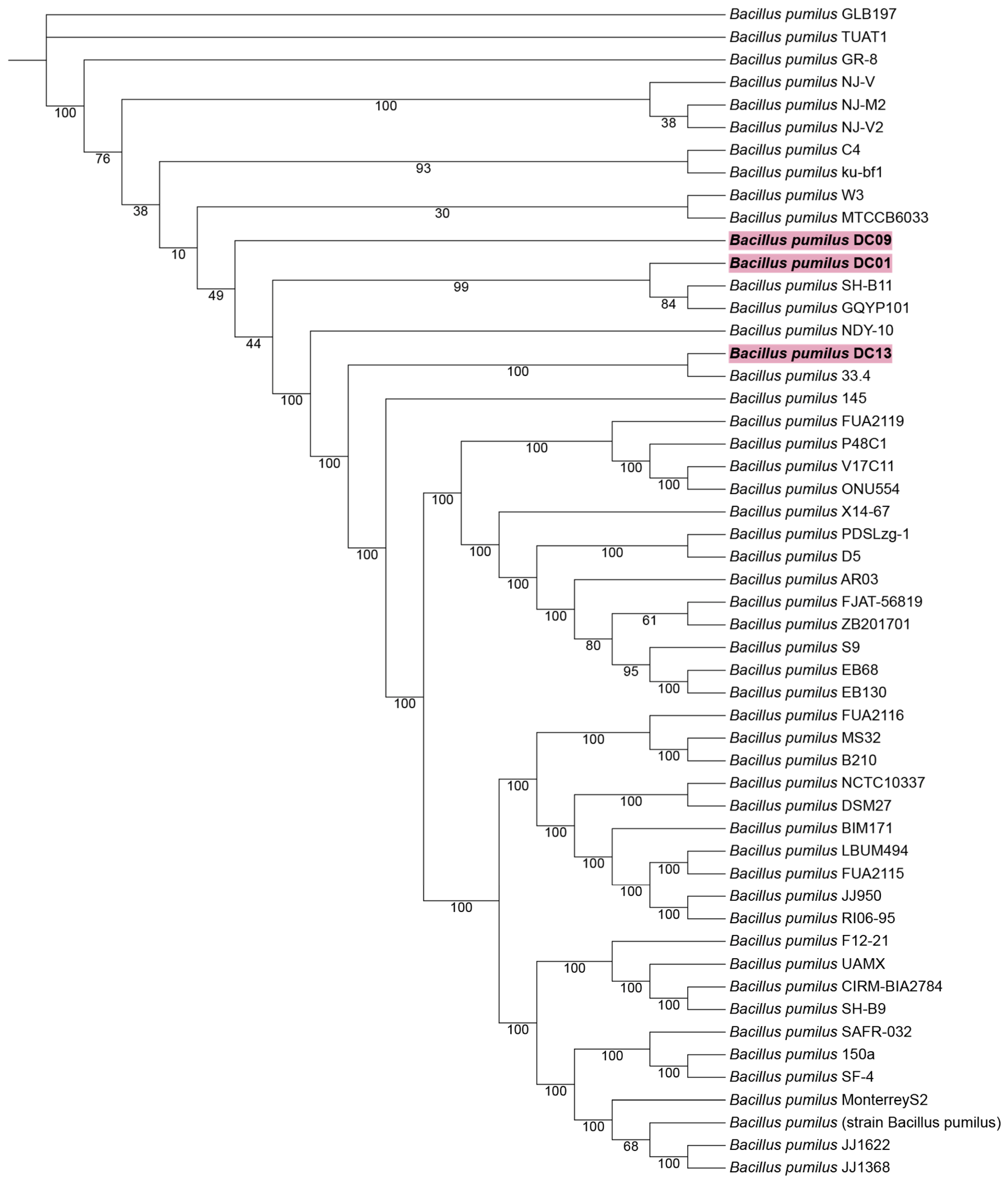
Moreover, phylogenetic analysis using a codon-based tree constructed from 100 single-copy core genes and including 49 publicly available B. pumilus genomes placed the DC isolates firmly within the B. pumilus clade, forming a well-supported subgroup (100% bootstrap support) (Figure 1). Together, these genomic analyses confirm that the soil isolates are bona fide B. pumilus strains and reveal a diverse suite of BGCs associated with potential antimicrobial production and PGP functions.

2.2. Soil-Derived B. pumilus Strains Strongly Inhibit M. oryzae Growth In Vitro

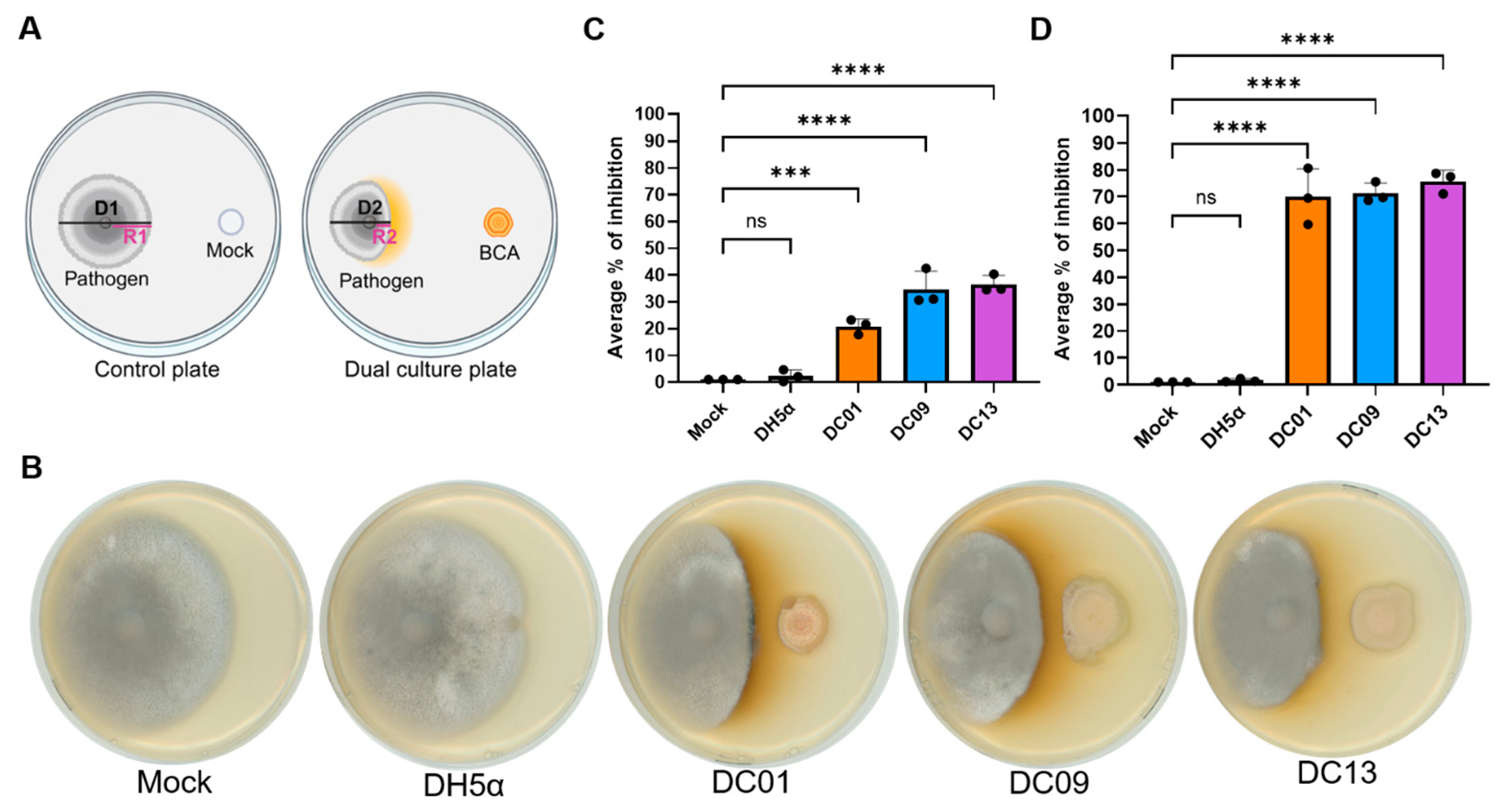
To assess whether the B. pumilus DC strains possess growth-inhibitory and antagonistic properties, we performed dual-culture assays on CM agar against M. oryzae. In this setup, each bacterial isolate (DC01, DC09, and DC13) was spotted opposite a central plug of the M. oryzae Guy11 strain, and fungal growth was monitored over a 10-day period (Figure 2A). At 10 days post-inoculation (dpi), all three isolates strongly suppressed fungal growth compared with uninoculated controls and the non-antagonistic control E. coli DH5α (Figure 2B). By 10 dpi, the overall colony diameter of M. oryzae was reduced by approximately 20–36% in the presence of DC01, DC09, or DC13 compared with untreated controls (Figure 2C). When fungal growth was quantified specifically along the axis facing the bacterial streak, inhibition was even more pronounced, reaching a 70–76% reduction in radial growth relative to mock-treatment plates (Figure 2D). At this stage, all three B. pumilus strains produced inhibition zones with a characteristic yellow-orange pigmentation at the bacterial-fungal interface. This pigmentation coincided with visibly reduced hyphal density, suggesting local accumulation of bacterial-derived inhibitory factor(s). These robust and reproducible antagonistic phenotypes supported the selection of DC strains for further characterization.

2.3. Cell-Free Supernatants of B. pumilus Strains Inhibit M. oryzae Growth

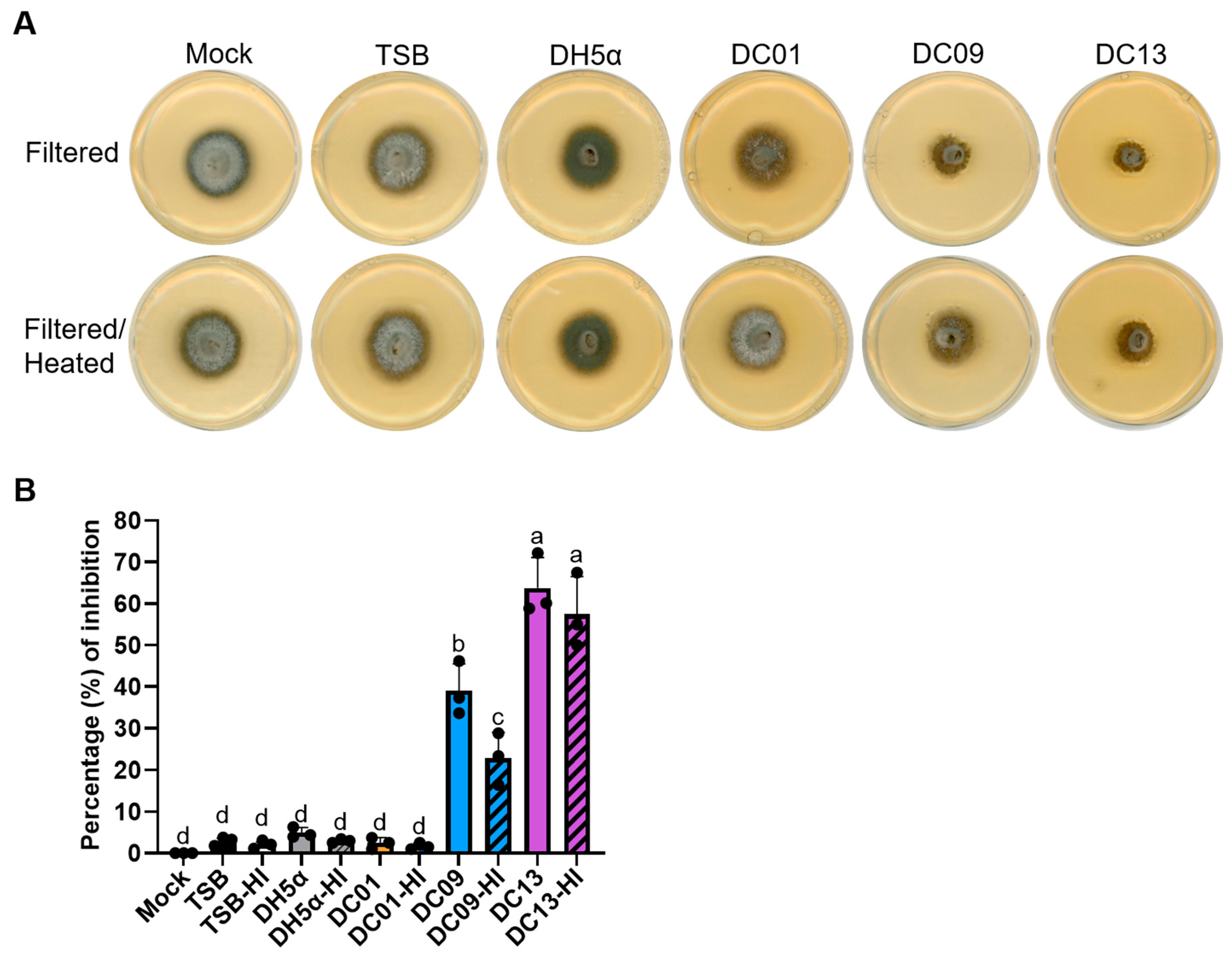
We next assessed whether cell-free supernatant (CFS) from the B. pumilus DC strains could inhibit M. oryzae growth in vitro, in the absence of living bacterial cells. Filtered CFS from DC01, DC09, and DC13, respectively, was incorporated into CM agar at a 1:6 ratio, and radial fungal growth was measured at 5 dpi. We observed that CFS from DC09 and DC13 significantly suppressed fungal colony expansion, whereas CFS from DC01 had little effect, comparable to the E. coli DH5α and mock controls (Figure 3). Quantitatively, DC13 CFS reduced radial growth by 62.6% ± 6.4%, and DC09 by 39.1% ± 6.4%. DC01 displayed only 2.5% ± 1.4% inhibition, which was not significantly different from the control treatments (Figure 3B).
To test whether the antifungal factors are heat-sensitive, CFS samples were heat-inactivated (HI) prior to incorporation. After HI treatment, DC13-HI maintained a strong inhibitory effect (57.5% ± 9.0%), and DC09-HI retained moderate activity (22.8% ± 6.1%), both significantly greater (p < 0.05) than controls (Figure 3A,B). By contrast, DC01-HI showed negligible inhibition (1.6% ± 0.7%), similar to mock. These results indicate that DC09 and DC13 secrete heat-stable extracellular factor(s) that inhibit M. oryzae growth in a cell-free context.
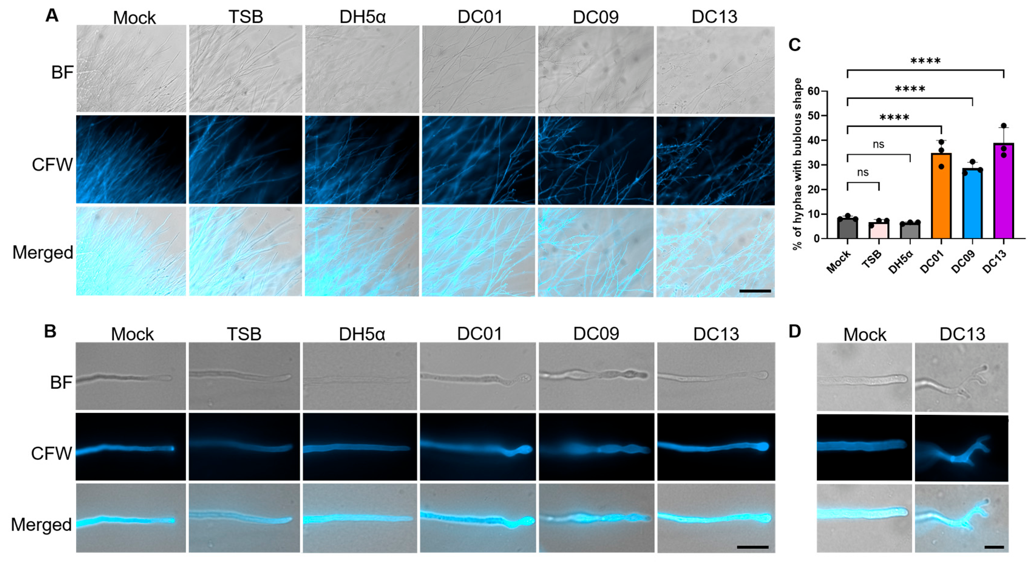
To further examine how diffusible extracellular factor(s) influence fungal development, M. oryzae was grown on thin CM agar layers supplemented with bacterial CFS and analyzed microscopically at 5 dpi. Hyphae exposed to DC isolate supernatants displayed sparse and irregular mycelial networks, with altered growth trajectories and reduced directional uniformity (Figure 4A). Among the treatments, DC13 produced the most pronounced effect, disrupting polarized hyphal extension and promoting extensive lateral branching. In contrast, hyphae grown on CM, CM supplemented with TSB, or CM containing E. coli DH5α supernatant exhibited dense and organized mycelial networks with continuous outward hyphal extension.
Visualization of individual hyphae further revealed that exposure to DC isolate supernatants induced hyphal swelling, resulting in a bulbous appearance at the apical regions (Figure 4B). DC01 and DC09 caused pronounced subapical swelling and localized tip abnormalities, whereas DC13 induced tip hyphal swelling and extensive branching defects, accompanied by apical curvature and subapical swelling (Figure 4B,D). Quantification of bulbous hyphae showed that DC01, DC09, and DC13 increased the proportion of abnormal hyphae to approximately 34.8% ± 5.0%, 28.6% ± 2.4%, and 38.9% ± 6.3%, respectively (Figure 4C). In contrast, mock, TSB, and E. coli DH5α treatments exhibited low frequencies of abnormal morphology, averaging approximately 8.4% ± 0.7%, 6.6% ± 1.15%, and 6.4% ± 0.3%, respectively, for bulbous hyphae. Together, these results indicate that extracellular factor(s) produced by DC isolates strongly alter hyphal growth patterns and morphogenesis in M. oryzae.

2.4. DC Strains Suppress M. oryzae Growth Through Volatile-Mediated Mechanisms

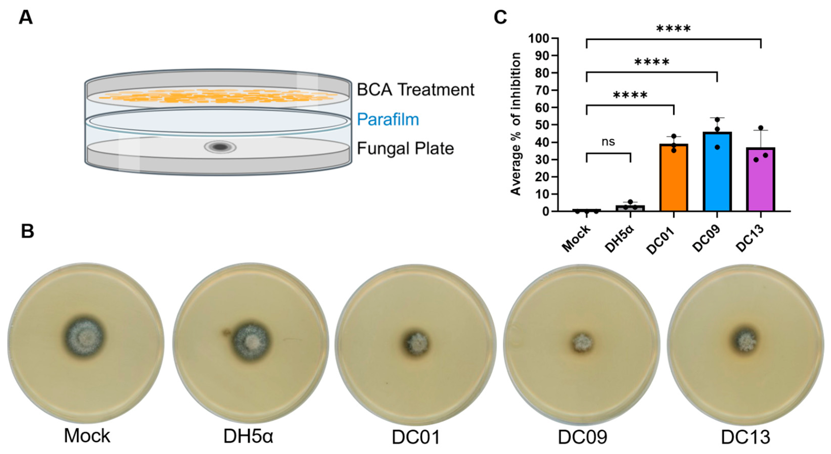
Once B. pumilus DC strains were confirmed to exhibit direct antagonism against M. oryzae in dual-culture assays, we next evaluated whether these strains also produced volatile organic compounds (VOCs) that could inhibit fungal growth. VOC activity was assessed using a sealed stacking plate assay (Figure 5A), in which the bacteria and fungus shared the same headspace but did not physically contact each other [19]. Radial growth was measured at 5 dpi to minimize the risk of cross-contamination between plates, including contamination from mature fungal spores or bacterial droplets. All three B. pumilus strains generated VOCs that reduced M. oryzae radial growth by approximately 30–45% compared with the untreated control (Figure 5B,C). Under the same conditions, the negative control E. coli DH5α did not measurably affect fungal growth. These results indicate that DC01, DC09, and DC13 release biologically active VOCs that suppress M. oryzae colony expansion. When combined with the dual-culture assays, these data demonstrate that the three B. pumilus isolates inhibit M. oryzae via both contact-dependent and volatile-mediated mechanisms, highlighting their potential as multifaceted biocontrol agents.

2.5. DC Strains Produce IAA and Exhibit Nitrogen Fixation Potential

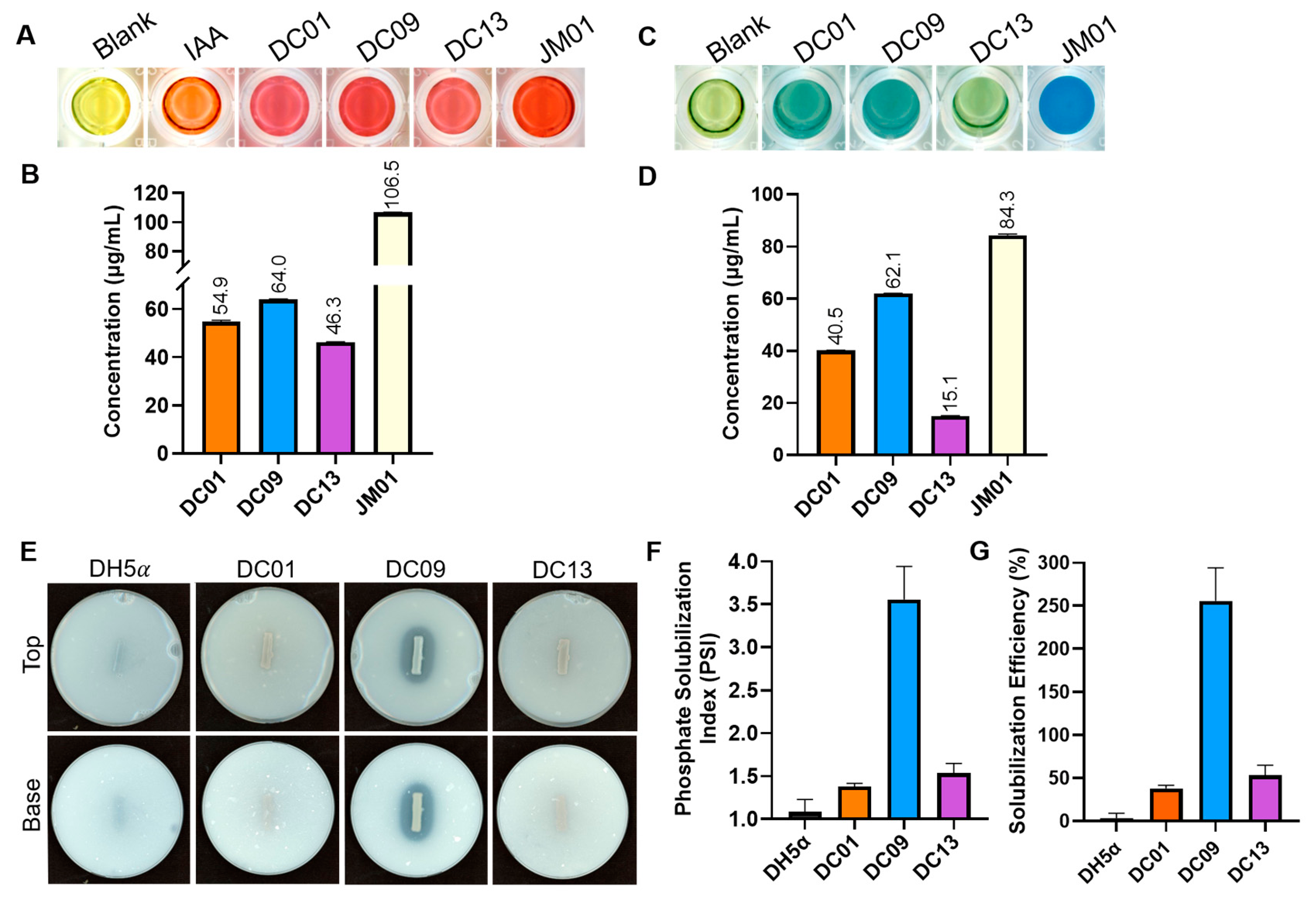
To evaluate the PGP traits of the B. pumilus DC strains, we conducted a series of assays targeting key plant-beneficial functions. IAA production by B. pumilus DC strains was quantified using the Salkowski reagent method. The development of pink/red coloration when CFS is incubated with Salkowski’s reagent indicates the presence of indole compounds, proportional to IAA concentration. All three strains produced a clear pink-to-red signal and measurable IAA levels (Figure 6A). DC01, DC09, and DC13 synthesized 54.9 ± 0.52, 64.0 ± 0.15, and 46.3 ± 0.14 µg/mL IAA, respectively, while the positive-control strain Azospirillum brasilense Sp7 (JM01) reached 106.5 ± 0.12 µg/mL (Figure 6B). No color change or IAA signal was detected in the reagent-only blank.
Next, to evaluate the nitrogen-fixing potential of the DC strains, each isolate was cultured in nitrogen-free bromothymol blue (NFb) medium. DC01 and DC09 produced a more intense blue coloration than DC13, suggesting stronger metabolic activity under nitrogen-limited conditions (Figure 6C). Nitrogen-containing metabolite accumulation was subsequently quantified using a colorimetric OD610 assay. DC01, DC09, and DC13 produced 40.5 ± 0.07, 62.1 ± 0.03, and 15.1 ± 0.06 µg/mL fixed-nitrogen equivalents, respectively, compared with 84.3 ± 0.50 µg/mL for the positive control A. brasilense Sp7 (JM01) (Figure 6D). These results indicate that DC strains exhibit measurable nitrogen-fixing potential in NFb medium, together with IAA production, supporting their classification as PGPB.

2.6. Phosphate Solubilization Differs Among the B. pumilus Isolates

To detect phosphate-solubilizing activity, B. pumilus DC strains were inoculated as lines on PKV media, and halo formation was monitored along the bacterial streak. DC09 produced a pronounced clearing zone, whereas DC13 and DC01 generated smaller and less distinct halos. In contrast, the negative-control strain E. coli DH5α showed no visible halo (Figure 6A). Quantitative measurements revealed that DC09 exhibited the strongest phosphate-solubilizing activity, with a Phosphate Solubilization Index (PSI) of 3.55 ± 0.4 and a Solubilization Efficiency (SE) of 255% ± 38%, indicating robust mobilization of insoluble phosphate (Figure 6F,G). DC01 and DC13 exhibited low solubilization, with PSI values of 1.37 ± 0.04 and 1.53 ± 0.11, respectively, and SE values of 37.7% ± 3.8% and 53.33% ± 11%. By comparison, E. coli DH5α displayed only minimal solubilization (PSI = 1.08; SE = 8.33%), close to the basal level observed for the water control. Together, these results demonstrate that B. pumilus DC09 can solubilize inorganic phosphate and exhibits the highest phosphate-solubilizing potential among the tested strains.

2.7. PEG-Induced Osmotic Stress Reveals Enhanced Tolerance in B. pumilus DC Strains

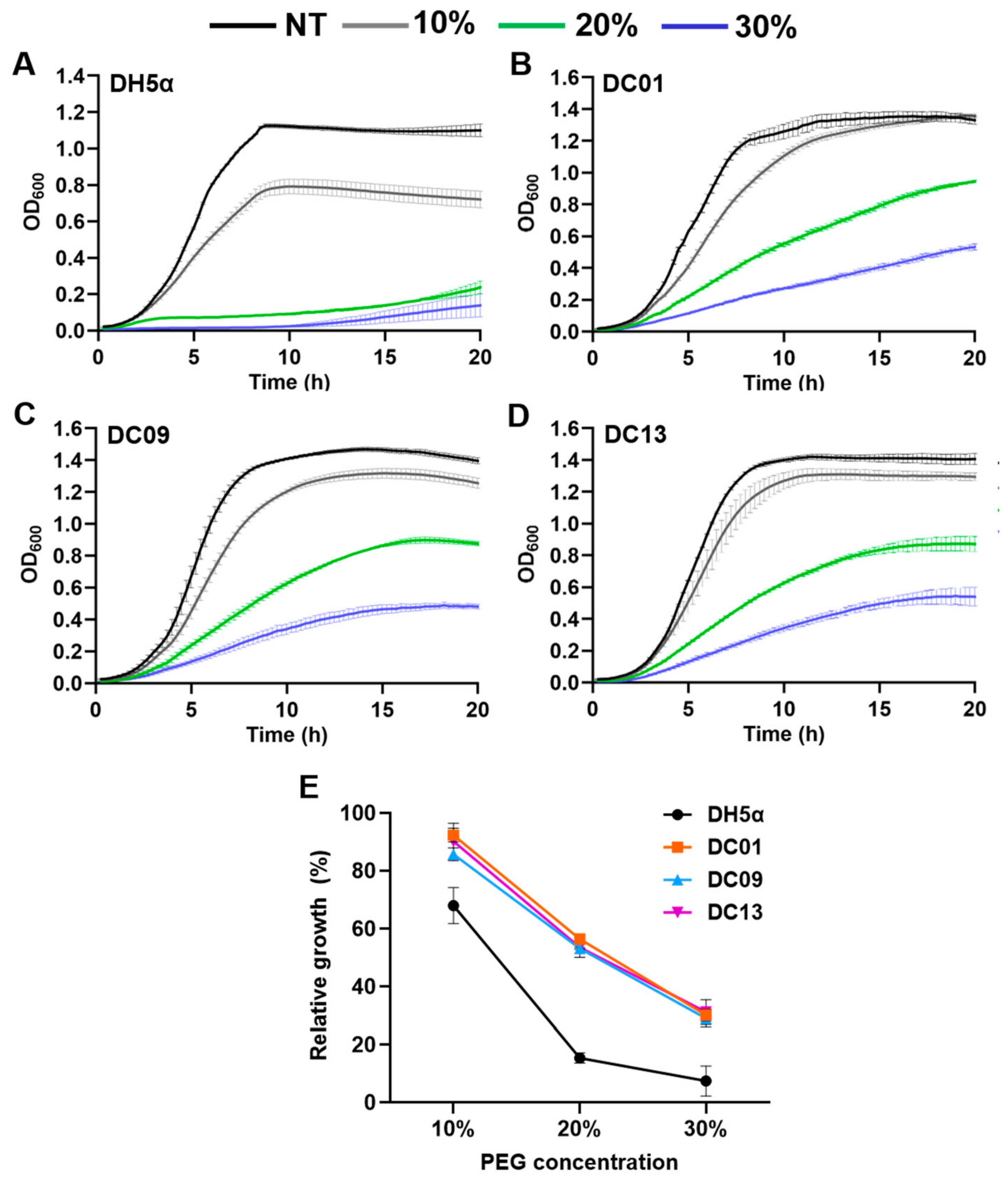
To assess whether the B. pumilus DC strains possess traits associated with drought resilience, we evaluated their growth under osmotic stress induced by polyethylene glycol (PEG). Growth kinetics monitored over 20 h revealed that all three DC strains grew substantially faster in greater PEG concentrations than E. coli DH5α (Figure 7A–D). While growth progressively declined as PEG concentration increased, DC01, DC09, and DC13 continued to proliferate under both 20% and 30% PEG conditions, whereas E. coli DH5α exhibited severe growth inhibition. To further quantify osmotic stress tolerance, the area under the curve (AUC) was calculated and normalized to untreated controls to determine relative growth (%) (Figure 7E). At 10% PEG, DC01 showed a relative growth of 92.3% ± 2.34%, DC09 85.7% ± 2.23%, and DC13 90.2% ± 6.24%, whereas E. coli DH5α reached only 67.8% ± 6.28%. At 20% PEG, growth decreased across strains; however, the DC isolates maintained significantly greater tolerance, with DC01 at 56.4% ± 1.57%, DC09 at 53.1% ± 0.29%, and DC13 at 53.5% ± 3.41%, compared to E. coli DH5α at 15.3% ± 1.74%. Under 30% PEG, DC01 (30.1% ± 1.95%), DC09 (29.1% ± 2.99%), and DC13 (31.2% ± 1.3%) retained markedly higher growth, whereas E. coli DH5α dropped to 11.78% ± 0.10%. While slight variations were observed among DC strains, their overall responses were highly similar across conditions, indicating no clear differentiation in drought tolerance among the isolates. Together, these findings demonstrate that all B. pumilus DC strains exhibit strong and consistent tolerance to PEG-induced osmotic stress, as supported by both growth kinetics and AUC-based quantification.

2.8. Soil Inoculation with B. pumilus DC Strains Reduces Rice Blast Severity and Induces Resistance

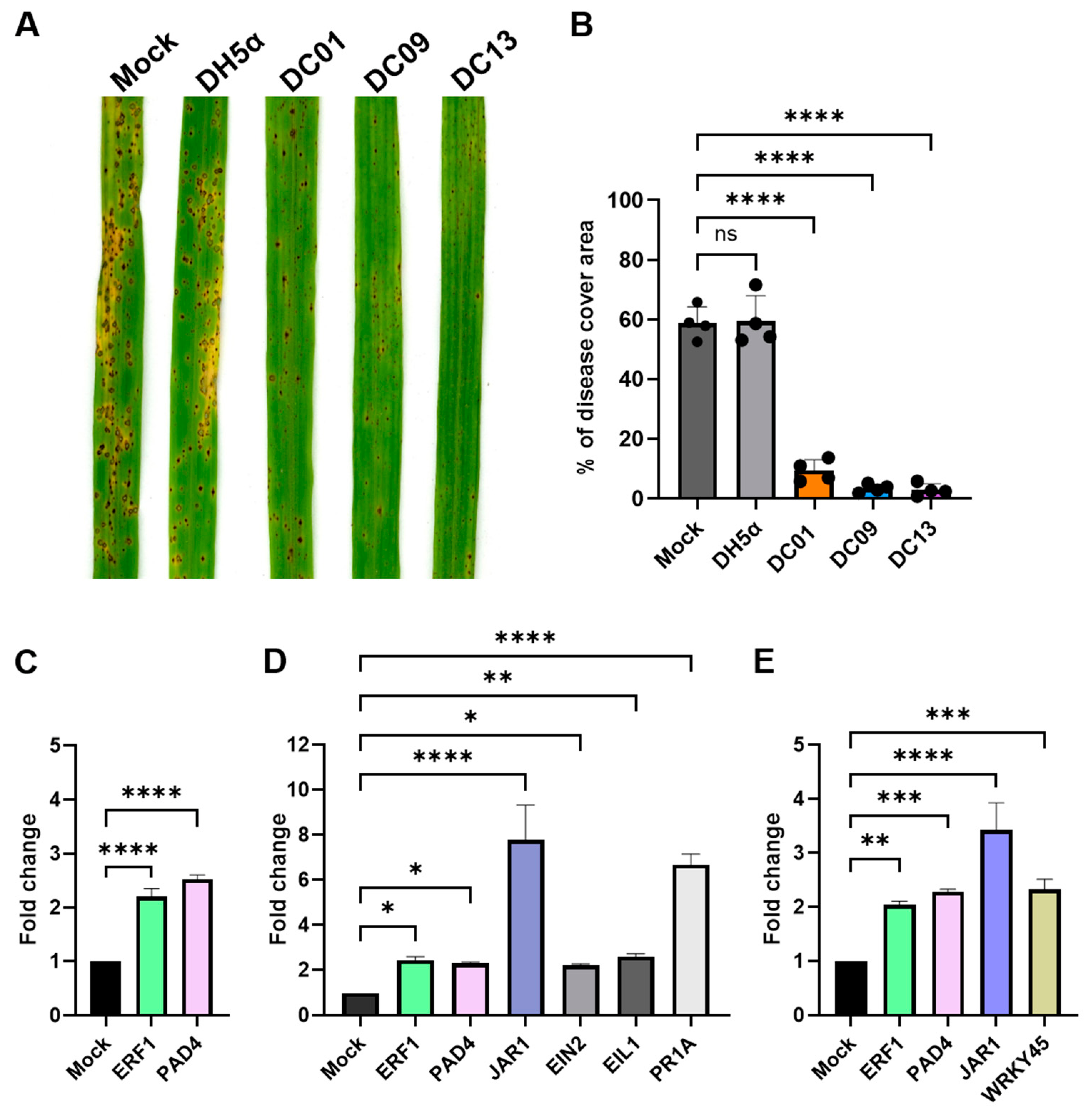
To evaluate whether the B. pumilus DC strains could reduce rice blast disease severity, we conducted a soil-treatment bioassay followed by foliar challenge with M. oryzae Guy11. Bacterial suspensions of DC01, DC09, or DC13 were applied directly to the soil surrounding the rice plants. After 24 h, the plants were spray-inoculated with M. oryzae spores. Plants inoculated with the DC strains developed substantially fewer lesions than the mock and E. coli DH5α treatments (Figure 8A). While mock- and E. coli DH5α-treated leaves displayed extensive necrotic lesions characteristic of severe blast infection, leaves from DC-treated plants exhibited markedly reduced lesion density and overall diseased leaf area (Figure 8A). Quantification of disease severity further confirmed that all three DC strains significantly reduced blast symptoms compared with the control treatments (Figure 8B). These results demonstrate that a 24 h pre-treatment with the DC isolates induces a protective response that restricts pathogen development in rice leaves.

2.9. Rice Defense Pathways Are Activated Following DC Strain Inoculation

To determine whether the DC strains trigger early immune signaling consistent with ISR, we quantified the expression of a panel of JA-, ET-, SA-, and defense-associated marker genes 24 h after root inoculation with B. pumilus DC01, DC09, or DC13. The genes examined included OsJAR1, OsERF1, OsEIN2, OsEIL2 (JA/ET signaling markers), OsWRKY45 and OsPR1a (SA-responsive markers), and OsPAD4 (a defense-associated marker). All three DC strains altered the expression of at least a subset of these markers relative to non-inoculated controls, but the magnitude and pattern of induction differed among isolates (Figure 8C–E). DC09 produced the strongest overall transcriptional response, with notable increases in JA/ET-associated genes (OsJAR1, OsERF1, OsEIN2, OsEIL2) (Figure 8D). OsJAR1 expression was also elevated in DC13-treated plants, but not in DC01-treated plants, suggesting strain-specific activation of JA-related signaling. DC01 and DC13 elicited more moderate or gene-specific expression changes, suggesting that early immune activation varies across strains (Figure 8C,E). SA-responsive genes showed modest induction across treatments. OsWRKY45 and OsPR1a increased primarily in DC09-treated plants, while OsERF1 and OsPAD4 were the only markers consistently upregulated by all three isolates (Figure 8C–E), suggesting a shared transcriptional response to DC strain treatment. These expression changes were detected before pathogen inoculation and therefore represent responses to the bacterial treatment.

2.10. DC Strains Exhibit Broad-Spectrum Antagonistic Activity

To assess whether the inhibitory activity of the DC strains extends across multiple taxa, we evaluated their antagonistic effects against selected fungal plant pathogens and bacterial human-associated pathogens. In dual-culture assays, all three B. pumilus DC strains inhibited the growth of agriculturally important fungi, although the magnitude of inhibition varied among strains and pathogens. DC01, DC09, and DC13 reduced the radial growth of Botrytis cinerea ME-1 by approximately 70%, 60%, and 55%, respectively, and of Colletotrichum orbiculare RH-18 by 20%, 30%, and 17%, as evidenced by restricted fungal expansion toward the bacterial streak (Table 2, Figure S1). These results indicate that the DC strains possess antifungal activity against multiple plant pathogens.
To evaluate antibacterial activity, DC01, DC09, and DC13 were tested against a panel of human-associated bacteria using cross-streak and spot-inoculation assays (Table 2), including the Gram-positive species Micrococcus luteus SK58 and Staphylococcus aureus ATCC 12600, and the Gram-negative species E. coli MG1655, Klebsiella pneumoniae KP35, and Pseudomonas aeruginosa PAO1. All three strains inhibited M. luteus SK58, producing measurable zones of inhibition ranging from 4.0 ± 1.9 mm (DC01) to 4.2 ± 0.99 mm (DC09), and a larger halo of 8.5 ± 1.34 mm for DC13. In contrast, inhibition of S. aureus ATCC 12600 was observed only for DC13 (5.2 ± 0.7 mm), whereas no detectable inhibition was observed for DC01 or DC09. No zones of inhibition were detected for any DC strain against the Gram-negative bacteria tested (E. coli MG1655, K. pneumoniae KP35, or P. aeruginosa PAO1). Together, these data indicate that the DC strains, particularly DC13, exhibit stronger antibacterial activity against Gram-positive taxa, consistent with the presence of predicted BGCs in their genomes.

3. Discussion

Rice blast caused by M. oryzae remains one of the most destructive diseases affecting rice production worldwide, underscoring the need for sustainable alternatives to chemical fungicides and the limited durability of resistance genes. In this study, we integrated comparative genomics, antagonistic assays, CFS-mediated inhibition, and host transcriptional profiling to characterize three B. pumilus isolates as multi-mechanistic biological control agents against M. oryzae. Collectively, our findings support a model in which these strains suppress rice blast through both direct antifungal activity and host immune modulation, while also revealing substantial strain-specific variation in biocontrol-associated traits. Similar multifunctional biocontrol strategies have been widely associated with B. pumilus and related Bacillus spp., including antibiosis, volatile production, hydrolytic enzyme secretion, and ISR [21].
Previous studies have shown that B. pumilus strains can suppress plant pathogens, promote plant growth, or induce ISR [27,30,31]. However, these properties are often examined separately. In the present study, the integrated comparison of three closely related isolates revealed distinct strain-specific profiles. DC09 produced the strongest induction of rice defense-associated genes, particularly JA/ET-related markers, whereas DC13 exhibited the strongest CFS-mediated inhibition and caused the most pronounced hyphal abnormalities. These findings suggest that DC09 and DC13 differ in the relative contributions of host defense activation and direct antifungal activity.
Whole-genome sequencing revealed that all three DC isolates harbor diverse NRPS and PKS BGCs, including clusters predicted to encode lipopeptides and siderophores commonly associated with Bacillus-mediated biocontrol. These genomic features are consistent with the experimentally observed antifungal and PGP traits and indicate potential genetic differences observed among DC01, DC09, and DC13. Previous studies have shown that B. pumilus strains frequently encode biosynthetic pathways involved in the production of surfactins, fengycins, bacilysin, and other antimicrobial metabolites that contribute to fungal suppression and plant colonization [21]. The repeated detection of siderophore-associated clusters across all three genomes further supports their potential role in nutrient competition and rhizosphere fitness. Because the DC13 genome remains partially assembled, its complete biosynthetic repertoire may not yet be fully represented.
Comparative analysis of the selected lichenysin- and fengycin-associated regions showed that DC01 and DC09 share broadly similar BGC architectures but contain sequence-level differences among several predicted proteins. These differences could potentially affect biosynthetic enzyme function, cluster regulation, or metabolite production and may contribute to the strain-specific phenotypes observed in this study. However, the presence of a predicted BGC or sequence variation within a cluster does not demonstrate that the corresponding metabolite was produced under the experimental conditions. Because the DC13 genome remains partially assembled, apparent cluster absence or incompleteness may also reflect assembly fragmentation rather than true biological differences.
The DC isolates inhibited M. oryzae through multiple antagonistic mechanisms, including direct interaction, diffusible extracellular factors, and VOC-mediated suppression. In dual-culture assays, all three strains significantly reduced fungal radial growth, demonstrating strong antagonistic activity during direct interaction. However, clear strain-specific differences emerged in the CFS assays. DC09 and DC13 retained strong inhibitory activity in the absence of living cells, whereas DC01 displayed minimal supernatant-mediated inhibition despite suppressing fungal growth in dual culture. The discrepancy between the dual-culture and CFS assays suggests that DC01-mediated inhibition may depend on direct bacterial–fungal interaction or localized metabolite production rather than freely diffusible compounds accumulated during growth in liquid culture. Similar observations have been reported in other Bacillus biocontrol systems, where strains displaying distinct lipopeptide signatures nevertheless exhibited comparable antifungal activity, suggesting that additional metabolites or interaction-dependent mechanisms may contribute substantially to pathogen suppression [32]. DC01 may therefore produce antifungal factors only in response to fungal proximity, or its inhibitory activity may be associated with cell-bound or surface-associated metabolites that are not efficiently recovered in the filtered supernatant. This distinction highlights mechanistic differences among the DC isolates despite their shared species classification.
An important observation was that antifungal activity persisted after heat treatment of the supernatants. Retention of inhibitory activity following heat inactivation suggests that at least part of the antifungal activity is mediated by heat-stable extracellular compounds, consistent with Bacillus-derived cyclic lipopeptides and related secondary metabolites reported in previous studies [21,33]. Cyclic lipopeptides such as fengycins, surfactins, and iturins are well known for their stability and membrane-disrupting activity against fungal pathogens [32,33,34,35,36]. Although the specific metabolites responsible for inhibition remain to be identified, the combination of predicted biosynthetic clusters and robust supernatant activity suggests a potential role for secreted secondary metabolites in the antagonistic activity of DC09 and DC13. However, the stronger CFS-mediated inhibition observed for these strains cannot yet be directly linked to individual BGCs. Future studies should integrate metabolomic characterization of the secreted compounds with detailed analyses of BGC completeness, gene organization, sequence variation, and associated regulatory regions.
Microscopic analysis further demonstrated that DC supernatants substantially altered the morphology of M. oryzae hyphae. Exposure to supernatants from DC01, DC09, and DC13 induced bulbous swelling, altered hyphal polarity, apical deformation, and increased branching, with DC09 and DC13 producing the most pronounced structural abnormalities. Similar morphological alterations have previously been associated with antifungal lipopeptides produced by Bacillus spp., particularly members of the iturin and fengycin families, which disrupt fungal membrane integrity by permeabilizing membranes and destabilizing lipid bilayers [34,35,36]. These compounds have been reported to inhibit several phytopathogenic fungi, including Botrytis cinerea, Fusarium spp., and other filamentous fungi. In contrast, the comparatively weaker phenotype induced by DC01 is consistent with its limited inhibitory activity in the CFS assays.
VOCs produced by DC isolates also contributed to fungal suppression. Physically separated VOC assays demonstrated that fungal growth inhibition could occur in the absence of direct contact or shared medium, indicating that volatile metabolites participate in antagonism. Similar VOC-mediated inhibition has been reported for several Bacillus species, including B. pumilus, in which volatile compounds contribute to the suppression of fungal phytopathogens in soil and rhizosphere environments [21,37,38]. The ability to inhibit pathogens through VOCs may be particularly advantageous under natural conditions, where volatile compounds can diffuse through soil pore spaces and influence microbial interactions at a distance. Although attempts were made to identify the volatile compounds produced by the DC isolates, these efforts were unsuccessful under the conditions tested. Future studies aimed at characterizing the volatile metabolite profiles of these strains will help clarify the contribution of VOCs to DC-mediated fungal inhibition.
Beyond M. oryzae, the DC isolates also displayed antagonistic activity against additional fungal and bacterial taxa, indicating that their inhibitory capacity is not restricted to a single pathogen. All three strains suppressed the growth of the phytopathogenic fungi B. cinerea and C. orbiculare, with varying degrees of inhibition observed among isolates. The DC isolates additionally inhibited the Gram-positive bacteria M. luteus, while DC13 uniquely suppressed S. aureus. In contrast, no detectable inhibition was observed against the Gram-negative bacteria tested. This differential activity spectrum is consistent with previous reports showing that Bacillus-derived antimicrobial compounds often display stronger activity against Gram-positive organisms due to differences in cell envelope structure and membrane susceptibility [21]. Collectively, these findings suggest that DC isolates possess antagonistic activities with varying target specificity. In addition to antifungal activity, all DC isolates displayed PGP traits, although these traits varied substantially among strains. Members of the genus Bacillus are widely recognized for their ability to enhance plant growth through phytohormone production, nutrient mobilization, and stress tolerance enhancement [14,39]. In the present study, all three isolates produced IAA and tolerated osmotic stress, while DC09 uniquely displayed phosphate-solubilizing activity. These findings reinforce the concept that plant-beneficial traits are highly strain-dependent, even among closely related isolates. Previous studies similarly demonstrate that B. pumilus strains vary considerably in their expression of phosphate solubilization, phytohormone production, siderophore secretion, and stress adaptation traits [21].
Importantly, soil inoculation with all three DC strains significantly reduced rice blast disease severity in planta, demonstrating that these isolates retain biocontrol activity under host-associated conditions. To investigate whether host immune activation contributed to disease suppression, we analyzed the expression of defense-associated genes involved in JA/ET signaling and SA-associated responses. All three DC strains altered host transcriptional responses relative to mock-treated plants, although the magnitude and composition of induction differed among isolates. A central observation was the consistent upregulation of OsERF1 and OsPAD4 across all DC treatments. In rice, OsERF1 is associated with JA/ET-responsive defense signaling, whereas OsPAD4 has been implicated in rice host–pathogen interactions and basal defense responses [40,41,42,43]. This suggests that root colonization by the DC isolates establishes an enhanced defensive state prior to pathogen challenge. Notably, DC09 induced the strongest activation of JA/ET-associated genes, including OsERF1, OsJAR1, OsEIN2, and OsEIL2. This transcriptional profile is consistent with ISR-like activation and aligns with previous studies demonstrating that Bacillus spp. can stimulate JA/ET-mediated defense pathways in plants [21,25,26,27]. In contrast, DC01 and DC13 induced more moderate transcriptional responses, suggesting that although all three strains engage host defense pathways, the magnitude and composition of immune activation differ between isolates.
Taken together, our genomic, in vitro, and in planta analyses support a model in which the DC isolates suppress rice blast through multiple complementary mechanisms, including diffusible antifungal activity, VOC-mediated inhibition, disruption of fungal development, and activation of host defense pathways. Among the isolates, DC09 and DC13 displayed the most integrated biocontrol profiles, combining strong CFS-mediated antagonism with activation of host defense-associated genes. More broadly, the substantial phenotypic differences observed among these closely related isolates highlight the importance of evaluating Bacillus strains individually rather than relying solely on species-level classifications when developing BCAs. To our knowledge, this study represents one of the most comprehensive characterizations of strain-specific antagonistic and plant-associated traits in B. pumilus isolates active against M. oryzae, integrating comparative genomics, CFS-associated inhibition, fungal morphogenesis, VOC-mediated antagonism, and host defense activation.
Future work should focus on identifying the metabolites responsible for antifungal activity, linking specific BGCs to inhibitory phenotypes, further characterizing the molecular basis of host immune activation, and evaluating the efficacy of the DC isolates under field conditions. Nonetheless, the present study demonstrates that the DC isolates, particularly DC09 and DC13, represent promising candidates for sustainable management of rice blast disease and further illustrate the potential of B. pumilus as a versatile platform for agricultural biocontrol.

4. Materials and Methods

4.1. Microbial Strains and Culture Conditions

The B. pumilus DC strains used in this study were recovered from various topsoil samples collected in the Gainesville, Florida area (29.643280° N, −82.331693° W; 29.624527° N, −82.361428° W; and 29.6241345° N, −82.3638663° W). Bacterial strains were routinely cultured and maintained in Luria–Bertani (LB) broth containing 10 g/L tryptone, 5 g/L yeast extract, and 5 g/L NaCl. Medium was autoclaved at 121 °C and 15 psi for 20 min prior to use. For routine culturing, bacterial strains were grown overnight in 3 mL LB broth at 30 °C with shaking at 200 revolutions per minute (rpm). Long-term stocks were maintained in 20% glycerol at −80 °C.
The fungal strain Magnaporthe oryzae Guy11, maintained in our laboratory collection, was used throughout this study and cultured on complete medium (CM). CM consisted of 50 mL of 20× nitrate salts, 1 mL trace elements, 10 g D-glucose, 2 g peptone, 1 g yeast extract, 1 g casamino acids, and 1 mL vitamin solution per liter of distilled water. The pH was adjusted to 6.5 using 5 M NaOH. For a solid medium, 15 g/L agar was added prior to autoclaving. Long-term fungal stocks were stored on sterile Whatman filter paper at −20 °C.
Additional fungal strains used in antagonism assays, including Botrytis cinerea RH-18 and Colletotrichum orbiculare ME-1, were kindly provided by Dr. Jeffrey Rollins’ laboratory. Additional bacterial strains used in antagonism assays, including Micrococcus luteus SK58, Staphylococcus aureus ATCC 12600, Escherichia coli MG1655, Klebsiella pneumoniae KP35, and Pseudomonas aeruginosa PAO1 were cultured in LB broth at 37 °C with shaking at 200 rpm. These additional fungal and bacterial strains were included to determine whether the antagonistic activity of the DC isolates was specific to M. oryzae or extended to other fungal pathogens and bacterial species.

4.2. Plant Growth Conditions

Seeds of Oryza sativa cultivar CO39 were dehusked and surface sterilized by immersion in 70% ethanol for 1 min, rinsed with sterile water for 1 min, followed by incubation in 5% bleach for 1 h. Seeds were rinsed three times with sterile water, dried on sterile paper towels, and planted in Magenta boxes containing 100 mL of half-strength Murashige and Skoog (½ MS) medium (2.25 g MS salts, 30 g sucrose, and 0.5 g MES per liter of distilled water). The pH was adjusted to 5.8 using KOH, followed by the addition of 3 g/L gelrite. The medium was autoclaved at 121 °C for 25 min, and 100 mL was dispensed aseptically into each autoclaved Magenta box. Plants were grown for 10 days at 25 °C under a 12 h light/dark photoperiod in a growth chamber. Ten-day-old seedlings were transplanted into 4-inch surface-sterilized pots containing autoclaved soil (Pro Mix HP Mycorrhizae: Michigan Peat Baccto General Purpose Potting Soil) and maintained in the greenhouse.

4.3. Whole-Genome Sequencing, Assembly, and Annotation of Bacterial Isolates

Bacterial isolates were grown overnight in 5 mL of fresh LB medium at 30 °C while shaking at 200 rpm. Genomic DNA was extracted from bacterial isolates using the AllPrep DNA/RNA/Protein Kit (QIAGEN, Hilden, Germany) according to the manufacturer’s protocol. DNA concentration and purity were assessed spectrophotometrically, and the samples were submitted to SeqCenter (Pittsburgh, PA, USA) for 16S rRNA gene amplicon sequencing and whole-genome sequencing (WGS) using the Illumina platform. Taxonomic identification was performed using 16S rRNA gene sequences and BLASTn against the NCBI nucleotide database. For WGS, raw Illumina reads were quality-checked using FastQC v0.12.1. [44] and then processed with the BBTools suite v39.06 [45] to remove adapters and phiX contamination. Cleaned reads were assembled de novo using SPAdes v3.15.5 [46], and assemblies were polished with two iterations of Pilon v1.24 [47]. Draft genome assemblies were uploaded to the Bacterial and Viral Bioinformatics Resource Center (BV-BRC) [48] for species-level identification and functional annotation using RASTtk. Biosynthetic gene clusters were predicted using antiSMASH v8.0 with relaxed detection strictness and all additional features enabled, and predicted coding sequences within secondary metabolite regions were assigned putative functions by BLASTp analysis [29]. To compare selected BGC regions, amino acid sequences from the predicted lichenysin- and fengycin-associated regions of DC01 were used as BLASTp queries against the corresponding proteins from DC09 and DC13. The highest-confidence matches and associated alignment statistics were recorded. Phylogenetic relationships were inferred using a codon-based tree constructed in BV-BRC with RAxML v82.12 based on 100 single-copy genes, including the isolates from this study and 49 reference Bacillus pumilus genomes; trees were visualized and annotated using the interactive Tree of Life (iTOL v7.6) web interface [49].

4.4. Dual Culture Assay

Antagonistic activity was assessed using a dual culture assay adapted from [19]. Overnight bacterial cultures were adjusted to an OD600 of 0.5 with sterile water. A UV-sterilized 5 mm core borer, forceps, and rod were used to collect agar plugs from the actively growing edge of 7-day-old M. oryzae cultures. Fungal plugs were placed approximately 1.5 cm from the center of a CM agar plate, ensuring mycelial contact with the media. Opposite the fungal plug and equidistant from the center, 5 µL of normalized bacterial culture was spotted onto the plate. As a negative control, sterile water was applied in place of the bacterial inoculum. Plates were incubated at 25 °C in the dark. Fungal growth was assessed at 10 dpi. Fungal colony diameters and radii were measured and averaged for each replicate. The percentage of growth inhibition was calculated using the formula: Inhibition (%) = [(average control − average experimental)/average control] × 100. This applies to the radius and diameter of the fungal colony.

4.5. Cell-Free Supernatant (CFS) Inhibition Assay

To evaluate the effect of bacterial metabolites on M. oryzae growth, we prepared cell-free supernatants (CFS) from the B. pumilus DC strains. Each DC isolate was grown in tryptic soy broth (TSB; Difco) at 30 °C with shaking at 180 rpm for 24 h, following the approach described in [50]. Because antifungal activity was detected in CFS collected at this time point, the 24 h incubation period was used for all subsequent assays. Cultures were then centrifuged at 3200× g for 60 min at 22 °C to remove bacterial cells, and the resulting supernatants were carefully collected and filter-sterilized through a 0.22 µm syringe filter (VWR) to obtain CFS. Sterility was confirmed by plating an aliquot of CFS onto LB agar plates and verifying the absence of bacterial growth after incubation. The pH of each CFS was measured after filtration and was approximately 7.0; no pH adjustment was performed before the assay. Sterile TSB medium was included as a medium control, and CM agar without added CFS or TSB was included as a mock control. For heat-inactivation assays, CFS were incubated at 95 °C for 10 min before being incorporated into the medium. For the plate assay, CFS or sterile TSB (control) were mixed with CM at a 1:6 (v/v) ratio, corresponding to a final CFS concentration of 14.3% (v/v), thoroughly mixed, and poured into Petri dishes [51]. After solidification, a 5 mm mycelial plug of M. oryzae strain Guy11 was placed in the center of each plate. Plates were incubated at 25 °C in the dark for 5 days. At the end of the incubation period, colony diameters were measured, and plates were scanned for documentation. The percentage of growth inhibition relative to the mock treatment was calculated using the same formula applied for the dual-culture assay. The experiment included three independent biological replicates, with four technical replicates per biological replicate.

4.6. Microscopic Analysis of Fungal Development

To further assess the effect of bacterial metabolites on fungal hyphal morphology, microscopic analysis was performed on CM agar and CM supplemented with CFS or TSB (control) at a 1:6 (v/v) ratio. Briefly, 250 µL of medium was applied to autoclaved microscope slides and allowed to dry prior to inoculation. A 5 µL spore suspension (5 × 10⁵ spores/mL) of M. oryzae was applied to each end of the agar smear. Slides were incubated at 25 °C in humid chambers under a 12 h light/12 h dark photoperiod and imaged at 5 dpi using a Carl Zeiss Axio Observer 7 microscope (Zeiss, Oberkochen, Germany). For Calcofluor White staining, fungal growth was treated with one drop of Calcofluor White (Sigma, Milwaukee, WI, USA) and one drop of 10% KOH, incubated for 1 min, and imaged immediately. Images were processed using ZEN Microscopy software (v3.13; CarlZeiss; Jena, Germany).

4.7. Volatile-Mediated Inhibition Assay

Volatile inhibition of M. oryzae by bacterial strains was assessed using a sealed dual-plate system. Bacterial cultures were grown and normalized to an OD600 of 0.5 using the same procedure as described for the dual culture assay. CM agar plates were inoculated with 5 µL of normalized bacterial culture and spread evenly using sterile glass beads. For the negative control, sterile water was used in place of the bacterial culture. Plates were incubated overnight at 28 °C in the dark to allow bacterial growth. Fungal plugs were obtained from the growing edge of 7-day-old M. oryzae cultures using sterilized tools and aseptically transferred to the center of fresh CM plates. The pre-inoculated bacterial plate (lid removed) was inverted and placed on top of the fungal plate, forming a sealed volatile chamber using parafilm. The assembled plates were incubated for 5 days at 25 °C in the dark. Following incubation, fungal growth was imaged, and colony diameter was measured in two perpendicular directions and averaged. The percentage of inhibition was calculated using the formula described in the dual-culture assay.

4.8. Indole-3-Acetic Acid (IAA) Production Quantification

To quantify IAA production, 3 mL cultures of half-strength TSB supplemented with 0.5% (w/v) L-tryptophan were inoculated with bacterial strains normalized to an OD600 of 0.5. Cultures were incubated with shaking at 200 rpm for two days, beginning at 37 °C for the first 24 h and then shifted to 28 °C for the remaining 48 h. IAA production was assessed at 72 h. The temperature shift was used to support initial bacterial growth at 37 °C, followed by IAA production at a temperature closer to the optimal growth temperature of the DC strains. At this time, 500 µL of culture was centrifuged at 10,000× g for 10 min, and 50 µL of the resulting supernatant was transferred to a 96-well plate. Control conditions included a negative control containing 50 µL of 0.5% L-tryptophan-supplemented 1/5 TSB, a chemical positive control made by mixing 25 µL of 0.5% L-tryptophan TSB with 25 µL of 100 µg/mL IAA stock solution, and a biological positive control using Azospirillum brasilense (kindly provided by Dr. Jean-Michel Ané), a known IAA-producing strain. To each well, 100 µL of freshly prepared Salkowski reagent (50 mL of 35% perchloric acid mixed with 1 mL of 0.5 M FeCl3) was added, and samples were incubated in the dark at room temperature for 30 min [52,53]. Color development was visually documented, with IAA-positive samples turning pink. Absorbance was measured at 536 nm using a microplate reader, and IAA concentrations were calculated using the standard curve trendline equation: y = 0.0069x − 0.0013, R2 = 0.9937, where y is the absorbance at 536 nm and x is the IAA concentration (µg/mL). Standard curve was generated using absorbance readings from 10 to 100 ug/mL IAA standards. The experiment included three independent biological replicates, each with three technical replicates.

4.9. Phosphate Solubilization Capacity

Bacterial cultures were prepared by inoculating 3 mL of LB broth with the bacterial strain of interest and incubating for 24 h at 30 °C with shaking at 200 rpm. Cultures were normalized to an OD600 of 0.5 using sterile distilled water, and cells were washed with water to remove any trace of LB. Phosphate solubilization was assessed using Pikovskaya’s agar (PVK; 10 g D-glucose, 5 g Ca3(PO4)2, 0.5 g (NH4)2SO4, 0.2 g KCl, 0.1 g MgSO4, trace amounts (~0.001 g each) of MnSO4 and FeSO4, and 0.5 g yeast extract per liter) [53,54]. A 5 µL aliquot of normalized culture was spotted at the center of PVK plates and streaked into a 3 cm line. Plates were incubated at 37 °C for seven days. After incubation, phosphate solubilization was evaluated by the formation of clear halos around bacterial growth. Plates were imaged, and halo and colony diameters were measured. Phosphate solubilization was quantified by calculating the Phosphate Solubilization Index (PSI) and Solubilization Efficiency (SE). PSI was calculated as: (Halo diameter + Colony diameter)/Colony diameter. SE was calculated as: SE (%) = [(Halo diameter − Colony diameter)/Colony diameter] × 100.

4.10. Nitrogen Fixation Assay

Nitrogen-free bromothymol blue (NFb) medium was prepared by combining the following components per liter of distilled water: 5 g malic acid, 0.5 g K2HPO4, 0.2 g MgSO4, 0.1 g NaCl, 0.02 g CaCl2·2H2O, 2 mL micronutrient solution (containing 0.04 g CuSO4·5H2O, 0.12 g ZnSO4·7H2O, 1.40 g H3BO3, 1.0 g Na2MoO4·2H2O, and 1.175 g MnSO4·H2O; brought to 1 L and stored at 4 °C), 2 mL bromothymol blue solution (5 g bromothymol blue in 0.2 M KOH), 4 mL Fe-EDTA solution (16.4 g Fe-EDTA per liter), 1 mL vitamin solution (10 mg biotin and 20 mg pyridoxine in 100 mL distilled water), and 4.5 g KOH, the pH was set to 6.5 using KOH [28,55]. After autoclaving, a 50 mL working stock was prepared using 49.833 mL of NFb medium and 166 µL of sodium lactate solution, then stored at 4 °C.
To assess nitrogen fixation, 3 mL of overnight bacterial cultures were diluted in LB to an OD600 of 0.5. In a sterile 96-well plate, three columns were filled with 180 µL of NFb medium, and 20 µL of normalized bacterial culture was added to each well. A. brasilense was included as a biological positive control under the same conditions. Three wells containing 200 µL of uninoculated NFb medium served as negative controls. The plate was incubated at 37 °C for 24 h while shaking at 200 rpm. Absorbance was measured at 610 nm using a microplate reader. Color changes from green to blue were recorded as indicative of ammonium ion production. Fixed nitrogen concentration was determined using a standard curve and trendline equation, as reported in [28].

4.11. Drought Tolerance Assay

Drought tolerance of bacterial isolates was assessed through culturing in liquid LB medium supplemented with 10%, 20%, and 30% (w/v) polyethylene glycol (PEG 6000) to simulate osmotic stress. Overnight bacterial cultures were grown in LB and normalized to an OD600 of 0.5. A 1:100 dilution of each culture was prepared in PEG-supplemented LB. In a sterile 96-well plate, 180 µL of each PEG-containing medium was aliquoted per well, and 20 µL of the diluted bacterial suspension was added. Control wells contained uninoculated LB medium. E. coli DH5α was included as a non-tolerant control. Plates were incubated at 37 °C with continuous shaking, and bacterial growth was monitored by measuring optical density at 600 nm (OD600) over a 20 h period using a microplate reader. Growth curves were generated over time, and total growth for each condition was quantified by calculating the area under the curve using Riemann sum approximation.

4.12. Plant Inoculations and Infections

Overnight bacterial cultures were prepared and normalized to an OD600 of 0.1. Cultures were centrifuged at 10,000× g for 3 min, washed twice with sterile water, and resuspended in 1 mL of sterile water. This suspension was diluted in 9 mL of sterile water to obtain a final inoculum. 10-day-old rice seedlings were submerged in the bacterial suspension for 20 min, then transferred to sterile soil-filled 4-inch pots. Control plants were mock-inoculated using sterile water. At three weeks post-germination, plants were re-inoculated with 20 mL of the normalized bacterial culture applied directly to the soil. After 24 h, a M. oryzae spore suspension (1 × 10⁵ conidia/mL in 0.2% gelatin) was sprayed onto the leaves of each plant [12]. Plants were covered with clear plastic bags to maintain high humidity and incubated at 25 °C under a 12 h light/dark cycle in a growth chamber. After two days, bags were partially opened to gradually reduce humidity. Five days post-inoculation, leaves were harvested, imaged, and disease severity was quantified as the percentage of diseased leaf area using ImageJ 1.53t [56]. Each treatment included four plants as technical replicates within each experiment, and the complete experiment was independently repeated four times, resulting in four biological replicates per treatment (n = 4).

4.13. Gene Expression Analysis

Total RNA was extracted from rice leaf samples using the RNeasy Plant Mini Kit (QIAGEN, Hilden, Germany) according to the manufacturer’s protocol. For each biological replicate, leaf material from four plants within the same treatment was pooled, flash-frozen in liquid nitrogen, and ground to a fine powder using a mortar and pestle. RNA concentration and purity were measured using a spectrophotometer, and RNA integrity was confirmed via agarose gel electrophoresis. DNA contamination was removed using DNase I treatment. First-strand complementary DNA (cDNA) was synthesized from 1 µg of total RNA in a final volume of 20 µL using the Quantabio qScript cDNA Synthesis it (Quantabio, Beverly, MA, USA) with oligo-dT primers, following the manufacturer’s instructions.
Quantitative reverse-transcription PCR was performed using Quantabio PerfeCTa SYBR Green FastMix (Beverly, MA, USA) in a Bio-Rad CFX96 thermal cycler (Bio-Rad Laboratories, Hercules, CA, USA) with gene-specific primers. Each reaction was carried out in a total volume of 10 µL, including 2 µL of diluted cDNA, 0.5 µM of each primer (Table S6), and 1× SYBR Green master mix (Quantabio, Beverly, MA, USA). The thermal cycling conditions were as follows: initial denaturation at 95 °C for 2 min, followed by 40 cycles of 95 °C for 15 s and 60 °C for 30 s. A melt curve analysis was included to confirm the specificity of amplification. GAPDH was used as the reference gene for normalization [57]. Relative gene expression levels were calculated using the 2−ΔΔCt method, and expression was reported as fold change relative to uninoculated control plants. Each biological replicate was analyzed in technical triplicate, and the experiment was independently performed three times, resulting in three biological replicates per treatment (n = 3).

4.14. Statistical Analysis

Statistical analyses and graphing were performed using GraphPad Prism v10.6.1 (GraphPad Software, Boston, MA, USA). Unless otherwise indicated, experiments were conducted using three independent biological replicates, each containing three technical replicates. Technical replicates were averaged to obtain one value for each biological replicate. Before ANOVA, the normality of residuals was assessed using the Shapiro–Wilk test, and homogeneity of variance was evaluated using the Brown–Forsythe test. No significant deviations from normality or differences in variance were detected. Statistical significance was determined using ordinary one-way or two-way ANOVA followed by Dunnett’s or Tukey’s post hoc multiple comparison tests, as appropriate (p < 0.05). Error bars represent the standard deviation (SD) of biological replicates.

Supplementary Materials

The following supporting information can be downloaded at: https://www.mdpi.com/article/10.3390/ijms27167197/s1.

Author Contributions

Conceptualization, J.F.; methodology, J.F., D.M.C., L.E.K., T.R.J., G.L.E., N.P. and R.E.K.; writing, J.F., D.M.C., G.L.E., L.E.K. and R.E.K.; supervision, J.F. and D.M.C. All authors have read and agreed to the published version of the manuscript.

Funding

L.E.K, T.R.J., and R.E.K. were supported by the University of Florida Office of Research through the Research Opportunity Seed Fund (ROSF). D.M.C was partially funded by the Innovative Teaching Award from the Association of Public and Land-grant Universities (APLU).

Institutional Review Board Statement

Not applicable.

Informed Consent Statement

Not applicable.

Data Availability Statement

The original data presented in the study are openly available in NCBI Sequence Read Archive (SRA) under BioProject accession PRJNA1480024. The corresponding SRA accession numbers are SRR39245188 (DC01), SRR39245187 (DC09), and SRR39245186 (DC13).

Acknowledgments

We thank members of the Fernandez laboratory for their constructive feedback and helpful discussions. We also thank the undergraduate students in the laboratory who indirectly supported this work by assisting with lab organization and maintaining supplies. We are grateful to Daniel Czyz and members of the Czyz laboratory for helpful edits and valuable feedback on the manuscript.

Conflicts of Interest

The authors declare no conflicts of interest.

Abbreviations

The following abbreviations are used in this manuscript:
PGPBPlant growth-promoting bacteria
IAAIndole-3-acetic acid
ISRInduced systemic resistance
VOCVolatile organic compounds
BCABiocontrol agent
BGCBiosynthetic gene clusters
SASalicylic acid
JAJasmonic acid
ETEthylene
T1PKSType I polyketide synthases
T3PKSType III polyketide synthases
NRPSNon-ribosomal peptide synthetase
CFSCell-free supernatant
dpiDays post-inoculation
CMComplete media
TSBTryptic soy broth
NFbNitrogen-free bromothymol blue
PVKPikovskaya medium
SESolubilization Efficiency
PSIPhosphate solubilization index
PEGPolyethylene glycol
MSMurashige and Skoog
LBLuria–Bertani

References

  1. Fernandez, J. The Phantom Menace: Latest findings on effector biology in the rice blast fungus. aBIOTECH 2023, 4, 140–154. [Google Scholar] [CrossRef] [Scilit] [PubMed]
  2. Tan, J.; Zhao, H.; Li, J.; Gong, Y.; Li, X. The devastating rice blast airborne pathogen Magnaporthe oryzae—A review on genes studied with mutant analysis. Pathogens 2023, 12, 379. [Google Scholar] [CrossRef] [Scilit] [PubMed]
  3. Kalicharan, R.E.; Fernandez, J. Triple Threat: How global fungal rice and wheat pathogens utilize comparable pathogenicity mechanisms to drive host colonization. Mol. Plant Microbe Interact. 2025, 38, 173–186. [Google Scholar] [CrossRef] [Scilit] [PubMed]
  4. Veneault-Fourrey, C.; Barooah, M.; Egan, M.; Wakley, G.; Talbot, N.J. Autophagic fungal cell death is necessary for Infection by the rice blast fungus. Science 2006, 312, 580–583. [Google Scholar] [CrossRef] [Scilit] [PubMed]
  5. Asibi, A.E.; Chai, Q.; Coulter, J.A. Rice Blast: A disease with implications for global food security. Agronomy 2019, 9, 451. [Google Scholar] [CrossRef] [Scilit]
  6. Fernandez, J.; Orth, K. Rise of a cereal killer: The biology of Magnaporthe oryzae biotrophic growth. Trends Microbiol. 2018, 26, 582–597. [Google Scholar] [CrossRef] [Scilit] [PubMed]
  7. Younas, M.U.; Ahmad, I.; Qasim, M.; Ijaz, Z.; Rajput, N.; Parveen Memon, S.; Ul Zaman, W.; Jiang, X.; Zhang, Y.; Zuo, S. Progress in the management of rice blast disease: The role of avirulence and resistance genes through gene-for-gene interactions. Agronomy 2024, 14, 163. [Google Scholar] [CrossRef] [Scilit]
  8. Dean, R.A.; Talbot, N.J.; Ebbole, D.J.; Farman, M.L.; Mitchell, T.K.; Orbach, M.J.; Thon, M.; Kulkarni, R.; Xu, J.R.; Pan, H.; et al. The genome sequence of the rice blast fungus Magnaporthe grisea. Nature 2005, 434, 980–986. [Google Scholar] [CrossRef] [Scilit] [PubMed]
  9. Lei, L.Y.; Xiong, Z.X.; Li, J.L.; Yang, D.Z.; Li, L.; Chen, L.; Zhong, Q.F.; Yin, F.Y.; Li, R.X.; Cheng, Z.Q.; et al. Biological control of Magnaporthe oryzae using natively isolated Bacillus subtilis G5 from Oryza officinalis roots. Front. Microbiol. 2023, 14, 1264000. [Google Scholar] [CrossRef] [Scilit] [PubMed]
  10. Fanai, A.; Bohia, B.; Lalremruati, F.; Lalhriatpuii, N.; Lalrokimi; Lalmuanpuii, R.; Singh, P.K.; Zothanpuia. Plant growth promoting bacteria (PGPB)-induced plant adaptations to stresses: An updated review. PeerJ 2024, 12, e17882. [Google Scholar] [CrossRef] [Scilit] [PubMed]
  11. Pérez-Montaño, F.; Aparicio, N.; Arenas, F.; Arjona, J.M.; Camacho, M.; Fernández-García, N.; García-Fraile, P.; Goicoechea, N.; Macías-Naranjo, S.; Matías, J.; et al. Emerging crops and plant growth-promoting bacteria (PGPB): A synergistic approach to climate-resilient agriculture. Microbiome 2025, 13, 228. [Google Scholar] [CrossRef] [Scilit] [PubMed]
  12. Spence, C.; Alff, E.; Johnson, C.; Ramos, C.; Donofrio, N.; Sundaresan, V.; Bais, H. Natural rice rhizospheric microbes suppress rice blast infections. BMC Plant Biol. 2014, 14, 130. [Google Scholar] [CrossRef] [Scilit] [PubMed]
  13. Salazar, B.; Ortiz, A.; Keswani, C.; Minkina, T.; Mandzhieva, S.; Pratap Singh, S.; Rekadwad, B.; Borriss, R.; Jain, A.; Singh, H.B.; et al. Bacillus spp. as bio-factories for antifungal secondary metabolites: Innovation beyond whole organism formulations. Microb. Ecol. 2023, 86, 1–24. [Google Scholar] [CrossRef] [Scilit] [PubMed]
  14. Khan, A.R.; Mustafa, A.; Hyder, S.; Valipour, M.; Rizvi, Z.F.; Gondal, A.S.; Yousuf, Z.; Iqbal, R.; Daraz, U. Bacillus spp. as bioagents: Uses and application for sustainable agriculture. Biology 2022, 11, 1763. [Google Scholar] [CrossRef] [Scilit] [PubMed]
  15. Zhang, N.; Wang, Z.; Shao, J.; Xu, Z.; Liu, Y.; Xun, W.; Miao, Y.; Shen, Q.; Zhang, R. Biocontrol mechanisms of Bacillus: Improving the efficiency of green agriculture. Microb. Biotechnol. 2023, 16, 2250–2263. [Google Scholar] [CrossRef] [Scilit] [PubMed]
  16. Figueiredo, J.E.F.; Diniz, G.F.D.; Marins, M.S.; Silva, F.C.; Ribeiro, V.P.; Lanza, F.E.; de Oliveira-Paiva, C.A.; Cruz-Magalhães, V. Bacillus velezensis CNPMS-22 as biocontrol agent of pathogenic fungi and plant growth promoter. Front. Microbiol. 2025, 16, 1522136. [Google Scholar] [CrossRef] [Scilit] [PubMed]
  17. Liu, L.; Jin, X.; Lu, X.; Guo, L.; Lu, P.; Yu, H.; Lv, B. Mechanisms of surfactin from Bacillus subtilis SF1 against Fusarium foetens: A novel pathogen inducing potato wilt. J. Fungi 2023, 9, 367. [Google Scholar] [CrossRef] [Scilit] [PubMed]
  18. Serrão, C.P.; Ortega, J.C.G.; Rodrigues, P.C.; de Souza, C.R.B. Bacillus species as tools for biocontrol of plant diseases: A meta-analysis of twenty-two years of research, 2000–2021. World J. Microbiol. Biotechnol. 2024, 40, 110. [Google Scholar] [CrossRef] [Scilit] [PubMed]
  19. Johnson, T.; Kemmerer, L.; Garcia, N.; Fernandez, J. Bacillus subtilis strain UD1022 as a biocontrol agent against Magnaporthe oryzae, the rice blast pathogen. Microbiol. Spectr. 2025, 13, e00797-00725. [Google Scholar] [CrossRef] [Scilit] [PubMed]
  20. Xue, Y.; Zhang, Y.; Huang, K.; Wang, X.; Xing, M.; Xu, Q.; Guo, Y. A novel biocontrol agent Bacillus velezensis K01 for management of gray mold caused by Botrytis cinerea. AMB Express 2023, 13, 91. [Google Scholar] [CrossRef] [Scilit] [PubMed]
  21. Dobrzyński, J.; Jakubowska, Z.; Kulkova, I.; Kowalczyk, P.; Kramkowski, K. Biocontrol of fungal phytopathogens by Bacillus pumilus. Front. Microbiol. 2023, 14, 1194606. [Google Scholar] [CrossRef] [Scilit] [PubMed]
  22. Dushku, E.; Kotzamanidis, C.; Kargas, A.; Fanara-Lolou, M.-E.; Giantzi, V.; Krystallidou, E.; Zdragas, A.; Malousi, A. Unveiling the genetic basis of biochemical pathways of plant growth promotion in Bacillus pumilus and the first genomic insights into B. pseudomycoides as a biostimulant. Curr. Res. Microb. Sci. 2025, 9, 100419. [Google Scholar] [CrossRef] [Scilit] [PubMed]
  23. Wang, Q.; Zhang, L.; Zhang, Y.; Chen, H.; Song, J.; Lyu, M.; Chen, R.; Zhang, L. Comparative genomic analyses reveal genetic characteristics and pathogenic factors of Bacillus pumilus HM-7. Front. Microbiol. 2022, 13, 1008648. [Google Scholar] [CrossRef] [Scilit] [PubMed]
  24. Pieterse, C.M.; van Wees, S.C.; Hoffland, E.; van Pelt, J.A.; van Loon, L.C. Systemic resistance in Arabidopsis induced by biocontrol bacteria is independent of salicylic acid accumulation and pathogenesis-related gene expression. Plant Cell 1996, 8, 1225–1237. [Google Scholar] [CrossRef] [Scilit] [PubMed]
  25. Pieterse, C.M.; Zamioudis, C.; Berendsen, R.L.; Weller, D.M.; Van Wees, S.C.; Bakker, P.A. Induced systemic resistance by beneficial microbes. Annu. Rev. Phytopathol. 2014, 52, 347–375. [Google Scholar] [CrossRef] [Scilit] [PubMed]
  26. Yu, Y.; Gui, Y.; Li, Z.; Jiang, C.; Guo, J.; Niu, D. Induced systemic resistance for improving plant immunity by beneficial microbes. Plants 2022, 11, 386. [Google Scholar] [CrossRef] [Scilit] [PubMed]
  27. Jeong, H.; Choi, S.K.; Kloepper, J.W.; Ryu, C.M. Genome sequence of the plant endophyte Bacillus pumilus INR7, triggering induced systemic resistance in field crops. Genome Announc. 2014, 2, e01093-14. [Google Scholar] [CrossRef] [Scilit] [PubMed]
  28. Cordova-Rodriguez, A.; Rentería-Martínez, M.E.; López-Miranda, C.A.; Guzmán-Ortíz, J.M.; Moreno-Salazar, S.F. Simple and sensitive spectrophotometric method for estimating the nitrogen-fixing capacity of bacterial cultures. MethodsX 2022, 9, 101917. [Google Scholar] [CrossRef] [Scilit] [PubMed]
  29. Blin, K.; Shaw, S.; Vader, L.; Szenei, J.; Weber, T.; Zachary, L.R.; Hannah, E.A.; José, D.D.C.-B.; de Crécy-Lagard, V.; Robert, A.K.; et al. AntiSMASH 8.0: Extended gene cluster detection capabilities and analyses of chemistry, enzymology, and regulation. Nucleic Acids Res. 2025, 53, W32–W38. [Google Scholar] [CrossRef] [Scilit] [PubMed]
  30. Li, W.; Lee, S.Y.; Cho, Y.J.; Ghim, S.Y.; Jung, H.Y. Mediation of induced systemic resistance by the plant growth-promoting rhizobacteria Bacillus pumilus S2-3-2. Mol. Biol. Rep. 2020, 47, 8429–8438. [Google Scholar] [CrossRef] [Scilit] [PubMed]
  31. Sha, Y.; Zeng, Q.; Sui, S. Screening and application of Bacillus strains isolated from nonrhizospheric rice soil for the biocontrol of rice blast. Plant Pathol. J. 2020, 36, 231–243. [Google Scholar] [CrossRef] [Scilit] [PubMed]
  32. Bouchard-Rochette, M.; Machrafi, Y.; Cossus, L.; Thuy An Nguyen, T.; Antoun, H.; Droit, A.; Tweddell, R.J. Bacillus pumilus PTB180 and Bacillus subtilis PTB185: Production of lipopeptides, antifungal activity, and biocontrol ability against Botrytis cinerea. Biol. Control 2022, 170, 104925. [Google Scholar] [CrossRef] [Scilit]
  33. Nikolić, I.; Berić, T.; Dimkić, I.; Popović, T.; Lozo, J.; Fira, D.; Stanković, S. Biological control of Pseudomonas syringae pv. aptata on sugar beet with Bacillus pumilus SS-10.7 and Bacillus amyloliquefaciens (SS-12.6 and SS-38.4) strains. J. Appl. Microbiol. 2019, 126, 165–176. [Google Scholar] [CrossRef] [Scilit] [PubMed]
  34. Moreno-Velandia, C.A.; Ongena, M.; Cotes, A.M. Effects of fengycins and Iturins on Fusarium oxysporum f. sp. physali and root colonization by Bacillus velezensis Bs006 protect Golden Berry against vascular wilt. Phytopathology 2021, 111, 2227–2237. [Google Scholar] [CrossRef] [Scilit] [PubMed]
  35. Toral, L.; Rodríguez, M.; Béjar, V.; Sampedro, I. Antifungal activity of lipopeptides from Bacillus XT1 CECT 8661 against Botrytis cinerea. Front. Microbiol. 2018, 9, 1315. [Google Scholar] [CrossRef] [Scilit] [PubMed]
  36. Bakker, C.; Graham, H.R.; Popescu, I.; Li, M.; McMullin, D.R.; Avis, T.J. Fungal membrane determinants affecting sensitivity to antifungal cyclic lipopeptides from Bacillus spp. Fungal Biol. 2024, 128, 2080–2088. [Google Scholar] [CrossRef] [Scilit] [PubMed]
  37. Arrebola, E.; Sivakumar, D.; Korsten, L. Effect of volatile compounds produced by Bacillus strains on postharvest decay in citrus. Biol. Control 2010, 53, 122–128. [Google Scholar] [CrossRef] [Scilit]
  38. Gotor-Vila, A.; Teixidó, N.; Di Francesco, A.; Usall, J.; Ugolini, L.; Torres, R.; Mari, M. Antifungal effect of volatile organic compounds produced by Bacillus amyloliquefaciens CPA-8 against fruit pathogen decays of cherry. Food Microbiol. 2017, 64, 219–225. [Google Scholar] [CrossRef] [Scilit] [PubMed]
  39. Fira, D.; Dimkić, I.; Berić, T.; Lozo, J.; Stanković, S. Biological control of plant pathogens by Bacillus species. J. Biotechnol. 2018, 285, 44–55. [Google Scholar] [CrossRef] [Scilit] [PubMed]
  40. Ke, Y.; Liu, H.; Li, X.; Xiao, J.; Wang, S. Rice OsPAD4 functions differently from Arabidopsis AtPAD4 in host-pathogen interactions. Plant J. 2014, 78, 619–631. [Google Scholar] [CrossRef] [Scilit] [PubMed]
  41. Fang, L.; Su, R.; Li, C.; Liu, X.; Song, Y.; Zeng, R.; Wang, Q.; Cui, H.; Chen, D. OsEDS1 and OsPAD4 Are involved in brown planthopper resistance in rice. Plants 2025, 14, 1612. [Google Scholar] [CrossRef] [Scilit] [PubMed]
  42. Ahmad Ganie, S.; Bhat, J.; Devoto, A. The influence of endophytes on rice fitness under environmental stresses. Plant Mol. Biol. 2021, 109, 447–467. [Google Scholar] [CrossRef] [Scilit] [PubMed]
  43. Duan, C.; Yu, J.; Bai, J.; Zhu, Z.; Wang, X. Induced defense responses in rice plants against small brown planthopper infestation. Crop J. 2014, 2, 55–62. [Google Scholar] [CrossRef] [Scilit]
  44. Andrews, S. FastQC: A Quality Control Tool for High-Throughput Sequence Data. Babraham Bioinformatics. 2023. Available online: http://www.bioinformatics.babraham.ac.uk/projects/fastqc (accessed on 13 June 2025).
  45. Bushnell, B. BBMap: A fast, accurate, splice-aware aligner. In Proceedings of the 9th Annual Genomics of Energy & Environment Meeting, Walnut Creek, CA, USA, 17–20 March 2014; Available online: https://sourceforge.net/projects/bbmap/ (accessed on 14 June 2025).
  46. Prjibelski, A.; Antipov, D.; Meleshko, D.; Lapidus, A.; Korobeynikov, A. Using SPAdes De Novo Assembler. Curr. Protoc. Bioinform. 2020, 70, e102. [Google Scholar] [CrossRef] [Scilit] [PubMed]
  47. Walker, B.J.; Abeel, T.; Shea, T.; Priest, M.; Abouelliel, A.; Sakthikumar, S.; Cuomo, C.A.; Zeng, Q.; Wortman, J.; Young, S.K.; et al. Pilon: An integrated tool for comprehensive microbial variant detection and genome assembly improvement. PLoS ONE 2014, 9, e112963. [Google Scholar] [CrossRef] [Scilit] [PubMed]
  48. Olson, R.D.; Assaf, R.; Brettin, T.; Conrad, N.; Cucinell, C.; Davis, J.J.; Dempsey, D.M.; Dickerman, A.; Dietrich, E.M.; Kenyon, R.W.; et al. Introducing the bacterial and viral bioinformatics resource center (BV-BRC): A resource combining PATRIC, IRD and ViPR. Nucleic Acids Res. 2023, 51, D678–D689. [Google Scholar] [CrossRef] [Scilit] [PubMed]
  49. Letunic, I.; Bork, P. Interactive Tree of Life (iTOL) v6: Recent updates to the phylogenetic tree display and annotation tool. Nucleic Acids Res. 2024, 52, W78–W82. [Google Scholar] [CrossRef] [Scilit] [PubMed]
  50. Hirozawa, M.T.; Ono, M.A.; de Souza Suguiura, I.M.; Bordini, J.G.; Hirooka, E.Y.; Ono, E.Y.S. Antifungal effect and some properties of cell-free supernatants of two Bacillus subtilis isolates against Fusarium verticillioides. Braz. J. Microbiol. 2024, 55, 2527–2538. [Google Scholar] [CrossRef] [Scilit] [PubMed]
  51. Li, L.; Li, Y.; Lu, K.; Chen, R.; Jiang, J. Bacillus subtilis KLBMPGC81 suppresses appressorium-mediated plant infection by altering the cell wall integrity signaling pathway and multiple cell biological processes in Magnaporthe oryzae. Front. Cell. Infect. Microbiol. 2022, 12, 983757. [Google Scholar] [CrossRef] [Scilit] [PubMed]
  52. Gang, S.; Sharma, S.; Saraf, M.; Buck, M.; Schumacher, J. Analysis of indole-3-acetic acid (IAA) production in Klebsiella by LC-MS/MS and the Salkowski method. Bio-Protocol 2019, 9, e3230. [Google Scholar] [CrossRef] [Scilit] [PubMed]
  53. Pramanik, R.S.; Chandwani, S.; Amaresan, N. Screening of endophytes for plant growth-promoting metabolites. In Endophytic Microbes: Isolation, Identification, and Bioactive Potentials; Sankaranarayanan, A., Amaresan, N., Dwivedi, M.K., Eds.; Springer Protocols: New York, NY, USA; Humana Press: New York, NY, USA, 2023; pp. 179–187. [Google Scholar] [CrossRef] [Scilit]
  54. Kalayu, G. Phosphate solubilizing microorganisms: Promising approach as biofertilizers. Int. J. Agron. 2019, 2019, 4917256. [Google Scholar] [CrossRef] [Scilit]
  55. Baldani, J.I.; Reis, V.M.; Videira, S.S.; Boddey, L.H.; Baldani, V.L.D. The art of isolating nitrogen-fixing bacteria from non-leguminous plants using N-free semi-solid media: A practical guide for microbiologists. Plant Soil 2014, 384, 413–431. [Google Scholar] [CrossRef] [Scilit]
  56. Schindelin, J.; Arganda-Carreras, I.; Frise, E.; Kaynig, V.; Longair, M.; Pietzsch, T.; Preibisch, S.; Rueden, C.; Saalfeld, S.; Schmid, B.; et al. Fiji: An open-source platform for biological-image analysis. Nat. Methods 2012, 9, 676–682. [Google Scholar] [CrossRef] [Scilit] [PubMed]
  57. Chen, X.; Laborda, P.; Dong, Y.; Liu, F. Evaluation of suitable reference genes for normalization of quantitative real-time PCR analysis in rice plants under Xanthomonas oryzae pv. oryzae infection and melatonin supplementation. Food Prod. Process. Nutr. 2020, 2, 21. [Google Scholar] [CrossRef] [Scilit]
Figure 1. Phylogenetic analysis of DC isolates and publicly available B. pumilus genomes. A maximum-likelihood phylogenetic tree was generated from an alignment of 100 single-copy core genes from the DC isolates and 49 publicly available B. pumilus genomes using the BV-BRC phylogenetic pipeline. Core gene sequences were aligned with MAFFT, and the tree was inferred using RAxML. The tree was visualized and edited in iTOL where branch colors were added for clarity. Bootstrap values are shown at branch nodes. The scale bar represents the number of nucleotide substitutions per site. The DC isolates clustered within the B. pumilus clade, supporting their classification as B. pumilus.
Ijms 27 07197 g001
Figure 2. Antagonistic activity of Bacillus DC strains against M. oryzae. (A) Schematic diagram of the dual culture setup, where the control plate contains the pathogen with a water mock treatment, and the assay plate contains the pathogen with DC strains. D1 and D2 represent perpendicular measurements of the fungal colony diameter. R1 and R2 represent the mycelial radii measured from the point of inoculation toward the control mock and bacterial treatment sides of the plate, respectively. (B) Dual culture assay plates showing fungal growth inhibition by DC strains compared to controls (water and E. coli DH5α) at 10 days post-inoculation (dpi). (C,D) Percentage of fungal growth inhibition calculated from diameter (C) and radial (D) measurements, showing inhibition by DC strains relative to controls. Plates were incubated at 25 °C, and images and measurements were taken at 10 dpi. Bars represent mean ± standard deviation (SD) from three independent biological replicates (n = 3). Statistical analysis was performed using one-way ANOVA followed by Dunnett’s multiple-comparisons test. ns, no significant; p < 0.001 (***), p < 0.0001 (****).
Ijms 27 07197 g002
Figure 3. Growth inhibition of M. oryzae by cell-free supernatants of B. pumilus DC strains. (A) Representative CM agar plates showing fungal growth in the presence of cell-free supernatants (CFS) from DC01, DC09, and DC13, including heat-inactivated (HI) preparations. CFSs were incorporated into CM agar at a 1:6 ratio, and M. oryzae was point-inoculated at the center. Plates were incubated at 25 °C, and colony diameter was measured at 5 dpi. (B) Quantification of M. oryzae growth inhibition. Bars represent mean ± SD of three biological replicates. Statistical significance was assessed by one-way ANOVA followed by Tukey’s multiple-comparisons test among all treatments. Different letters indicate statistically significant differences among treatments (p < 0.05).
Ijms 27 07197 g003
Figure 4. Effects of DC strain cell-free supernatants on M. oryzae growth. (A) Brightfield, fluorescence, and merged images showing altered mycelial architecture and branching patterns following exposure to supernatants from DC01, DC09, and DC13, compared with mock, TSB, and DH5α controls. (B) Representative images of hyphal swelling and apical deformation induced by DC isolate supernatants. (C) Quantification of bulbous hyphae following treatment with the indicated supernatants. Data represent mean ± SD. Statistical significance was determined by one-way ANOVA followed by Dunnett’s multiple-comparisons test relative to mock. p < 0.0001 (****); ns, not significant. (D) Representative images of hyperbranching and polarity defects observed following DC13 treatment. Scale bars: 100 μm (A), 10 μm (B), and 5 μm (D).
Ijms 27 07197 g004
Figure 5. Volatility assay of DC strains against M. oryzae. (A) Diagram illustrating the volatile-mediated assay using a stacked plate method. (B) Fungal plates from the volatile-mediated assay after 5 dpi. (C) Quantification of the percentage of fungal growth inhibition in the presence of DCs and control samples (mock-water inoculation and E. coli DH5α). Bars represent the mean ± SD from three independent biological replicates (n = 3). Statistical significance was determined by one-way ANOVA followed by Dunnett’s multiple-comparisons test against mock control. ns, not significant; p < 0.0001 (****).
Ijms 27 07197 g005
Figure 6. Plant growth-promoting traits of B. pumilus DC isolates. (A,B) IAA production was detected using Salkowski’s reagent; pink/red coloration indicates IAA synthesis. Values are expressed as µg/mL IAA equivalents. The 50 µg/mL IAA standard and the positive-control strain A. brasilense Sp7 (JM01) were included as positive controls, and the blank sample consisted of reagent only. (C,D) Nitrogen fixation potential is expressed as µg/mL of nitrogen-containing compounds produced in a nitrogen-free medium. (E) Qualitative phosphate solubilization on Pikovskaya’s medium plates, showing clear halos around bacterial colonies. (F) Phosphate Solubilization Index (PSI) for each strain. (G) Solubilization Efficiency (SE, %) of each strain. Bars represent mean ± SD from n = 3 biological replicates.
Ijms 27 07197 g006
Figure 7. Drought tolerance of B. pumilus DC strains. (AD) Growth curves of DC01, DC09, DC13, and E. coli DH5α cultured in LB medium supplemented with 0%, 10%, 20%, or 30% PEG 6000 to simulate osmotic stress. NT indicates the no-treatment control (0% PEG). Growth was monitored by OD600 measurements over 20 h. (E) Relative bacterial growth (%) calculated from the area under the growth curve (AUC) and normalized to the corresponding NT condition for each strain. Values represent mean ± SD of three biological replicates. Statistical significance was determined by two-way ANOVA followed by Dunnett’s multiple-comparisons test comparing each B. pumilus strain with E. coli DH5α at the same PEG concentration (p < 0.05).
Ijms 27 07197 g007
Figure 8. DC bacterial isolates induce systemic resistance in rice against M. oryzae. (A) Representative disease symptoms on CO39 rice leaves from mock- and DC-treated plants. Roots were inoculated with bacterial suspensions, and 24 h later, plants were spray-inoculated with M. oryzae (1 × 10⁵ spores/mL). (B) Disease severity was quantified as the percentage of leaf area covered by blast lesions. Bars represent mean ± SD with individual biological replicates shown as dots (n = 4). (CE) Relative expression of defense-related genes in rice measured by qRT-PCR, normalized to GAPDH, and calculated using the 2−ΔΔCt method. (C) DC01; (D) DC09; and (E) DC13. Data are mean ± SD (n = 3 biological replicates). Statistical significance was determined by one-way ANOVA followed by Dunnett’s multiple comparisons test against the mock control. p < 0.05 (*), p < 0.01 (**), p < 0.001 (***), p < 0.0001 (****).
Ijms 27 07197 g008
Table 1. Biosynthetic gene clusters identified in AntiSMASH [29].
B. pumilus DC01
RegionTypeStartEndSimilarity ConfidenceMost Similar Known Cluster
Region 1.1Terpene56,96078,837NANA
Region 1.2T3PKS117,021158,118NANA
Region 1.3Terpene-precursor326,752347,648NANA
Region 1.4Beta-lactone631,551663,966NANA
Region 2.1NI-siderophore, Terpene290,148327,687MediumSchizokinen
Region 2.2RiPP-like363,837374,181NANA
Region 2.3NRP-metallophore, NRPS441,665493,394HighBacillibactin/E/F
Region 3.1Beta-lactone432,663461,071MediumFengycin
Region 4.1Other146,734188,155HighBacilysin
Region 5.1NRPS130,626214,350HighLichenysin
Region 5.2NRPS-like351,382393,932MediumSporulation Killing Factor
Region 6.1Terpene-precursor261,732282,655NANA
B. pumilus DC09
RegionTypeStartEndSimilarity ConfidenceMost Similar Known Cluster
Region 1.1Beta-lactone970338,114MediumFengycin
Region 1.2NI-siderophore, terpene791,568829,161MediumSchizokinen
Region 2.1RiPP-like35,78346,127NANA
Region 2.2Other211,975253,396HighBacilysin
Region 3.1NRPS159,150242,874HighLichenysin
Region 4.1Terpene56,55778,434NANA
Region 4.2T3PKS117,948159,045NANA
Region 4.3Terpene-precursor328,929349,819NANA
Region 5.1Beta-lactone262,418285,615NANA
Region 8.1NRP-metallophore, NRPS23,21974,948HighBacillibactin/E/F
B. pumilus DC13
RegionTypeStartEndSimilarity ConfidenceMost Similar Known Cluster
Region 1NRPS-like, NRP-metallophore173,147HighBacillibactin
Region 2NRPS132,026189,607MediumLichenysin
Region 3NRPS260,123322,362LowSurfactin
Region 4RRE-containing441,304462,209NANA
Region 5NRPS, T1PKS882,426963,387LowZwittermicin A
Region 6Beta-lactone1,936,9361,969,187NANA
Abbreviations used in Table 1 are as follows: (NA) Not applicable, (T3PKS) Type III polyketide synthases, (NI) NRPS-independent, (RiPP-like), (NRP) Non-ribosomal peptide, (NRPS) Non-ribosomal peptide synthetase, (T1PKS) Type I polyketide synthases.
Table 2. Broad-spectrum antimicrobial activity of B. pumilus strains against phytopathogenic fungi and human-associated bacteria.
StrainsDC01DC09DC13
Colletotrichum orbiculare RH-1820%30%17%
Botrytis cinerea ME-170%60%55%
Micrococcus luteus SK584.0 ± 1.9 mm4.2 ± 0.99 mm8.5 ± 1.34 mm
Staphylococcus aureus ATCC12600nono5.2 ± 0.7 mm
Escherichia coli MG1655nonono
Klebsiella pneumoniae KP35nonono
Pseudomonas aeruginosa PAO1nonono
Table note: Percent inhibition (%) indicates reduction in fungal radial growth relative to the untreated control. Inhibition against bacterial pathogens is reported as the diameter of the inhibition zone in millimeters (mm). Values are presented as mean ± standard deviation. “no” indicates no detectable inhibition under the assay conditions tested.
Disclaimer/Publisher’s Note: The statements, opinions and data contained in all publications are solely those of the individual author(s) and contributor(s) and not of MDPI and/or the editor(s). MDPI and/or the editor(s) disclaim responsibility for any injury to people or property resulting from any ideas, methods, instructions or products referred to in the content.