Review

Integrating Thyroid Hormone Signaling in Hypothalamic Control of Metabolism: Crosstalk Between Nuclear Receptors

by
Soumaya Kouidhi
1,2 and
Marie-Stéphanie Clerget-Froidevaux
3,*
1
Laboratory BVBGR, LR11ES31, Higher Institute of Biotechnology of SidiThabet (ISBST), Department of Biotechnology, University of Manouba, Sidi Thabet 2020, Tunisia
2
Laboratory of Genetics, Immunology and Human Pathology, Faculty of Sciences of Tunis, Department of Biology, University Tunis El Manar, Tunis 2020, Tunisia
3
CNRS UMR 7221 “Evolution of Endocrine Regulations” Department of “Life Adaptations” MuséumNational d’HistoireNaturelle 57, rue Cuvier CP 32, 75231 Paris, CEDEX 05, France
*
Author to whom correspondence should be addressed.
Int. J. Mol. Sci. 2018, 19(7), 2017; https://doi.org/10.3390/ijms19072017
Submission received: 7 June 2018 / Revised: 6 July 2018 / Accepted: 6 July 2018 / Published: 11 July 2018
(This article belongs to the Special Issue Molecular Biology of Nuclear Receptors)

Abstract

:
The obesity epidemic is well recognized as a significant global health issue. A better understanding of the energy homeostasis mechanisms could help to identify promising anti-obesity therapeutic strategies. It is well established that the hypothalamus plays a pivotal role governing energy balance. The hypothalamus consists of tightly interconnected and specialized neurons that permit the sensing and integration of several peripheral inputs, including metabolic and hormonal signals for an appropriate physiological response. Current evidence shows that thyroid hormones (THs) constitute one of the key endocrine factors governing the regulation and the integration of metabolic homeostasis at the hypothalamic level. THs modulate numerous genes involved in the central control of metabolism, as TRH (Thyrotropin-Releasing Hormone) and MC4R (Melanocortin 4 Receptor). THs act through their interaction with thyroid hormone receptors (TRs). Interestingly, TH signaling, especially regarding metabolic regulations, involves TRs crosstalk with other metabolically linked nuclear receptors (NRs) including PPAR (Peroxisome proliferator-activated receptor) and LXR (Liver X receptor). In this review, we will summarize current knowledge on the important role of THs integration of metabolic pathways in the central regulation of metabolism. Particularly, we will shed light on the crosstalk between TRs and other NRs in controlling energy homeostasis. This could be an important track for the development of attractive therapeutic compounds.

1. Introduction

According to the World Health Organization (WHO, available online: http://www.who.int/gho/ncd/risk_factors/obesity_text/en/), obesity is no longer viewed as simply a major health problem giving rise to a global “obesity epidemic”. Indeed, obesity has increased worldwide over recent years and represents a major contributor to morbidity and mortality [1,2], which increases healthcare costs. The metabolic diseases associated with obesity, such as type 2 diabetes mellitus and cardiovascular diseases, are closely linked to perturbations in lipid and glucose metabolism [3,4]. However, the mechanisms controlling the major regulatory pathways are not yet fully understood. Thus, it is interesting to further decrypt the processes that underlie energy homeostasis, as their dysregulations can promote the metabolic diseases described above by unbalancing energy intake and energy expenditure could lead to overweight, and thus, to the comorbidities of obesity.
Obesity has long been considered the result of a long-term disproportion between energy intake and energy expenditure. However, the regulation of metabolism is an intricate process coordinated by the central nervous system (CNS) where specialized neurons control and integrate peripheral signals including nutrient and hormone signals, such as insulin and leptin, to control energy balance [5,6]. These complex biological programs could be also influenced by multiple factors, including environmental, genetic, and epigenetic mechanisms. The hypothalamus is the key brain center controlling feeding behavior, and whose dysfunction is thereby involved in the energy imbalance and its subsequent metabolic disorders [7]. The hypothalamus resides in the medial basal region of the brain. It encloses neurons of the arcuate nucleus (ARC), tightly interconnected with other hypothalamic centers such as the paraventricular nucleus (PVN), the lateral hypothalamic area (LHA), the dorsomedial nucleus (DMN), and the ventromedial nucleus (VMN). These specialized hypothalamic nuclei are able to sense and to integrate diverse nutrients and hormone signals resulting in a change in the expression, secretion and activity of specific neurotransmitters and neuromodulators [8]. As a consequence, energy intake and expenditure are modulated [9].
As a key driver of metabolism, increasing evidence highlights the important role played by thyroid hormones (THs) in the hypothalamus, acting centrally to regulate food intake and energy expenditure [10]. Indeed, several studies have reported that thyroid dysfunction correlates with alterations in energy balance and body weight [11,12], but the exact mechanisms and interactions of the various TH signaling pathways by which the metabolism is integrated and modulated in the brain are still not fully understood. In this review we will examine the current knowledge on the roles played by THs on the regulation of energy balance at the central level, focusing on the interaction among the related metabolic pathways and the nuclear receptor crosstalk.

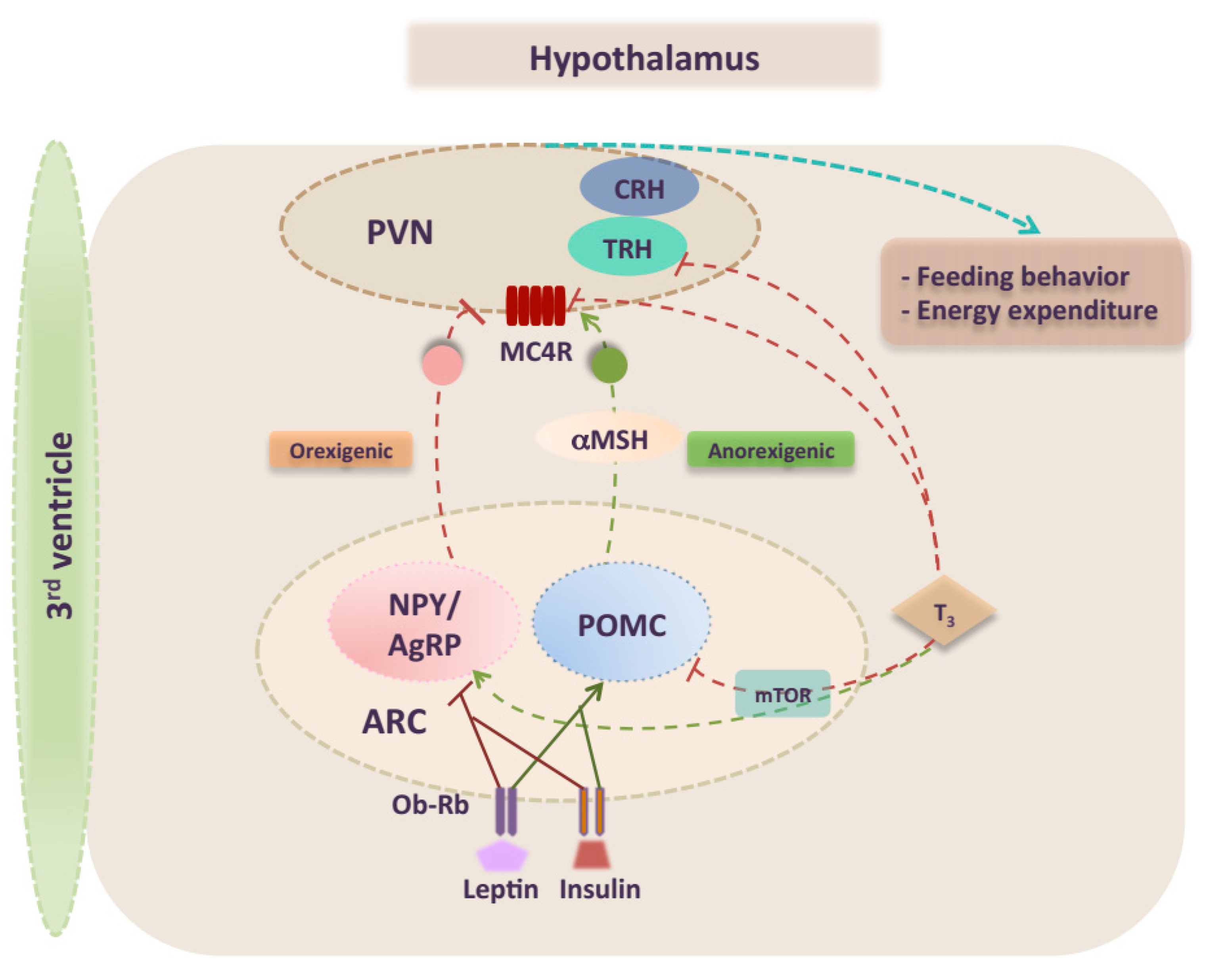
2. Overview of CNS Control of Energy Homeostasis

During the last decades, a significant number of review articles elegantly summarized the knowledge on the mechanisms and signaling pathways underlying the regulation of energy balance. Even a small dysregulation in one of these pathways can lead to obesity [9]. The hypothalamus is a key brain area that plays a pivotal role integrating whole-body signals and controlling food intake and energy expenditure [13,14]. The hypothalamus is organized into well-structured nuclei [15]. In particular, ARC is the best-characterized nucleus for the regulation of feeding. The ARC has a privileged location in the brain that allows it to tightly sense several signals from the periphery [8,16]. Specifically, in the ARC, there are two well-characterized antagonistic neuronal subpopulations with opposite effects. The NPY–AgRP neurons express orexigenic neuropeptides, agouti-related peptide (AgRP), neuropeptide Y (NPY), and the inhibitory neurotransmitter γ-aminobutyric acid (GABA) [17,18]. POMC neurons express the anorexigenic neuropeptides α-melanocyte-stimulating hormone (α-MSH), a proteolytic product of pro-opiomelanocortin (POMC), and cocaine- and amphetamine-regulated transcript (CART) [19]. Both NPY–AgRP and POMC neurons exert their antagonistic effects by projection to second-order neurons mainly in the PVN (Figure 1), but also in other hypothalamic nuclei (i.e., DMN, LHA, VMN) to modulate feeding behavior. It is well recognized that upon nutrient ingestion α-MSH acts on second order neurons located in the PVN and activates melanocortin 3 (MC3R) and melanocortin 4 (MC4R) receptors, which reduce energy intake and induce energy expenditure [20]. Of note, the PVN neurons display the highest MC4R expression within the hypothalamic area and ligand modulation of MC4R signaling in the PVN profoundly affects feeding. Taken together, the melanocortin pathway is considered as a major anorexigenic circuit in the brain [21,22]. Consistent with this idea, human and mice studies showed that deletion of POMC neurons or their peptide product as well as MC4R deficiency results in obesity [23,24].
Both NPY–AgRP and POMC neurons are directly targeted by circulating hormones such as insulin and leptin (Figure 1) [25,26]. It is well established that leptin, a satiety hormone, plays a key role in the central regulation of energy homeostasis. Leptin is an adipokine encoded by the LEP gene, liberated in the circulation by the adipose tissue proportionally to the whole-body fat content and acting as an afferent satiety signal [27]. A significant attention has been given to leptin signaling in ARC, where NPY–AgRP and POMC neurons are the main targets of leptin action via their expression of high levels of leptin receptors (LEPRs) [28,29]. Leptin directly stimulates POMC neurons and activates melanocortin–receptors, while inhibiting the activity of AgRP neurons. Thus, the net effect of leptin signaling within the hypothalamus is increased energy expenditure and reduced body weight. Although it has been believed that melanocortin signaling is a distal mediator of the leptin pathway, there is now evidence that leptin and melanocortin signaling are independent [30]. Emphasizing the importance of leptin, it has been shown that chronic administration of leptin into the brain reduced caloric intake, body weight, and improved glucose sensing in high fat-fed animals [31,32]. Conversely, deficiency of leptin or the ablation of LEPR in the CNS induces a morbid obese phenotype [33,34]. Hence, over-reactivation of the LEPR in hypothalamic neurons, including POMC neurons, may induce obesity in high-fat fed mice [35].

3. Effects of TH on Central Metabolism

For more than a century, THs have been recognized through clinical observations and experimental studies as a main regulator of the metabolic rate of the whole organism [12]. Actually, THs profoundly affect key metabolic pathways that control energy [36,37]. Thus, it becomes evident that dysregulation of TH levels markedly impacts body weight and metabolism homeostasis in Human [38,39]. Reduced energy expenditure leading to weight gain is observed in hypothyroid patients. Conversely, hyperthyroidism (excess of thyroid hormone), promotes a hypermetabolic state characterized by increased energy expenditure and weight loss [40]. These beneficial effects of TH have led to the development of selective thyroid hormone mimetics as powerful new tools against atherosclerosis and obesity [41,42]. Moreover, the role of TH has been demonstrated particularly in glucose homeostasis [43]. Indeed, hyperthyroid rats showed reduced glucose tolerance and reduced insulin-secretory capacity of β cells, in addition to an increased hepatic insulin resistance [44]. Similarly, hypothyroidism promotes glucose intolerance in hypothyroid non-diabetic mice [45]. These observations are clinically relevant given the increased prevalence of diabetes mellitus in both hypo- and hyperthyroidism [46].
At the peripheral level, TH has been characterized for a long time as directly affecting metabolic tissues (such as liver, white and brown adipose tissue (WAT and BAT), heart, skeletal muscle, …) and controlling the bulk of physiological processes implicated in the modulation of energy expenditure [47,48]. However nowadays, it is well established that THs also promote whole body metabolism and modulate food intake by acting at the central level, in the hypothalamus [49].
TH plasma levels are maintained at the appropriate level to preserve energy homeostasis. This adjustment is due to the integration of a range of metabolic pathways at the hypothalamic level. TH is secreted from the thyroid gland under a flexible and dynamic regulation of the hypothalamic-pituitary axis (HPT) [50,51]. Under normal physiological conditions, the intact HPT axis maintains stable serum TH levels, resulting in a steady TH contribution to energy homeostasis. In the PVN, TRH-producing neurons are sensitively affected by changes in circulating TH. In turn, they define the set point of thyroid gland function by regulating pituitary thyroid-stimulating hormone (TSH) secretion and thus the circulating levels of TH [52,53,54]. Importantly, The TRH-TSH-TH feedback loop is mainly mediated by a direct activation of TR isoform β-dependent signaling to decrease TRH and TSH secretion [55].
Given its crucial role in metabolism regulation that affects all the tissues in the body, TH availability and signaling are tightly controlled by several mechanisms [37,52]. Two THs are derived from the thyroid gland: Thyroxine (T4), the most abundant form and Triiodothyronine (T3), the transcriptionally active form. The TH cellular availability is modulated by enzymes known as deiodinases. Local activation of T4 to the active form T3 by the type 2 5-deiodinase (D2), constitutes a key mechanism of the TH regulation of metabolism. D2 is both expressed at the peripheral and central levels [56,57]. Alongside D2, trans-membrane transporters and TH receptors also control TH signaling pathways, by regulating respectively the TH entrance to the cells and its transcriptional action [58]. Nevertheless, integration of TH signaling occurs both peripherally, in liver, white fat, and BAT, and centrally, in the hypothalamus [11,47].
In addition to mediating the feedback mechanisms regulating THs levels, hypothalamic TH signaling also regulates energy homeostasis by influencing appetite [10,59]. Recent evidence indicates that both orexigenic and anorexigenic neurons are targets of the TH feedback loop in the ARC (Figure 1) [60,61]. It has been shown that centrally-mediated actions of T3 dampens anorexigenic signals by inhibiting POMC mRNA expression. In the fasted state, T3 signaling increases uncoupling protein 2 (UCP2) levels in the hypothalamus thereby stimulating orexigenic pathways by increasing NPY and AgRP levels [62,63]. In the PVN, the T3-induced AgRP release suppresses TRH mRNA expression by inhibiting MC4R, a key relay in leptin signaling [64]. This mechanism alters the setpoint of the HPT axis and severely disrupts feeding circuit homeostasis. Recent data have elucidated this link between TH and energy balance, reporting a particular TH-modulation of melanocortin pathway in the hypothalamus [65,66]. Decherf et al. have demonstrated in mice that thyroid status was associated with change of Mc4r expression in the hypothalamus. Both qPCR and in situ hybridization showed hypothyroidism to increase endogenous Mc4r expression in the PVN, whereas hyperthyroidism repressed Mc4r expression in the ARC [65]. Clear evidence from mutagenesis and ChIP assays suggests that T3 can mediate repression of Mc4r levels by direct binding of TR to its responsive elements (TREs) on the Mc4r promoter. Interestingly, in vivo knockdown or over-expression assays and use of TR isoform-specific knock-in mouse models showed that both TRα and TRβ isoforms play a key role in the Mc4r regulation. Thus, the thyroid may be regulated through another negative feedback loop, where MC4R stimulates TH release, which in turn down-regulates MC4R expression [65,67,68]. A first physiological relevance of MC4R repression caused by T3-negative feedback is an induced weight gain in hypothyroid mice treated with T4. Altogether, these results consolidate the important role of thyroid hormone to tightly drive metabolism in a key energy-related brain area.
It is important to highlight the fact that the central effects of THs are interrelated with master energy sensors in the brain such as AMP-activated protein kinase (AMPK) and mammalian target of rapamycin (mTOR) [69,70]. Over the past few years many studies have focused on AMPK and mTOR pathways that act as global regulators of cellular metabolism in both central nervous system and peripheral organs [71,72]. Specifically, both AMPK and mTOR coexist and interact in the same specific hypothalamic nuclei to respond to nutrient availability and hormonal milieu, regulating energy homeostasis. Cross-regulation between these two signaling pathways is thought to be modulated by TH. It has been shown that hyperthyroidism or central administration of T3 reduces hypothalamic AMPK phosphorylation [73]. In turn, this may upregulate the thermogenic program in BAT and increase weight-gain. In contrast, the hyperthyroid state activates the hypothalamic mTOR signaling pathway associated with upregulation of orexigenic peptides AgRP and NPY in the ARC and increases feeding (Figure1). Interestingly, specific treatment with mTOR inhibitor reversed hyperthyroidism-induced increase in food intake. Notably, it has been suggested that THs directly regulate mTOR in the ARC since it is highly co-expressed with thyroid hormone receptor-α (TRα) in this brain region [74].

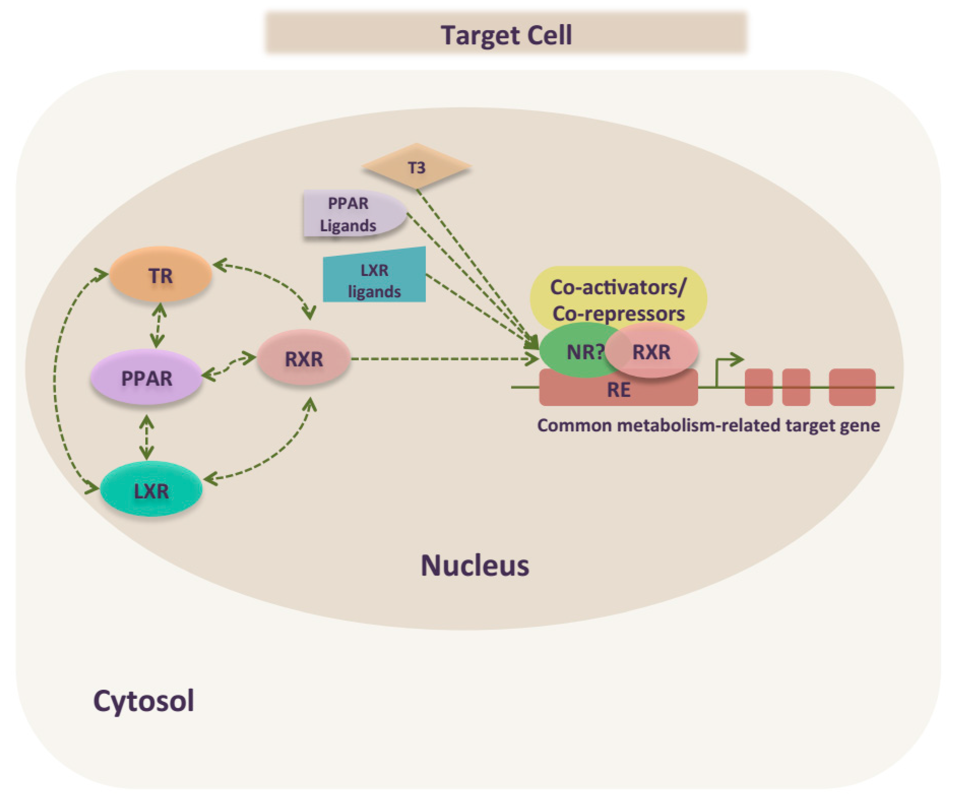
4. Nuclear Receptors, Integrators of Metabolic Regulation

NRs constitute a major target for hormones and metabolites, which makes them fundamental players in the most important biological process, as metabolism regulation. Indeed, NRs are key metabolic sensors that properly integrate environment changes and energy homeostasis [75,76,77]. Ultimately, a dysfunction of that intricate machinery leads to obesity and type 2-diabetes. Given this important role of NRs, they become increasingly interesting therapeutic targets for various metabolic diseases. In this context, we will particularly focus on: Thyroid hormone receptors (TRs), Nuclear receptors partners (RXR), and nuclear receptor coregulators (PPAR and LXR).

4.1. Thyroid Hormone Receptors

At the cellular level, TH transcriptional regulation of metabolic target genes is achieved through hormone-responsive nuclear transcription factors, TRs. NR1A1 and NR1A2 are two different genes coding for TRs, which are alternatively spliced to generate four main isoforms: respectively TRα1, TRα2 (which is unable to bind T3) for NR1A1 and TRβ1and TRβ2 for NR1A2. The TR isoforms exhibit varying expression levels both developmentally and spatially within TH target tissues, suggesting a specific tissue-dependent role for each TR isoform. TR transcriptional regulation is modulated by ligand interactions [36,78]. Indeed, a series of highly controlled intracellular processes occurs to ultimately allow for TH binding to its receptors and lead to TH induced target gene transcriptional regulations. These processes include TH transport into the cells, activation or inactivation by deiodinases, and differential expression levels of TR isoforms, nuclear corepressors and coactivators [36,79]. TRs bind to target genes on TREs mainly as functional heterodimeric complexes notably with retinoid X receptors (RXRs) [36,78]. Many studies analyzing TREs sequences in the promoters of T3 target genes have shown that such DNA response elements consist of a core consensus sequence of the hexanucleotide “half-site” (A/G)GGT(C/A/G)A, existing generally in pairs. Such TREs are qualified as positive TREs (pTRE) or negative TREs (nTRE) [79].
At the brain level, all TR isoforms are highly coexpressed throughout the brain, especially in areas related to energy balance such as the ARC, the PVN and the VMN hypothalamic nuclei. TH is the master of HPT axis control by regulating hypothalamic TRH gene expression and production in a classical negative feedback loop [80,81]. All the functional TRs are colocalized in the T3-responsive TRH neurons in the PVN [82,83]. Indeed, TRs play isoform-specific roles in T3-dependent repression of Trh gene [84]. Specifically, TRβ isoforms play important differential roles in the regulation of Trh gene. Both TRβ isoforms bind to the Trh promoter at an unusual TRE as a functional heterodimer complex with RXR [85,86], but they induce isoform-specific differential transcriptional regulations.
Three separate nTRE half-sites were characterized in the Trh proximal promoter (site 4 from −55 to −60 base pairs (bp); site 5 from +14 to +19 bp; and site 6 from +37 to +42 bp), all of them acting in combination to allow T3-dependent negative regulations [87,88]. Among these nTREs, the Trh promoter site 4 appears to be the most important for TH-induced regulation, as it transduces both T3-independent transcriptional activation and T3-dependent repression [89]. Moreover, unlike the other sites, the Trh site 4 preferentially binds TR/RXR heterodimers [90].
Likewise, TH directly represses Mc4r in the hypothalamus. The T3-induced Mc4r repression is achieved via a putative nTRE (half-site), the TRE1, a non-classical sequence identified in the Mc4r promoter, different to Trh site 4, and thought to bind monomeric nuclear receptors to mediate their transcriptional activities [65]. According to Chip results, Mc4r TRE1 recruited high levels of TRβ in presence of T3, whereas low levels were recruited in absence of T3. Indeed, overexpression of TRβ1 or TRβ2 enhanced the repressive effects of T3 on Mc4r transcription, independently of the isoforms. Furthermore, the same inhibitory TRβ mediated effect was observed in newborn and adult mice. Besides, TRα isoform is also involved in Mc4r regulation. Overexpression of TRα reduced T3-independent transcription. This result was confirmed in TRα knockout mice TRα °/°, where Mc4r expression was increased [65].
Collectively, extensive studies demonstrate that TRs are major physiological regulators of food intake and energy homeostasis, not only peripherally, but also centrally through their action regulating HPT axis. It is well documented that mice and humans with negative mutations of TRα and TRβ display a variety of metabolic phenotypes of induced weight gain and reduced energy expenditure. Consistent with these observations, recent data showed that selective TRβ knockdown specifically in the VMN, a key brain region for the control of energy homeostasis, results in marked obesity similarly to murine models with the most extreme forms of monogenic obesity [91]. Furthermore, in vivo studies showed that activation of the TRβ isoform with selective agonists increased the metabolic rate and prevents glucose intolerance, hyper-triglyceridaemia and body weight gain in obese or high-fat diet fed rats [92]. Moreover, mutant mice with specific TRα mutation developed visceral adiposity and insulin resistance [93]. Finally, it has been advanced that targeting TR subtypes improve global metabolic outcomes [94].

4.2. Retinoid X Receptor (RXR)

Molecular endocrinology has known great progress after the characterization of orphan receptors, in particular, the RXR. Indeed, the discovery of RXR and its ligand led to stupendous concepts in the nuclear receptor research area [95,96]. Mainly, this important finding reveals a not yet defined signaling pathway, which can be modulated by specific ligands. Furthermore, it led to define the interconnection of multiple signaling pathways, especially by the discovery of RXR heterodimerization with different nuclear receptors [97,98]. Three RXR subtypes were identified: RXRα, RXRβ and RXRγ, each encoded by different genes [99]. The three isoforms are highly conserved and share the same mechanisms of heterodimerization with partners. To mediate transcriptional regulation, RXR binds as homodimer, to a direct repeat of half-sites separated by one nucleotide (i.e., a DR1 element) in response to RXR ligands, which might be 9-cis retinoic acid (9cRA). Because of the unique nucleotide in the spacer between the twoDR1 half-sites, it seems that the DR1 sequence underwent Evolution to generate novel binding motifs (DR2, DR3, DR4, etc.). This flexibility of RXR allows it to adopt multiple conformations and thereby dimerize with different nuclear receptors [97,99]. Thus, RXR could play dual roles in NR signaling.
Interestingly, RXRs are involved in several metabolic pathways because of their heterodimerization with nuclear receptor partners, where they can modulate transcription through ligand activation. Hence, the modulation of transcription could be achieved either by the RXR ligand or the partner receptor ligand. This kind of dual-ligand regulation resulted into two categories of heterodimers: permissive and non-permissive [100]. Permissive receptor partners like PPARs, LXRs and FXR are those that can be activated either by ligands of RXR or its partner, and the presence of both ligands results in a cooperative response [101,102]. Non-permissive partners like TRs, VDRs and RARs, are those that can be only activated by the partner’s ligand and function primarily as hormone receptors, while RXR is silent [103,104].
An example of RXR in regulating metabolic pathway is its strong heterodimerization with TR, increasing thereby TR binding to the TRE and amplifying its transcriptional activity [105]. Indeed, it has been shown that RXR increases stimulatory TR responses of TH target genes [106,107]. Moreover, RXR/TR heterodimers play roles in both basal transactivation and T3 suppression of negatively regulated genes. Nevertheless, the role of RXRs in T3-dependent repression showed more complexity than T3-dependent activation. In a context of T3 negative regulation, in vitro results showed that RXR subtypes improve T3 dependent Trh regulation, independently of their DNA-binding properties. Likewise, RXRs increase the dominant negative effect of some mutant TRs on specific nTREs [108]. The activation of endogenous RXR by specific ligands increased Tsh mRNA levels [109] but did not show any effect on Trh expression or preproTRH levels [110]. A functional study demonstrates that knockdown and overexpression of RXRα and RXRβ change hypothalamic Trh levels, suggesting differential roles of both RXR subtypes to modulate T3-dependent Trh transcription [111].

4.3. Peroxisome Proliferator-Activated Receptors (PPARs)

PPARs are nuclear transcription factors belonging to the steroid receptor superfamily. They regulate target genes by binding to peroxisome proliferator hormone response elements (PPREs), generally as a heterodimer with RXR [112]. There are three known PPAR isoforms, PPARα, PPARγ and PPARδ, differentially expressed among tissues. PPARα is abundant in the liver, brown adipose tissue, heart, and kidney; PPARγ is mainly enriched in the adipose tissue and PPARβ/δis ubiquitously expressed throughout the body [113]. PPARs act as fundamental players in various physiological and pathological processes, especially in energy metabolism. Particularly, PPARα and PPARγ isoforms have been extensively documented mainly because they are activated by clinically-used molecules [114]. PPARα is described as a master regulator of lipid metabolism as it controls genes involved in hepatic fatty acid oxidation. PPARα is increasingly described as a potential molecular target of the hypolipidemic drugs for the treatment of dyslipidemia and fibrates. Its exogenous activation decreases both circulating triglyceride levels and reduces lipid stores in liver, muscle, and adipose tissue [115,116]. PPARγ is highly expressed in adipose tissue and plays key roles in adipogenesis, promoting the expression of specific adipocyte markers such as adipocyte lipid binding protein (aP2), phosphoenolpyruvate carboxylase (PEPCK) or lipoprotein lipase (LPL) [117,118]. Moreover, PPARγ is also involved in fatty acid uptake and storage and in glucose metabolism in many other peripheral tissues [119]. Interestingly, PPARγ is the target of thiazolidinedione (TZD), the only current class of insulin-sensitizing drugs in patients with type 2 diabetes [120]. However, several side effects are caused by long-term use of these drugs (mainly an increase of weight gain in both patients and rodent models). Finally, the less-described PPAR isotype, PPARδ, appears as an attractive therapeutic target in metabolic syndrome. PPARδ is ubiquitously expressed and is involved in fatty acid catabolism, energy uncoupling in adipose tissue and muscle, insulin sensitizing, and the reduction of inflammation [121].
Recent evidence supports a new potential role for PPARs in central energy homeostasis regulation. In the CNS, all PPAR subtypes, PPARα, PPARδ and PPARγ, have been involved in the regulation of energy homeostasis. Particularly, PPARγ seems to play a key role in the regulation of energy balance. New studies suggest that exogenous activation of central PPARγ by its ligand TZD leads to weight gain which may contribute to obesity [122,123,124].
Consistently, hypothalamic activation of central PPARγ by either specific agonists or overexpression, leads to enhance positive energy balance. However, inhibition of PPARγ activity in the brain with antagonists or by shRNA-mediated knockdown results in negative energy balance [123]. Interestingly, specific inhibition of PPARγ in the CNS improves the sensitivity of the leptin pathway in the hypothalamus of high-fat diet-(HFD)-fed animals. Recently, a model of transgenic mice lacking PPARγ in POMC neurons showed increased energy expenditure, while body weight and food intake were reduced. Furthermore, these models showed improved glucose metabolism when exposed to HFD [124]. Besides, peripheral administration of either a PPARγ activator or inhibitor failed to affect food intake of mice with POMC-specific Pparγ ablation. Taken together, PPARγ signaling in the brain seems to profoundly impact energy balance and to promote the obesity phenotype [125]. The same obesogenic effects have been demonstrated for activation of PPARα in the brain. Conversely, PPARδ seems to play inverse role than PPARγ. In mutant mice lacking Pparδ via genetic deletion, Pparγ and Pparα are highly expressed in the hypothalamus which would potentiate diet induced obesity [126].

4.4. Liver X Receptors (LXRs)

Dysregulated cholesterol levels and metabolism represent hallmarks of diseases such as diabetes and atherosclerosis. The LXRs are members of the nuclear receptor family and are considered as major sensors of cholesterol and lipid homeostasis in mammals [127]. Two related LXRs isoforms have been identified: LXRα (encoded by NR1H3) and LXRβ (encoded by NR1H2), which are a part of the emerging significant newer drug targets within the NR family [128]. Both LXRs isoforms are structurally similar and are activated by the same ligands, however, their tissue expression differs. LXRα is highly expressed in metabolically active tissues and cell types such as liver, intestine, adipose and macrophages, whereas LXRβ is expressed ubiquitously. These transcription factors, activated by cholesterol metabolites, control the expression of a panel of genes involved in cellular cholesterol traffic in a tissue-dependent manner, thereby protecting the cell from cholesterol overload [129,130]. In addition to their central role in cholesterol metabolism, LXRs are key regulators of lipogenesis and have an impact on systemic glucose homeostasis [128]. Thus, inhibiting hepatic LXR seems unlikely to be a successful therapeutic strategy for type 2 diabetes.
Both LXR isoforms are expressed in the brain [131]. However, their central functions are not as well understood as their roles in peripheral organs such as the liver. Neurons need a tightly controlled cholesterol rate regulation for an appropriate synaptic functioning. There is evidence to support that LXRs in the brain regulate pathways that maintain cholesterol balance and activate anti-inflammatory pathways [132]. In fact, studies in isolated murine neurons and glial cells have generally confirmed the ability of LXRs to regulate the expression of genes linked to cholesterol transport, including ABCA1 and APOE24. Thus, the brain LXRs signaling exhibits neuroprotective mechanisms and anti-inflammatory effects [133]. However, a perturbation of such pathway increased both cellular cholesterol and amyloid-β levels in the brain. Such deregulation constitutes a fundamental mechanism in the development of Alzheimer’s disease. Thereby, a particular link between LXR signaling and Alzheimer’s disease has been established [134].
Recent data indicate that LXR could modulate set-points of the HPT axis andMC4R pathways in the hypothalamus [135]. Thus, activated LXR represses TRH levels and induces the orexigenic peptides, which may promote weight gain and obesity. In contrast, specific inactivation of LXRs enhances Trh expression in the hypothalamus [135] and induces the browning of WAT, thereby ameliorating obesity outcomes [136,137].

5. TR Crosstalk with PPAR and LXR to Regulate Metabolism

It is well established that the thyroid hormone is a key factor regulating basal metabolic rate, thermogenesis, glucose metabolism, lipolysis, and HPT axis. Increasing data have focused on specific actions of TH in metabolic regulations. These include the molecular mechanisms of TRs actions on cholesterol and carbohydrate metabolism, through direct actions on gene expression, as monomers, homodimers or heterodimers with RXR. TRs could also mediate indirect actions by interfering with other NRs signaling pathways to regulate common target genes [138]. Indeed, TH signaling, especially in metabolic regulation, involves TR crosstalk with other nuclear hormone receptors including PPARα, PPARγ and LXR [138]. Such crosstalk has not only been demonstrated in vitro but also in vivo, using mouse models. They could impact different molecular levels of gene regulation (Figure 2) [139,140].

5.1. TR Peripheral Crosstalk in Regulating Metabolism

Although, NRs interaction remains a complex mechanism that requires further investigation, it could be explained at least by the structural similarity in the DNA and ligand binding domains among NRs. All TR, PPAR and LXR are ligand-activated NRs that share similar structure and mode of action by binding to DNA response elements to form heterodimers with RXR [96]. First, the DNA binding domains are highly conserved among these NRs, containing two zinc fingers and arranged as direct repeats of hexameric half-sites, although the spacing of the hexamers varies among the different receptors. Second, NRs also share a very conserved leucine zipper, which is the interface for RXR heterodimerization [96]. Thus, NRs may compete for limiting amounts of RXR. Such a competition for RXR influences gene expression [141]. Moreover, TRs bind to TH with a higher affinity than PPAR and LXR bind to their natural ligands, conferring to TR a dominance in its interaction with RXR, and reflected by a greater effect on coregulated genes [138]. Third, this competition could be extended to common coactivators and corepressors. Indeed, the ligand binding domain (LBD), apart its ligand sequences specific for each receptor, contains regions interacting with other receptors as well as coactivators and corepressors [138].
Crosstalk between TR, PPAR and LXR has been reported, especially on cholesterol, lipid and glucose metabolism-related genes. Previous in vitro studies have underlined interactions between PPAR signaling and TR-dependent pathways [142]. Crosstalk between TR and PPAR signals could involve competition for their common heterodimeric-partner (RXR) [143], as well as for their respective responsive elements in target gene promoters. Indeed, TR has been shown to bind PPRE [144,145]. Nevertheless, the response to PPAR agonists may depend on the interactions between PPARs and TRs. In most cases, non-ligand binding TR mutant represses PPAR transcriptional activity [139].
Both TR and PPAR regulate expression of key enzymes of the fatty acid oxidation carnitine palmitoyl transferase Ia (CPT-Ia) and acyl–CoA oxidase (ACO). Crosstalk between TR and PPARα has been observed in regulation of CPT-Ia and ACO, especially in a mouse model of TRα (P398H) mutant [139]. This TRα mutant isoform is still able to bind to PPRE, thereby inhibiting PPARα-induced enzyme expression and, causing fatty acid accumulation in the livers of these mice [139]. Furthermore, lipoprotein lipase is regulated by both PPARγ. It was demonstrated that the mutant TRβ PV represses the PPARγ induced lipoprotein lipase gene expression, by binding to PPRE and by recruiting corepressors [137].
The role of LXR as a coordinator of both lipid and carbohydrate metabolism suggests the potential for interactions with TR. ATP-binding cassette transporter A1 (ABCA1) is a transporter of cellular non-esterified cholesterol and phospholipids in the liver, mainly regulated by LXR. TR competes with LXR for binding to the LXRE and inhibiting LXR-mediated Abca1 gene expression [146,147]. Such examples of crosstalk and interaction are also observed in carbohydrate metabolism. A recent study describes that carbohydrate-response element-binding protein (ChREBP), a major transcription factor controlling the activation of glucose-induced lipogenesis in the liver, was characterized as a direct target of TRβ and LXR regulated in a tissue-selective manner. Both in vivo and in vitro assays showed a crosstalk between LXR and TRβ signaling on the ChREBP promoter, especially in the liver [148,149].
Furthermore, recent investigations support a complex interaction between LXR and PPARα in the regulation of glucose and lipid metabolism. The two NRs may either cooperate or have opposite effects on gene expression [150,151]. Yet the exact mechanisms underlying such crosstalk remain to be determined.

5.2. TR Crosstalk in the Regulation of Central Metabolism

Recent studies have demonstrated the expression of NRs within different brain regions, in particular at the hypothalamic level, the integrator of whole body energy homeostasis [77]. However, little is known about their role in the central control of energy homeostasis. Increasing data show interactions between TR and PPAR or LXR in the peripheral tissues (described above). Besides, such crosstalk at the central level remains less investigated. In this context, several studies focused on hypothalamic interactions between the different signaling pathways controlling metabolism.
Recent evidence supports a potential role for PPARs in the central energy homeostasis regulation [152]. In the CNS, all PPAR subtypes are expressed at different levels, however their function in the brain is not well elucidated. Of the three, PPARγ signaling pathway is the best characterized in the brain. Indeed, PPARγ and its cognate agonists appear to be attractive therapeutic targets for various disorders of the central nervous system [153,154]. PPARγ agonists have shown promising results in animal models of Alzheimer’s disease, stroke, multiple sclerosis, Parkinson’s disease, amyotrophic lateral sclerosis, and pituitary adenoma [155]. Further, it was confirmed that PPARγ isoform is expressed in key neuronal subsets regulating energy homeostasis [156]. More importantly, a recent study has demonstrated that mice lacking PPARγ in POMC neurons displayed increased energy expenditure and decreased fat mass and food intake. The absence of PPARγ was also associated with improved glucose metabolism and increased insulin sensitivity. Peripheral administration of either a PPARγ activator or inhibitor failed to affect food intake of mice with POMC-specific PPARγ ablation [124]. Besides, PPARα showed also an important effect on the brain. Intra-cerebroventricular (icv) and Intra-hypothalamic administration of the PPARα activator Wy14643 reduced glucose utilization and increased food intake in wild-type but not in PPARα-deficient mice [157]. Importantly, a previous report showed that the peripheral activation of PPARγ by rosiglitazone treatment affects hypothalamic-pituitary-thyroid axis and thyroid hormone release [143]. Taken together, these results suggest a pivotal role of hypothalamic PPARγ signaling pathway on central metabolic homeostasis, particularly on TH-dependent gene regulations.
Accordingly, an in vivo study tested the hypothesis of crosstalk between hypothalamic PPARγ and TRβ on T3-dependent regulation of the Trh promoter [158]. Our results showed first, that icv administration of PPARγ agonists leads to increased T3-independent Trh transcription and increased circulating T4 levels. In contrast, PPARγ antagonist did not affect Trh transcriptional activity in the absence of T3, but interfered with T3-dependent Trh repression. Then, silencing PPARγ protein levels by using small hairpin RNA (shRNA) increased T3-independent Trh transcription, whilst PPARγ overexpression abrogated T3-dependent Trh repression. Interestingly, the effect of PPARγ overexpression was reversed by co-expression of either TRβ1 or RXR [158]. These results suggest that PPARγ may interfere with TR signaling at the hypothalamic level, through a competition for limiting amounts of RXR.
LXRs are one class of nuclear receptors which are believed to be master integrators of cholesterol metabolism in the periphery [159]. Recently, their activation with a specific agonist has been shown to enhance cholesterol metabolism also in the CNS [160]. Furthermore, LXRs are expressed in the CNS, especially in the hypothalamus [161]. Thus, it suggests that LXR could play physiological and metabolic functions in the brain. Indeed, several arguments are in favor of this hypothesis, particularly via a crosstalk between the LXR and TR signaling pathways.
A recent study has revealed a crosstalk between TR and LXR in the regulation of Selective Alzheimer’s disease (AD) indicator-1 (Seladin-1) gene expression in an AD mouse model. Overexpressing Seladin-1 in the neurons increases the amount of cholesterol and avoids β-amyloid accumulation, oxidative stress and neurons apoptosis [162]. Both NRs have been shown to be involved in Seladin-1 gene expression, TR-β and LXR-α competitively up-regulating the human Seladin-1 promoter [140]. These results suggest that TR and LXR would co-regulate lipid metabolism in CNS. Interestingly LXR could be an attractive therapeutic target for neurodegenerative diseases [163,164].
Also, a crosstalk between TR and LXR has been recently reported, in the context of the hypothalamic TH negative feedback loop regulation: key target genes involved in the central control of metabolism, Trh and Mc4r, were impacted by this crosstalk between TR and LXR [135]. Indeed, using in vivo gene transfer, we explored the involvement of LXR in the hypothalamic metabolic pathways, analyzing the interference of LXR with the transcriptional regulation induced by TRs. Our results showed that activation of LXR by its specific agonist GW3965 repressed the transcriptional activity of both Trh and Mc4r promoters, and this only occurred in euthyroid mice. This repression was restored by TH treatment in hypothyroid mice, yet only in the Trh promoter [135]. Conversely, LXR knocked-down abrogated this repression, leading to a relative activation of the Trh promoter in the PVN. Further, in vivo ChIP results, showed that LXR was recruited to the Trh promoter region only in the presence of T3. Yet, no simultaneous recruitment of RXR and LXR on the Trh promoter region were observed [135]. Nevertheless, LXR KO mice showed enhanced secretion of TSH, thereby stimulating THs levels. Collectively, these results provide evidence that depletion of LXRs would abrogate the TH negative feedback loop in the hypothalamus and cause a loss of TRs in the PVN area [136,137].
Furthermore, the T3 signaling pathway could affect LXR transcriptional regulation. Indeed, qPCR results showed that T3 treatment of newborn mice induced the hypothalamic regulation of a number of LXR target genes implicated in metabolism and inflammation. Interestingly, key genes of inflammation, Pparα, Tnfα and Il1 showed significant hypothalamic mRNA levels increase afterT3 treatment [135]. Thus, as in the periphery, the later genes would be LXR/TR targets in the hypothalamus. These results suggest that crosstalk between LXR and TR may be involved in the central regulation of inflammation. Since LXR activation by its specific ligand showed anti-inflammatory effects [165,166,167], this property could be exploited also in the CNS.

6. Conclusions

It is well demonstrated that TH are endocrine messengers with a profound impact on energy expenditure and appetite regulation. Accumulating evidences obtained from genetic mouse models and pharmacologic approaches pinpoint the TH signaling pathway as a master driver of metabolism regulation by acting, to a large extent, at the central level. Several studies have elegantly elucidated molecular mechanisms of action of TH in the brain. A continuous interaction between TH and key regulatory mechanisms coexist in the hypothalamus for a tightly controlled body weight and optimal energy balance. Remarkably, effects of THs are interrelated with key energy sensors in the brain. In addition, TH-mediated action is absolutely dependent upon its cognate receptors TRs that directly bind to target genes. Interestingly, TRs isoforms interact with other nuclear receptors that play a key role in metabolic regulation such as PPAR and LXR. Thus a deeper understanding of the mechanisms and interactions of TH signaling pathways in the hypothalamic control of metabolism will lead to identifying biomarkers and effective and selective targets that will improve the therapy of energy balance disorders, such as obesity.

Funding

This work was supported by a PHC-Utique program/CMCU grant No. 06G0910 for S. Kouidhi, EU grant number 602757 Human FP7-Health-2013-Innovation-1 and EU contract No. LSHM-CT-2005-018652 “CRESCENDO”.

Conflicts of Interest

The authors declare no conflict of interest.

References

  1. Lavie, C.J.; Arena, R.; Alpert, M.A.; Milani, R.V.; Ventura, H.O. Management of cardiovascular diseases in patients with obesity. Nat. Rev. Cardiol. 2018, 15, 45–56. [Google Scholar] [CrossRef] [PubMed]
  2. González-Muniesa, P.; Mártinez-González, M.-A.; Hu, F.B.; Després, J.-P.; Matsuzawa, Y.; Loos, R.J.F.; Moreno, L.A.; Bray, G.A.; Martinez, J.A. Obesity. Nat. Rev. Dis. Primers 2017, 3, 17034. [Google Scholar] [CrossRef] [PubMed]
  3. Jarolimova, J.; Tagoni, J.; Stern, T.A. Obesity: Its Epidemiology, Comorbidities, and Management. Primacy Care Companion CNS Disord. 2013, 15. [Google Scholar] [CrossRef] [PubMed] [Green Version]
  4. Campanella, G.; Gunter, M.J.; Polidoro, S.; Krogh, V.; Palli, D.; Panico, S.; Sacerdote, C.; Tumino, R.; Fiorito, G.; Guarrera, S.; et al. Epigenome-wide association study of adiposity and future risk of obesity-related diseases. Int. J. Obes. 2018. [Google Scholar] [CrossRef] [PubMed]
  5. Williams, K.W.; Elmquist, J.K. From neuroanatomy to behavior: Central integration of peripheral signals regulating feeding behavior. Nat. Neurosci. 2012, 15, 1350–1355. [Google Scholar] [CrossRef] [PubMed]
  6. Gao, Q.; Horvath, T.L. Neurobiology of feeding and energy expenditure. Annu. Rev. Neurosci. 2007, 30, 367–398. [Google Scholar] [CrossRef] [PubMed]
  7. Sohn, J.-W. Network of hypothalamic neurons that control appetite. BMB Rep. 2015, 48, 229–233. [Google Scholar] [CrossRef] [PubMed] [Green Version]
  8. Joly-Amado, A.; Cansell, C.; Denis, R.G.P.; Delbes, A.-S.; Castel, J.; Martinez, S.; Luquet, S. The hypothalamic arcuate nucleus and the control of peripheral substrates. Best Pract. Res. Clin. Endocrinol. Metab. 2014, 28, 725–737. [Google Scholar] [CrossRef] [PubMed]
  9. Timper, K.; Brüning, J.C. Hypothalamic circuits regulating appetite and energy homeostasis: Pathways to obesity. Dis. Models Mech. 2017, 10, 679–689. [Google Scholar] [CrossRef] [PubMed]
  10. Zhang, Z.; Boelen, A.; Bisschop, P.H.; Kalsbeek, A.; Fliers, E. Hypothalamic effects of thyroid hormone. Mol. Cell. Endocrinol. 2017, 458, 143–148. [Google Scholar] [CrossRef] [PubMed]
  11. López, M.; Alvarez, C.V.; Nogueiras, R.; Diéguez, C. Energy balance regulation by thyroid hormones at central level. Trends Mol. Med. 2013, 19, 418–427. [Google Scholar] [CrossRef] [PubMed]
  12. Mullur, R.; Liu, Y.-Y.; Brent, G.A. Thyroid hormone regulation of metabolism. Physiol. Rev. 2014, 94, 355–382. [Google Scholar] [CrossRef] [PubMed]
  13. Coll, A.P.; Yeo, G.S. The hypothalamus and metabolism: Integrating signals to control energy and glucose homeostasis. Curr. Opin. Pharmacol. 2013, 13, 970–976. [Google Scholar] [CrossRef] [PubMed]
  14. Yu, J.H.; Kim, M.-S. Molecular Mechanisms of Appetite Regulation. Diabetes Metab. J. 2012, 36, 391–398. [Google Scholar] [CrossRef] [PubMed]
  15. Bouret, S.G. Development of Hypothalamic Circuits That Control Food Intake and Energy Balance. In Appetite and Food Intake: Central Control; Harris, R.B.S., Ed.; CRC Press/Taylor & Francis: Boca Raton, FL, USA, 2017; ISBN 978-1-4987-2316-9. [Google Scholar]
  16. Waterson, M.J.; Horvath, T.L. Neuronal Regulation of Energy Homeostasis: Beyond the Hypothalamus and Feeding. Cell Metab. 2015, 22, 962–970. [Google Scholar] [CrossRef] [PubMed]
  17. Luquet, S.; Perez, F.A.; Hnasko, T.S.; Palmiter, R.D. NPY/AgRP neurons are essential for feeding in adult mice but can be ablated in neonates. Science 2005, 310, 683–685. [Google Scholar] [CrossRef] [PubMed]
  18. Gropp, E.; Shanabrough, M.; Borok, E.; Xu, A.W.; Janoschek, R.; Buch, T.; Plum, L.; Balthasar, N.; Hampel, B.; Waisman, A.; et al. Agouti-related peptide-expressing neurons are mandatory for feeding. Nat. Neurosci. 2005, 8, 1289–1291. [Google Scholar] [CrossRef] [PubMed]
  19. Millington, G.W. The role of proopiomelanocortin (POMC) neurones in feeding behaviour. Nutr. Metab. 2007, 4, 18. [Google Scholar] [CrossRef] [PubMed] [Green Version]
  20. Dores, R.M.; Liang, L.; Davis, P.; Thomas, A.L.; Petko, B. 60 YEARS OF POMC: Melanocortin receptors: Evolution of ligand selectivity for melanocortin peptides. J. Mol. Endocrinol. 2016, 56, T119–T133. [Google Scholar] [CrossRef] [PubMed]
  21. Butler, A.A. The melanocortin system and energy balance. Peptides 2006, 27, 281–290. [Google Scholar] [CrossRef] [PubMed] [Green Version]
  22. Shen, W.-J.; Yao, T.; Kong, X.; Williams, K.W.; Liu, T. Melanocortin neurons: Multiple routes to regulation of metabolism. Biochim. Biophys. Acta 2017, 1863, 2477–2485. [Google Scholar] [CrossRef] [PubMed] [Green Version]
  23. Butler, A.A.; Cone, R.D. Knockout studies defining different roles for melanocortin receptors in energy homeostasis. Ann. N. Y. Acad. Sci. 2003, 994, 240–245. [Google Scholar] [CrossRef] [PubMed]
  24. Lensing, C.J.; Adank, D.N.; Doering, S.R.; Wilber, S.L.; Andreasen, A.; Schaub, J.W.; Xiang, Z.; Haskell-Luevano, C. The Ac-Trp-DPhe(p-I)-Arg-Trp-NH2 250-Fold Selective Melanocortin-4 Receptor (MC4R) Antagonist over the Melanocortin-3 Receptor (MC3R) Affects Energy Homeostasis in Male and Female Mice Differently. ACS Chem. Neurosci. 2016, 7, 1283–1291. [Google Scholar] [CrossRef] [PubMed] [Green Version]
  25. Gautron, L.; Elmquist, J.K. Sixteen years and counting: An update on leptin in energy balance. J. Clin. Investig. 2011, 121, 2087–2093. [Google Scholar] [CrossRef] [PubMed]
  26. Belgardt, B.F.; Brüning, J.C. CNS leptin and insulin action in the control of energy homeostasis. Ann. N. Y. Acad. Sci. 2010, 1212, 97–113. [Google Scholar] [CrossRef] [PubMed] [Green Version]
  27. Rosenbaum, M.; Leibel, R.L. Role of leptin in energy homeostasis in humans. J. Endocrinol. 2014, 223, T83–T96. [Google Scholar] [CrossRef] [PubMed]
  28. Park, H.-K.; Ahima, R.S. Physiology of leptin: Energy homeostasis, neuroendocrine function and metabolism. Metabolism 2015, 64, 24–34. [Google Scholar] [CrossRef] [PubMed]
  29. Cowley, M.A.; Smart, J.L.; Rubinstein, M.; Cerdán, M.G.; Diano, S.; Horvath, T.L.; Cone, R.D.; Low, M.J. Leptin activates anorexigenic POMC neurons through a neural network in the arcuate nucleus. Nature 2001, 411, 480–484. [Google Scholar] [CrossRef] [PubMed]
  30. Shimizu, H.; Inoue, K.; Mori, M. The leptin-dependent and -independent melanocortin signaling system: Regulation of feeding and energy expenditure. J. Endocrinol. 2007, 193, 1–9. [Google Scholar] [CrossRef] [PubMed]
  31. Da Silva, A.A.; Hall, J.E.; do Carmo, J.M. Leptin reverses hyperglycemia and hyperphagia in insulin deficient diabetic rats by pituitary-independent central nervous system actions. PLoS ONE 2017, 12, e0184805. [Google Scholar] [CrossRef] [PubMed]
  32. Abraham, M.A.; Rasti, M.; Bauer, P.V.; Lam, T.K.T. Leptin enhances hypothalamic lactate dehydrogenase A (LDHA)-dependent glucose sensing to lower glucose production in high-fat-fed rats. J. Biol. Chem. 2018, 293, 4159–4166. [Google Scholar] [CrossRef] [PubMed]
  33. Brown, J.A.; Bugescu, R.; Mayer, T.A.; Gata-Garcia, A.; Kurt, G.; Woodworth, H.L.; Leinninger, G.M. Loss of Action via Neurotensin-Leptin Receptor Neurons Disrupts Leptin and Ghrelin-Mediated Control of Energy Balance. Endocrinology 2017, 158, 1271–1288. [Google Scholar] [CrossRef] [PubMed]
  34. Engin, A. Diet-Induced Obesity and the Mechanism of Leptin Resistance. Adv. Exp. Med. Biol. 2017, 960, 381–397. [Google Scholar] [PubMed]
  35. Gamber, K.M.; Huo, L.; Ha, S.; Hairston, J.E.; Greeley, S.; Bjørbæk, C. Over-expression of leptin receptors in hypothalamic POMC neurons increases susceptibility to diet-induced obesity. PLoS ONE 2012, 7, e30485. [Google Scholar] [CrossRef] [PubMed]
  36. Brent, G.A. Mechanisms of thyroid hormone action. J. Clin. Investig. 2012, 122, 3035–3043. [Google Scholar] [CrossRef] [PubMed] [Green Version]
  37. Song, Y.; Yao, X.; Ying, H. Thyroid hormone action in metabolic regulation. Protein Cell 2011, 2, 358–368. [Google Scholar] [CrossRef] [PubMed] [Green Version]
  38. Bjergved, L.; Jørgensen, T.; Perrild, H.; Laurberg, P.; Krejbjerg, A.; Ovesen, L.; Rasmussen, L.B.; Knudsen, N. Thyroid Function and Body Weight: A Community-Based Longitudinal Study. PLoS ONE 2014, 9, e93515. [Google Scholar] [CrossRef] [PubMed] [Green Version]
  39. Abdi, H.; Kazemian, E.; Gharibzadeh, S.; Amouzegar, A.; Mehran, L.; Tohidi, M.; Rashvandi, Z.; Azizi, F. Association between Thyroid Function and Body Mass Index: A 10-Year Follow-Up. Ann. Nutr. Metab. 2017, 70, 338–345. [Google Scholar] [CrossRef] [PubMed]
  40. Laurberg, P.; Knudsen, N.; Andersen, S.; Carlé, A.; Pedersen, I.B.; Karmisholt, J. Thyroid Function and Obesity. Eur. Thyroid J. 2012, 1, 159–167. [Google Scholar] [CrossRef] [PubMed] [Green Version]
  41. Elbers, L.P.; Kastelein, J.J.; Sjouke, B. Thyroid Hormone Mimetics: The Past, Current Status and Future Challenges. Curr. Atheroscler. Rep. 2016, 18, 14. [Google Scholar] [CrossRef] [PubMed]
  42. Baxter, J.D.; Webb, P. Thyroid hormone mimetics: Potential applications in atherosclerosis, obesity and type 2 diabetes. Nat. Rev. Drug Discov. 2009, 8, 308–320. [Google Scholar] [CrossRef] [PubMed]
  43. Bos, M.M.; Smit, R.A.J.; Trompet, S.; van Heemst, D.; Noordam, R. Thyroid Signaling, Insulin Resistance, and 2 Diabetes Mellitus: A Mendelian Randomization Study. J. Clin. Endocrinol. Metab. 2017, 102, 1960–1970. [Google Scholar] [CrossRef] [PubMed]
  44. Karbalaei, N.; Noorafshan, A.; Hoshmandi, E. Impaired glucose-stimulated insulin secretion and reduced β-cell mass in pancreatic islets of hyperthyroid rats. Exp. Physiol. 2016, 101, 1114–1127. [Google Scholar] [CrossRef] [PubMed]
  45. Lin, Y.; Sun, Z. Thyroid hormone potentiates insulin signaling and attenuates hyperglycemia and insulin resistance in a mouse model of type 2 diabetes. Br. J. Pharmacol. 2011, 162, 597–610. [Google Scholar] [CrossRef] [PubMed] [Green Version]
  46. Vanderpump, M.P.; Ahlquist, J.A.; Franklyn, J.A.; Clayton, R.N. Consensus statement for good practice and audit measures in the management of hypothyroidism and hyperthyroidism. The Research Unit of the Royal College of Physicians of London, the Endocrinology and Diabetes Committee of the Royal College of Physicians of London, and the Society for Endocrinology. BMJ 1996, 313, 539–544. [Google Scholar] [PubMed]
  47. McAninch, E.A.; Bianco, A.C. Thyroid hormone signaling in energy homeostasis and energy metabolism. Ann. N. Y. Acad. Sci. 2014, 1311, 77–87. [Google Scholar] [CrossRef] [PubMed] [Green Version]
  48. Alkemade, A. Central and peripheral effects of thyroid hormone signalling in the control of energy metabolism. J. Neuroendocrinol. 2010, 22, 56–63. [Google Scholar] [CrossRef] [PubMed]
  49. Donangelo, I. Thyroid Hormone and Central Control of Metabolic Homeostasis. J. Endocrinol. Diabetes Obes. 2014, 2, 1047. [Google Scholar]
  50. Ortiga-Carvalho, T.M.; Chiamolera, M.I.; Pazos-Moura, C.C.; Wondisford, F.E. Hypothalamus-Pituitary-Thyroid Axis. Compr. Physiol. 2016, 6, 1387–1428. [Google Scholar] [PubMed]
  51. Fliers, E.; Boelen, A.; van Trotsenburg, A.S.P. Central regulation of the hypothalamo-pituitary-thyroid (HPT) axis: Focus on clinical aspects. Handb. Clin. Neurol. 2014, 124, 127–138. [Google Scholar] [PubMed]
  52. Fliers, E.; Alkemade, A.; Wiersinga, W.M.; Swaab, D.F. Hypothalamic thyroid hormone feedback in health and disease. Prog. Brain Res. 2006, 153, 189–207. [Google Scholar] [PubMed]
  53. Joseph-Bravo, P.; Jaimes-Hoy, L.; Uribe, R.M.; Charli, J.L. 60 YEARS OF NEUROENDOCRINOLOGY: TRH, the first hypophysiotropic releasing hormone isolated: Control of the pituitary–thyroid axis. J. Endocrinol. 2015, 226, T85–T100. [Google Scholar] [CrossRef] [PubMed]
  54. Fliers, E.; Kalsbeek, A.; Boelen, A. Beyond the fixed setpoint of the hypothalamus-pituitary-thyroid axis. Eur. J. Endocrinol. 2014, 171, R197–R208. [Google Scholar] [CrossRef] [PubMed]
  55. Nikrodhanond, A.A.; Ortiga-Carvalho, T.M.; Shibusawa, N.; Hashimoto, K.; Liao, X.H.; Refetoff, S.; Yamada, M.; Mori, M.; Wondisford, F.E. Dominant role of thyrotropin-releasing hormone in the hypothalamic-pituitary-thyroid axis. J. Biol. Chem. 2006, 281, 5000–5007. [Google Scholar] [CrossRef] [PubMed]
  56. Arrojo E Drigo, R.; Fonseca, T.L.; Werneck-de-Castro, J.P.S.; Bianco, A.C. Role of the type 2 iodothyronine deiodinase (D2) in the control of thyroid hormone signaling. Biochim. Biophys. Acta 2013, 1830, 3956–3964. [Google Scholar] [CrossRef] [PubMed] [Green Version]
  57. Werneck de Castro, J.P.; Fonseca, T.L.; Ueta, C.B.; McAninch, E.A.; Abdalla, S.; Wittmann, G.; Lechan, R.M.; Gereben, B.; Bianco, A.C. Differences in hypothalamic type 2 deiodinase ubiquitination explain localized sensitivity to thyroxine. J. Clin. Investig. 2015, 125, 769–781. [Google Scholar] [CrossRef] [PubMed] [Green Version]
  58. Van der Spek, A.H.; Fliers, E.; Boelen, A. The classic pathways of thyroid hormone metabolism. Mol. Cell. Endocrinol. 2017, 458, 29–38. [Google Scholar] [CrossRef] [PubMed]
  59. Martínez-Sánchez, N.; Alvarez, C.V.; Fernø, J.; Nogueiras, R.; Diéguez, C.; López, M. Hypothalamic effects of thyroid hormones on metabolism. Best Pract. Res. Clin. Endocrinol. Metab. 2014, 28, 703–712. [Google Scholar] [CrossRef] [PubMed]
  60. Iwen, K.A.; Oelkrug, R.; Brabant, G. Effects of thyroid hormones on thermogenesis and energy partitioning. J. Mol. Endocrinol. 2018, 60, R157–R170. [Google Scholar] [CrossRef] [PubMed] [Green Version]
  61. Herwig, A.; Ross, A.W.; Nilaweera, K.N.; Morgan, P.J.; Barrett, P. Hypothalamic Thyroid Hormone in Energy Balance Regulation. Obes. Facts 2008, 1, 71–79. [Google Scholar] [CrossRef] [PubMed]
  62. Ishii, S.; Kamegai, J.; Tamura, H.; Shimizu, T.; Sugihara, H.; Oikawa, S. Triiodothyronine (T3) stimulates food intake via enhanced hypothalamic AMP-activated kinase activity. Regul. Pept. 2008, 151, 164–169. [Google Scholar] [CrossRef] [PubMed]
  63. Coppola, A.; Liu, Z.-W.; Andrews, Z.B.; Paradis, E.; Roy, M.-C.; Friedman, J.M.; Ricquier, D.; Richard, D.; Horvath, T.L.; Gao, X.-B.; et al. A central thermogenic-like mechanism in feeding regulation: An interplay between arcuate nucleus T3 and UCP2. Cell Metab. 2007, 5, 21–33. [Google Scholar] [CrossRef] [PubMed]
  64. Nillni, E.A. Regulation of the Hypothalamic Thyrotropin Releasing Hormone (TRH) Neuron by Neuronal and Peripheral Inputs. Front. Neuroendocrinol. 2010, 31, 134–156. [Google Scholar] [CrossRef] [PubMed]
  65. Decherf, S.; Seugnet, I.; Kouidhi, S.; Lopez-Juarez, A.; Clerget-Froidevaux, M.-S.; Demeneix, B.A. Thyroid hormone exerts negative feedback on hypothalamic type 4 melanocortin receptor expression. Proc. Natl. Acad. Sci. USA 2010, 107, 4471–4476. [Google Scholar] [CrossRef] [PubMed] [Green Version]
  66. Wu, Z.; Martinez, M.E.; St. Germain, D.L.; Hernandez, A. Type 3 Deiodinase Role on Central Thyroid Hormone Action Affects the Leptin-Melanocortin System and Circadian Activity. Endocrinology 2016, 158, 419–430. [Google Scholar] [CrossRef] [PubMed]
  67. Vella, K.R.; Ramadoss, P.; Lam, F.S.; Harris, J.C.; Ye, F.D.; Same, P.D.; O’Neill, N.F.; Maratos-Flier, E.; Hollenberg, A.N. NPY and MC4R signaling regulate thyroid hormone levels during fasting through both central and peripheral pathways. Cell Metab. 2011, 14, 780–790. [Google Scholar] [CrossRef] [PubMed]
  68. Preston, E.; Cooney, G.J.; Wilks, D.; Baran, K.; Zhang, L.; Kraegen, E.W.; Sainsbury, A. Central neuropeptide Y infusion and melanocortin 4 receptor antagonism inhibit thyrotropic function by divergent pathways. Neuropeptides 2011, 45, 407–415. [Google Scholar] [CrossRef] [PubMed] [Green Version]
  69. Martínez-Sánchez, N.; Seoane-Collazo, P.; Contreras, C.; Varela, L.; Villarroya, J.; Rial-Pensado, E.; Buqué, X.; Aurrekoetxea, I.; Delgado, T.C.; Vázquez-Martínez, R.; et al. Hypothalamic AMPK-ER Stress-JNK1 Axis Mediates the Central Actions of Thyroid Hormones on Energy Balance. Cell Metab. 2017, 26, 212–229. [Google Scholar] [CrossRef] [PubMed]
  70. Oh, T.S.; Cho, H.; Cho, J.H.; Yu, S.-W.; Kim, E.-K. Hypothalamic AMPK-induced autophagy increases food intake by regulating NPY and POMC expression. Autophagy 2016, 12, 2009–2025. [Google Scholar] [CrossRef] [PubMed] [Green Version]
  71. López, M.; Nogueiras, R.; Tena-Sempere, M.; Diéguez, C. Hypothalamic AMPK: A canonical regulator of whole-body energy balance. Nat. Rev. Endocrinol. 2016, 12, 421–432. [Google Scholar] [CrossRef] [PubMed]
  72. Martínez de Morentin, P.B.; Urisarri, A.; Couce, M.L.; López, M. Molecular mechanisms of appetite and obesity: A role for brain AMPK. Clin. Sci. 2016, 130, 1697–1709. [Google Scholar] [CrossRef] [PubMed]
  73. López, M.; Varela, L.; Vázquez, M.J.; Rodríguez-Cuenca, S.; González, C.R.; Velagapudi, V.R.; Morgan, D.A.; Schoenmakers, E.; Agassandian, K.; Lage, R.; et al. Hypothalamic AMPK and fatty acid metabolism mediate thyroid regulation of energy balance. Nat. Med. 2010, 16, 1001–1008. [Google Scholar] [CrossRef] [PubMed] [Green Version]
  74. Varela, L.; Martínez-Sánchez, N.; Gallego, R.; Vázquez, M.J.; Roa, J.; Gándara, M.; Schoenmakers, E.; Nogueiras, R.; Chatterjee, K.; Tena-Sempere, M.; et al. Hypothalamic mTOR pathway mediates thyroid hormone-induced hyperphagia in hyperthyroidism. J. Pathol. 2012, 227, 209–222. [Google Scholar] [CrossRef] [PubMed]
  75. Bantubungi, K.; Prawitt, J.; Staels, B. Control of metabolism by nutrient-regulated nuclear receptors acting in the brain. J. Steroid Biochem. Mol. Biol. 2012, 130, 126–137. [Google Scholar] [CrossRef] [PubMed]
  76. Liu, S.; Downes, M.; Evans, R.M. Metabolic Regulation by Nuclear Receptors. In Innovative Medicine; Springer: Tokyo, Japan, 2015; pp. 25–37. ISBN 978-4-431-55650-3. [Google Scholar]
  77. Xu, Y.; O’Malley, B.W.; Elmquist, J.K. Brain nuclear receptors and body weight regulation. J. Clin. Investig. 2017, 127, 1172–1180. [Google Scholar] [CrossRef] [PubMed] [Green Version]
  78. Yen, P.M.; Ando, S.; Feng, X.; Liu, Y.; Maruvada, P.; Xia, X. Thyroid hormone action at the cellular, genomic and target gene levels. Mol. Cell. Endocrinol. 2006, 246, 121–127. [Google Scholar] [CrossRef] [PubMed]
  79. Cheng, S.-Y.; Leonard, J.L.; Davis, P.J. Molecular aspects of thyroid hormone actions. Endocr. Rev. 2010, 31, 139–170. [Google Scholar] [CrossRef] [PubMed]
  80. Zoeller, R.T.; Tan, S.W.; Tyl, R.W. General background on the hypothalamic-pituitary-thyroid (HPT) axis. Crit. Rev. Toxicol. 2007, 37, 11–53. [Google Scholar] [CrossRef] [PubMed]
  81. Guissouma, H.; Ghorbel, M.T.; Seugnet, I.; Ouatas, T.; Demeneix, B.A. Physiological regulation of hypothalamic TRH transcription in vivo is T3 receptor isoform specific. FASEB J. 1998, 12, 1755–1764. [Google Scholar] [CrossRef] [PubMed] [Green Version]
  82. Clerget-Froidevaux, M.S.; Seugnet, I.; Demeneix, B.A. Thyroid status co-regulates thyroid hormone receptor and co-modulator genes specifically in the hypothalamus. FEBS Lett. 2004, 569, 341–345. [Google Scholar] [CrossRef] [PubMed] [Green Version]
  83. Lechan, R.M.; Qi, Y.; Jackson, I.M.; Mahdavi, V. Identification of thyroid hormone receptor isoforms in thyrotropin-releasing hormone neurons of the hypothalamic paraventricular nucleus. Endocrinology 1994, 135, 92–100. [Google Scholar] [CrossRef] [PubMed]
  84. Guissouma, H.; Becker, N.; Seugnet, I.; Demeneix, B.A. Transcriptional repression of TRH promoter function by T3: Analysis by in vivo gene transfer. Biochem. Cell Biol. 2000, 78, 155–163. [Google Scholar] [CrossRef] [PubMed]
  85. Dupré, S.M.; Guissouma, H.; Flamant, F.; Seugnet, I.; Scanlan, T.S.; Baxter, J.D.; Samarut, J.; Demeneix, B.A.; Becker, N. Both thyroid hormone receptor (TR)β1 and TR β2 isoforms contribute to the regulation of hypothalamic thyrotropin-releasing hormone. Endocrinology 2004, 145, 2337–2345. [Google Scholar] [CrossRef] [PubMed]
  86. Abel, E.D.; Ahima, R.S.; Boers, M.E.; Elmquist, J.K.; Wondisford, F.E. Critical role for thyroid hormone receptor β2 in the regulation of paraventricular thyrotropin-releasing hormone neurons. J. Clin. Investig. 2001, 107, 1017–1023. [Google Scholar] [CrossRef] [PubMed] [Green Version]
  87. Hollenberg, A.N.; Monden, T.; Flynn, T.R.; Boers, M.E.; Cohen, O.; Wondisford, F.E. The human thyrotropin-releasing hormone gene is regulated by thyroid hormone through two distinct classes of negative thyroid hormone response elements. Mol. Endocrinol. 1995, 9, 540–550. [Google Scholar] [PubMed]
  88. Satoh, T.; Monden, T.; Ishizuka, T.; Mitsuhashi, T.; Yamada, M.; Mori, M. DNA binding and interaction with the nuclear receptor corepressor of thyroid hormone receptor are required for ligand-independent stimulation of the mouse preprothyrotropin-releasing hormone gene. Mol. Cell. Endocrinol. 1999, 154, 137–149. [Google Scholar] [CrossRef]
  89. Satoh, T.; Yamada, M.; Iwasaki, T.; Mori, M. Negative regulation of the gene for the preprothyrotropin-releasing hormone from the mouse by thyroid hormone requires additional factors in conjunction with thyroid hormone receptors. J. Biol. Chem. 1996, 271, 27919–27926. [Google Scholar] [CrossRef] [PubMed]
  90. Guissouma, H.; Dupré, S.M.; Becker, N.; Jeannin, E.; Seugnet, I.; Desvergne, B.; Demeneix, B.A. Feedback on hypothalamic TRH transcription is dependent on thyroid hormone receptor N terminus. Mol. Endocrinol. 2002, 16, 1652–1666. [Google Scholar] [CrossRef] [PubMed]
  91. Hameed, S.; Patterson, M.; Dhillo, W.S.; Rahman, S.A.; Ma, Y.; Holton, C.; Gogakos, A.; Yeo, G.S.H.; Lam, B.Y.H.; Polex-Wolf, J.; et al. Thyroid Hormone Receptor βin the Ventromedial Hypothalamus Is Essential for the Physiological Regulation of Food Intake and Body Weight. Cell Rep. 2017, 19, 2202–2209. [Google Scholar] [CrossRef] [PubMed]
  92. Amorim, B.S.; Ueta, C.B.; Freitas, B.C.G.; Nassif, R.J.; de Azevedo Gouveia, C.H.; Christoffolete, M.A.; Moriscot, A.S.; Lancelloti, C.L.; Llimona, F.; Barbeiro, H.V.; et al. A TRβ-selective agonist confers resistance to diet-induced obesity. J. Endocrinol. 2009, 203, 291–299. [Google Scholar] [CrossRef] [PubMed] [Green Version]
  93. Liu, Y.-Y.; Schultz, J.J.; Brent, G.A. A Thyroid Hormone Receptor α Gene Mutation (P398H) Is Associated with Visceral Adiposity and Impaired Catecholamine-stimulated Lipolysis in Mice. J. Biol. Chem. 2003, 278, 38913–38920. [Google Scholar] [CrossRef] [PubMed] [Green Version]
  94. Grover, G.J.; Mellström, K.; Malm, J. Therapeutic potential for thyroid hormone receptor-β selective agonists for treating obesity, hyperlipidemia and diabetes. Curr. Vasc. Pharmacol. 2007, 5, 141–154. [Google Scholar] [CrossRef] [PubMed]
  95. Evans, R.M.; Mangelsdorf, D.J. Nuclear Receptors, RXR, and the Big Bang. Cell 2014, 157, 255–266. [Google Scholar] [CrossRef] [PubMed]
  96. Kojetin, D.J.; Matta-Camacho, E.; Hughes, T.S.; Srinivasan, S.; Nwachukwu, J.C.; Cavett, V.; Nowak, J.; Chalmers, M.J.; Marciano, D.P.; Kamenecka, T.M.; et al. Structural mechanism for signal transduction in RXR nuclear receptor heterodimers. Nat. Commun. 2015, 6, 8013. [Google Scholar] [CrossRef] [PubMed] [Green Version]
  97. Ricci, C.G.; Silveira, R.L.; Rivalta, I.; Batista, V.S.; Skaf, M.S. Allosteric Pathways in the PPARγ-RXRα nuclear receptor complex. Sci. Rep. 2016, 6, 19940. [Google Scholar] [CrossRef] [PubMed] [Green Version]
  98. Mangelsdorf, D.J.; Borgmeyer, U.; Heyman, R.A.; Zhou, J.Y.; Ong, E.S.; Oro, A.E.; Kakizuka, A.; Evans, R.M. Characterization of three RXR genes that mediate the action of 9-cis retinoic acid. Genes Dev. 1992, 6, 329–344. [Google Scholar] [CrossRef] [PubMed]
  99. Chandra, V.; Wu, D.; Li, S.; Potluri, N.; Kim, Y.; Rastinejad, F. The quaternary architecture of RARβ–RXRα heterodimer facilitates domain–domain signal transmission. Nat. Commun. 2017, 8, 868. [Google Scholar] [CrossRef] [PubMed]
  100. Forman, B.M.; Umesono, K.; Chen, J.; Evans, R.M. Unique response pathways are established by allosteric interactions among nuclear hormone receptors. Cell 1995, 81, 541–550. [Google Scholar] [CrossRef]
  101. Leblanc, B.P.; Stunnenberg, H.G. 9-cis retinoic acid signaling: Changing partners causes some excitement. Genes Dev. 1995, 9, 1811–1816. [Google Scholar] [CrossRef] [PubMed]
  102. Yue, L.; Ye, F.; Gui, C.; Luo, H.; Cai, J.; Shen, J.; Chen, K.; Shen, X.; Jiang, H. Ligand-binding regulation of LXR/RXR and LXR/PPAR heterodimerizations: SPR technology-based kinetic analysis correlated with molecular dynamics simulation. Protein Sci. 2005, 14, 812–822. [Google Scholar] [CrossRef] [PubMed] [Green Version]
  103. Shulman, A.I.; Mangelsdorf, D.J. Retinoid x receptor heterodimers in the metabolic syndrome. N. Engl. J. Med. 2005, 353, 604–615. [Google Scholar] [CrossRef] [PubMed]
  104. Li, D.; Li, T.; Wang, F.; Tian, H.; Samuels, H.H. Functional Evidence for Retinoid X Receptor (RXR) as a Nonsilent Partner in the Thyroid Hormone Receptor/RXR Heterodimer. Mol. Cell. Biol. 2002, 22, 5782–5792. [Google Scholar] [CrossRef] [PubMed] [Green Version]
  105. Velasco, L.F.R.; Togashi, M.; Walfish, P.G.; Pessanha, R.P.; Moura, F.N.; Barra, G.B.; Nguyen, P.; Rebong, R.; Yuan, C.; Simeoni, L.A.; et al. Thyroid hormone response element organization dictates the composition of active receptor. J. Biol. Chem. 2007, 282, 12458–12466. [Google Scholar] [CrossRef] [PubMed]
  106. Castillo, A.I.; Sánchez-Martínez, R.; Moreno, J.L.; Martínez-Iglesias, O.A.; Palacios, D.; Aranda, A. A Permissive Retinoid X Receptor/Thyroid Hormone Receptor Heterodimer Allows Stimulation of Prolactin Gene Transcription by Thyroid Hormone and 9-cis-Retinoic Acid. Mol. Cell. Biol. 2004, 24, 502–513. [Google Scholar] [CrossRef] [PubMed]
  107. Leng, X.; Tsai, S.Y.; O’Malley, B.W.; Tsai, M.J. Ligand-dependent conformational changes in thyroid hormone and retinoic acid receptors are potentially enhanced by heterodimerization with retinoic X receptor. J. Steroid Biochem. Mol. Biol. 1993, 46, 643–661. [Google Scholar] [CrossRef]
  108. Yen, P.M.; Sugawara, A.; Refetoff, S.; Chin, W.W. New insights on the mechanism(s) of the dominant negative effect of mutant thyroid hormone receptor in generalized resistance to thyroid hormone. J. Clin. Investig. 1992, 90, 1825–1831. [Google Scholar] [CrossRef] [PubMed]
  109. Janssen, J.S.; Sharma, V.; Pugazhenthi, U.; Sladek, C.; Wood, W.M.; Haugen, B.R. A rexinoid antagonist increases the hypothalamic-pituitary-thyroid set point in mice and thyrotrope cells. Mol. Cell. Endocrinol. 2011, 339, 1–6. [Google Scholar] [CrossRef] [PubMed]
  110. Sharma, V.; Hays, W.R.; Wood, W.M.; Pugazhenthi, U.; St Germain, D.L.; Bianco, A.C.; Krezel, W.; Chambon, P.; Haugen, B.R. Effects of rexinoids on thyrotrope function and the hypothalamic-pituitary-thyroid axis. Endocrinology 2006, 147, 1438–1451. [Google Scholar] [CrossRef] [PubMed]
  111. Decherf, S.; Seugnet, I.; Becker, N.; Demeneix, B.A.; Clerget-Froidevaux, M.-S. Retinoic X receptor subtypes exert differential effects on the regulation of Trh transcription. Mol. Cell. Endocrinol. 2013, 381, 115–123. [Google Scholar] [CrossRef] [PubMed]
  112. Chandra, V.; Huang, P.; Hamuro, Y.; Raghuram, S.; Wang, Y.; Burris, T.P.; Rastinejad, F. Structure of the intact PPAR-γ–RXR-α nuclear receptor complex on DNA. Nature 2008, 456, 350–356. [Google Scholar] [CrossRef] [PubMed] [Green Version]
  113. Janani, C.; Kumari, B.R. PPAR γ gene—A review. Diabetes Metab. Syndr. 2015, 9, 46–50. [Google Scholar] [CrossRef] [PubMed]
  114. Tyagi, S.; Gupta, P.; Saini, A.S.; Kaushal, C.; Sharma, S. The peroxisome proliferator-activated receptor: A family of nuclear receptors role in various diseases. J. Adv. Pharm. Technol. Res. 2011, 2, 236–240. [Google Scholar] [CrossRef] [PubMed]
  115. Kersten, S.; Stienstra, R. The role and regulation of the peroxisome proliferator activated receptor α in human liver. Biochimie 2017, 136, 75–84. [Google Scholar] [CrossRef] [PubMed]
  116. Pawlak, M.; Lefebvre, P.; Staels, B. Molecular mechanism of PPARα action and its impact on lipid metabolism, inflammation and fibrosis in non-alcoholic fatty liver disease. J. Hepatol. 2015, 62, 720–733. [Google Scholar] [CrossRef] [PubMed]
  117. Siersbaek, R.; Nielsen, R.; Mandrup, S. PPARγ in adipocyte differentiation and metabolism—Novel insights from genome-wide studies. FEBS Lett. 2010, 584, 3242–3249. [Google Scholar] [CrossRef] [PubMed]
  118. Lefterova, M.I.; Haakonsson, A.K.; Lazar, M.A.; Mandrup, S. PPARγ and the global map of adipogenesis and beyond. Trends Endocrinol. Metab. 2014, 25, 293–302. [Google Scholar] [CrossRef] [PubMed] [Green Version]
  119. Rosen, E.D.; Spiegelman, B.M. PPARγ: A Nuclear Regulator of Metabolism, Differentiation, and Cell Growth. J. Biol. Chem. 2001, 276, 37731–37734. [Google Scholar] [CrossRef] [PubMed]
  120. Soccio, R.E.; Chen, E.R.; Lazar, M.A. Thiazolidinediones and the Promise of Insulin Sensitization in Type 2 Diabetes. Cell Metab. 2014, 20, 573–591. [Google Scholar] [CrossRef] [PubMed]
  121. Cox, R.L. Rationally designed PPARδ-specific agonists and their therapeutic potential for metabolic syndrome. Proc. Natl. Acad. Sci. USA 2017, 114, 3284–3285. [Google Scholar] [CrossRef] [PubMed] [Green Version]
  122. Ryan, K.K.; Li, B.; Grayson, B.E.; Matter, E.K.; Woods, S.C.; Seeley, R.J. A role for central nervous system PPAR-γ in the regulation of energy balance. Nat. Med. 2011, 17, 623–626. [Google Scholar] [CrossRef] [PubMed] [Green Version]
  123. Lu, M.; Sarruf, D.A.; Talukdar, S.; Sharma, S.; Li, P.; Bandyopadhyay, G.; Nalbandian, S.; Fan, W.; Gayen, J.R.; Mahata, S.K.; et al. Brain PPAR-γ promotes obesity and is required for the insulin-sensitizing effect of thiazolidinediones. Nat. Med. 2011, 17, 618–622. [Google Scholar] [CrossRef] [PubMed]
  124. Stump, M.; Guo, D.-F.; Lu, K.-T.; Mukohda, M.; Liu, X.; Rahmouni, K.; Sigmund, C.D. Effect of selective expression of dominant-negative PPARγ in pro-opiomelanocortin neurons on the control of energy balance. Physiol. Genom. 2016, 48, 491–501. [Google Scholar] [CrossRef] [PubMed]
  125. Long, L.; Toda, C.; Jeong, J.K.; Horvath, T.L.; Diano, S. PPARγ ablation sensitizes proopiomelanocortin neurons to leptin during high-fat feeding. J. Clin. Investig. 2014, 124, 4017–4027. [Google Scholar] [CrossRef] [PubMed] [Green Version]
  126. Kocalis, H.E.; Turney, M.K.; Printz, R.L.; Laryea, G.N.; Muglia, L.J.; Davies, S.S.; Stanwood, G.D.; McGuinness, O.P.; Niswender, K.D. Neuron-specific deletion of peroxisome proliferator-activated receptor delta (PPARδ) in mice leads to increased susceptibility to diet-induced obesity. PLoS ONE 2012, 7, e42981. [Google Scholar] [CrossRef] [PubMed]
  127. Korach-André, M.; Archer, A.; Barros, R.P.; Parini, P.; Gustafsson, J.-Å. Both liver-X receptor (LXR) isoforms control energy expenditure by regulating brown adipose tissue activity. Proc. Natl. Acad. Sci. USA 2011, 108, 403–408. [Google Scholar] [CrossRef] [PubMed]
  128. Korach-André, M.; Gustafsson, J.-Å. Liver X receptors as regulators of metabolism. Biomol. Concepts 2015, 6, 177–190. [Google Scholar] [CrossRef] [PubMed]
  129. Oosterveer, M.H.; Grefhorst, A.; Groen, A.K.; Kuipers, F. The liver X receptor: Control of cellular lipid homeostasis and beyond Implications for drug design. Prog. Lipid Res. 2010, 49, 343–352. [Google Scholar] [CrossRef] [PubMed]
  130. Zhang, Y.; Breevoort, S.R.; Angdisen, J.; Fu, M.; Schmidt, D.R.; Holmstrom, S.R.; Kliewer, S.A.; Mangelsdorf, D.J.; Schulman, I.G. Liver LXRα expression is crucial for whole body cholesterol homeostasis and reverse cholesterol transport in mice. J. Clin. Investig. 2012, 122, 1688–1699. [Google Scholar] [CrossRef] [PubMed] [Green Version]
  131. Gofflot, F.; Chartoire, N.; Vasseur, L.; Heikkinen, S.; Dembele, D.; Le Merrer, J.; Auwerx, J. Systematic gene expression mapping clusters nuclear receptors according to their function in the brain. Cell 2007, 131, 405–418. [Google Scholar] [CrossRef] [PubMed]
  132. Courtney, R.; Landreth, G.E. LXR Regulation of Brain Cholesterol: From Development to Disease. Trends Endocrinol. Metab. 2016, 27, 404–414. [Google Scholar] [CrossRef] [PubMed] [Green Version]
  133. Mouzat, K.; Raoul, C.; Polge, A.; Kantar, J.; Camu, W.; Lumbroso, S. Liver X receptors: From cholesterol regulation to neuroprotection—A new barrier against neurodegeneration in amyotrophic lateral sclerosis? Cell. Mol. Life Sci. 2016, 73, 3801–3808. [Google Scholar] [CrossRef] [PubMed]
  134. Cao, G.; Bales, K.R.; DeMattos, R.B.; Paul, S.M. Liver X receptor-mediated gene regulation and cholesterol homeostasis in brain: Relevance to Alzheimer’s disease therapeutics. Curr. Alzheimer Res. 2007, 4, 179–184. [Google Scholar] [CrossRef] [PubMed]
  135. Ghaddab-Zroud, R.; Seugnet, I.; Steffensen, K.R.; Demeneix, B.A.; Clerget-Froidevaux, M.-S. Liver X receptor regulation of thyrotropin-releasing hormone transcription in mouse hypothalamus is dependent on thyroid status. PLoS ONE 2014, 9, e106983. [Google Scholar] [CrossRef] [PubMed]
  136. Miao, Y.; Wu, W.; Dai, Y.; Maneix, L.; Huang, B.; Warner, M.; Gustafsson, J.-Å. Liver X receptor β controls thyroid hormone feedback in the brain and regulates browning of subcutaneous white adipose tissue. Proc. Natl. Acad. Sci. USA 2015, 112, 14006–14011. [Google Scholar] [CrossRef] [PubMed] [Green Version]
  137. Miao, Y.; Warner, M.; Gustafsson, J.-K. Liver X receptor β: New player in the regulatory network of thyroid hormone and “browning” of white fat. Adipocyte 2016, 5, 238–242. [Google Scholar] [CrossRef] [PubMed]
  138. Liu, Y.-Y.; Brent, G.A. Thyroid hormone crosstalk with nuclear receptor signaling in metabolic regulation. Trends Endocrinol. Metab. 2010, 21, 166–173. [Google Scholar] [CrossRef] [PubMed] [Green Version]
  139. Liu, Y.-Y.; Heymann, R.S.; Moatamed, F.; Schultz, J.J.; Sobel, D.; Brent, G.A. A mutant thyroid hormone receptor α antagonizes peroxisome proliferator-activated receptor α signaling in vivo and impairs fatty acid oxidation. Endocrinology 2007, 148, 1206–1217. [Google Scholar] [CrossRef] [PubMed]
  140. Hashimoto, K.; Cohen, R.N.; Yamada, M.; Markan, K.R.; Monden, T.; Satoh, T.; Mori, M.; Wondisford, F.E. Cross-talk between thyroid hormone receptor and liver X receptor regulatory pathways is revealed in a thyroid hormone resistance mouse model. J. Biol. Chem. 2006, 281, 295–302. [Google Scholar] [CrossRef] [PubMed]
  141. Hsu, J.H.; Zavacki, A.M.; Harney, J.W.; Brent, G.A. Retinoid-X receptor (RXR) differentially augments thyroid hormone response in cell lines as a function of the response element and endogenous RXR content. Endocrinology 1995, 136, 421–430. [Google Scholar] [CrossRef] [PubMed]
  142. Hyyti, O.M.; Portman, M.A. Molecular mechanisms of cross-talk between thyroid hormone and peroxisome proliferator activated receptors: Focus on the heart. Cardiovasc. Drugs Ther. 2006, 20, 463–469. [Google Scholar] [CrossRef] [PubMed]
  143. Juge-Aubry, C.E.; Gorla-Bajszczak, A.; Pernin, A.; Lemberger, T.; Wahli, W.; Burger, A.G.; Meier, C.A. Peroxisome proliferator-activated receptor mediates cross-talk with thyroid hormone receptor by competition for retinoid X receptor. Possible role of a leucine zipper-like heptad repeat. J. Biol. Chem. 1995, 270, 18117–18122. [Google Scholar] [CrossRef] [PubMed]
  144. Araki, O.; Ying, H.; Furuya, F.; Zhu, X.; Cheng, S.-Y. Thyroid hormone receptor β bmutants: Dominant negative regulators of peroxisome proliferator-activated receptor γ action. Proc. Natl. Acad. Sci. USA 2005, 102, 16251–16256. [Google Scholar] [CrossRef] [PubMed]
  145. Ying, H.; Suzuki, H.; Zhao, L.; Willingham, M.C.; Meltzer, P.; Cheng, S.-Y. Mutant thyroid hormone receptor βrepresses the expression and transcriptional activity of peroxisome proliferator-activated receptor γ during thyroid carcinogenesis. Cancer Res. 2003, 63, 5274–5280. [Google Scholar] [PubMed]
  146. Hashimoto, K.; Mori, M. Crosstalk of thyroid hormone receptor and liver X receptor in lipid metabolism and beyond. Endocr. J. 2011, 58, 921–930. [Google Scholar] [CrossRef] [PubMed]
  147. Huuskonen, J.; Vishnu, M.; Pullinger, C.R.; Fielding, P.E.; Fielding, C.J. Regulation of ATP-binding cassette transporter A1 transcription by thyroid hormone receptor. Biochemistry 2004, 43, 1626–1632. [Google Scholar] [CrossRef] [PubMed]
  148. Gauthier, K.; Billon, C.; Bissler, M.; Beylot, M.; Lobaccaro, J.-M.; Vanacker, J.-M.; Samarut, J. Thyroid hormone receptor β (TRβ) and liver X receptor (LXR) regulate carbohydrate-response element-binding protein (ChREBP) expression in a tissue-selective manner. J. Biol. Chem. 2010, 285, 28156–28163. [Google Scholar] [CrossRef] [PubMed] [Green Version]
  149. Poupeau, A.; Postic, C. Cross-regulation of hepatic glucose metabolism via ChREBP and nuclear receptors. Biochim. Biophys. Acta 2011, 1812, 995–1006. [Google Scholar] [CrossRef] [PubMed] [Green Version]
  150. Ducheix, S.; Podechard, N.; Lasserre, F.; Polizzi, A.; Pommier, A.; Murzilli, S.; Di Lisio, C.; D’Amore, S.; Bertrand-Michel, J.; Montagner, A.; et al. A systems biology approach to the hepatic role of the oxysterol receptor LXR in the regulation of lipogenesis highlights a cross-talk with PPARα. Biochimie 2013, 95, 556–567. [Google Scholar] [CrossRef] [PubMed]
  151. Gao, M.; Bu, L.; Ma, Y.; Liu, D. Concurrent Activation of Liver X Receptor and Peroxisome Proliferator-Activated Receptor α Exacerbates Hepatic Steatosis in High Fat Diet-Induced Obese Mice. PLoS ONE 2013, 8, e65641. [Google Scholar] [CrossRef] [PubMed]
  152. Di Giacomo, E.; Benedetti, E.; Cristiano, L.; Antonosante, A.; d’Angelo, M.; Fidoamore, A.; Barone, D.; Moreno, S.; Ippoliti, R.; Cerù, M.P.; et al. Roles of PPAR transcription factors in the energetic metabolic switch occurring during adult neurogenesis. Cell Cycle 2017, 16, 59–72. [Google Scholar] [CrossRef] [PubMed]
  153. Wang, J.; Shen, Y.; Zhang, Y.; Zhang, R.; Tang, X.; Fang, L.; Xu, Y. Recent Evidence of the Regulatory Role of PPARs in Neural Stem Cells and Their Underlying Mechanisms for Neuroprotective Effects. Curr. Stem Cell Res Ther. 2016, 11, 188–196. [Google Scholar] [CrossRef] [PubMed]
  154. Agarwal, S.; Yadav, A.; Chaturvedi, R.K. Peroxisome proliferator-activated receptors (PPARs) as therapeutic target in neurodegenerative disorders. Biochem. Biophys. Res. Commun. 2017, 483, 1166–1177. [Google Scholar] [CrossRef] [PubMed]
  155. Rolland, B.; Bordet, R. The first theme issue on PPARs for brain disorders. Curr. Drug Targets 2013, 14, 723. [Google Scholar] [CrossRef] [PubMed]
  156. Moreno, S.; Farioli-Vecchioli, S.; Cerù, M.P. Immunolocalization of peroxisome proliferator-activated receptors and retinoid X receptors in the adult rat CNS. Neuroscience 2004, 123, 131–145. [Google Scholar] [CrossRef] [PubMed]
  157. Chakravarthy, M.V.; Zhu, Y.; López, M.; Yin, L.; Wozniak, D.F.; Coleman, T.; Hu, Z.; Wolfgang, M.; Vidal-Puig, A.; Lane, M.D.; et al. Brain fatty acid synthase activates PPARα to maintain energy homeostasis. J. Clin. Investig. 2007, 117, 2539–2552. [Google Scholar] [CrossRef] [PubMed] [Green Version]
  158. Kouidhi, S.; Seugnet, I.; Decherf, S.; Guissouma, H.; Elgaaied, A.B.; Demeneix, B.; Clerget-Froidevaux, M.-S. Peroxisome proliferator-activated receptor-γ (PPARγ) modulates hypothalamic Trh regulation in vivo. Mol. Cell. Endocrinol. 2010, 317, 44–52. [Google Scholar] [CrossRef] [PubMed]
  159. Zhao, C.; Dahlman-Wright, K. Liver X receptor in cholesterol metabolism. J. Endocrinol. 2010, 204, 233–240. [Google Scholar] [CrossRef] [PubMed]
  160. Stukas, S.; May, S.; Wilkinson, A.; Chan, J.; Donkin, J.; Wellington, C.L. The LXR agonist GW3965 increases apoA-I protein levels in the central nervous system independent of ABCA1. Biochim. Biophys. Acta 2012, 1821, 536–546. [Google Scholar] [CrossRef] [PubMed]
  161. Kruse, M.S.; Suarez, L.G.; Coirini, H. Regulation of the expression of LXR in rat hypothalamic and hippocampal explants. Neurosci. Lett. 2017, 639, 53–58. [Google Scholar] [CrossRef] [PubMed]
  162. Ishida, E.; Hashimoto, K.; Okada, S.; Satoh, T.; Yamada, M.; Mori, M. Thyroid hormone receptor and liver X receptor competitively up-regulate human selective Alzheimer’s disease indicator-1 gene expression at the transcriptional levels. Biochem. Biophys. Res. Commun. 2013, 432, 513–518. [Google Scholar] [CrossRef] [PubMed]
  163. Xu, P.; Li, D.; Tang, X.; Bao, X.; Huang, J.; Tang, Y.; Yang, Y.; Xu, H.; Fan, X. LXR agonists: New potential therapeutic drug for neurodegenerative diseases. Mol. Neurobiol. 2013, 48, 715–728. [Google Scholar] [CrossRef] [PubMed]
  164. Paterniti, I.; Campolo, M.; Siracusa, R.; Cordaro, M.; Di Paola, R.; Calabrese, V.; Navarra, M.; Cuzzocrea, S.; Esposito, E. Liver X receptors activation, through TO901317 binding, reduces neuroinflammation in Parkinson’s disease. PLoS ONE 2017, 12, e0174470. [Google Scholar] [CrossRef] [PubMed]
  165. Steffensen, K.R.; Jakobsson, T.; Gustafsson, J.-Å. Targeting liver X receptors in inflammation. Expert Opin. Ther. Targets 2013, 17, 977–990. [Google Scholar] [CrossRef] [PubMed]
  166. Fessler, M.B. The challenges and promise of targeting the Liver X Receptors for treatment of inflammatory disease. Pharmacol. Ther. 2018, 181, 1–12. [Google Scholar] [CrossRef] [PubMed]
  167. Yu, S.; Li, S.; Henke, A.; Muse, E.D.; Cheng, B.; Welzel, G.; Chatterjee, A.K.; Wang, D.; Roland, J.; Glass, C.K.; et al. Dissociated sterol-based liver X receptor agonists as therapeutics for chronic inflammatory diseases. FASEB J. 2016, 30, 2570–2579. [Google Scholar] [CrossRef] [PubMed] [Green Version]
Figure 1. Schematic illustration of hypothalamic regulation of energy homeostasis. Peripheral signals such as leptin and insulin enter the CNS, and act on their specific receptors in key hypothalamic regions that regulate food intake and energy expenditure. Leptin stimulates POMC neurons and inhibits NPY/AgRP neurons in the ARC, resulting in the inhibition of food intake via the action of MC4R-expressing neurons in the PVN, and other brain areas. TH also regulates a number of other metabolic processes by acting on hypothalamic metabolic sensors. Central T3 regulates feeding through mTOR signaling pathway targeting orexigenic and anorexigenic neurons in the ARC, and exerts a negative feedback on TRH and MC4R expression in the PVN. αMSH: melanocyte stimulating hormone; ARC: arcuate nucleus; MC4R: melanocortin 4 receptor; mTOR: mammalian target of rapamycin; Ob-Rb: leptin receptor; NPY: neuropeptide Y; POMC: proopiomelanocortin; PVN: paraventricular nucleus; T3: triiodothyronine; TRH: Thyrotropin-releasing hormone; CRH: corticotropin-releasing hormone; green arrow: activation; red blind-ended arrow: inhibition; solid line: direct action; dashed line: indirect action or other pathway intervention.
Ijms 19 02017 g001
Figure 2. Nuclear receptors crosstalk targeting metabolic pathways. Thyroid hormone signaling involves TR crosstalk with other nuclear hormone receptor including PPAR and LXR, for the transcriptional control of metabolic gene expression. Although NRs interaction is an intricate mechanism that needs further investigations, it could be explained at least by the competition to bind similar DNA response elements (RE) on common metabolism-related target genes and to form heterodimers with RXR that exists in limiting amounts. Also, another interaction could be a reciprocal effect on their expression. Dashed line: direct or indirect crosstalk between signaling pathways.
Ijms 19 02017 g002

