Review

Biological Retention and Accumulation of Inhaled Environmental Particles Disrupt Immune Homeostasis: Implications for Chronic Lung Disease


Department of Pharmaceutical Sciences, Kobe Gakuin University, Kobe 650-8586, Japan
Int. J. Environ. Med. 2026, 1(2), 7; https://doi.org/10.3390/ijem1020007
Submission received: 23 February 2026 / Revised: 28 March 2026 / Accepted: 9 April 2026 / Published: 10 April 2026

Abstract

Environmental exposure to particulate matter, including PM2.5 and engineered nanomaterials, is a major global health concern. Although acute toxic effects have been widely documented, new evidence suggests that the retained particle burden arising from incomplete clearance, tissue retention, and redistribution plays a key role in long-term health outcomes. This review synthesizes knowledge on particle accumulation at multiple biological levels. It examines how particles are retained in pulmonary and lymphoid tissues, their uptake by immune cells, and their sequestration within organelles, particularly the endo-lysosomal system. The mechanisms by which lysosomal dysfunction can cause mitochondrial stress, redox and metabolic disturbances, and impaired autophagy are also discussed. These disruptions can alter the status of immune cells and disturb immune homeostasis. This review also examines how immune perturbation from accumulation may contribute to chronic lung diseases. Understanding these mechanisms explains the persistent health effects associated with low-dose exposure and supports more effective risk assessment and prevention.

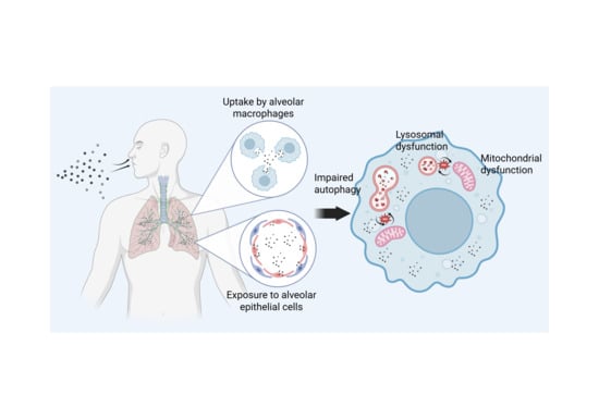
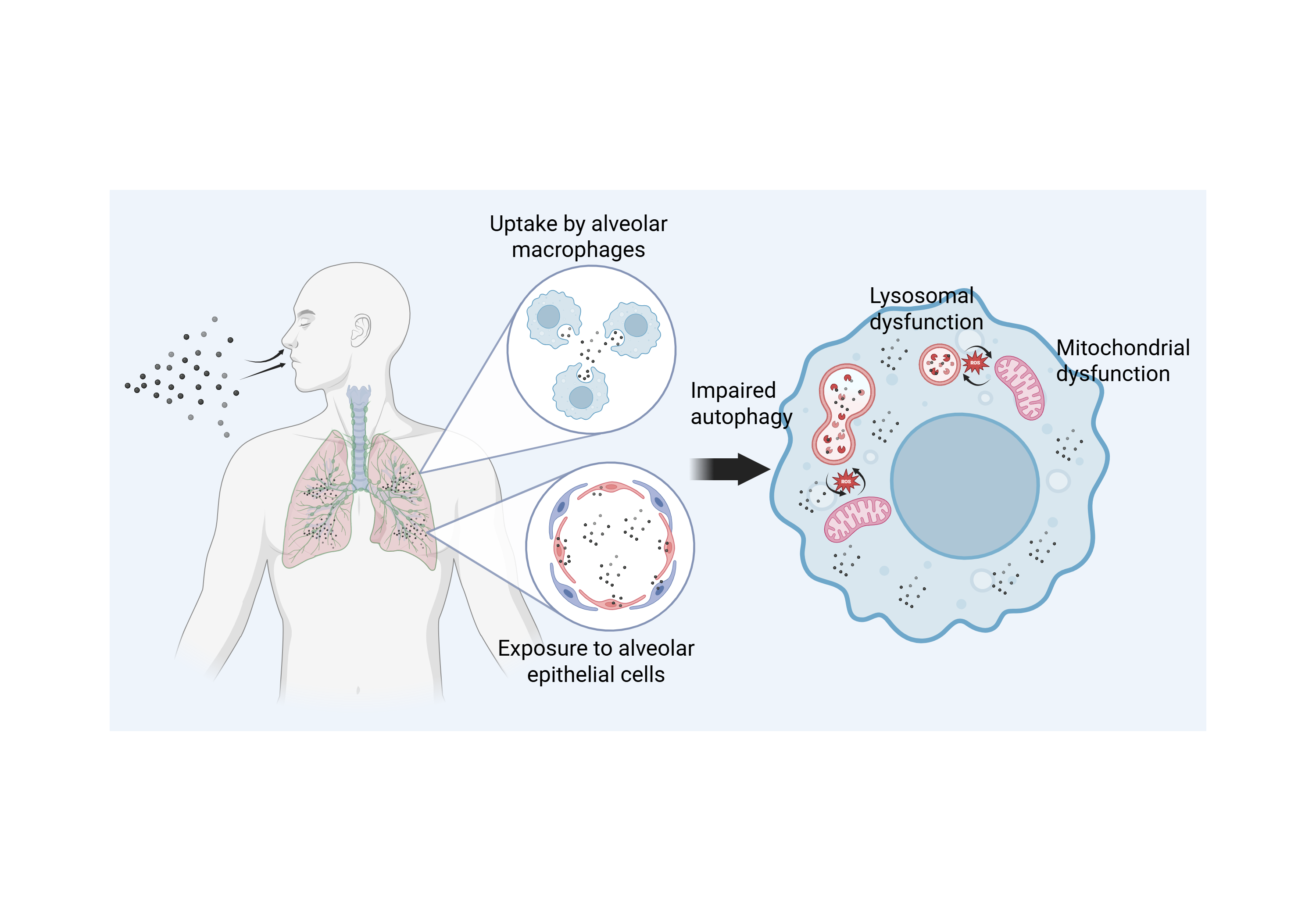
Graphical Abstract

1. Introduction

Exposure to environmental particles in the atmosphere is an important global public health issue. Fine particulate matter (PM) with an aerodynamic diameter of 2.5 µm or less (PM2.5) has various emission sources, including fossil fuel combustion, traffic-related emissions, industrial activity, and naturally derived dust. Global analyses integrating satellite observations, ground monitoring, and chemical transport models have shown that PM2.5 is widely distributed on a global scale, with a significant proportion of the worldwide population chronically exposed to mean PM2.5 concentrations exceeding WHO guidelines, though with clear regional variation [1]. In urban environments, combustion-derived particles, including traffic-related emissions, are major contributors to PM2.5 concentrations and are strongly associated with population exposure [2]. In particular, quantitative data from Beijing suggest that PM2.5 concentrations are significantly higher at monitoring sites with heavy traffic, with road traffic-related emissions an important determinant of PM2.5 concentrations in urban air [3].
Following exposure, PM2.5 has the ability to reach the deep respiratory tract, particularly the alveolar region, though its deposition patterns depend strongly on the particle size. Deposition analysis in humans has reported that, for fine particles with a diameter of approximately 1–2.5 µm, about 20–40% of inhaled particles are deposited in the pulmonary region, whereas larger coarse particles are deposited primarily in the upper airway and bronchial regions [4,5]. For this reason, PM2.5 is an important environmental risk factor for both respiratory and cardiovascular diseases. In particular, epidemiological evidence suggests that an increase in long-term PM2.5 exposure of 10 µg/m3 is associated with a 4–6% higher risk of all-cause mortality and a 6–10% higher risk of cardiovascular and cardiopulmonary mortality [6,7,8]. Long-term exposure is also associated with declines in lung function and the onset and progression of respiratory diseases such as chronic obstructive pulmonary disease (COPD) [5,6,7,8].
In addition to conventional air pollutants, exposure to engineered nanomaterials used for industrial and medical applications and ultrafine particles (UFPs) generated during combustion and abrasion processes has increased. Greater production volume is expected to increase the environmental release of engineered nanomaterials in the future, with model-based assessments consequently suggesting that the environmental concentrations of nanomaterials may increase over time [9]. Similarly, while traffic exhaust-derived PM has been reduced with the introduction of stronger regulations on exhaust particles, the contribution of non-exhaust particles, such as those from brakes, tires, and road surface wear and resuspension, has increased [10]. These non-exhaust particles are small and have become an important component of PM exposure in urban environments, thus they have also attracted significant research attention.
Environmental particles, including PM2.5 and nanomaterials, vary greatly in their size, shape, and physicochemical characteristics; thus, their behavior in the body and toxicity mechanisms are not uniform. Therefore, it has become important to develop a comprehensive understanding of the health effects of environmental particle exposure. This exposure not only causes localized lung injury but can also induce systemic health effects. The American Heart Association has reported that exposure to PM may contribute to the development and progression of cardiovascular diseases via pulmonary inflammation, endothelial dysfunction, autonomic nervous system imbalance, and the activation of coagulation pathways [11,12]. Advances in epidemiological and experimental studies have also found that PM2.5 exposure is associated with chronic inflammation and endothelial dysfunction, while fine particle exposure has been associated with reduced endothelium-dependent vasodilation responses in humans [13]. In addition, animal experimental studies have reported that particle exposure reduces innate immune function in the lungs and weakens bacterial clearance, suggesting that environmental particle exposure can induce immune dysfunction [14].
This inflammation and immune dysregulation have also been implicated in the onset and progression of other chronic diseases, including metabolic diseases, with increasing evidence of an association between PM2.5 exposure and type 2 diabetes [15]. Moreover, large prospective cohort studies have shown that long-term air pollution exposure significantly increases the risk of cardiovascular events, illustrating that environmental particle exposure is an important environmental risk factor for systemic chronic diseases [16].
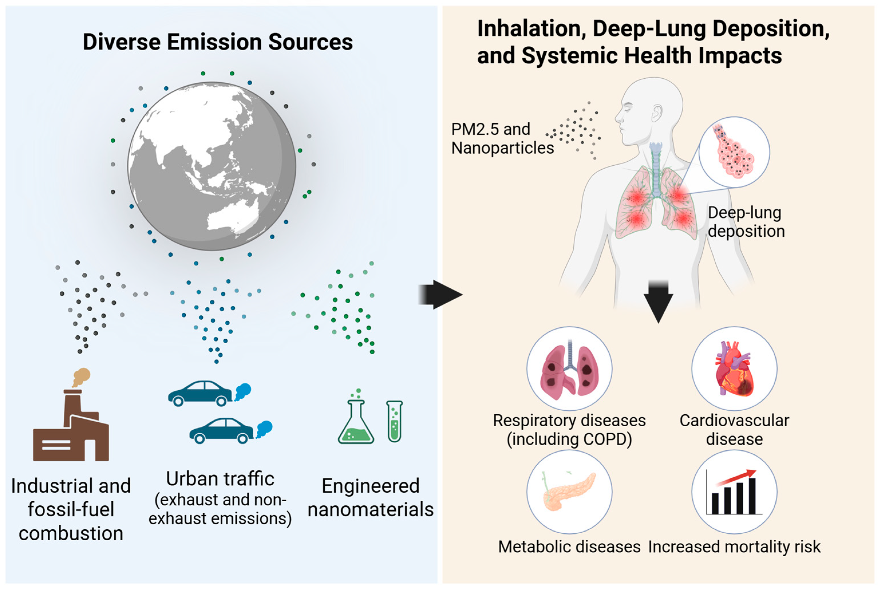
Because environmental particle exposure is an unavoidable health risk in modern society, there is a need to comprehensively evaluate its biological effects from the perspectives of environmental medicine and public health. In this review, the health effects of environmental particle exposure are discussed from the perspectives of biological retention, redistribution, and accumulation, together with their links to chronic lung diseases and systemic effects through disruption of immune homeostasis. The aim of this review is to provide an integrated framework for understanding how retained particles contribute to long-term pathological processes by linking particle biokinetics with intracellular stress, immune dysregulation, and chronic disease progression. However, despite growing evidence that inhaled particles can persist in the body and exert long-term effects, current understanding remains fragmented across separate fields, including particle toxicology, pulmonary inflammation, nanotoxicology, and systemic health research. In particular, the biological significance of particle retention, redistribution, and accumulation has not been sufficiently integrated into a unified framework linking intracellular stress, immune dysregulation, chronic lung disease progression, and systemic consequences. Unlike previous reviews, which have mainly focused on acute toxicity, particle composition, or organ-specific outcomes, this review highlights biological retention and accumulation as central determinants of long-term health effects. On this basis, an integrative perspective is proposed in which retained particles act not only as local toxicants but also as persistent biological stressors that reshape immune homeostasis and contribute to chronic pathology within and beyond the lungs. As shown in Figure 1, environmental particles derived from industrial and fossil fuel combustion, urban traffic (including exhaust and non-exhaust emissions), and engineered nanomaterials can be inhaled and deposited in the deep lung, where they contribute not only to respiratory diseases but also to systemic outcomes such as cardiovascular and metabolic diseases. Figure 1 provides an overview of these major emission sources, deep lung deposition, and representative respiratory and systemic health impacts that form the background and rationale for this review.
This review was prepared as a narrative synthesis of literature. References were selected based on their relevance to the conceptual framework of this review, with particular emphasis on studies addressing particle retention, redistribution, accumulation, intracellular dysfunction, immune dysregulation, chronic lung disease, and systemic effects. Priority was given to representative mechanistic, experimental, translational, and epidemiological studies that contributed to the integration of these themes.

2. Biological Fate and Accumulation of Environmental Particles

2.1. Deposition and Clearance of Inhaled Particles

The deposition sites of inhaled environmental particles depend strongly on the particle size. Previous research using a human airway model (G3–G6) has shown that, within the range of 1–10 µm, deposition patterns are affected by the particle size [17]. A measurement-based study of healthy adults also quantified the total respiratory tract deposition for fine particles with diameters of 1, 3, and 5 µm as an empirical function based on the breathing conditions (e.g., ventilation) and particle size, demonstrating that particle size is a major determinant of the deposition levels [18]. Furthermore, an experimental setup has been used to evaluate the size-resolved lung deposition fractions for children and adults over a range of 15–5000 nm, with particle sizes determined by aerodynamic particle sizers and scanning mobility particle sizers [19].
Deposition patterns are also influenced by the particle shape. The deposition of fibrous particles has been quantified for the human nasal airway [20], while an integrated experimental and numerical study using a realistic female airway model suggested that the handling shape (including rotational motion) is an important factor dictating fiber deposition [21]. The specific gravity of particles is also known to affect deposition, particularly mass deposition, and it has been shown that, for inhaled nanoparticles, deposition and dosimetry estimates can be biased if effective density is not considered [22]. Theoretical deposition models for non-spherical particles such as combustion-derived aggregates support this, indicating that density- and shape-related parameters can influence deposition estimates [23].
Research also suggests that particle surface properties affect deposition patterns. It has been reported that, when the hygroscopic growth of urban atmospheric aerosols is considered, modeled regional deposition patterns can change [24]. Similarly, in a human upper airway model for pharmaceutical aerosols, electrostatic properties of particles were found to influence deposition [25]. Studies using alveolate microchannels that mimic alveolar structures have also examined the contribution of electrostatic deposition, suggesting that surface electrical properties are involved in deposition mechanisms [26].
These differences in deposition patterns influence the cell types and local microenvironments that particles come into contact with and strongly affect subsequent biological responses and toxicity expression patterns. In particular, particle removal does not rely on a single pathway, with lung homeostasis maintained by multiple pathways, including airway clearance and lymphatic translocation. A majority of the particles deposited in the alveolar region are phagocytosed by alveolar macrophages (AMs), removing them from the lungs [27,28]. AMs are the resident innate immune cells in the alveolar space and represent the first line of defense against inhaled foreign matter. They have been shown to rapidly recognize and phagocytose particles and microorganisms [29]. These phagocytosed particles can be transported toward the airway via the movement of AMs and subsequently removed through pathways connected to the mucociliary escalator [27,28]. This mucociliary clearance is an important defense mechanism that transfers inhaled particles toward the pharynx via ciliary motion and mucus secretion of the airway epithelium, and it is a core element of adverse outcome pathways related to the decline in lung function after inhalation exposure [30]. In addition, at the airway level, the cough reflex can function as an auxiliary particle expulsion mechanism, suggesting that macrophage phagocytosis and physical expulsion mechanisms may cooperate to contribute to particle clearance [28,31]. Animal and human studies have also shown that some particles can be translocated and accumulate in draining lymph nodes via the lymphatic system [32,33].
However, these clearance mechanisms have limitations. Poorly soluble, low-toxicity particles (PSLTs) are often bio-persistent and difficult to eliminate. Under lung overload conditions, macrophage phagocytic and migratory capacities decline, impairing particle removal [34]. The removal of PSLTs by AMs may thus be incomplete, leading to long-term retention of particles in the lungs [35,36]. In an in vitro overload model of TiO2 and carbon black deposition, macrophage gene expression differed markedly and functional alterations were observed, including inflammation-related responses [37]. As a result, particle accumulation occurs and the retained lung burden increases; for example, in a rat model with whole-body inhalation exposure to poorly soluble nanoparticles, the lung particle burden increased over time, while reduced clearance and greater particle retention were observed [38]. Particle-induced lung overload shares common mechanisms that can cause tissue injury via chronic inflammatory responses, and macrophages are thought to play a central role [39,40]. It has also been suggested that particle-laden macrophages that remain in the tissue may be associated with persistent inflammatory stimulation [34,39]. In support of this, human epidemiological studies have shown that individuals with higher particle loads in their AMs have elevated systemic inflammatory markers [41]. Thus, while deposition and clearance function as defense mechanisms, removal can be incomplete depending on the particle properties and exposure conditions, leading to retention and accumulation in the lungs.

2.2. Biological Retention and Redistribution

PSLTs deposited in the lung can accumulate over time, leading to lung particle overload [34,42]. In animal models, it has been reported that carbon black nanoparticles and polymer particles are retained in the lungs for a long period even after inhalation, and residual particles are observed after several weeks or more [35,36]. This long-term lung retention exhibits multiphasic clearance behavior that cannot be explained by a single time constant [35] and is closely related to the poor solubility and high durability of the particles. Incomplete clearance by AMs due to disrupted macrophage-mediated transport and elimination is considered to be a major cause of this [34,35]. While these residual particles are less likely to be observed in toxicity evaluations based on acute exposure, the persistence of particles retained in the lungs becomes an internal burden and is responsible for chronic inflammatory stimulation [42,43].
Inflammatory responses in the lungs thus need to be interpreted not only in terms of their presence and/or intensity but also in terms of persistence and reversibility. It has been pointed out that short-term inflammatory indicators alone may not appropriately evaluate long-term adverse effects due to inflammation that persists beyond the adaptive range [44]. The total surface area of particles retained in the lungs has been reported to be related to the strength of inflammatory responses, while the retained particle amount contributes to the persistence of inflammation for acute, subacute, and chronic exposure conditions [43]. In addition to chronic inflammatory responses, lung particle overload conditions have been reported to trigger responses related to tissue remodeling, including epithelial changes and changes in tissue architecture [42]. It has also been suggested that the molecular responses of macrophages observed in in vitro overload models using TiO2 and carbon black may be consistent with chronic inflammatory responses that occur in vivo due to long-term particle retention [37].
The retention and redistribution of particles is not limited to the lungs. It has been suggested that some inhaled environmental particles, after being taken up by AMs and other pulmonary cells, are transported outside the lungs via cell migration. In recent studies, it has been reported that pulmonary cells such as lung megakaryocytes take up inhaled particles, induce pulmonary inflammation, and can be involved in distribution to extrapulmonary tissues [45]. In particular, the migration of inhaled particles to draining lymph nodes via pulmonary lymph flow has attracted attention as an important redistribution pathway. In inhalation exposure experiments using mice, it has been shown that ultrafine carbon, after being deposited in the lungs, can accumulate in lung-associated lymph nodes and the spleen, with extrapulmonary translocation via the lymphatic system as one of the major pathways [32]. Particles have also been shown to accumulate in pulmonary lymph nodes in humans, with age-related particle accumulation potentially affecting the structure and immune function of lymph nodes [33]. These findings indicate that inhaled particles are not only eliminated locally in the lungs but can directly interact with the immune system via the lymphatic system. In fact, particle accumulation in pulmonary lymph nodes has been shown to be associated with changes in the composition and spatial organization of immune cells and the modulation of immune responses, suggesting that inhaled particles can modify the immune environment [32,33].
In addition to lymphatic translocation, distribution to extrapulmonary organs has been reported. In experimental model studies, it has been observed that some inhaled environmental particles are not only retained in the lungs after initial deposition and pulmonary clearance, but some are also detected in extrapulmonary organs such as the spleen and liver. In an animal model exposed via inhalation to crystalline silica, it has been quantitatively demonstrated that particles, after being deposited in the lungs, are distributed in a time-dependent manner to extrapulmonary sites such as the mediastinal lymph nodes, spleen, and liver, suggesting the presence of early and late translocation mechanisms [46]. These findings indicate the possibility that inhaled environmental particles are not limited to the local sites in the lungs but can be redistributed at the systemic level. In line with this, in a review focusing on ultrafine PM, it has been reported that inhaled particles can translocate to extrapulmonary organs via lymphatic or hematogenous pathways after lung deposition and that particle redistribution is an important element of biokinetics [47].
Previous research has suggested that inter-tissue translocation is influenced by the physicochemical properties of the deposited particles and the types of cells used as carriers. In a study using silica particles, indicators related to particle surface properties were found to be associated with extrapulmonary translocation behavior over time [46]. Similarly, classical studies have reported that extrapulmonary translocation of UFPs depends on particle size, indicating that size is an important factor influencing redistribution efficiency [48]. Supporting this concept, in vivo imaging studies using size-dependent fluorescent particles as a model of fine dust have demonstrated that smaller particles exhibit distinct biodistribution patterns and a greater potential for extrapulmonary translocation [49]. These findings further support the view that particle size is a critical determinant of systemic organ exposure after inhalation. Recent studies have also shown that specific cell types present in the lungs take up inhaled particles and can be involved in pulmonary inflammation and extrapulmonary distribution, with differences in carrier cell types potentially affecting modes of particle transport [45]. Nevertheless, the molecular mechanisms that control extrapulmonary translocation and redistribution processes and their long-term impacts on organisms remain unclear. In particular, the pathways and cells involved in the translocation of particles to extrapulmonary organs and how this process affects inflammatory responses and organ function remain issues that require further investigation [45,47].
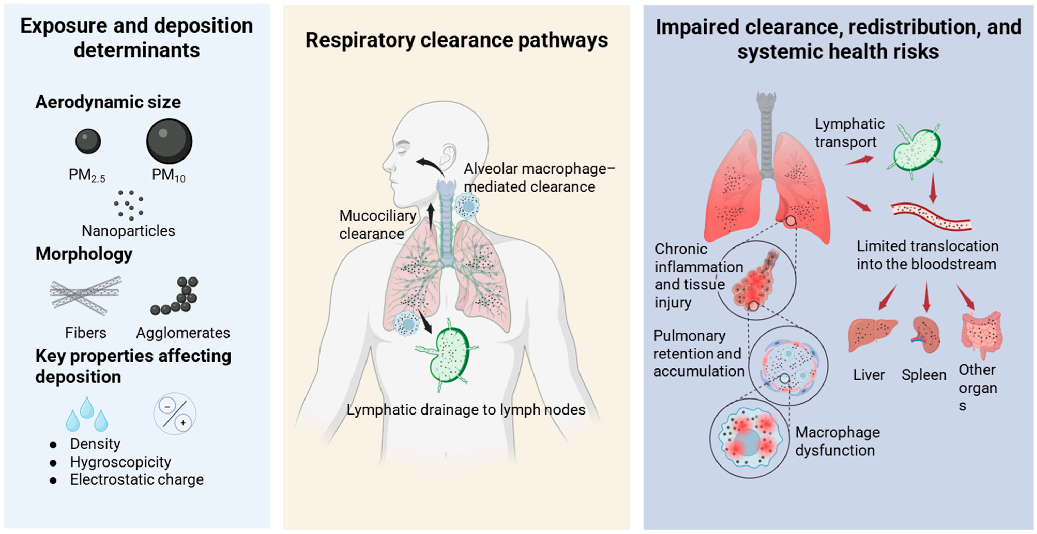
This biological retention and redistribution highlight the need to re-evaluate the effects of particle exposure outside of the conventional framework that focuses on the local toxicity of a single organ. In addition to these physicochemical and cellular determinants, biologically active materials associated with environmental particles, including bacteria, fungi, lipopolysaccharides, and extracellular vesicles, may further modulate their inflammatory and pathogenic potential [50]. These components may enhance pulmonary inflammation and alter host immune responses, thereby contributing to the progression of chronic lung diseases. As summarized in Figure 2, inhaled particles are not always rapidly cleared after deposition but may be retained in the lungs, redistributed via lymphatic and limited extrapulmonary pathways, and persist while altering the immune system and tissue environment. These processes should be taken into account when considering the chronic health effects of environmental particle exposure.

3. Accumulation and Functional Impairment at the Intracellular Organelle Level

3.1. Endo-Lysosomal Accumulation and Dysfunction

Representative studies discussed in this section are summarized in Table 1 to highlight the major experimental approaches, principal findings, and their relevance to the framework of this review.
After being taken up by cells, inhalation-derived environmental particles can enter the intracellular space via endocytosis and/or endocytosis-related pathways. These internalized particles can then be transported to lysosomes through the maturation process of the endosomal system. This has been demonstrated by previous research on the uptake mechanisms of nanoparticles, analytical methods for intracellular trafficking, and the visualization of trafficking dynamics at the single-cell level [51,62]. It has been shown that particles tend to accumulate in the endo-lysosomal system, which can become a major intracellular compartment for particle accumulation, leading to functional disruption [51,52,63,64]. In particular, for poorly soluble or highly durable nanomaterials, dissolution under lysosomal conditions is slow, leading to intracellular retention and accumulation [63]. Lysosome functional perturbations and related mechanisms associated with particle exposure have consequently been reported, with perturbation of the endo-lysosomal system an important component of particle toxicity [52,64]. Furthermore, lysosomal membrane permeabilization (LMP) has been reported for silica particles, suggesting that lysosomes can become a major site of particle effects [65]. Taken together, inhaled particles accumulate intracellularly in the endo-lysosomal system, and if these particles have poor solubility and/or high durability, this system is highly likely to be a major site of particle accumulation and functional impacts [51,52,62,63,64,65,66,67].
In dendritic cells (DCs), intracellular particle accumulation and the modulation of immune function have been reported even under subtoxic DEP/PM2.5 exposure conditions [53]. In these cells, it has been reported that particle exposure can increase LMP, and LMP-mediated cellular injury due to PM2.5 exposure has also been reported [54]. Similarly, it has been reported that, in systems centered on AMs, cellular injury associated with lysosomal membrane damage/LMP occurs with carbon black nanoparticle exposure, subsequently leading to pulmonary inflammatory responses [55]. Related to this, following exposure to particles/nanoparticles (e.g., ZnO and SiO2), it has been shown that hyperpolarization of lysosomal membrane potential precedes LMP and NLRP3 activation [68]. Another mechanism responsible for lysosomal dysfunction that has been suggested is the disruption of acidification (including the proton pump/v-ATPase), which leads to an increase in lysosomal pH [69]. Related to this, for inorganic silica nanoparticles, it has been reported that functional changes can occur, including lysosomal biology and protease activity [70]. Collectively, LMP, disturbance of acidification, and changes in enzyme activity can modulate the degradative capacity and trigger intracellular stress and inflammatory responses [52,64]. In addition to foreign body processing, lysosomal dysfunction is also associated with autophagy and immune response regulation (e.g., cellular homeostasis and signal regulation based on lysosomes); thus, its impacts are broad [71].
Abnormalities of the endo-lysosomal system (e.g., lysosomal accumulation of particles, disturbance of acidification, LMP, and modulation of degradative function) have been proposed as toxicity mechanisms associated with inhaled particles/nanoparticles, and these can be observed in multiple cell types, including AMs, DCs, and epithelial cells [52,64]. On the other hand, macrophages and DCs, which are responsible for foreign body processing and antigen presentation in the innate immune system, can differ in their functional adaptation and processing capacity depending on the tissue environment, leading to differences in their particle uptake and processing dynamics, thus affecting the intracellular burden and the impact on the endo-lysosomal system [72]. Therefore, in immune cells with phagocytic capacity, the lysosomal burden can increase due to particle retention [52,53,64]. In contrast, it has been reported that, in airway epithelial cells (BEAS-2B), the acidic pH environment of lysosomes is affected by the intracellular accumulation of vehicle exhaust particles, illustrating that lysosomal functional perturbation is a potential cellular response even under conditions that do not necessarily incur high-burden phagocytosis [56]. Furthermore, in neuronal cell models, an association between cell death pathways via LMP and impairment of autophagy function due to PM2.5 exposure has been reported, indicating that lysosomal functional perturbation can be linked to downstream stress responses and cellular functional abnormalities [54].
Overall, while abnormalities of the endo-lysosomal system can occur for multiple cell types, the manifestation of the lysosomal burden and functional perturbation can differ in immune cells (AMs/DCs) and epithelial cells due to differences in their uptake modes and processing capacity. These commonalities and differences can determine cellular responses after particle exposure, including inflammation, stress responses, and immune modulation.

3.2. Mitochondrial Stress, Metabolic Reprogramming, Organelle Crosstalk, and Intracellular Trafficking

In addition to the impact on the endo-lysosomal system, intracellular effects of particle exposure can be observed in multiple organelles, including mitochondria. Here, emphasis is placed on the major intracellular pathways most consistently implicated in retained particle toxicity, particularly organelle crosstalk linking endo-lysosomal dysfunction with downstream immune and inflammatory consequences. In BEAS-2B cells, the uptake of PM2.5 can cause oxidative injury, inflammatory cytokine production, mitochondrial structural damage (e.g., cristae disruption), activation of mitophagy-related pathways (e.g., Bnip3L/NIX), and accumulation of autophagic vesicles/autolysosomes [57]. In addition, repeated PM2.5 exposure in an airway epithelial air–liquid interface model confirmed the intracellular translocation of particles, persistent inflammatory responses, and changes in mitochondrial activity, together with potential p62 accumulation and inhibition of the autophagic flux [58]. As a framework to understand this multiorgan propagation, reviews summarizing lysosome–mitochondria crosstalk have also been presented [73].
Regarding the process by which particles reach the intracellular space, some reviews have presented the endocytosis pathways for nanoparticles/submicron particles and the basic principles and methods employed to analyze uptake mechanisms, and these have been positioned as general principles supporting the intracellular entry of particles [52,74]. Furthermore, for traffic-derived UFPs, exposure has been shown to inhibit mitochondrial oxidative phosphorylation, reduce respiratory function and ATP levels, and increase reactive oxygen species (ROS)/augmented oxidative stress. accompanied by disruption of the redox balance [59].
Mitochondrial functional decline can also occur through secondary stress triggered by lysosomal damage. Using silver nanoparticles, it has been shown that lysosome–mitochondria interactions are involved in the breakdown of mitochondrial homeostasis [60]. In addition, acute cell necrosis triggered by lysosomal disruption (i.e., LMP or rupture) has been reported following exposure to carbon black nanoparticles, with the release of mtDNA mediating neutrophilic inflammation in the lungs [55]. Disruption of the autophagic flux and p62-dependent responses associated with lysosomal dysfunction (e.g., alkalinization, LMP, and cathepsin release) have been demonstrated for PM2.5 exposure, illustrating that lysosomal damage can amplify downstream cellular stress responses [54].
In addition to reports that the toxicity of PM components is mitochondria-dependent and that metals and polycyclic aromatic hydrocarbons can be involved [75], disruption of the redox balance and increased ROS have been reported following UFP exposure [59]. Mitochondrial-derived ROS (mtROS) have also been proposed as a secondary messenger involved in the regulation of immune responses, including macrophages and T cells, thus linking ROS to inflammatory signaling/transcriptional regulation [76]. Based on these reports, mitochondria-derived oxidative stress generated by particle exposure can affect not only cellular injury but also the strength and persistence of inflammatory responses [55,58,76].
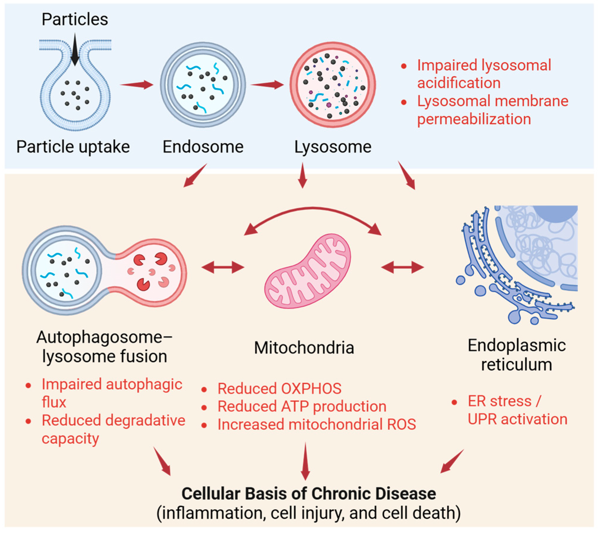
Mitochondrial damage can also have deleterious metabolic consequences. For example, DEP exposure not only disrupts the mitochondrial morphological network but also decreases the respiration parameters based on oxygen consumption (e.g., basal/ATP-linked respiration), reduces the ATP production rate, and weakens glycolysis-related indices [77]. UFP exposure has also been observed to damage respiratory chain function and reduce ATP production, while also modulating NAD and glutathione metabolism, thus providing evidence for the rewiring of energy and redox metabolism [59]. In addition, the close linkage between immune function regulation via mtROS and the metabolic state of cells has been presented in past reviews, supporting the theory that mitochondria–redox modulation generated by particle exposure is associated with bias in immune phenotypes [76]. Particle exposure also extends to the autophagy system and ER stress responses. Reports of mitophagy-related responses and accumulation of autophagic vesicles/autolysosomes [57], p62 accumulation and flux inhibition under repeated exposure conditions [58], and flux disruption via lysosomal damage from PM2.5 exposure [54] provide evidence that stagnation of the lysosome–autophagy axis can act as a bottleneck for the processing of abnormal proteins and damaged organelles. Similarly, PM2.5 has been shown to activate ER stress pathways and is associated with inflammation and mucus production via IRE1α/NOD1/NF-κB [61], while silver nanoparticles are known to be involved in ER stress responses and the IRE1–XBP1 axis [78]. Reviews emphasizing the functional linkage between lysosomes and mitochondria and suggesting that dysfunction of both can contribute to pathogenesis have also been presented [73,79], providing a basis for considering crosstalk in which ER, mitochondria, and lysosomal stress mutually influence each other following particle exposure. As summarized in Figure 3, particle-induced endo-lysosomal dysfunction can propagate to other organelles, including mitochondria and the endoplasmic reticulum, thereby promoting inflammation, cellular injury, and cell death. These interconnected organelle stresses may provide a cellular basis for the development and progression of chronic disease.

4. Disease Relevance

4.1. Chronic Lung Diseases as Accumulation-Driven Disorders

This section discusses how persistent retained particle burden in the lungs may contribute to chronic lung disease progression by sustaining immune dysregulation, epithelial injury, and tissue remodeling.
COPD is thought to derive from persistent airway inflammation and immunopathology. Within the framework of immune-mediated inflammation, the pathophysiological cascade leading to airway remodeling and emphysematous changes has been described [80,81,82]. In particular, pulmonary fibrosis (particularly idiopathic pulmonary fibrosis; IPF) is an aberrant wound-healing response centered on repetitive epithelial injury and abnormal repair, together with fibroblast activation and extracellular matrix (ECM) accumulation [83]. However, in recent years, based on the analysis of the residual retention of inhaled particles and lung particle overload, a framework has also been proposed to reinterpret chronic lung diseases not only as the persistence of inflammation but also from the perspective that the particle burden remaining in the lungs can have long-term effects on immune status. In this framework, lung clearance is restricted due to an overload of poorly soluble particles, and the retained particles act as persistent stimuli [34].
It has also been shown in AM overload models using TiO2 and carbon black that the molecular responses of AMs can be reprogrammed depending on the particle burden [37]. Environmental particles, particularly poorly soluble and highly durable particles, are not completely eliminated after deposition in the alveolar region and can remain for long periods within biphasic clearance kinetics involving AMs, as demonstrated by lung clearance analysis of carbon black nanoparticles [35]. Similarly, the distribution and retention of inhaled polymer particles in the lungs have been evaluated, and it has been reported that these particles can be retained in the lungs for a certain period [36]. For example, cellular injury triggered by LMP due to exposure to carbon black has been linked to inflammatory responses [55]. Carbon in AMs has also been associated with gene expression characteristics, indicating that particle retention can be reflected in the molecular state of immune cells [84]. From this perspective, the functional status and molecular responses of immune cells (particularly AMs) in the lungs can shift under persistent stimulation by retained particles during particle overload [34]. The reprogramming of transcriptional responses observed in overloaded AMs may illustrate this transition at the cellular level [37].
In line with this, it has been reported that the function of AMs is altered due to smoking and COPD, including the release of tissue-injurious mediators such as MMP-12 and the lower clearance via phagocytosis or efferocytosis of respiratory pathogens and apoptotic cells. Therefore, it has been suggested that functional abnormalities of immune cells that take up particles play a role in chronic pathology [85]. In addition, it has been shown that the spatial distribution of carbon black in the lungs is associated with COPD exacerbations, with the particle burden linked to clinical outcomes [86]. More broadly, epidemiological studies have consistently shown that long-term exposure to particulate air pollution is associated with the development and progression of chronic respiratory diseases, including COPD and fibrotic lung conditions. From a clinical perspective, these findings support the relevance of retained particle burden not only as a mechanistic concept but also as a factor potentially linked to disease severity, exacerbation risk, and long-term respiratory health outcomes. This supports the framework positing that COPD progresses from sustained immunopathology to remodeling and tissue destruction [80,81,82], from the perspective of particle retention.
In addition to the assumption that epithelial injury and abnormal repair are central to the pathology [83], it has been reported that PM2.5 can contribute to pulmonary fibrosis via cell fate control and autophagy [87], indicating that persistent stress derived from environmental particles can generate a fibrotic environment. Therefore, long-term particle retention may chronically alter the interactions among immune, epithelial, and mesenchymal cells, which underlies the progression of COPD and pulmonary fibrosis [35,36,37,55,80,81,82,83,84,85,86,87]. This framework, in addition to an inflammation-centered understanding, can improve the understanding of these diseases and potential intervention targets by incorporating retained burden and clearance failure as evaluation axes [34,80,81,82].

4.2. Systemic Implications

A framework for the systemic effects that arise after the inhalation of nanoparticles has been presented in which indirect mediators arising from local lung responses are involved in the outcomes for distant organs [88]. In particular, PM can induce oxidative stress and inflammation that leads to tissue injury, thus inducing the “systemicization” of an inflammatory environment originating in the lungs [89]. The relationship between PM and cardiovascular risk and potential intervention strategies have previously been reviewed [90,91,92], with lung-derived immune/inflammatory signals possibly connected to systemic pathogenesis [90,91]. Therefore, even though the exact molecular mechanisms are still under investigation, it has been proposed that persistent immune activation in the lungs can contribute to a systemic inflammatory or oxidative stress environment via the circulatory system [88,89,90,91]. In this framework, it is assumed that, when the particles remain in the lungs, there is a chronic increase in the production of inflammatory cytokines and immune-modulatory factors, which may affect distant organs via the circulatory system [88,89]. It has also been reported that mtROS can act as a secondary messenger in the regulation of immune responses [76], while chronic immune stimulation within the lungs can be sustained and amplified via intracellular signaling.
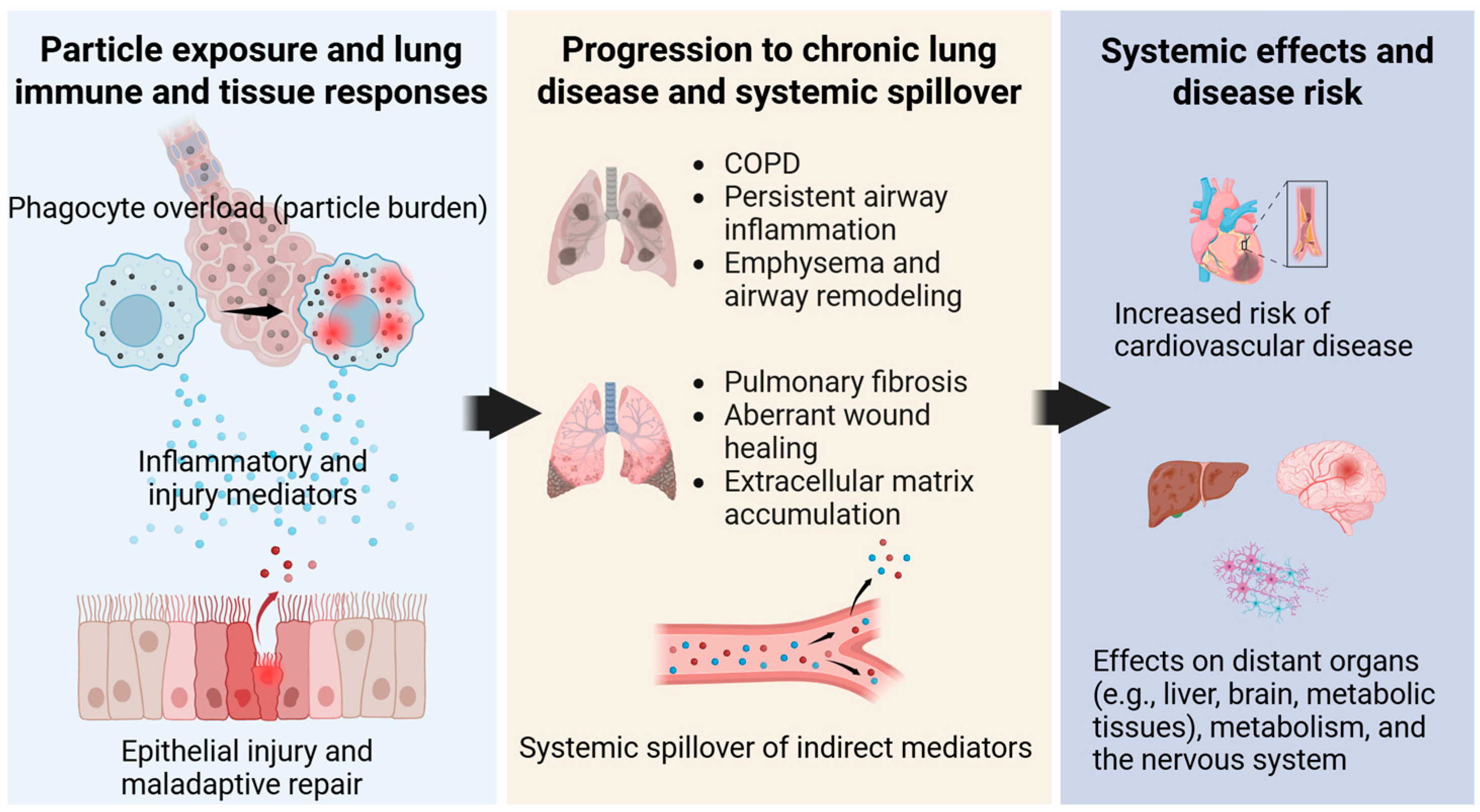
From this perspective, the lungs represent an exposure organ that can subsequently influence outcomes in distant organs as a source of inflammatory and oxidative stress mediators generated in response to particle exposure [88,89]. Changes in lung immune status after particle exposure can affect various organ systems, including the cardiovascular system, as a result of chronic immune stimulation due to particles remaining in the lungs [34,35,88,89,90,91,92]. As summarized in Figure 4, retained particle burden in the lungs may drive chronic lung pathology by sustaining phagocyte overload, tissue injury, and immune dysregulation, while also promoting systemic spillover of lung-derived mediators. This framework links persistent particle retention with chronic respiratory disease progression and broader systemic health risks.

5. Conclusions

In this review, the health effects of environmental particle exposure were summarized from the perspectives of biological retention, redistribution, and accumulation. Retained particles can induce lysosomal accumulation and dysfunction, which may subsequently propagate to mitochondria, cellular metabolism, and the autophagy system, thereby exerting long-term effects on immune cell function. On this basis, chronic lung diseases may arise not only from persistent inflammation but also from continuous stimulation by retained particles, which disrupts immune homeostasis and promotes tissue remodeling. In addition, chronic immune activation originating in the lungs may extend beyond the respiratory system and contribute to systemic effects through indirect mediators and oxinflammation.
Despite the growing body of mechanistic evidence, several limitations of the current literature should be acknowledged. First, many available studies are based on in vitro systems or short-term animal experiments and may not fully capture the complexity of long-term, repeated human exposure. Second, substantial heterogeneity exists across studies in terms of particle type, size, composition, dose, exposure duration, and biological endpoints, making direct comparison and generalization difficult. Third, although mechanistic studies provide important insights into intracellular dysfunction and immune modulation, direct longitudinal human evidence linking retained particle burden to chronic disease progression remains limited. These methodological and translational limitations should be considered when interpreting the current evidence base.
At the same time, several important knowledge gaps remain. First, the determinants of long-term particle retention and reduced clearance under repeated or chronic exposure conditions are still incompletely understood, particularly in relation to particle size, composition, surface properties, and biological interactions. Second, the mechanisms linking retained particle burden to intracellular organelle stress, immune dysfunction, and chronic tissue remodeling have not yet been fully integrated across experimental systems. Third, the pathways and biological significance of particle redistribution to lymphatic and extrapulmonary sites remain insufficiently characterized. In addition, longitudinal human evidence directly connecting retained particle burden with disruption of immune homeostasis and chronic disease progression is still limited.
Future research should therefore move beyond conventional short-term toxicity testing and incorporate retained dose, clearance kinetics, overload conditions, and repeated exposure scenarios into risk assessment frameworks. More specifically, long-term inhalation models, quantitative imaging approaches for particle redistribution, and integrated analyses combining pathology, immune phenotyping, and organelle stress markers will be needed. Human longitudinal studies and translational approaches are also required to clarify the relevance of retained particle burden to disease susceptibility and progression, especially in vulnerable populations. Furthermore, future work should address mixed and real-world exposures, including non-exhaust particles and biologically active particle-associated materials, and should develop integrated models that link particle biokinetics with immune homeostasis and chronic pathology. Such efforts will be essential not only for mechanistic understanding but also for prevention, exposure reduction, and the development of more realistic public health strategies.

Funding

This research received no external funding.

Institutional Review Board Statement

Not applicable.

Informed Consent Statement

Not applicable.

Data Availability Statement

No new data were created or analyzed in this study. Data sharing is not applicable.

Acknowledgments

Figures were created in BioRender. Onodera, A. (2026). https://BioRender.com/0ee26n1 (accessed on 13 February 2026).

Conflicts of Interest

The author declares no conflicts of interest.

Abbreviations

The following abbreviations are used in this manuscript:
PM2.5Fine particulate matter with aerodynamic diameter ≤ 2.5 µm
PM10Particulate matter with aerodynamic diameter ≤ 10 µm
PMParticulate matter
WHOWorld Health Organization
AHAAmerican Heart Association
COPDChronic obstructive pulmonary disease
IPFIdiopathic pulmonary fibrosis
ECMExtracellular matrix
SMPSScanning mobility particle sizer
APSAerodynamic particle sizer
PSLTsPoorly soluble, low-toxicity particles
DC(s)Dendritic cell(s)
DEPDiesel exhaust particles
AMAlveolar macrophage(s)
TiO2Titanium dioxide
LMPLysosomal membrane permeabilization
v-ATPaseVacuolar-type H+-ATPase
ZnOZinc oxide
SiO2Silicon dioxide
NLRP3NOD-, LRR- and pyrin domain-containing protein 3
BEAS-2BBronchial Epithelium transformed with Ad12-SV40 2B
ALIAir–liquid interface
p62 (SQSTM1)p62/SQSTM1 (sequestosome 1)
UFPUltrafine particles
OXPHOSOxidative phosphorylation
ATPAdenosine triphosphate
ROSReactive oxygen species
mtROSMitochondrial reactive oxygen species
mtDNAMitochondrial DNA
PAHsPolycyclic aromatic hydrocarbons
NADNicotinamide adenine dinucleotide
EREndoplasmic reticulum
UPRUnfolded protein response
AgNPSilver nanoparticles
IRE1αInositol-requiring enzyme 1 alpha
XBP1X-box binding protein 1
NOD1Nucleotide-binding oligomerization domain-containing protein 1
NF-κBNuclear factor kappa B
MMP-12Matrix metalloproteinase-12
Bnip3L/NIXBCL2/adenovirus E1B 19 kDa protein-interacting protein 3-like (NIX)

References

  1. van Donkelaar, A.; Hammer, M.S.; Bindle, L.; Brauer, M.; Brook, J.R.; Garay, M.J.; Hsu, N.C.; Kalashnikova, O.V.; Kahn, R.A.; Lee, C.; et al. Monthly global estimates of fine particulate matter and their uncertainty. Environ. Sci. Technol. 2021, 55, 15287–15300. [Google Scholar] [CrossRef]
  2. Karagulian, F.; Belis, C.A.; Dora, C.F.C.; Prüss-Ustün, A.M.; Bonjour, S.; Adair-Rohani, H.; Amann, M. Contributions to cities’ ambient particulate matter (PM): A systematic review of local source contributions at global level. Atmos. Environ. 2015, 120, 475–483. [Google Scholar] [CrossRef]
  3. Fang, X.; Li, R.; Xu, Q.; Bottai, M.; Fang, F.; Cao, Y. A two-stage method to estimate the contribution of road traffic to PM2.5 concentrations in Beijing, China. Int. J. Environ. Res. Public Health 2016, 13, 124. [Google Scholar] [CrossRef] [PubMed]
  4. Deng, Q.; Deng, L.; Miao, Y.; Guo, X.; Li, Y. Particle deposition in the human lung: Health implications of particulate matter from different sources. Environ. Res. 2019, 169, 237–245. [Google Scholar] [CrossRef] [PubMed]
  5. Guo, C.; Zhang, Z.; Lau, A.K.; Lin, C.Q.; Chuang, Y.C.; Chan, J.; Jiang, W.K.; Tam, T.; Yeoh, E.-K.; Chan, T.-C.; et al. Effect of long-term exposure to fine particulate matter on lung function and chronic obstructive pulmonary disease: A longitudinal cohort study. Lancet Planet. Health 2018, 2, e114–e125. [Google Scholar] [CrossRef]
  6. Dockery, D.W.; Pope, C.A.; Xu, X.; Spengler, J.D.; Ware, J.H.; Fay, M.E.; Ferris, B.G.J.; Speizer, F.E. An association between air pollution and mortality in six U.S. cities. N. Engl. J. Med. 1993, 329, 1753–1759. [Google Scholar] [CrossRef]
  7. Pope, C.A.I.; Burnett, R.T.; Thun, M.J.; Calle, E.E.; Krewski, D.; Ito, K.; Thurston, G.D. Lung cancer, cardiopulmonary mortality, and long-term exposure to fine particulate air pollution. JAMA 2002, 287, 1132–1141. [Google Scholar] [CrossRef]
  8. Pope, C.I.; Burnett, R.; Krewski, D.; Jerrett, M.; Shi, Y.; Calle, E.; Thun, M. Cardiovascular mortality and exposure to airborne fine particulate matter and cigarette smoke: Shape of the exposure-response relationship. Circulation 2009, 120, 941–948. [Google Scholar] [CrossRef]
  9. Giese, B.; Klaessig, F.; Park, B.; Kaegi, R.; Steinfeldt, M.; Wigger, H.; von Gleich, A.; Gottschalk, F. Risks, release and concentrations of engineered nanomaterial in the environment. Sci. Rep. 2018, 8, 1565. [Google Scholar] [CrossRef]
  10. Harrison, R.M.; Allan, J.; Carruthers, D.; Heal, M.R.; Lewis, A.C.; Marner, B.; Murrells, T.; Williams, A. Non-exhaust vehicle emissions of particulate matter and VOC from road traffic: A review. Atmos. Environ. 2021, 262, 118592. [Google Scholar] [CrossRef]
  11. Brook, R.D.; Rajagopalan, S.; Pope, C.A.I.; Brook, J.R.; Bhatnagar, A.; Diez-Roux, A.V.; Holguin, F.; Hong, Y.; Luepker, R.V.; Mittleman, M.A.; et al. Particulate matter air pollution and cardiovascular disease: An update to the scientific statement from the American Heart Association. Circulation 2010, 121, 2331–2378. [Google Scholar] [CrossRef]
  12. Brook, R.D.; Franklin, B.; Cascio, W.; Hong, Y.; Howard, G.; Lipsett, M.; Luepker, R.; Mittleman, M.; Samet, J.; Smith, S.C.J.; et al. Air Pollution and cardiovascular disease: A statement for healthcare professionals from the Expert Panel on Population and Prevention Science of the American Heart Association. Circulation 2004, 109, 2655–2671. [Google Scholar] [CrossRef]
  13. Pope, C.A.I.; Bhatnagar, A.; McCracken, J.P.; Abplanalp, W.; Conklin, D.J.; O’Toole, T. Exposure to fine particulate air pollution is associated with endothelial injury and systemic inflammation. Circ. Res. 2016, 119, 1204–1214. [Google Scholar] [CrossRef]
  14. Sigaud, S.; Goldsmith, C.-A.W.; Zhou, H.; Yang, Z.; Fedulov, A.; Imrich, A.; Kobzik, L. Air pollution particles diminish bacterial clearance in the primed lungs of mice. Toxicol. Appl. Pharmacol. 2007, 223, 1–9. [Google Scholar] [CrossRef]
  15. Rajagopalan, S.; Brook, R.D. Air pollution and type 2 diabetes: Mechanistic insights. Diabetes 2012, 61, 3037–3045. [Google Scholar] [CrossRef] [PubMed]
  16. Miller, K.A.; Siscovick, D.S.; Sheppard, L.; Shepherd, K.; Sullivan, J.H.; Anderson, G.L.; Kaufman, J.D. Long-term exposure to air pollution and incidence of cardiovascular events in women. N. Engl. J. Med. 2007, 356, 447–458. [Google Scholar] [CrossRef] [PubMed]
  17. Ou, C.; Hang, J.; Deng, Q. Particle deposition in human lung airways: Effects of airflow, particle size, and mechanisms. J. Aerosol Sci. 2020, 20, 2846–2858. [Google Scholar] [CrossRef]
  18. Kim, C.S.; Hu, S.-C. Total respiratory tract deposition of fine micrometer-sized particles in healthy adults: Empirical deposition equations. J. Appl. Physiol. 2006, 101, 401–412. [Google Scholar] [CrossRef]
  19. Rissler, J.; Nicklasson, H.; Gudmundsson, A.; Wollmer, P.; Swietlicki, S.; Löndahl, J. A set-up for respiratory tract deposition efficiency measurements (15–5000 nm) and first results for a group of children and adults. Aerosol Sci. Technol. 2017, 17, 1244–1255. [Google Scholar] [CrossRef]
  20. Su, W.-C.; Cheng, Y. Deposition of fiber in the human nasal airway. Aerosol Sci. Technol. 2005, 39, 888–901. [Google Scholar] [CrossRef]
  21. Prinz, F.; Kánská, J.; Elcner, J.; Hájek, O.; Kummerländer, A.; Krause, M.; Jícha, M.; Lízal, F. Transport and deposition of inhaled fibers in a realistic female airway model: A combined experimental and numerical study. Comput. Biol. Med. 2025, 194, 110473. [Google Scholar] [CrossRef]
  22. Lizonova, D.; Nagarkar, A.; Demokritou, P.; Kelesidis, G. Effective density of inhaled environmental and engineered nanoparticles and its impact on the lung deposition and dosimetry. Part. Fibre Toxicol. 2024, 21, 7. [Google Scholar] [CrossRef]
  23. Sturm, R. Theoretical models for dynamic shape factors and lung deposition of small particle aggregates originating from combustion processes. Z. Med. Phys. 2010, 20, 226–234. [Google Scholar] [CrossRef] [PubMed]
  24. Farkas, Á.; Füri, P.; Thén, W.; Salma, I. Effects of hygroscopic growth of ambient urban aerosol particles on their modelled regional and local deposition. Sci. Total Environ. 2022, 806, 151202. [Google Scholar] [CrossRef] [PubMed]
  25. Xi, J.; Si, X.; Longest, W. Electrostatic charge effects on pharmaceutical aerosol deposition in human nasal–laryngeal airways. Pharmaceutics 2014, 6, 26–35. [Google Scholar] [CrossRef] [PubMed]
  26. Bessler, R.; Bhardwaj, S.; Malka, D.; Fishler, R.; Sznitman, J. Exploring the role of electrostatic deposition on inhaled aerosols in alveolated microchannels. Sci. Rep. 2023, 13, 23069. [Google Scholar] [CrossRef]
  27. Sou, T.; Bergström, C. Contemporary formulation development for inhaled pharmaceuticals. J. Pharm. Sci. 2021, 110, 66–86. [Google Scholar] [CrossRef]
  28. Patel, B.; Gupta, N.; Ahsan, F. Barriers that inhaled particles encounter. J. Aerosol Med. Pulm. Drug Deliv. 2024, 37, 299–306. [Google Scholar] [CrossRef]
  29. Lim, P.; Cervantes, M.; Pham, L.; Rothchild, A. Alveolar macrophages: Novel therapeutic targets for respiratory diseases. Expert Rev. Mol. Med. 2021, 23, e18. [Google Scholar] [CrossRef]
  30. Luettich, K.; Sharma, M.; Yepiskoposyan, H.; Breheny, D.; Lowe, F. An adverse outcome pathway for decreased lung function focusing on mechanisms of impaired mucociliary clearance following inhalation exposure. Front. Toxicol. 2021, 3, 750254. [Google Scholar] [CrossRef]
  31. Geiser, M. Update on macrophage clearance of inhaled micro- and nanoparticles. J. Aerosol Med. Pulm. Drug Deliv. 2010, 23, 207–217. [Google Scholar] [CrossRef] [PubMed]
  32. Kim, G.; Jeon, S.; Park, B.; Jo, S.-G.; Moon, H.-G.; Kim, S.-H.; Huh, Y.; Lee, K.; Duffin, R.; Cho, W.-S. Lung-associated lymph nodes and spleen are major secondary accumulating organs in mice following inhaled black carbon as a surrogate of ultrafine ambient particles. Environ. Int. 2025, 199, 109475. [Google Scholar] [CrossRef] [PubMed]
  33. Ural, B.; Caron, D.; Dogra, P.; Wells, S.; Szabo, P.; Granot, T.; Senda, T.; Poon, M.; Lam, N.; Thapa, P.; et al. Inhaled particulate accumulation with age impairs immune function and architecture in human lung lymph nodes. Nat. Med. 2022, 28, 2622–2632. [Google Scholar] [CrossRef] [PubMed]
  34. Driscoll, K.; Borm, P. Expert workshop on the hazards and risks of poorly soluble low toxicity particles. Inhal. Toxicol. 2020, 32, 53–62. [Google Scholar] [CrossRef]
  35. Lee, D.-K.; Kim, G.; Maruthupandy, M.; Lee, K.; Cho, W.-S. Multimodal pulmonary clearance kinetics of carbon black nanoparticles deposited in the lungs of rats: The role of alveolar macrophages. Part. Fibre Toxicol. 2024, 21, 32. [Google Scholar] [CrossRef]
  36. Thá, E.; Ma-Hock, L.; Rueckel, M.; Gruendling, T.; Wohlleben, W.; Reck, B.; Landsiedel, R. Lung retention, distribution and persistence of polymer particles in rats exposed via inhalation. Part. Fibre Toxicol. 2026, 23, 2. [Google Scholar] [CrossRef]
  37. Perez, L.; Ambroise, J.; Bearzatto, B.; Froidure, A.; Pilette, C.; Yakoub, Y.; Palmai-Pallag, M.; Bouzin, C.; Ryelandt, L.; Pavan, C.; et al. Unique transcriptomic responses of rat and human alveolar macrophages in an in vitro model of overload with TiO2 and carbon black. Part. Fibre Toxicol. 2025, 22, 8. [Google Scholar] [CrossRef]
  38. Kasai, T.; Hirai, S.; Furukawa, Y.; Misumi, K.; Takeda, T.; Goto, Y.; Takanobu, K.; Yoneyama, K.; Yamano, S.; Senoh, H.; et al. Lung carcinogenicity by whole body inhalation exposure to anatase-type nano-titanium dioxide in rats. J. Toxicol. Sci. 2024, 49, 359–383. [Google Scholar] [CrossRef]
  39. Borm, P. The parallels between particle induced lung overload and particle induced periprosthetic osteolysis (PPOL). Open Res. Eur. 2022, 1, 16. [Google Scholar] [CrossRef]
  40. Hamanaka, R.; Mutlu, G. Particulate matter air pollution: Effects on the respiratory system. J. Clin. Investig. 2025, 135, e194312. [Google Scholar] [CrossRef]
  41. Son, Y.; Carranza, C.; Subramhanya, S.; Gardner, C.; Black, K.; Jones, L.; Meng, Q.; Torres, M.; Vargas, A.O.; Zhang, J.; et al. Correlations between human alveolar macrophage particulate matter load, air pollution particulate matter levels, and systemic inflammation markers in Mexico City. Sci. Rep. 2025, 15, 29903. [Google Scholar] [CrossRef] [PubMed]
  42. Driscoll, K. Review of lung particle overload, rat lung cancer, and the conclusions of the Edinburgh Expert Panel-It’s time to revisit cancer hazard classifications for titanium dioxide and carbon black. Front. Public Health 2022, 10, 907318. [Google Scholar] [CrossRef] [PubMed]
  43. Cosnier, F.; Seidel, C.; Valentino, S.; Schmid, O.; Bau, S.; Vogel, U.; Devoy, J.; Gaté, L. Retained particle surface area dose drives inflammation in rat lungs following acute, subacute, and subchronic inhalation of nanomaterials. Part. Fibre Toxicol. 2021, 18, 29. [Google Scholar] [CrossRef] [PubMed]
  44. Poland, C.; Duffin, R.; Weber, K.; Dekant, W.; Borm, P. Is pulmonary inflammation a valid predictor of particle induced lung pathology? The case of amorphous and crystalline silicas. Toxicol. Lett. 2024, 399, 18–30. [Google Scholar] [CrossRef]
  45. Qiu, J.; Ma, J.; Dong, Z.; Ren, Q.; Shan, Q.; Liu, J.; Gao, M.; Liu, G.; Zhang, S.; Qu, G.; et al. Lung megakaryocytes engulf inhaled airborne particles to promote intrapulmonary inflammation and extrapulmonary distribution. Nat. Commun. 2024, 15, 7396. [Google Scholar] [CrossRef]
  46. Leinardi, R.; Sanchez-Calero, C.L.; Ibouraadaten, S.; Uwambayinema, F.; Yakoub, Y.; Pavan, C.; Claus, R.; Lemaire, F.; Ronsmans, S.; Ghosh, M.; et al. Dynamic biodistribution of inhaled silica particles to extrapulmonary sites: Early and late translocation mechanisms with implication for particle biomonitoring. Environ. Int. 2025, 199, 109473. [Google Scholar] [CrossRef]
  47. Han, D.; Chen, R.; Kan, H.; Xu, Y. The bio-distribution, clearance pathways, and toxicity mechanisms of ambient ultrafine particles. Eco-Environ. Health 2023, 2, 95–106. [Google Scholar] [CrossRef]
  48. Kreyling, W.; Semmler, M.; Erbe, F.; Mayer, P.; Takenaka, S.; Schulz, H.; Oberdörster, G.; Ziesenis, A. Translocation of ultrafine insoluble iridium particles from lung epithelium to extrapulmonary organs is size dependent but very low. J. Toxicol. Environ. Health A 2002, 65, 1513–1530. [Google Scholar] [CrossRef]
  49. Son, T.; Cho, Y.J.; Lee, H.; Cho, M.Y.; Goh, B.; Kim, H.M.; Ngoc Hoa, P.T.; Cho, S.-H.; Park, Y.-J.; Park, H.S.; et al. Monitoring in vivo behavior of size-dependent fluorescent particles as a model fine dust. J. Nanobiotechnol. 2022, 20, 227. [Google Scholar] [CrossRef]
  50. Yang, J.; Kim, E.K.; Park, H.J.; McDowell, A.; Kim, Y.-K. The impact of bacteria-derived ultrafine dust particles on pulmonary diseases. Exp. Mol. Med. 2020, 52, 338–347. [Google Scholar] [CrossRef]
  51. Nie, B.; Liu, X.; Lei, C.; Liang, X.; Zhang, D.; Zhang, J. The role of lysosomes in airborne particulate matter-induced pulmonary toxicity. Sci. Total Environ. 2024, 919, 170893. [Google Scholar] [CrossRef] [PubMed]
  52. Sydor, M.; Kendall, R.; Holian, A. Cholesterol content regulates silica-induced lysosomal membrane permeability. Front. Toxicol. 2023, 5, 1112822. [Google Scholar] [CrossRef] [PubMed]
  53. Nakahira, Y.; Otomo, D.; Okuda, T.; Onodera, A. Sub-toxic exposure to DEPs and PM2.5 impairs dendritic cell function through intracellular particle accumulation. J. Xenobiot. 2025, 15, 142. [Google Scholar] [CrossRef] [PubMed]
  54. Wei, M.; Bao, G.; Li, S.; Yang, Z.; Cheng, C.; Le, W. PM2.5 exposure triggers cell death through lysosomal membrane permeabilization and leads to ferroptosis insensitivity via the autophagy dysfunction/p62-KEAP1-NRF2 activation in neuronal cells. Ecotoxicol. Environ. Saf. 2022, 248, 114333. [Google Scholar] [CrossRef]
  55. Yuan, X.; Nie, Z.; Yang, J.; Shao, B.; Ma, X.; Zhang, X.; Bi, B.; Sun, L.; Liang, X.; Tie, Y.; et al. Carbon black nanoparticles induce cell necrosis through lysosomal membrane permeabilization and cause subsequent inflammatory response. Theranostics 2020, 10, 4589–4605. [Google Scholar] [CrossRef]
  56. Onodera, A.; Shimomura, T.; Ochi, H.; Sunada, R.; Fukutomi, E.; Hidaka, K.; Kawai, Y. The cellular accumulation of vehicle exhaust particulates changes the acidic pH environment of lysosomes in BEAS-2B airway epithelial cells. J. Xenobiot. 2023, 13, 653–661. [Google Scholar] [CrossRef]
  57. Zhai, X.; Wang, J.; Sun, J.; Xin, L. PM2.5 induces inflammatory responses via oxidative stress-mediated mitophagy in human bronchial epithelial cells. Toxicol. Res. 2022, 11, 195–205. [Google Scholar] [CrossRef]
  58. Chivé, C.; Eon-Bertho, A.; Martín-Faivre, L.; Tête, A.; Izabelle, C.; Sallenave, S.; Garcia-Verdugo, I.; Baeza-Squiban, A. Repeated exposures to PM2.5 alter the mitochondria and the influenza-mediated interferon response in an air-liquid interface bronchial epithelium. Environ. Pollut. 2025, 382, 126687. [Google Scholar] [CrossRef]
  59. Mussalo, L.; Lampinen, R.; Avesani, S.; Závodná, T.; Krejčík, Z.; Kalapudas, J.; Penttilä, E.; Löppönen, H.; Koivisto, A.; Malm, T.; et al. Traffic-related ultrafine particles impair mitochondrial functions in human olfactory mucosa cells—Implications for Alzheimer’s disease. Redox Biol. 2024, 75, 103272. [Google Scholar] [CrossRef]
  60. Liu, J.; Huang, Z.; Yin, S.; Zhou, X.; Jiang, Y.; Shao, L. The lysosome-mitochondrion crosstalk engaged in silver nanoparticles-disturbed mitochondrial homeostasis. Sci. Total Environ. 2023, 889, 164078. [Google Scholar] [CrossRef]
  61. Hu, L.; Xu, C.; Tang, X.; Yu, S.; Wang, L.; Li, Q.; Zhou, X. Fine particulate matter promotes airway inflammation and mucin production by activating endoplasmic reticulum stress and the IRE1α/NOD1/NF κB pathway. Int. J. Mol. Med. 2023, 52, 96. [Google Scholar] [CrossRef]
  62. Uzhytchak, M.; Smolková, B.; Lunova, M.; Frtús, A.; Jirsa, M.; Dejneka, A.; Lunov, O. Lysosomal nanotoxicity: Impact of nanomedicines on lysosomal function. Adv. Drug Deliv. Rev. 2023, 197, 114828. [Google Scholar] [CrossRef] [PubMed]
  63. Feng, Y.; Fu, H.; Zhang, X.; Liu, S.; Wei, X. Lysosome toxicities induced by nanoparticle exposure and related mechanisms. Ecotoxicol. Environ. Saf. 2024, 286, 117215. [Google Scholar] [CrossRef] [PubMed]
  64. Rennick, J.; Johnston, A.; Parton, R. Key principles and methods for studying the endocytosis of biological and nanoparticle therapeutics. Nat. Nanotechnol. 2021, 16, 266–276. [Google Scholar] [CrossRef] [PubMed]
  65. de Almeida, M.S.; Susnik, E.; Drasler, B.; Taladriz-Blanco, P.; Petri-Fink, A.; Rothen-Rutishauser, B. Understanding nanoparticle endocytosis to improve targeting strategies in nanomedicine. Chem. Soc. Rev. 2021, 50, 5397–5434. [Google Scholar] [CrossRef]
  66. Vtyurina, N.; Åberg, C.; Salvati, A. Imaging of nanoparticle uptake and kinetics of intracellular trafficking in individual cells. Nanoscale 2021, 13, 10436–10446. [Google Scholar] [CrossRef]
  67. Zanoni, I.; Keller, J.; Sauer, U.; Müller, P.; Ma-Hock, L.; Jensen, K.; Costa, A.; Wohlleben, W. Dissolution rate of nanomaterials determined by ions and particle size under lysosomal conditions. Chem. Res. Toxicol. 2022, 35, 963–980. [Google Scholar] [CrossRef]
  68. Ziglari, T.; Wang, Z.; Holian, A. Contribution of particle-induced lysosomal membrane hyperpolarization to lysosomal membrane permeabilization. Int. J. Mol. Sci. 2021, 22, 2277. [Google Scholar] [CrossRef]
  69. Colacurcio, D.; Nixon, R. Disorders of lysosomal acidification-The emerging role of v-ATPase in aging and neurodegenerative disease. Ageing Res. Rev. 2016, 32, 75–88. [Google Scholar] [CrossRef]
  70. Syrocheva, A.O.; Gorbacheva, V.I.; Egorova, V.S.; Zamyatnin, A.A., Jr.; Parodi, A.; Kolesova, E.P. Inorganic silica nanoparticles increase lysosomal biology and protease activity. Int. J. Mol. Sci. 2025, 26, 8291. [Google Scholar] [CrossRef]
  71. Zhang, Z.; Yue, P.; Lu, T.; Wang, Y.; Wei, Y.; Wei, X. Role of lysosomes in physiological activities, diseases, and therapy. J. Hematol. Oncol. 2021, 14, 79. [Google Scholar] [CrossRef] [PubMed]
  72. Roquilly, A.; Mintern, J.; Villadangos, J. Spatiotemporal adaptations of macrophage and dendritic cell development and function. Annu. Rev. Immunol. 2022, 40, 525–557. [Google Scholar] [CrossRef] [PubMed]
  73. Kiraly, S.; Stanley, J.; Eden, E. Lysosome–mitochondrial crosstalk in cellular stress and disease. Antioxidants 2025, 14, 125. [Google Scholar] [CrossRef] [PubMed]
  74. Manzanares, M.; Ceña, V. Endocytosis: The nanoparticle and submicron nanocompounds gateway into the cell. Pharmaceutics 2020, 12, 371. [Google Scholar] [CrossRef]
  75. Pardo, M.; Qiu, X.; Zimmermann, R.; Rudich, Y. Particulate matter toxicity is Nrf2 and mitochondria dependent: The roles of metals and polycyclic aromatic hydrocarbons. Chem. Res. Toxicol. 2020, 33, 1110–1120. [Google Scholar] [CrossRef]
  76. Weinberg, S.; Chandel, N. Mitochondria reactive oxygen species signaling-dependent immune responses in macrophages and T cells. Immunity 2025, 58, 1904–1921. [Google Scholar] [CrossRef]
  77. Cattani-Cavalieri, I.; Trombetta-Lima, M.; Yan, H.; Manzano-Covarrubias, A.L.; Baarsma, H.A.; Oun, A.; van der Veen, M.M.; Oosterhout, E.; Dolga, A.M.; Ostrom, R.S.; et al. Schmidt Diesel exhaust particles alter mitochondrial bioenergetics and cAMP producing capacity in human bronchial epithelial cells. Front. Toxicol. 2024, 6, 1412864. [Google Scholar] [CrossRef]
  78. Quan, J.-H.; Gao, F.; Lee, M.; Yuk, J.-M.; Cha, G.-H.; Chu, J.-Q.; Wang, H.; Lee, Y.-H. Involvement of endoplasmic reticulum stress response and IRE1-mediated ASK1/JNK/Mcl-1 pathways in silver nanoparticle-induced apoptosis of human retinal pigment epithelial cells. Toxicology 2020, 442, 152540. [Google Scholar] [CrossRef]
  79. Guerra, F.; Girolimetti, G.; Beli, B.; Mitruccio, M.; Pacelli, C.; Ferretta, A.; Gasparre, G.; Cocco, T.; Bucci, C. Synergistic effect of mitochondrial and lysosomal dysfunction in Parkinson’s disease. Cells 2019, 8, 452. [Google Scholar] [CrossRef]
  80. Christenson, S.; Smith, B.; Bafadhel, M.; Putcha, N. Chronic obstructive pulmonary disease. Lancet 2022, 399, 2227–2242. [Google Scholar] [CrossRef]
  81. Caramori, G.; Casolari, P.; Barczyk, A.; Durham, A.; Di Stefano, A.; Adcock, I. COPD immunopathology. Semin. Immunopathol. 2016, 38, 497–515. [Google Scholar] [CrossRef]
  82. Cass, S.; Cope, A.; Nicolau, D.; Russell, R.; Bafadhel, M. Moving the pathway goalposts: COPD as an immune-mediated inflammatory disease. Lancet Respir. Med. 2022, 10, 1110–1113. [Google Scholar] [CrossRef] [PubMed]
  83. Moss, B.J.; Ryter, S.W.; Rosas, I.O. Pathogenic mechanisms underlying idiopathic pulmonary fibrosis. Annu. Rev. Pathol. 2022, 17, 515–546. [Google Scholar] [CrossRef]
  84. Baker, J.; Booth, S.; Dungwa, J.; Higham, A.; Singh, D.; Lea, S. Alveolar macrophage carbon is associated with COPD severity. ERJ Open Res. 2025, 11, 00933-2024. [Google Scholar] [CrossRef] [PubMed]
  85. Hiemstra, P. Altered macrophage function in chronic obstructive pulmonary disease. Ann. Am. Thorac. Soc. 2013, 10, S180–S185. [Google Scholar] [CrossRef] [PubMed]
  86. Tejwani, V.; Woo, H.; Liu, C.; Tillery, A.; Gassett, A.; Kanner, R.; Hoffman, E.; Martinez, F.; Woodruff, P.; Barr, R.; et al. Black carbon content in airway macrophages is associated with increased severe exacerbations and worse COPD morbidity in SPIROMICS. Respir. Res. 2022, 23, 310. [Google Scholar] [CrossRef]
  87. Liu, B.; Han, Y.; Ye, Y.; Wei, X.; Li, G.; Jiang, W. Atmospheric fine particulate matter (PM2.5) induces pulmonary fibrosis by regulating different cell fates via autophagy. Sci. Total Environ. 2024, 923, 171396. [Google Scholar] [CrossRef]
  88. Mostovenko, E.; Canal, C.; Cho, M.; Sharma, K.; Erdely, A.; Campen, M.; Ottens, A. Indirect mediators of systemic health outcomes following nanoparticle inhalation exposure. Pharmacol. Ther. 2022, 235, 108120. [Google Scholar] [CrossRef]
  89. Valacchi, G.; Magnani, N.; Woodby, B.; Ferreira, S.; Evelson, P. Particulate matter induces tissue oxInflammation: From mechanism to damage. Antioxid. Redox Signal. 2020, 33, 308–326. [Google Scholar] [CrossRef]
  90. Aryal, A.; Harmon, A.C.; Dugas, T.R. Particulate matter air pollutants and cardiovascular disease: Strategies for intervention. Pharmacol. Ther. 2021, 223, 107890. [Google Scholar] [CrossRef]
  91. Sagheer, U.; Al-Kindi, S.; Abohashem, S.; Phillips, C.; Rana, J.; Bhatnagar, A.; Gulati, M.; Rajagopalan, S.; Kalra, D. Environmental pollution and cardiovascular disease: Part 1 of 2: Air pollution. JACC Adv. 2023, 3, 100805. [Google Scholar] [CrossRef]
  92. Krittanawong, C.; Qadeer, Y.; Hayes, R.; Wang, Z.; Thurston, G.; Virani, S.; Lavie, C. PM2.5 and cardiovascular diseases: State-of-the-art review. Int. J. Cardiol. Cardiovasc. Risk Prev. 2023, 19, 200217. [Google Scholar] [CrossRef]
Figure 1. Overview of environmental particle sources, deep lung deposition, and systemic health impacts. Environmental particles from industrial and fossil fuel combustion, urban traffic, and engineered nanomaterials can be inhaled and deposited in the deep lung, where they are associated with respiratory and systemic health effects. The black arrows indicate the overall flow from emission sources to deep-lung deposition and subsequent health outcomes, while the red upward arrow indicates increased mortality risk.
Ijem 01 00007 g001
Figure 2. Determinants of inhaled particle deposition, respiratory clearance pathways, and downstream retention and redistribution. Particle deposition is influenced by aerodynamic size, morphology, and physicochemical properties. Clearance occurs through mucociliary transport, alveolar macrophage–mediated removal, and lymphatic drainage. When clearance is impaired, particles may be retained in the lungs and redistributed to lymph nodes and extrapulmonary organs. Black arrows indicate the reverse direction representing particle clearance and elimination, whereas red arrows indicate particle translocation from the lung to extrapulmonary organs.
Ijem 01 00007 g002
Figure 3. Particle-induced endo-lysosomal accumulation and organelle crosstalk as a cellular basis for chronic disease. Following cellular uptake, particles traffic through the endosomal system and accumulate in lysosomes. Lysosomal dysfunction can impair degradative homeostasis and autophagic flux and propagate stress to other organelles, including mitochondria and the endoplasmic reticulum. Arrows indicate the overall progression of intracellular trafficking and downstream organelle dysfunction following particle uptake.
Ijem 01 00007 g003
Figure 4. Framework for the retained particle burden leading to chronic lung disease and systemic health risks. Retained particle burden may overload pulmonary phagocytes and promote inflammatory and injury signals, leading to epithelial injury and maladaptive repair. Persistent immune activation in the lungs can contribute to chronic lung disease and to systemic effects through spillover of lung-derived mediators. Arrows indicate the overall progression from particle exposure and lung immune and tissue responses to chronic lung disease development, systemic spillover, and increased systemic disease risk.
Ijem 01 00007 g004
Table 1. Representative studies on intracellular particle accumulation, organelle dysfunction, and downstream cellular consequences relevant to Section 3.
Key StudyModel/MethodologyMain FindingsRelevance to This Review
Particle/Exposure
Nie et al. (2024) [51]ReviewReviewed lysosome-centered mechanisms in PM-induced pulmonary toxicity, including lysosomal accumulation, membrane destabilization, and downstream inflammatory responsesSupports the concept of endo-lysosomal dysfunction as a central early event after intracellular particle uptake
Airborne particulate matter
Sydor et al. (2023) [52]Experimental cell studyShowed that silica-induced lysosomal membrane permeability is regulated by lysosomal membrane composition, especially cholesterol contentProvides mechanistic evidence linking particle exposure to lysosomal membrane permeabilization (LMP)
Silica particles
Nakahira et al. (2025) [53]In vitro dendritic cell studyDemonstrated that intracellular particle accumulation impairs dendritic cell function even under sub-toxic exposure conditionsConnects intracellular particle retention with immune dysfunction rather than overt cytotoxicity alone
DEPs and PM2.5 under sub-toxic exposure conditions
Wei et al. (2022) [54]In vitro neuronal cell studyReported LMP-mediated cell death, autophagy dysfunction, and altered stress-response signaling after PM2.5 exposureSupports the link between lysosomal injury and downstream disruption of cellular homeostasis
PM2.5
Yuan et al. (2020) [55]Experimental cell studyShowed that carbon black nanoparticles induce LMP, necrotic cell death, and subsequent inflammatory responsesDemonstrates that lysosomal damage can initiate inflammatory and injurious downstream effects
Carbon black nanoparticles
Onodera et al. (2023) [56]In vitro airway epithelial cell studyShowed that intracellular accumulation of vehicle exhaust particulates alters the acidic lysosomal environment in BEAS-2B cellsSupports the concept that retained particles disrupt lysosomal function even in epithelial cells without high-burden phagocytosis
Vehicle exhaust particulates
Zhai et al. (2022) [57]In vitro bronchial epithelial cell studyReported oxidative stress, inflammatory responses, and mitophagy-related mitochondrial dysfunction after PM2.5 exposureLinks particle exposure to mitochondria-related stress downstream of intracellular accumulation
PM2.5
Chivé et al. (2025) [58]Repeated-exposure air–liquid interface bronchial epithelium modelShowed that repeated PM2.5 exposure alters mitochondrial activity and antiviral interferon responses and is associated with p62 accumulation and impaired autophagic fluxDemonstrates how repeated particle exposure affects mitochondrial function, autophagy, and epithelial immune competence
PM2.5
Mussalo et al. (2024) [59]Human cell-based experimental studyShowed that ultrafine particles impair mitochondrial respiration, ATP production, and redox metabolismSupports the idea that particle-induced mitochondrial dysfunction contributes to broader chronic cellular stress
Traffic-related ultrafine particles
Liu et al. (2023) [60]Experimental cell studyDemonstrated that lysosome–mitochondrion crosstalk contributes to disruption of mitochondrial homeostasis after silver nanoparticle exposureProvides mechanistic support for organelle crosstalk linking lysosomal stress to mitochondrial dysfunction
Silver nanoparticles
Hu et al. (2023) [61]In vitro airway epithelial cell studyShowed that fine particulate matter promotes airway inflammation and mucin production through endoplasmic reticulum stress and the IRE1α/NOD1/NF-κB pathwayExtends the framework from lysosomal and mitochondrial dysfunction to ER stress and inflammatory signaling
Fine particulate matter
Disclaimer/Publisher’s Note: The statements, opinions and data contained in all publications are solely those of the individual author(s) and contributor(s) and not of MDPI and/or the editor(s). MDPI and/or the editor(s) disclaim responsibility for any injury to people or property resulting from any ideas, methods, instructions or products referred to in the content.