1. Introduction
The monoclonal antibody (mAb) market has expanded significantly over the past few decades. In 1986, the first mAb was approved in the United States (muromonab-CD3) for the prevention of kidney-transplant rejection [
1]. In 2010, two new mAbs were approved, and more than 20 are expected in 2025 [
2]. There is a consistent growth also in the number of new applications and modalities, such as Antibody-Drug Conjugates (ADC) and bispecifics, which are growing consistently each year.
In 2024, 21 mAbs were commercially approved, including 10 targeting different types of cancer, while in 2025, a further 10 novel drugs containing an antibody were approved by the FDA [
3]. A further indication of growing investments in antibody-based therapies is the increase in the first-in-human (FIH) studies of antibody therapeutics stratified by general molecular category. From approximately 50 total FIH mAbs in 2010, the number increased to around 350 FIH large biomolecules tested in 2023, including mAbs, ADC, and bispecific [
2].
The commercial production and purification of mAbs follows a well-defined, standardized process that includes upstream fermentation followed by downstream processing (DSP). DSP typically includes centrifugation, microfiltration, affinity chromatography, virus inactivation, ion exchange chromatography, and hydrophobic chromatography [
4].
The affinity chromatography step is the most expensive and often involves a Protein A affinity resin [
5].
Native Protein A is a 42 kDa protein anchored in the cell wall of
Staphylococcus aureus [
6] that strongly interacts with all classes of human IgG except for IgG3 [
7]. The protein contains five homologous domains (referred to as E, D, A, B, and C, in order of their arrangement from the N-terminus) and one cell wall-associated domain [
8,
9]. Initially, protein A was produced in
S. aureus by extracting the protein from the bacterial cell walls [
10], and subsequently, a new strain of
S. aureus was discovered that secreted protein A into its culture supernatant [
11]. Nowadays, the technology has evolved, and protein A is expressed as a fragment without its cell-wall domain using
Escherichia coli as an expression host [
12,
13,
14].
Protein A binds to mAbs through the Fc region and Fab regions of most mammalian IgG with a very specific and strong interaction in nature, including hydrophobic interaction, hydrogen bonds, and two salt bridges [
7,
15,
16]. Therefore, when protein A is coupled to a solid-phase support, it can be efficiently used in the chromatographic separation and purification of antibodies during production.
The high specificity enables protein A affinity chromatography to remove as much as 98% of impurities from complex solutions such as cell harvest media in a single purification step [
17].
With advances in upstream processing leading to increased cell culture titres exceeding 5–10 g/L [
18], downstream processing has emerged as the bottleneck in mAb manufacture. Chromatographic separations represent 64.1% of the total processing time and 55.8% of the overall cost associated with mAb downstream processing [
19]. A key contributor to this cost is the protein A resin, which ranges from
$8000 up to
$15,000 per litre for variants stable up to 0.5 M NaOH, and can exceed
$20,000 per litre for variants stable up to 1 M NaOH [
20]. This represents a significant investment, also taking into consideration that the size of chromatographic columns is increasing to meet the increasing demand of mAbs [
21].
With the continuous expansion of antibody purification scale in the market, there is an increasing demand for affinity protein A chromatography resins with higher loading capacity, improved flow properties, and enhanced alkaline resistance.
One effective strategy to mitigate the cost implications of protein A chromatography resin in DSP is to enhance its alkaline stability during Cleaning-In-Place (CIP) procedures using NaOH. Fouling is one of the greatest issues contributing to reduced reusability of the protein A chromatography resins [
22,
23], which can be efficiently reduced by applying suitable CIP procedures. Most CIPs require a controlled contact of the resin with NaOH solution for a minimum of 15 min after each cycle [
24]. Increasing the reusability of the resin directly correlates with an extended service life, thereby reducing overall production costs [
21,
25,
26]. However, native protein A exhibits limited stability in the presence of NaOH at concentrations exceeding 0.1 M [
4]. Consequently, researchers have focused on the alkaline stabilization of protein A through rational site-directed mutations. This approach aims to increase its resistance to NaOH, the preferred CIP agent, which is critical for improving service life and achieving cost-effective drug production [
24,
27].
To address these challenges, recent efforts have been focused on developing recombinant protein A variants with improved alkaline stability and durability during repeated operational cycles. For industrial use, alkaline stability must be combined with robust recycling, as modern manufacturing standards typically require more than 150 cycles [
28].
There is a variety of chromatography resins based on recombinant protein A on the market [
20]. Protein A can be covalently bonded to agarose, cellulose, or synthetic polymers [
18,
29]. However, agarose-based resins remain widely used chromatography media for the commercial production of mAbs due to their high DBC and reduced nonspecific binding.
In this study, we report the development of a novel recombinant Protein A (rProtein A) that was specifically engineered for enhanced alkaline resistance and long-term operational stability. Through targeted modifications to the C domain of native Protein A and evaluating engineered multimeric fusion variants for structural and functional improvements, we were able to develop a protein A with outstanding NaOH stability for 40 h in 1 M NaOH and 22 h in 1.5 M NaOH for long service life in a cost-efficient process. Furthermore, the inclusion of a His-tag facilitated an efficient and scalable purification process, reducing the overall production cost of the rProtein A ligand and consequently of the chromatography media. Lifetime cycling and mAb purification experiments demonstrate the performance of the processes employing the rProtein A agarose chromatography resin. Such advances provide a promising path towards more durable and cost-effective affinity resins suitable for high-throughput and large-scale monoclonal antibody manufacturing and purification.
2. Materials and Methods
The amino acid abbreviations in this work follow the general guidelines for peptides and proteins [
30].
All chemicals were analytical reagent grade and were purchased from Sinopharm Chemical Reagent Co. Ltd., Shanghai, China, unless otherwise specified, and all FPLC work was performed using ÄKTA Pure, Cytiva, Upsala, Sweden chromatography systems. Most experiments have been performed in duplicates or triplicates.
2.1. Amino Acid Substitutions Within the Native C Domain
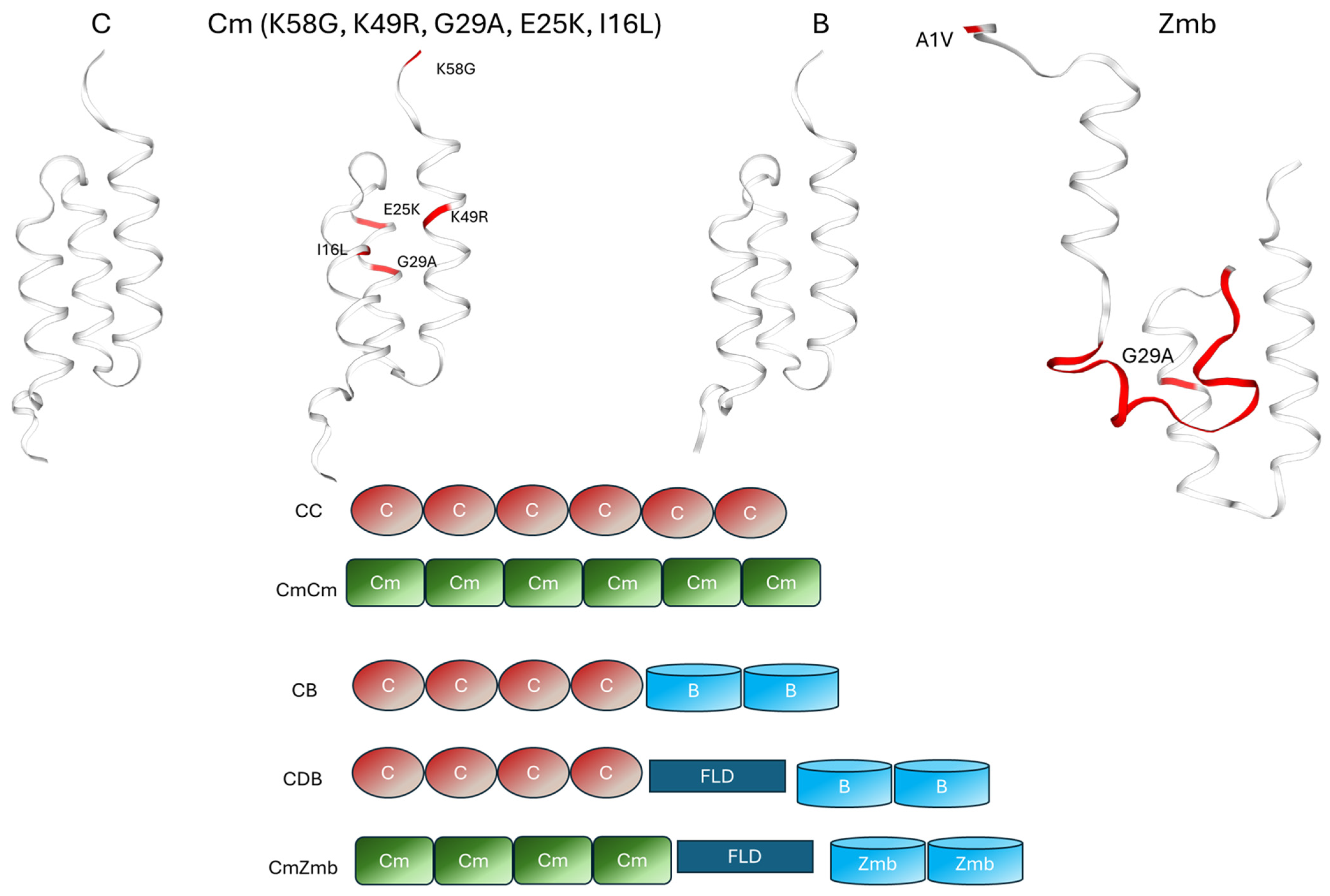
To improve IgG-binding capacity and alkali resistance, targeted amino acid substitutions were introduced at five key residues within the native C domain (positions 16, 25, 29, 49, and 58), generating modified variants denoted Cm. The specific substitutions were as follows: position 16 was replaced with leucine or valine; position 25 with lysine, arginine, histidine, or tryptophan; position 29 with alanine, leucine, or threonine; position 49 with arginine or histidine; and position 58 with glycine, isoleucine, or alanine.
Multiple tandem repeats of these engineered Cm domains (four or six repeats) were created and tested for their properties. This multivalent design aims to increase affinity effects, thereby boosting IgG-binding capacity.
2.1.1. Inclusion of Functional Peptides
In addition to these point mutations, to further improve the functionality of the fusion-type proteins, two additional engineered peptide components were included.
- (1)
Zmb Domain. Based on the engineered B domain, the new Zmb module is formed of 73 amino acids containing a 15-residue polypeptide insertion between amino acids 20 and 21, the first and second helices, resulting in the formation of an additional small helix within the structure. It has been shown that loop engineering of the three helix protein formed by the B domain by the insertion of glycine-rich polypeptide can affect the stability of the IgG interaction [
31]. The inserted sequence GGHLIAGGSDSERKE in the Zmb domain is enriched in glycine and histidine residues, which not only enhance solubility but may also contribute to improved metal coordination, providing an advantageous feature for purification via immobilized metal affinity chromatography (IMAC).
- (2)
FLD Peptide. This is a 14-amino acid peptide enriched with hydrophobic residues, such as Gly, designed to reduce steric hindrance within the overall structure, improving spatial reorientation and facilitating recombination with IgG molecules. This, in turn, enhances the overall IgG-binding capacity of the fusion protein.
2.1.2. Construct Design and Nomenclature
Various fusion-type multimeric proteins were generated using various combinations of native or modified domains, with or without the FLD linker and the Zmb domain. The abbreviations of all the modifications studied are listed in
Table 1:
2.2. Construction of Recombinant Vector
The DNA sequence encoding the fusion-type multimeric protein and the expression vector pET-30a(+) were both digested with restriction enzymes NdeI and HindIII (Sangon Biotechnology Co., Ltd., Shanghai, China). The digested fragments were purified using a DNA recovery kit (Biyuntian Biotechnology Co., Ltd., Shanghai, China) and ligated. The resulting recombinant plasmid was transformed into Escherichia coli DH5α by incubation on ice for 30 min, followed by heat-shock in a water bath at 42 °C for 90 s, and immediately cooled on ice for 3 min. After this transformation, 100 µL of room temperature Luria–Bertani (LB) liquid medium was added to the cells, which were then incubated in a shaker at 37 °C, 220 rpm for 60 min. The bacterial suspension was spread onto LB plates containing 50 mg/L kanamycin (Sangon Bioengineering Co., Ltd., Shanghai, China). The plates were inverted and incubated overnight at 37 °C. Kanamycin-resistant colonies were screened, and positive clones expressing a recombinant vector with the fusion-type multimeric protein were selected. For example, one recombinant vector expressing the fusion-type multimeric protein CmZmb was named pET-30a-CmZmb.
2.3. Construction of Recombinant Bacteria
Fresh E. coli BL21 (DE3) cells were incubated on ice for 30 min at 4 °C, then centrifuged at 1000× g for 10 min. The resulting pellet was resuspended in 0.1 M MgCl2-CaCl2 (80 mM MgCl2 and 80 mM CaCl2) at 4 °C, followed by centrifugation at 1000× g for 10 min. The pellet was resuspended in 0.1 M CaCl2 solution to prepare BL21 (DE3) competent cells.
The recombinant vector (such as pET-30a-CmZmb) was transformed into the competent E. coli BL21 (DE3). The transformed cells were plated on LB agar containing 50 mg/L kanamycin, and colonies were screened for successful transformation. Positive transformants were confirmed by sequencing and named BL21-pET30a-CmZmb. Recombinant transformants for other fusion-type multimeric proteins were named following the same naming convention.
2.4. Fermentation of Recombinant Bacteria
The recombinant transformants (such as BL21-pET30a-CmZmb) confirmed by sequencing were inoculated into LB medium with an inoculum volume of 0.5% (v/v) and cultured overnight at 37 °C until the OD600 reached 0.8. Protein expression was induced for 3–5 h by the addition of 10 g/L lactose; cells were then collected by centrifugation. The expression level of the recombinant protein was approximately 400 mg/L.
To analyze protein expression, 40 µL of bacterial supernatant, collected both before and after lactose induction, were mixed with 10 µL of loading buffer (Proanti Biotechnology Development Co., Ltd., Shanghai, China) and boiled for 5 min. A total of 20 µL of each sample was loaded onto a gel for SDS-PAGE gel electrophoresis.
SDS-PAGE was performed using a Mini-Protean (Bio-Rad, San Francisco, CA, USA) electrophoresis system with a 12% polyacrylamide gel. Electrophoresis was carried out at 150 V. The protein ladder, RealBand pre-stained protein marker (10–180 kDa), and the reducing buffer 5× Protein Loading Dye used to treat the protein sample were provided by Sangon Biotechnology Co., Ltd., China. Gels were stained with Coomassie Brilliant Blue R250 solution (Beijing Vokai Biotechnology Co. Ltd., Beijing, China) to visualize protein bands.
2.5. Purification of Fusion-Type Multimeric Protein Variants
A solution containing 0.2 M NaCl in 20 mM phosphate buffer, pH 7.4, was used as buffer A. Bacterial pellets obtained after fermentation were resuspended in buffer A at a 1:10 (w/v) ratio. Cell lysis was performed using an Ultrasonic cell disruptor (Scientz Biotechnology Co., Ltd., Ningbo, China), with a cycle of 20 s sonication followed by 10 s rest, repeated for a total time of 40 min. The lysate was centrifuged at room temperature, and the supernatant was subjected to nickel affinity column purification. The target protein was eluted with 0.25 M imidazole following the procedure described below, and the eluate was collected and analyzed by SDS-PAGE.
The nickel column affinity purification was performed by FPLC using a 5 × 30 cm column packed with Seplife® 6AG NTA/Ni/90 (Sunresin New Materials Co. Ltd., Xi’an, China) agarose resin with a bed height of 23 cm. A 10 mL/min flow rate was applied to equilibrate the column with 10 column volumes (CV) of buffer A. UV absorbance was monitored at 280 nm. After equilibration to baseline, a 1000–2000 mL sample of lysate supernatant was loaded onto the column, followed by washing with buffer A to reach baseline (approx. 10 CV). impurities were washed with approx. 10 CV of 20 mM imidazole solution (Sinopod Chemical Reagents Co., Ltd., Beijing, China). To elute the protein of interest, 2 CV of a 0.25 M imidazole solution was used, and then 0.5 M imidazole solution was used to regenerate the column (approx. 3CV), followed by column equilibration with Buffer A. The elution fractions were collected for further analysis.
Total protein content was determined using the biuret method, following the procedure described by Rodger, A et al. [
32]. rProtein A was purified with a recovery rate of about 80% and was used in the immobilization step on the agarose resin without further processing.
2.6. Preparation of Agarose Beads Functionalized with rProtein A
The fusion-type multimeric proteins obtained after the Ni affinity purification were used to prepare affinity chromatography media as follows: Seplife 4AG/90 agarose beads (Sunresin New Materials Co., Ltd., China) were activated with epoxy groups using epichlorohydrin in a NaOH alkaline solution. The epoxy-activated beads were rinsed with 0.1 M phosphate buffer, pH 8.6, followed by the addition of 10 mg fusion-type multimeric protein per mL functionalized agarose beads, ensuring the pH remained at 8.6. Anhydrous sodium sulphate was also added to the mixture to ensure correct protein folding during the coupling process. The same amount of protein and agarose resin was used for each preparation. The reaction was allowed to proceed until >95% of the protein was immobilized on the resin. The protein was covalently attached to the epoxy groups on the resin via the thiol groups located at the C-terminal cysteine introduced in the sequence. Following coupling, the beads were sequentially washed with phosphate buffer pH 8.6, acetic acid, and water to remove unbound protein. The prepared affinity chromatography medium was stored in 20% (v/v) ethanol until further use.
2.7. Alkaline Stability and Application Testing
Free recombinant proteins of different C and Cm hexamer constructs (not immobilized on agarose) have been incubated in 1 M NaOH solution for up to 24 h at 25 °C, and samples were tested by SDS-PAGE to determine the protein degradation in solution. This was used as a qualitative method to confirm the alkaline stability of the free recombinant protein as an intermediate step before further modifications.
The different rProtein A variants produced and immobilized on agarose beads were then tested by FPLC for alkaline stability and affinity towards human IgG. To perform the alkaline stability tests, the agarose resin modified with a specific rProtein A variant was packed in a 1 mL column (7 × 25 mm). The chromatography columns were equilibrated using a solution of 0.15 M NaCl in 20 mM phosphate buffer, pH 7.4 (Buffer A1) at a flow rate of 0.5 mL/min for 10 min followed by CIP with 0.5 M NaOH at a flow rate of 0.2 mL/min for 15 min, then the column was rinsed with equilibration buffer at a flow rate of 0.5 mL/min for 10 min and the above step was repeated 100 cycles. The dynamic binding capacity (DBC) of the resin was determined initially and after every 10 cycles.
To test the DBC of the resin packed in the chromatography column (7 × 25 mm), the flow rate was set to 1.0 mL/min, and the column was rinsed with 10 CV of 0.1 M citric acid-sodium citrate buffer, pH 3.3 (Buffer B). Then the column was equilibrated with 15 CV equilibration buffer A1, and then a 2 mg/mL human IgG sample (Suo Laibao Technology Co., Ltd., Shenzhen, China) was loaded at a flow rate of 0.2 mL/min (5 min contact time—CT). When the outlet absorbance at 280 nm (A280) reached 90 mAu (10% of the initial IgG solution absorbance), the IgG sample application was stopped, and the column was rinsed with buffer A1. The IgG bound to the resin was eluted using buffer B at a flow rate of 1.0 mL/min until A280 reached baseline. The column was finally equilibrated using buffer A1. The eluted IgG was collected and concentration measured by a UV-VIS microplate reader (Perlang New Technology Co., Ltd., Shunyi, China).
The DBC was calculated as: DBC = (VA × C0)/VC (mg/mL resin). Where C0 is the concentration of the target protein in the sample, VA is the total volume of the sample loaded when the concentration of the target protein in the breakthrough curve reaches 10% C0, and VC is the total column bed volume.
Additional alkaline stability studies were done using the CmZmb modified chromatography resin packed in a 5 mL column (0.8 × 10 cm) using an ÄKTA Pure, Cytiva chromatography system. The 0.5 M NaOH, 1.0 M NaOH, and 1.5 M NaOH solutions were circulated through individual columns at a flow of 1 mL/min (5 min CT) for a total incubation time of up to 60 h at 25 °C. DBC tests were performed at specified times, using a 2 mg/mL IgG sample, applying a flow of 1 mL/min; the DBC values were compared to the untreated resin.
2.8. Long-Term Performance of CmZmb-Modified Chromatography Resin
The long-term performance (lifetime cycling) of the CmZmb affinity chromatography resin was tested using an FPLC system. The chromatography resin was packed in a column with dimensions 1 × 18 cm (14 mL), and a contact time of 6 min was applied for the sample loading. For this test, the sample used was a human IgG1-type monoclonal antibody produced in CHO-K1 cells (purchased from AtaGenix, Wuhan, China). The CHO-K1 cells were cultured in serum-free medium (Youyi Biotechnology, Guangzhou, China) and perfused-cultured in a 20 L wave bioreactor (Cekg Tech. Co. Ltd., Wuhan, China). After collecting, the culture supernatant was filtered through a 0.22 µm hollow fibre membrane (Bona Biotechnology Group Co., Ltd., Jinan, China) and then aliquoted and stored for subsequent use. The concentration of the antibody in the fermentation broth was 2.9 g/L. The CIP performed every cycle was done with 0.1 M NaOH, and every sixth cycle, an additional CIP with 0.5 M NaOH was completed.
The long-term performance of the CmZmb affinity agarose resin was evaluated over 120 full cycles; DBC, column pressure, and rProtein A leaching were measured periodically to assess performance. rProtein A leaching was measured in the mAb elution fraction using Mix-N-Go Protein A assay F610, while the host cell protein (HCP) residue was measured using CHO HCP ELISA Kit 3G-(F550-1), both from Cygnus Technologies, Leland, NC, USA.
3. Results
The present paper focuses on the design and construction of a fusion-type multimeric protein derived from the native Staphylococcus aureus Protein A sequence, with the aim of achieving higher IgG loading capacity and enhanced alkaline resistance upon immobilization on agarose beads for applications in affinity chromatography.
Building upon the native sequence, targeted amino acid substitutions were introduced to generate engineered Cm domains. These modified domains were fused with functional peptide elements to construct a multimeric fusion protein, named CmZmb, which successfully met the desired requirements [
33].
All recombinant constructs were cloned into the pET-30a(+) expression vector, which inserted a N-terminal hexa-histidine tag. This His-tag facilitates the downstream purification of the recombinant fusion proteins via IMAC (Ni-NTA) affinity chromatography. In the process of expanding the production of protein A ligands, using a purification method with lower costs is the optimal choice. The introduction of His-tag can effectively reduce production costs while ensuring the stability of protein purification and reducing batch-to-batch variations. The recombinant fusion-type multimeric protein constructs were expressed using E. coli BL21 (DE3) as the host strain. This strain was selected due to its suitability for high-level expression of recombinant proteins under the control of the T7 promoter, compatibility with the pET-30a(+) expression system, and its deficient protease background, which minimizes degradation of the target protein. Additionally, BL21 (DE3) facilitates easy scaling of fermentation and is widely used for the production of His-tagged proteins, which aligns with our purification strategy via Ni-NTA affinity chromatography.
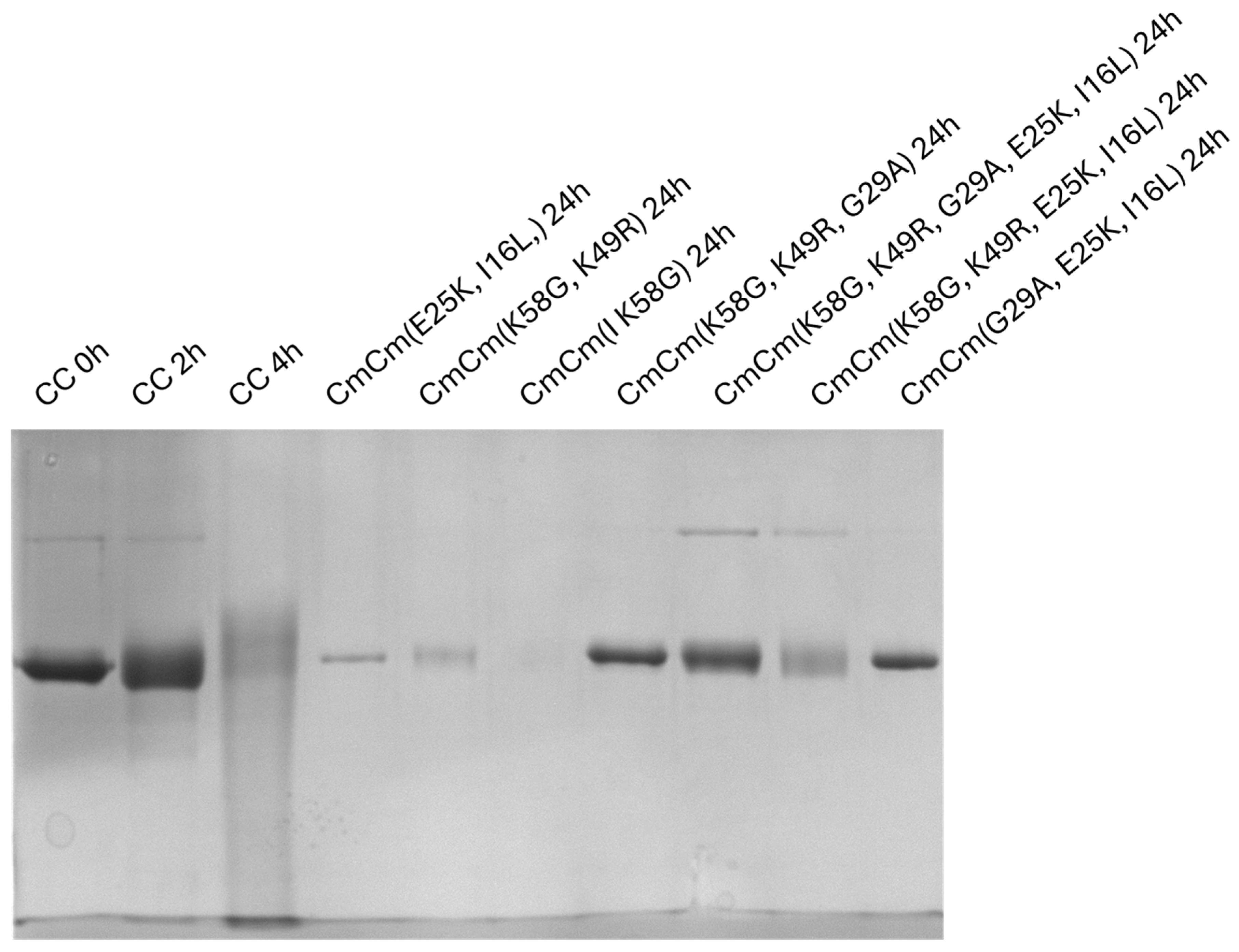
As an intermediate step in designing rProtein A with high alkaline stability, recombinant proteins containing C and Cm hexamer constructs were produced and subjected to alkaline treatment with 1 M NaOH at 25 °C for up to 24 h. The SDS-PAGE of various Cm hexamer constructs was compared with the fusion protein made of six native C domains (CC), and the results are presented in
Figure 2. Cm constructs containing 5 mutations (I16L, E25K, G29A, K49R, K58G) demonstrated significantly higher resistance to alkaline degradation compared to the native C domain constructs. In contrast, constructs with fewer amino acid mutations displayed moderate to low stability under the same conditions. This consisted of an initial qualitative assessment of the alkaline stability of the Cm hexamer constructs.
To further evaluate the alkaline stability and DBC of the various designed protein constructs, the purified fusion proteins were covalently attached onto agarose beads through thiol coupling of the terminal Cys to epoxy groups on the resin [
37]. The resulting affinity resins were further evaluated for DBC and alkaline stability as described below.
The initial 10% DBC at 5 min contact time was measured with commercial IgG and repeated after 100 CIP cycles with 0.5 M NaOH, 15 min treatment per cycle. The results are shown in
Table 2. Among all tested constructs, the CmCm (K58G, K49R, G29A, E25K, I16L) variant exhibited the best performance, combining a high initial DBC of 56.0 mg/mL with the highest DBC retention of 73.7% after 100 CIP cycles. This construct clearly outperformed the native C-domain fusion (CC), which lost nearly 40% of its binding capacity over the same number of cleaning cycles.
Constructs with fewer substitutions, such as CmCm (K58G, K49R) or CmCm (E25K, I16L), showed intermediate performance, highlighting the synergistic effect of multiple rationally designed mutations in enhancing the alkaline resistance while maintaining the IgG-binding affinity.
Building on the improvements in the alkaline stability displayed by the top-performing CmCm variant-specific constructs shown in
Table 2, CmCm (K58G, K49R, G29A, E25K, I16L) and the native CC were used to further modify the fusion protein by incorporating the B domain and/or functional peptide domains into the fusion protein constructs. Specifically, the construct CmZmb was designed with the domain arrangement as Cm–Cm–Cm–Cm–FLD–Zmb–Zmb, where each Cm domain includes the five single point mutations (K58G, K49R, G29A, E25K, I16L). The addition of FLD and Zmb was hypothesized to confer further improvements in both binding kinetics and additional resilience during repetitive alkaline CIP cycles [
38].
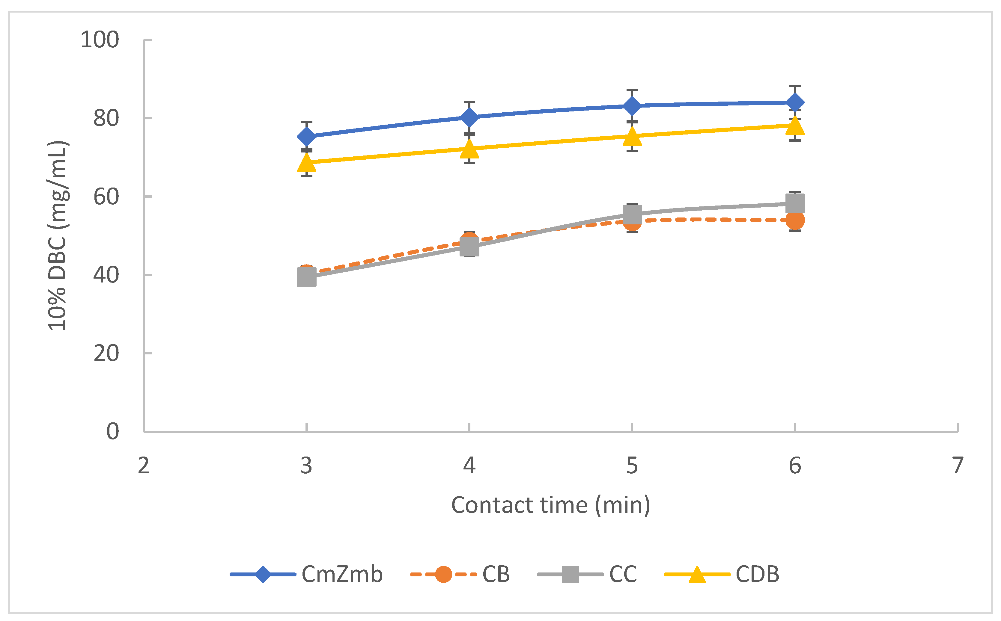
The performance of CmZmb was compared against that of native CC, CC with added B domains (CB), and native C domain with added FLD and B domains (CDB). As shown in
Figure 3, the presence of the FLD peptide resulted in a high DBC across different contact times. This indicates favourable binding kinetics and improved accessibility of the binding domains due to the FLD spacer. Notably, CmZmb maintained high DBC even at short contact times, outlining its suitability for high-throughput purification processes.
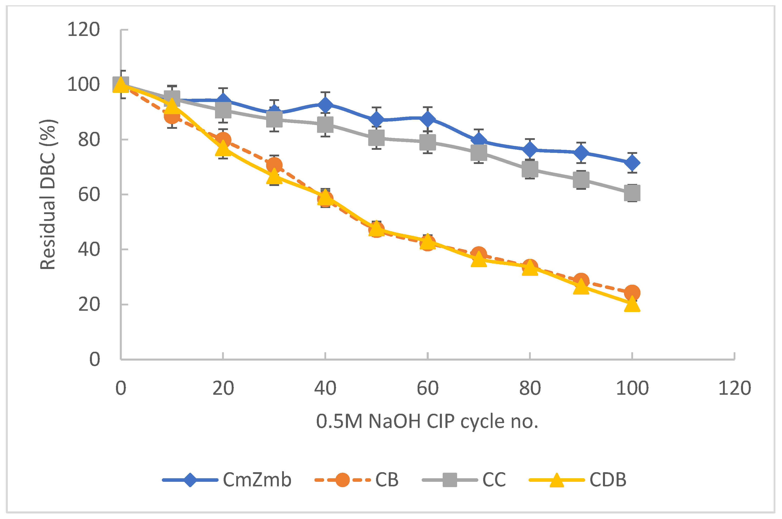
Figure 4 shows that CmZmb retained a high proportion of its binding capacity after 100 CIP cycles in 0.5 M NaOH (15 min/cycle). In contrast, CDB, which contains the FLD and B domains but lacks the C domain, showed poor alkaline stability upon point mutations. This comparison highlights the critical contribution of the engineered mutations within the Cm domains to alkaline tolerance. Together, these findings demonstrate that the combined inclusion of FLD and Zmb, when paired with rationally designed point mutations in the C domain, results in a highly functional and alkali-stable fusion ligand ideal for industrial antibody purification applications.
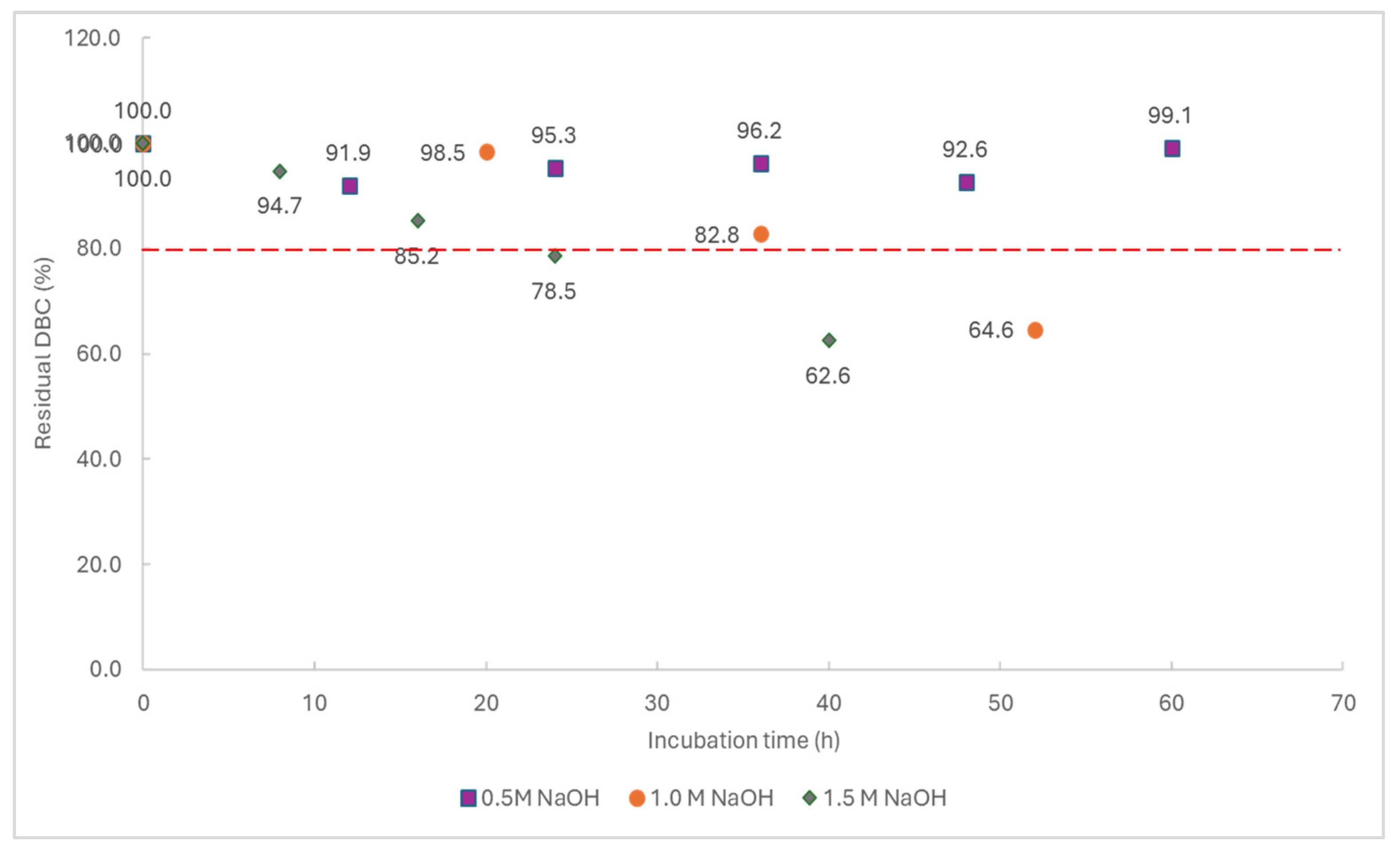
To further validate the alkaline stability of the selected fusion ligand, agarose resin functionalised with CmZmb was subjected to accelerated testing by incubation in NaOH solutions of increasing concentrations (0.5 M, 1.0 M, and 1.5 M NaOH, respectively) at 25 °C. The 10% DBC value was tested at selected time points using commercial IgG. A generally accepted benchmark for resin longevity is the ability to maintain 80% of the initial DBC before it becomes obsolete [
18]. As shown in
Figure 5 the CmZmb-functionalised resin (now commercialized under the name of rProtein A Seplife
® Suno) retained a high percentage of its original DBC across all tested NaOH concentrations; 80% DBC was retained for up to 22 h in 1.5 M NaOH and up to 40 h in 1.0 M NaOH while DBC remained stable >90% for 60 h incubation with 0.5 M NaOH. These results further confirm the robust alkali resistance conferred by the engineered Cm mutations and structural design of the fusion construct.
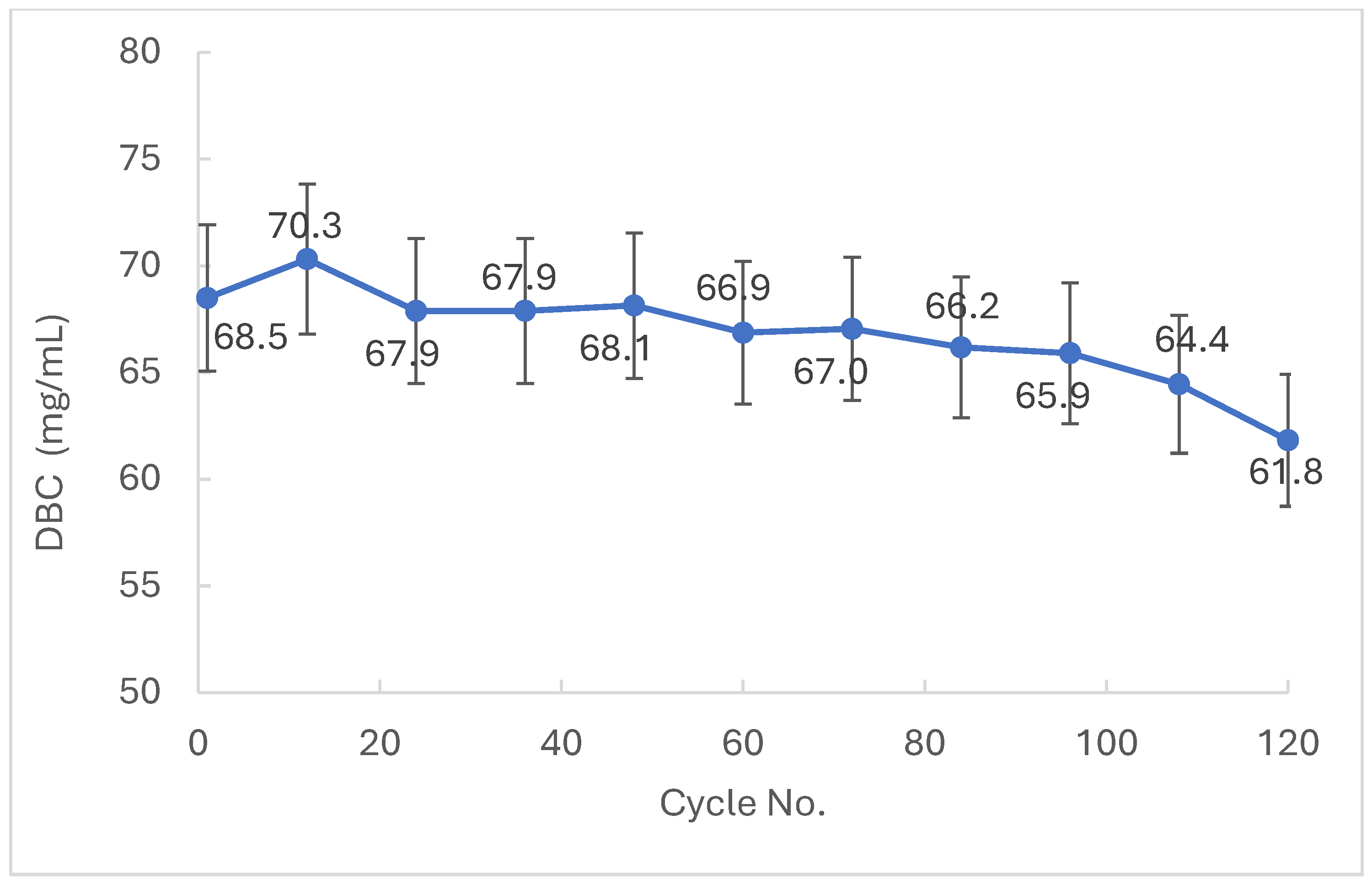
To assess the performance and long-term service of the chromatography media modified with CmZmb fusion protein, the resin was submitted to intensive cycling using clarified monoclonal antibody (mAb) fermentation broth. The 10% DBC was measured initially and after every 10 cycles. The data presented in
Figure 6 illustrates the gradual reduction in resin performance over time due to operational stress and repeated CIP with 0.5 M NaOH. The CmZmb functionalised agarose resin demonstrated high initial binding capacity and retained a substantial proportion of its initial performance across 120 CIP cycles, confirming its strong potential for repeated use in industrial-scale mAb purification processes.
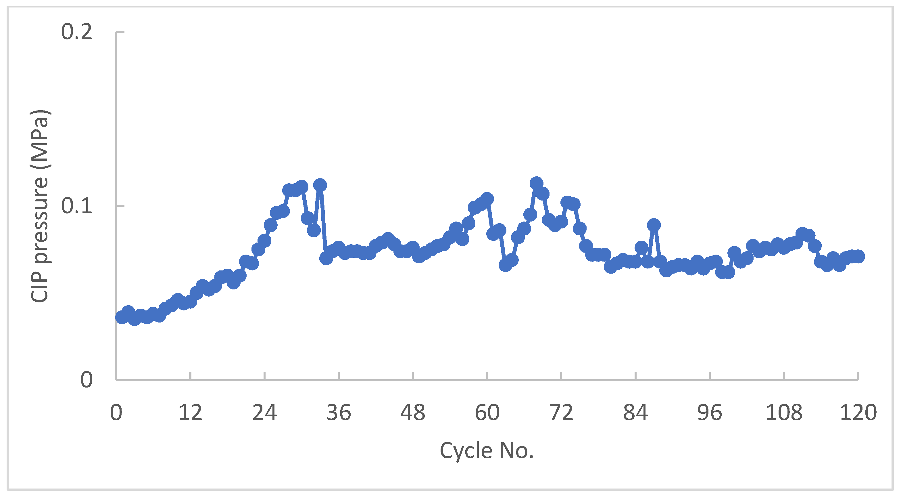
Alongside quantifying the DBC of the affinity chromatography media, the delta-column pressure was monitored for the duration of the study to evaluate the physical integrity and flow performance of the agarose resin. The change in column pressure over time provides insight into fouling, blockage, or structural degradation of the resin. As shown in
Figure 7, the CmZmb-functionalised resin maintained a relatively stable column pressure profile across the entire study, indicating minimal compression or particulate accumulation. This suggests that the resin not only retains functional performance but also preserves its physical robustness under repeated high-stringency cleaning conditions.
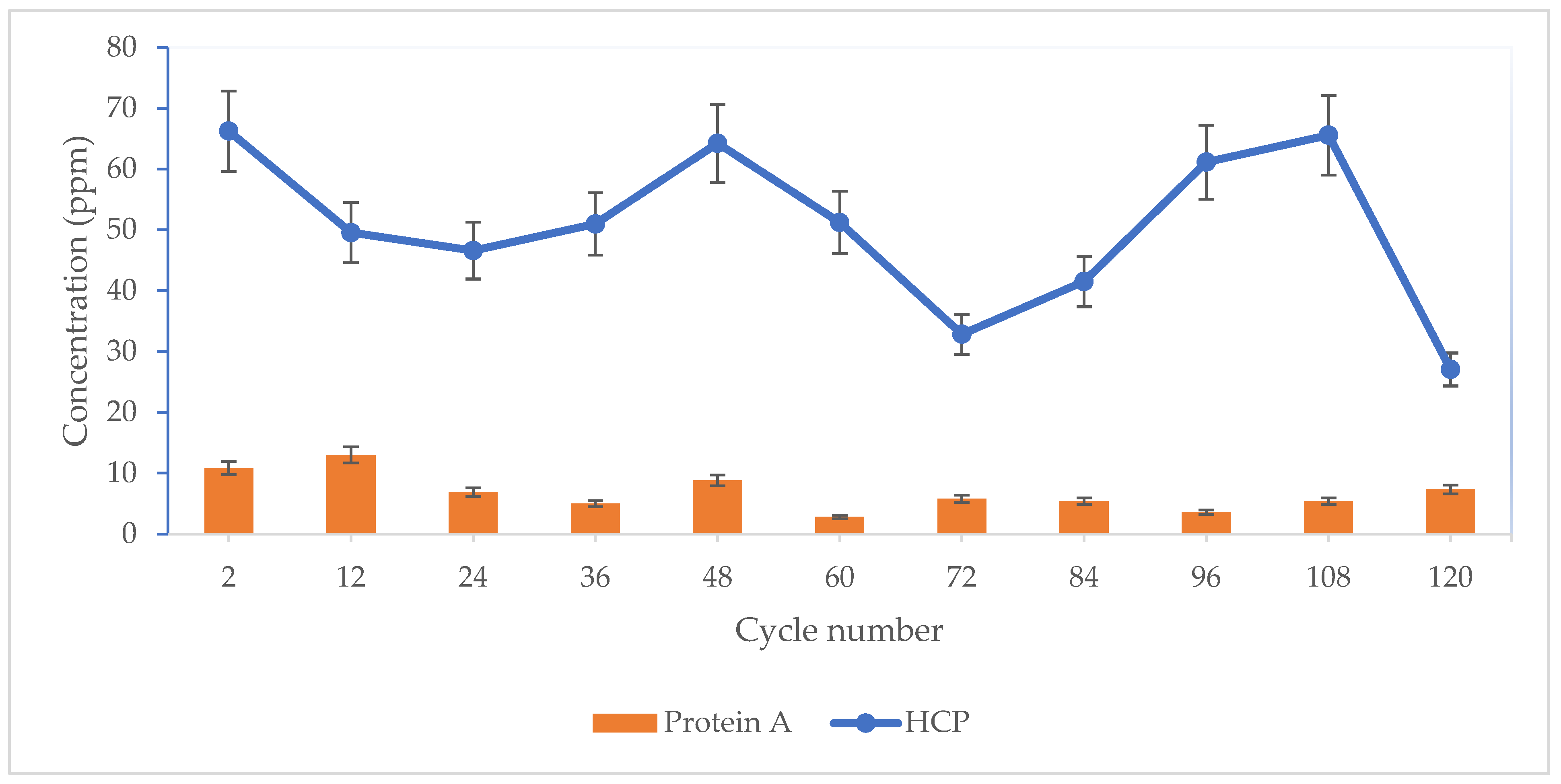
Another important parameter quantified during affinity chromatography purification is the level of affinity ligand leaching in the elution fraction and the remaining host cell proteins (HCP); they are considered process-related impurities, are closely monitored, and form part of the mAb drug substance specification [
39]. To evaluate the stability of the immobilized CmZmb-functionalised agarose resin and the efficiency of the mAb affinity purifications step, the concentration of rProtein A was measured in the mAb elution fraction every 12th cycle over the course of the 120-cycle purification process. As shown in
Figure 8, the level of CmZmb ligand leaching and HCP remained consistently low throughout the study, with all measurements below 15 ppm rProtein A and <70 ppm HCP. This confirms the structural stability and secure immobilization of the CmZmb ligand on the agarose matrix under repetitive alkaline cleaning conditions and the efficiency of the mAb chromatography purification.
4. Discussion
Protein A affinity chromatography is a critical capture step applied in the industrial-scale purification of mAbs. The development of recombinant protein A ligands with enhanced alkaline resistance and IgG-binding capacity is critical for increasing resin performance, extending operational lifetime, and reducing the overall process cost. The majority of protein A ligands are based on the full-length or multimeric constructs consisting of repeats of the B and C domains, or modified versions thereof [
40,
41,
42,
43].
The aim of this study was to develop an engineered, multimeric Protein A-derived ligand based on the native Staphylococcus aureus with enhanced IgG-binding capacity, superior alkaline resistance, and long-term stability under conditions mimicking industrial antibody purification processes. Through rational design incorporating specific amino acid substitutions, as well as the addition of functional polypeptides, we successfully created and validated a novel fusion construct—CmZmb—that meets these criteria.
Initial efforts focused on targeted amino acid substitutions within the native C domain of protein A (Cm variants) to improve its resistance to alkaline degradation while preserving or enhancing IgG binding. The targeted mutations I16L is a conserved substitution that may affect the protein folding and stabilize the hydrophobic core; E25K has the role to reduce nucleophilic attacks, G29A may increase the peptide rigidity, while K49R may stabilize basic residues, and K58G may reduce the alkaline degradation of the peptide. Collectively, these substitutions largely preserve antibody-binding capacity and alkaline resistance, with only minimal reduction in some cases, thereby establishing a structural and functional basis for the enhanced performance observed in fusion constructs containing the FLD and Zmb domains.
The results clearly demonstrate that constructs that have multiple amino acid substitutions (e.g., CmCm containing I16L, E25K, G29A, K49R, and K58G) significantly outperformed both single-mutation variants and the unmodified C domain (CC) in terms of DBC retention after 100 CIP cycles with 0.5 M NaOH. Importantly, the CmCm (I16L, E25K, G29A, K49R, and K58G) variant retained nearly 74% of its initial DBC, compared to just over 60% for the native construct, highlighting the combined effect of multiple point mutations. These mutations likely contribute to enhanced conformational stability or reduced chemical reactivity under alkaline conditions, as previously observed in similar rational protein engineering strategies targeting Protein A analogues [
33,
44,
45,
46,
47].
To enhance binding performance and operational robustness, the top-performing Cm variant Cm (K58G, K49R, G29A, E25K, I16L) was fused with additional domains FLD and Zmb to produce the CmZmb construct. The FLD linker was designed to reduce steric hindrance and improve spatial flexibility, while the Zmb domain incorporated structural alterations known to influence IgG interaction stability and enable potential metal-responsive elution characteristics. Following the fusion protein immobilization on agarose beads, the evaluation of DBC at varying contact times confirmed that the inclusion of FLD and Zmb improved IgG-binding kinetics and accessibility, as CmZmb achieved high binding even under short contact times. Notably, CmZmb also retained superior DBC after extensive CIP cycles compared to variants without mutations (e.g., CDB), underlining the critical role of engineered Cm domains in conferring true alkaline resistance. This finding confirms that the Zmb and FLD modules alone are insufficient to protect the ligand from degradation without accompanying structural stabilization within the core IgG-binding region.
The ability of CmZmb-functionalised agarose resin to withstand extended incubation in 0.5–1.5 M NaOH further reinforces its industrial viability. Maintaining over 80% of its initial DBC after 22 h in 1.5 M NaOH represents a significant improvement over native or commercial Protein A-based resins, many of which degrade substantially under such conditions. This data is particularly valuable for downstream bioprocessing, where prolonged cleaning cycles and sanitization procedures are common.
Importantly, when used with clarified monoclonal antibody fermentation broth, the CmZmb resin maintained functional integrity over 120 purification cycles, each including a 15 min 0.5 M NaOH CIP, with 88% DBC retention. Column pressure data indicated stable flow characteristics throughout, suggesting negligible fouling or matrix degradation. This physical robustness is critical for real-world process scalability.
Moreover, ligand leaching remained consistently below 15 ppm, meeting regulatory thresholds, confirming secure covalent immobilization and protein stability under repetitive elution and CIP conditions.
This positions CmZmb-functionalised agarose resin as a competitive candidate for GMP-compliant affinity resins in large-scale mAb purification.
5. Conclusions
This study successfully demonstrates the rational design and construction of a fusion-type multimeric Protein A ligand with enhanced IgG-binding capacity and superior alkaline stability. By introducing targeted amino acid substitutions within the native C domain and incorporating engineered functional peptide modules (FLD and Zmb), the resulting CmZmb fusion protein exhibited significantly improved performance compared to the native Protein A constructs.
The multi-point mutated Cm domains conferred markedly increased resistance to cleaning-in-place. The rProtein A ligand shows extremely high stability after 60 h incubation at 0.5 M NaOH by maintaining more than >90% initial DBC and up 80% DBC after 40 h in 1.0 M NaOH. However, the most impressive result was observed at 1.5 M NaOH, where the ligand preserved 80% binding capacity after 22 h incubation and 60% DBC after 40 h. To the best of our knowledge, such high alkaline stability has not been previously reported. These findings open the possibility to perform flexible resin CIP cycles using NaOH in the range 0.1–1.5 M. Alternating between low and high NaOH strengths could help maintain optimal resin performance while maximizing operational lifespan and reducing production costs.
In addition to improved alkaline stability, the engineered rProtein A ligand exhibited outstanding DBC values. The initial mAb 10% DBC was approx. 70 mg/mL and an 88% retention after 120 purification cycles. This performance was mainly due to the incorporation of the FLD spacer peptide that enhanced binding kinetics and accessibility, resulting in high and stable DBC values even at short contact times. The addition of the Zmb domain was key in further contributing to functional robustness without compromising alkaline tolerance. Finally, CmZmb-functionalised resin demonstrated excellent long-term durability and stable physical properties under repetitive cycling with clarified monoclonal antibody broth, showing minimal ligand leaching (<15 ppm) and pressure buildup.
Together, these advances highlight the potential of the CmZmb engineered fusion ligand as a highly effective affinity chromatography medium for industrial-scale monoclonal antibody purification. The enhanced alkali stability not only extends resin lifespan but also reduces process costs by enabling more stringent and frequent cleaning-in-place procedures without compromising operational longevity. Future work could focus on large-scale production and integrating this ligand into commercial purification platforms.
Collectively, these features offer a compelling strategy for reducing mAb production cost by extending resin lifetime, combined with the ability to shorten the CIP cycles using increased NaOH concentrations.




 ,
,