


Abstract
Background: Accidents caused by the Loxosceles laeta spider constitute a health problem in South America. Envenomation can lead to severe systemic manifestations, eventually compromising the patient’s life. Most regional health authorities consider antivenom administration the basis of effective treatment in the most serious cases. The availability of spider venom is the primary bottleneck for antivenom production. Herein, we present a novel biotechnological approach, based on the expression of recombinant versions of the most relevant toxin in loxoscelism, sphingomyelinase D (SphD), in insect larvae (Spodoptera frugiperda). Methods: We produced two versions of SphD: one conserving its biological activities (wtSphD) and a second alternative that was designed to be genetically detoxified (dSphD). Two horses were subjected to three consecutive hyperimmunization cycles with dSphD. The horses’ plasma was extracted at the end of each cycle and used to produce Active Pharmaceutical Ingredients (APIs) of antivenoms at a pilot scale. Results: Dermonecrotic activity of wtSphD was completely neutralized with the sera obtained from one horse and partially with that of the other. In contrast, the APIs derived in both cases completely neutralized wtSphD dermonecrotic activity. Direct hemolysis of human red blood cells by wtSphD was also neutralized by sera and APIs. Conclusions: These results show venom replacement or complementation potential by recombinant dSphD produced in this novel platform.
1. Introduction
Loxoscelism—envenomation by Loxosceles spiders—presents a significant threat to public health in South America, where Loxosceles laeta is associated with the most severe clinical presentations and cases range from localized cutaneous necrosis to severe systemic disease (hemolysis, coagulopathy, and, rarely, acute kidney injury) [1]. In severe presentations, antivenom administration is widely regarded as the cornerstone of treatment, although clinical benefit depends on timing and disease evolution [2]. The burden, the potential for life-threatening complications, and the need for timely, effective interventions make improvements in antivenom production and access a matter of regional and global importance [1,2]. However, despite decades of research, antivenom production remains critically constrained by the extremely limited and variable supply of Loxosceles venom, creating a structural bottleneck that prevents consistent, scalable and safe manufacturing. Dependence on native venom limits production capacity and introduces batch-to-batch variability, biosafety concerns, and operational barriers. Together, these factors restrict antivenom availability in public health systems.
Loxosceles’ venom is composed of a complex mixture of toxins that can be divided between abundant components (sphingomyelinase D, astacin-like metalloproteases, and peptide members of the inhibitor cystine knot family) and less abundant components (members of the serine proteases family, protease inhibitors and translationally controlled tumor proteins) [3]. Mechanistically, a single venom component—sphingomyelinase D (SphD; now classified among phospholipase D enzymes)—reproduces most hallmarks of loxoscelism. Purified SphD induces dermonecrosis, neutrophil infiltration, increased vascular permeability, and hemolysis in experimental models [4,5,6,7]. Biochemically, SphD acts as an acid–base catalyst, promoting the hydrolysis of phospholipids, mainly sphingomyelin and lisophospholipids such as lisophosphatidylcholine (LPC), producing choline and 1-phospho-ceramide or lisophosphatidic acid (LPA), respectively [4]. The fact that both substrates display a free OH group at position 2 (glycerol) or 3 (sphingosine), further supported by experimental observations, has led to the hypothesis that actually a transphosphatidylation reaction occurs forming cyclic phosphate products. Chemical modification of albumin-bound plasma LPC by SphD thus produces LPA and/or its cyclic derivative, both potent lipid mediators of different biological activities, including platelet aggregation, endothelial permeability and pro-inflammatory responses. Also, SphD association to plasma membranes of keratinocytes and its action on sphingomyelin induces the expression of matrix metalloproteases, complementing pathway activation while inhibiting epidermal growth factor receptor (EGFR) expression, thus promoting the degradation of collagen fibers and the influx of neutrophils to site of the lesion, thus promoting dermonecrosis while hindering wound healing [8]. Its association and the changes in the lipid composition of erythrocytes, activate membrane-bound metalloproteases, inducing the degradation of glycophorins and the activation of the alternative complement pathway. Also, the changes in the erythrocytes’ membranes structures promote the activation of the classical complement pathway by phosphatidyl serine exposure on its external surface. Both activities justify the indirect hemolytic activity described for SphD [4]. Furthermore, the association of SphD to the erythrocytes’ membranes ultimately lead to an increase in transporter mediated Ca+2 influx that also causes direct hemolysis [9,10].
Structurally, SphD is arranged in a slightly distortioned (α/β)8 TIM barrel, where its interfacial face is characterized by the presence of a catalytic loop, a variable loop, a flexible loop and short hydrophobic loops. Mechanistically, SphD acid–base catalysis is based on the coordinated action of two histidine residues localized in its catalytic loop. A Mg+2 ion is also present at his active site, bound to three charged amino acid residues, who participates on substrate binding directly or indirectly (through two water molecules) [11]. Based on their structural characteristics, SphD variants can be classified into two classes [12]. Class I SphDs have one disulfide bridge, are composed exclusively of the two most relevant L. laeta isoforms and are associated with venoms with higher toxicities [8]. In contrast, class II SphDs display two disulfide bridges, are further divided into two subclasses and are composed of proteins mostly described in Loxosceles species other than L. laeta, i.e., L. intermedia, L. reclusa, L. gaucho, L. similis, L. boneti and L. arizonica [8,13]. The extra disulfide bridge present in Class II SphDs links the flexible loop with the catalytic loop, affecting its position and reducing its volume about three times when compared to that of Class I SphDs [11]. These structural differences could justify the observed differences between both SphD Classes on their biochemical activities and their pathological consequences [8]. Thus, this classification is relevant not only for understanding clinical severity but also for antigen selection in recombinant antivenom strategies.
The clinical field includes controversies and diverging views. The only proven antidote for Loxosceles envenomation is the use of antivenom [2,14,15]. Although some drugs like dapsone [16] and tetracycline are usually suggested as non-specific treatments for loxoscelism [17,18], no prospective clinical trials that support their usefulness are available. Additional treatments, such as plasmapheresis or hyperbaric oxygen, have also been suggested in the literature. The use of plasmapheresis is promissory but rather scarce, and limited case reports are available to support its use [19]. There is no consensus about the effectiveness of hyperbaric oxygen use for treating Loxosceles envenoming, and it has only been suggested as an adjuvant for treatment [20]. Finally, although available information from clinical studies on the use of antivenom are still scarce, there is a consensus, in the countries where the Loxosceles antivenom is used, that its correct application is useful for favorable outcomes [14,15,21,22,23,24].
At the pathogenic level, while SphD is the principal determinant, the contribution of other venom components to lesion progression is still subject to debate, particularly in species/isoforms with differing biochemical profiles [4]. From a manufacturing standpoint, venom availability is the primary bottleneck for antivenom production: adult spiders yield minute volumes of venom, captive maintenance is difficult, and electrostimulation workflows pose safety and scaling constraints [3]. To address this, several groups have explored venom replacement or complementation strategies: using recombinant antigens—particularly SphD—to substitute for native venom during hyperimmunization. Proof-of-concept studies demonstrated that recombinant SphD can elicit neutralizing antibodies and support antivenom fabrication at laboratory scale [25,26,27]. In this context, recombinant strategies centered on SphD as a defined immunogen emerge not only as a response to venom scarcity but also as a rational approach to improve consistency and functional relevance of antivenoms, particularly against severe systemic outcomes such as hemolysis.
Multiple recombinant platforms have been evaluated. Bacterial expression (Escherichia coli) often produces inclusion bodies, requiring denaturation/refolding that reduces yield and complicates downstream processing [28]. Insect cell systems (baculovirus expression vector system, BEVS) improve folding and post-translational processing and are already used at an industrial scale for biopharmaceuticals [29,30]. A promising, cost-effective alternative is to use insect larvae as biofactories, enabling high-level expression with simple infrastructure, linear scale-out and lower fixed costs, albeit with specific downstream purification challenges [31,32,33,34].
A further refinement is the genetic detoxification of SphD—engineering mutations that abolish dermonecrotic/hemolytic activity while preserving immunogenic epitopes. Mutations targeting catalytic/recognition residues (e.g., Trp and Asp residues in the GDPD domain) can inactivate enzymatic activity without disrupting antigenic surfaces [35]. This approach improves animal welfare during hyperimmunization, focuses the immune response on the principal pathogenic determinant, and fits well with biofactory-based manufacturing.
This study introduces a biotechnological strategy to overcome the critical bottleneck in antivenom production: the limited availability of Loxosceles venom. We developed a platform based on S. frugiperda larvae to express recombinant SphD, the principal toxin responsible for loxoscelism. Two variants were produced: a biologically active wild-type form (wtSphD) and a genetically detoxified version (dSphD) designed to abolish dermonecrotic and hemolytic activity while preserving immunogenic epitopes. This work addresses not only venom scarcity, but also the intrinsic biological variability of native venom-based immunization
2. Materials and Methods
2.1. Gene and Vector Construction
The synthetic gene encoding sphingomyelinase D (wtSphD) from L. laeta (GenBank accession AY093599.1) was obtained from GenScript. The native signal peptide was removed by PCR amplification, and a 6 × His tag was added at the N-terminus to facilitate purification. The amplified fragment was cloned into the pAcGP67-B vector under the control of the polyhedrin promoter and fused to the GP64 signal sequence. The expression cassette was subsequently subcloned into the donor vector pFastBac™ Dual (Thermo Fisher Scientific, Waltham, MA, USA) between XbaI and HindIII restriction sites, generating pFBD-SphD-GP64. Correct insertion and sequence integrity were confirmed by colony PCR and Sanger sequencing.
2.2. Generation of Recombinant Baculovirus
Recombinant bacmids were generated using the Bac-to-Bac™ baculovirus expression system (Thermo Fisher Scientific, Waltham, MA, USA). The donor plasmid pFBD-SphD-GP64 was transformed into E. coli DH10Bac™ competent cells, and recombinant colonies were selected by blue–white screening on LB agar containing kanamycin, gentamicin, tetracycline, IPTG, and X-gal (Sigma-Aldrich, St. Louis, MO, USA). Bacmid DNA was isolated by alkaline lysis and transfected into Sf9 insect cells using a liposome formulation (CellFECTIN® reagent, cat. 10362-010, Invitrogen, Carlsbad, CA, USA), following the manufacturer’s instructions. Recombinant Autographa californica multiple nucleopolyhedrovirus (AcMNPV-SphD) was amplified through three successive passages in Sf9 cells and titrated by endpoint dilution assay to obtain working viral stocks of approximately 1 × 10⁸ PFU/mL.
2.3. Site-Directed Mutagenesis
A genetically detoxified variant of sphingomyelinase D (dSphD) was generated by site-directed mutagenesis using the QuickChange II kit (Agilent Technologies, Santa Clara, CA, USA). A single amino acid substitution (D259G) was introduced within the glycerophosphoryl diester phosphodiesterase (GDPD) domain, a region essential for substrate recognition and enzymatic activity. Mutagenic primers (25–45 bases) were designed with 10–15 nucleotide flanking regions and a calculated melting temperature ≥ 78 °C. PCR amplification was carried out using PfuUltra DNA polymerase under the following cycling conditions: initial denaturation at 95 °C for 30 s, followed by 12–18 cycles of 95 °C for 30 s, 55 °C for 1 min, and 68 °C for 1 min per kb of plasmid length. After amplification, parental methylated DNA was digested with DpnI for 1 h at 37 °C. The mutated construct was confirmed by Sanger sequencing. Recombinant baculoviruses carrying the dSphD gene were generated following the same Bac-to-Bac™ procedure described for wtSphD.
2.4. Larvae Rearing and Infection
Larvae of S. frugiperda and Rachiplusia nu (provided by AgIdea, Pergamino, Argentina) were reared under controlled conditions (23–25 °C, 70% relative humidity, 16:8 h light/dark photoperiod) and fed a wheat germ-based artificial diet. Fifth-instar larvae were anesthetized on ice and injected intrahemocoelically with 50 µL of recombinant baculovirus suspension (5 × 10⁵ PFU per larva) using a 1 mL syringe. Infected larvae were harvested at different days post-infection (dpi) to evaluate expression kinetics and to define the optimal harvest time for each species. Larvae expressing EGFP under UV illumination were selected and stored at −20 °C until processing.
2.5. Preparation of Larval Homogenate
Pools of larvae were homogenized using a BagMixer® 400 (Interscience, Saint Nom la Bretèche, France) with 1 mL of extraction buffer per larva (20 mM phosphate buffer, pH 7.2), supplemented with glutathione crystals and a protease inhibitor cocktail (product code P8465, Sigma-Aldrich, St. Louis, MO, USA). Homogenates were clarified by centrifugation (10,000 rpm, 10 min, 4 °C) and filtered through a grade 2 qualitative filter paper membrane (Whatman code 1002-047, Cytiva, Marlborough, MA, USA) to remove lipid material. Clarified supernatants were used for protein quantification and purification.
2.6. Purification of Recombinant SphD
Both wtSphD and dSphD were purified by Immobilized Metal Affinity Chromatography (IMAC). Clarified larval homogenates were loaded onto a Fast Protein Liquid Chromatography (FPLC) column (XK50, GE Healthcare, Uppsala, Sweden), prepacked with 75 mL of an IMAC media (Ni2+-NTA Sepharose, GE Healthcare, Uppsala, Sweden) equilibrated with 50 mM phosphate buffer, 500 mM NaCl, and 40 mM imidazole, pH 7.0, connected to a semiautomated FPLC equipment (Äkta pure 150, GE Healthcare, Uppsala, Sweden). After washing with 10 column volumes of equilibration buffer, bound proteins were eluted with 500 mM imidazole in the same buffer. The column was operated at a linear flow rate of approximately 2.0 cm/min during equilibration, washing, and elution steps. Eluted fractions were concentrated by ultrafiltration using 10 kDa molecular weight cutoff membranes. Protein concentration in the final materials was estimated by the Bradford assay, using commercial kit reagents (Quick start® Bradford dye reagent, cat. 5000205, Bio-Rad, Hercules, CA, USA) [30,36].
2.7. Reverse-Phase High Performance Liquid Chromatography (RP-HPLC)
Final product purity was assessed by RP-HPLC using a prepacked RP-HPLC (Jupiter 5 µm C4 300 Å, LC Column 250 × 4.6 mm, Phenomenex, Torrance, CA, USA) connected to an HPLC chromatographer (Prominence series, Shimadzu, Kyoto, Japan). Samples were acidified (0.1% v/v TFA), clarified (centrifuged at 15 kG per 20 min) and 15 µL aliquots of the supernatant were injected to the column, previously equilibrated in a 10% binary mixture of buffer B (0.1% v/v TFA in acetonitrile) in buffer A (0.1% v/v TFA in water). The chromatography was operated at a constant flow of 1 mL/min. After ten minutes from sample injection, a linear gradient was applied from 10% B to 85% B in 25 min. The absorbance at 280 nm wavelength in the eluates was registered, and the chromatograms were analyzed with the aid of specific software (LabSolutions, Shimadzu, Kyoto, Japan).
2.8. PolyAcrylamide Gel Electrophoresis (SDS-PAGE) and Western Blot
Sample purity was analyzed by SDS-PAGE using 15% w/v polyacrylamide gels, according to Laemmli’s technique [31,37] (Mini-Protean® Tetra Cell, Bio-Rad, Hercules, CA, USA). After electrophoresis, the protein bands in gels were specifically stained using Coomassie brilliant blue R250 (Coomassie brilliant blue R-250 0.1% w/v, methanol 50% v/v, acetic acid 10% v/v in water) or electrophoretically transferred to nitrocellulose membranes (Protran® 0.45 NC, GE Healthcare, Chicago, IL, USA) for Western blot analyses. In this latter case, the nitrocellulose membranes were incubated overnight at 4 °C in blocking solution (PBS with 0.05% v/v Tween detergent and 3% w/v skim milk), washed with washing solution (PBS with 0.05% v/v Tween) and incubated for 1 h at 37 °C, under gentle agitation in a 1:2500 dilution of an anti-polyhistidine antibody (6xHis monoclonal antibody, cat. MA1 21315, Invitrogen, Rockford, IL, USA) in blocking buffer. After incubation, the membrane was washed 3 times and incubated for 1 h at 37 °C, under gentle agitation in a 1:20,000 dilution of a rabbit anti-mouse IgG antibody conjugated to horseradish peroxidase (product code P0260, CiteAb, Bath, UK) in blocking buffer. The secondary antibody solution was removed, the membrane was washed three times and revealed by chemoluminescence (Pierce ECL®, Thermo Scientific, Waltham, MA, USA) on exposure films (CL-X Posure®, Thermo Scientific, Waltham, MA, USA), following the manufacturer’s instructions.
2.9. MALDI-TOF Mass Spectrometric Analysis
Selected bands from the SDS-PAGE gels were excised and submitted to trypsin digestion for posterior identification of their protein contents by Peptide Mass Fingerprint (PMF) analysis, following standard procedures [38]. MALDI-TOF MS spectra of the resulting peptides were then recorded on a 4700 Proteomics Analyzer Instrument (Applied Biosystems, Foster City, CA, USA). Samples of IMAC-purified wtSphD were loaded with sinapinic acid as the matrix in 30% (v/v) ACN and 0.1% v/v TFA in water onto a stainless-steel target. Selected m/z peaks were further fragmented in the Collision Induced Disociation (CID) cell of the instrument and further analyzed by MS/MS. The MS and MS/MS spectra were then analyzed with the aid of MASCOT software 2.5 (Matrix Science, London, UK) [39].
2.10. Biological Activity and Deglycosylation Assays
SphD enzymatic activity was evaluated using a commercial kit (colorimetric sphingomyelinase assay, cat. MAK152, Sigma-Aldrich, St. Louis, MO, USA) following the manufacturer’s instructions. Glycosylation was assessed by a combination of periodic acid-Schiff staining, using a commercial kit (GelCode® glycoprotein staining kit, cat. 24562, Pierce, Thermo Scientific, Waltham, MA, USA) and N-glycosilation removal from proteins by Peptide-N-Glycosidase F (PNGase F) treatment. Briefly, after SDS-PAGE electrophoresis, gels were fixed after incubation for 30 min in a 50% v/v methanol solution, washed two times in 3% v/v acetic acid in water, the oxidant reagent from the kit was added, and after a 15 min incubation period, the gel was washed three times in a 3% v/v acetic acid solution. The glycoprotein bands’ positions were then revealed after the addition of the colorimetric and reducing reagents of the kit.
N-glycosides presence was analyzed by comparing SDS-PAGE resolution and Western blot profiles from samples treated or not with PNGase F (N-Glycosidase F, cat. 11 365 185 001, Roche diagnostics GmbH, Mannheim, Germany). Briefly, 72 μL sample aliquots were mixed with denaturation buffer (SDS 2,5% w/v, DTT 0,4 M) and incubated for 10 min at 100 °C in a water bath. After treatment, 0.1 mL of the reaction buffer was added (50 mM Na phosphate, pH 7.5, 10% v/v NP40 detergent), the contents were homogenized and divided in equal volumes into two microtubes, adding 3 μL of PNGase F working solution (10 U/mL PNGase F in 100 mM sodium phosphate buffer, 25 mM EDTA, pH 7.2) to one of them (test tube) and nothing to the other (blank tube). Both were incubated ON at 37 °C and later analyzed for glycoprotein contents or Western blot.
2.11. Horses’ Immunization
Two mixed-breed 5-year-old, 300–450 kg horses, from INPB’s production ranch, were used in this study. Three immunization cycles were applied to these animals, with a formulation based on dSphD antigen. During the first cycle, horses were primed subcutaneously with 0.5 mg of dphD in 30% (v/v) Complete Freund Adjuvant (CFA) (F5881, Sigma-Aldrich, St. Louis, MO, USA) emulsions in saline, boosted two weeks later with 1.0 mg of dSphD in 30% (v/v) Incomplete Freund Adjuvant (IFA) (F5506, Sigma-Aldrich, St. Louis, MO, USA) (v/v) emulsions in saline and boosted 3–4 times at weekly intervals with 1.5 mg wtSphD in a 1.5% (w/v) Al(OH)3 gel suspension in saline. About two months later, a second immunization cycle was performed, where horses were initially primed subcutaneously with 0.5 mg dSphD resuspended in 30% (v/v) IFA emulsions in saline, followed by 4 boost doses, spaced by weekly intervals, with 1.5 mg dSphD solutions in a 1.5% Al(OH)3 gel. One-and-a-half months later, both horses were submitted to a third immunization cycle, with identical characteristics to the second one.
2.12. Blood Extraction and Equine Sera and Plasma Separation
Samples of hyperimmunized horse’s serum were prepared from blood samples collected at each immunization date and stored at −80 °C until use. When required, sera were decomplemented by heat treatment (56 °C exposure for 1 h, in a water bath). Seven days after the last immunization dose of each cycle, approximately 6 L to 8 L of blood were extracted on two consecutive days from both animals, and plasma was produced by citrate addition, followed by red blood cells separation by sedimentation and aspiration of the cell-free supernatant. Hyperimmunized horses’ plasma was preserved by refrigeration until use.
2.13. Hyperimmune Plasma Treatment for F(ab’)2 Fragments Production and Purification
Production of experimental APIs from hyperimmune horses’ plasma was performed at pilot scale in the antivenom production facility of the INPB, Buenos Aires, Argentina, following proprietary Standard Operating Procedures (SOPs), which are based on traditional Harms and Pope protocols [40,41].
2.14. Indirect ELISA
The specific anti-dSphD immune response in horses’ sera and plasma was estimated basically by the indirect ELISA technique described by Gonzalez Viacava et al., with minor adaptations [36,42]. The capture antigen (dSphD) was immobilized to the positions of 96-wells ELISA microplates (Maxisorp, Thermo Fisher Scientific, Waltham, MA, USA) by overnight incubation at 4 °C in a humid chamber with 100 µL of a resuspension of 100 ng dSphD in carbonate buffer (0.03 M Na2CO3, 0.07 M NaHCO3, pH 9.6). The following day, the liquid was removed from the wells by aspiration, the wells were washed six times with 200 µL of a Phosphate Buffered Saline (PBS, 1.44 M NaCl, 0.05 M sodium phosphate, pH 7.4) solution containing Tween 0.05% (v/v), and inespecific binding was prevented by incubation 1 h at 37 °C with 200 µL of a 4% (w/v) suspension of skim milk in PBS Tween 0.05% (v/v). The skim milk suspension was then removed; wells were washed six times and then incubated 1 h at 37 °C with 100 µL of 1:8000 dilution of the test samples (horses’ sera or plasma) in the same skim milk suspension used for blocking. After incubation, the wells’ contents were removed, washed six times and incubated for 1 h at 37 °C with a 1:75,000 dilution of anti-equine F(ab’)2 ovine antibody, conjugated to horse radish peroxidase (SAB3700130, Sigma-Aldrich, St. Louis, MO, USA) in the same skim milk suspension used for blocking. After discarding the secondary antibody, the wells were washed, incubated at room temperature for 7 to 10 min with 100 µL of 3,3′,5,5′-TetraMethylBenzidine (TMB, code T0440, Sigma-Aldrich, St. Louis, MO, USA), and color development was stopped by adding 100 µL of 2 M sulfuric acid. Color development was quantified by 450 nm Optical Density reading (OD450 nm) in an ELISA plate reader (Infinite F50, Tecan Trading AG, Männedorf, Switzerland).
2.15. In Vivo Testing of Dermonecrotic Activity and of the Neutralizing Potency Against This Activity in Sera and API Samples
Neutralization of the dermonecrotic effect of L. laeta venom in the rabbit model is the standard potency test used at the INPB during loxoscelic antivenom production and for the approval and release of the final product. Thus, dermonecrotic activity of the recombinant proteins was determined following the SOPs used during loxoscelic antivenom in the INPB, which are based on the technique described by Furlanetto [43]. Basically, six male New Zealand White (NZW) rabbits (weighing approximately 2 kg) were used, equally divided into two groups for performing two sequential trials. An initial screening was conducted on a first group of three rabbits. Five serial dilutions of SphD in physiological saline solution (0.85% w/v NaCl in water for injection) were done, starting at 200 µg/mL with a dilution factor of 1.5. Each of these dilutions were administered intradermally in the inner surface of each rabbit’s ear (one dose per ear). A sixth point was added, where only physiological saline was administered (control). The rabbits were then returned to their cages and monitored for 72 h for signs of distress, general health status, and for signs of dermonecrotic, hemorrhagic lesions or edemas at the site of inoculation. After this time point, the area of the dermonecrotic lesions, when present, were measured using calipers in two perpendicular directions. The arithmetic mean was considered the mean diameter of the lesion, expressed in mm. A second trial was then performed on a new group of three rabbits, to obtain a more precise estimate of the Mean Dermonecrotic Dose (MDD). The MDD is defined in this opportunity as the amount of wtSphD that, when inoculated as described before, can produce a dermonecrotic lesion of 100 mm2 area 72 h post injection. In this second trial, six inoculation points were used, at a rate of one point per dilution, with a factor of 1.2 for the serial dilutions. For choosing the starting point of these dilutions, the results of the initial trial were analyzed, and the dilutions were then centered at the minimum dose where a dermonecrotic area of at least 100 mm2 was observed in this initial trial. Again, one point was reserved for the inoculation of physiological saline and was considered the negative control for this experiment. The rabbits were again housed and observed for 72 h after injection, monitoring their health status, looking for any sign of distress. At this point, their ears are analyzed and registration was undertaken of signs of dermonecrotic lesions, hemorrhagic lesions or edema. The MDD of wtSphD was then estimated by linear interpolation of the results obtained during this second set of experiments.
For neutralizing potency estimation, aliquots of sera or F(ab’)2 fragments samples were diluted in PBS, mixed with 1 MDD dose of wtSphD, complemented to a final volume of 200 µL with PBS and incubated 1 h at 37 °C in a water bath (Vicking SRL, Buenos Aires, Argentina). After the incubation period, the mixture was intradermically inoculated in the ears of White New Zealand rabbits, following the same procedure as previously described for dermonecrotic testing. The Mean Neutralizing Dose was defined as the minimum sample volume required to completely neutralize one MDD of wtSphD following this methodology. In order to reduce the use of experimental animals, the neutralizing activity of all the samples was tested in a single opportunity and at a simple dose, i.e., only a volume equivalent to 200 MND/mL was used per sample. This potency level is the minimum for approving and releasing INPB’s loxoscelic antivenoms. The rationale of this design was based on detecting only strong neutralizing responses, comparable to those used for antivenom production purposes. Thus, sample potency was considered satisfactory only when complete neutralization was observed at this dilution level. A total of seven NZW rabbits were used for potency estimation. In an initial experiment a group of four rabbits were used in the analysis of sera samples from both horses at the end of every immunization cycle (six samples in total), administering one sample per site of inoculation, adding one sample where venom alone was used (positive control) and another where only saline was used (negative control). In a second round of experiments, three rabbits were used, and all F(ab’)2 fragments samples, with the exception of the one prepared from horse 2 plasma extracted at the end of cycle 1, were analyzed. A positive control was added at the remaining inoculation site.
During the course of these experiments, animals were allocated individually in stainless steel cages, with availability of fresh water and rabbit food ad libitum. The cages were accommodated in racks, housed at the experimental bioterium facility of the INPB. Housing conditions also included twelve hours light/dark cycles, with temperature control (18–22 °C), fifteen air changes per hour and bed changes three times per week.
2.16. Estimation of the Neutralizing Potency Against Direct wtSphD-Induced Hemolysis in Sera and F(ab’)2 Preparations
Blood was collected from voluntary healthy human donors in tubes containing an equal volume of Alsever’s solution (NaCl 0.42% w/v, sodium citrate · 2H2O 0.8% w/v, citric acid · 1H2O 0.055% w/v, D-glucose 2.05% w/v), at least one day before each experiment and conserved refrigerated (4–8 °C) for up to seven days. The day of the experiments, samples of red blood cells were separated by centrifugation (200–500× g, 10 min), followed by aspiration of the residual buffy coat and supernatant by gentle aspiration with a pipette. The erythrocytes were then washed three times with an equal volume of a Tris Buffered isotonic Sucrose solution (TBS, 250 mM sucrose, 10 mM Tris. HCl, pH 7.4). Washed erythrocytes were then resuspended and diluted 1:40 in lysis solution (TBS with 1 mM CaCl2). Direct hemolysis and neutralization testing protocol was based on a slightly modified version of Pereira da Silva et al. methodology, as follows [44]. Two hundred microliter aliquots of resuspended red blood cells were deposited in microtubes, and an equal volume of the following solutions was added to each microtube. For positive controls of direct hemolysis, 3 µg of wtSphD diluted in 200 µL lysis buffer were added. For samples where the neutralizing capacity of decomplemented horses’ sera or F(ab’)2 concentrates were assessed, a 1:200 dilution of the sample was done in a solution with the same composition of the positive direct hemolysis control, incubated 1 h at 37 °C in a water bath (Vicking SRL, Buenos Aires, Argentina), and then 200 µL of these mixtures were added to the resuspended red blood cells. Negative controls were composed only by lysis buffer. All reaction mixtures were then simultaneously incubated for 3 h at 37 °C, under gentle agitation. At the end of the incubation period, the microtubes were centrifuged for 5 min at 1000× g and 4 °C (Universal 320R, Hettich, Tüttingen, Germany), and the supernatant, free from intact red blood cells and fragments, was transferred to new microtubes. Two hundred microliters of each supernatant were deposited in the wells of clear, flat-bottomed 96 wells polystyrene microplates, and 450 nm wavelength absorbance was estimated with the aid of a microplate reader (Infinite F50, Tecan Trading AG, Männedorf, Switzerland). Absorbance readings were converted to relative hemolysis (expressed as percentage), considering the positive control of direct hemolysis as 100%. Each sample was tested in duplicate, and the mean hemolysis of two independent experiments were considered for further statistical analysis. Significant differences between decomplemented horses’ sera or F(ab’)2 concentrates were analyzed separately by ANOVA (p < 0.05), and, when detected, contrast analysis was performed by Tukey HSD post hoc test. Significant pairwise differences are depicted in the figures with their respective p-values. Mean neutralization levels are expressed in tables as percentages, with their estimated standard errors.
3. Results
3.1. Expression and Purification of wtSphD and dSphD in Biofactories
3.1.1. SphD Cloning and Expression in Insect Larvae
A master bank of recombinant baculoviruses was constructed, based on an AcMNPV backbone, where the mature sequence of a wild-type SphD was inserted under the expression control of a polyhedrin promoter and fused both to the GP64 signal sequence and a polyHistidine tag. The ability of this recombinant baculovirus to induce wtSphD expression was evaluated in two lepidopteran species, S. frugiperda and R. nu. To define the optimal harvest time and to interpret yield differences between species, recombinant wtSphD expression was evaluated at different days post-infection (dpi) in both R. nu and S. frugiperda larvae. In R. nu, maximal recoverable wtSphD levels were obtained at 4 dpi; beyond this time point, larvae showed reduced tolerance to baculovirus infection, which limited further protein recovery. Notably, no evidence of recombinant protein degradation was observed, but rather a decreased capacity of R. nu larvae to sustain infection for longer periods. In contrast, S. frugiperda larvae tolerated infection more efficiently, allowing wtSphD expression to be sustained up to 6 dpi without a significant loss in recoverable protein. This difference is consistent with the greater physiological tolerance of S. frugiperda to baculovirus infection and with its larger larval biomass, which in our experience routinely results in higher recoverable amounts of recombinant toxins compared with R. nu. This broader and more robust expression window enabled later harvesting and resulted in higher overall yields per larva and per gram of biomass in S. frugiperda, contributing to its selection as the preferred biofactory species for subsequent experiments.
After expression, wtSphD was purified from larvae lysates based on immobilized metal affinity chromatography (IMAC), exploiting the engineered His-tag. SDS-PAGE and Western blot showed the presence of a single major band at approximately 33 kDa in the eluates, harboring a poly-His tag (Figure 1, panels A and B). The identity of this band as wtSphD was confirmed by an additional Western blot using anti-loxoscelic antivenom as the primary antibody and by mass spectrometry (Figure S1 and Tables S1 and S2). Thus, by basing the purification strategy on a single IMAC step, a roughly 80% purity level was achieved, with wtSphD recovery of ~40 µg per larva (159 ± 7 µg/g) and ~13 µg per larva (83 ± 24 µg/g) for S. frugiperda and R. nu, respectively. The main co-eluting protein (~80–89 kDa) was identified by mass spectrometry as a methionine-rich storage protein (hexamerin), a common and abundant component of lepidopteran hemolymph, consistent with the biological matrix of the larval expression platform. Reverse-phase HPLC further confirmed the high purity of IMAC eluates (Figure 1, panel C).
Figure 1.
Analysis of wtSphD purification by IMAC. This figure shows representative images obtained during wtSphD recovery and purification from S. frugiperda extract. Similar results were obtained during expression in R. nu. (A) Western blot wtSphD revealed with an anti-polyhistidine specific antibody, lanes: 1, molecular weight marker; 2, total homogenate from infected larvae; 3, eluate from IMAC purification; 4, positive control of a glycoprotein bearing a polyhistidine tag. (B) SDS-PAGE analysis from selected fractions taken during wtSphD purification by IMAC. Lanes: 1, molecular weight marker; 2, total homogenate from infected larvae; 3, Pass through; 4, first Wash step; 5, final Wash step; 6, eluate with 500 mM imidazole; 7, eluate with 500 mM imidazol seven-fold ultracentrifuged and concentrated. (C) RP-HPLC profile of wtSphD purified by IMAC. The complete chromatogram is depicted in the left image, with the amplified region corresponding to the wtSphD peak in the right image.
The identity of wtSphD was confirmed by mass spectrometry and, despite the existence of a potential N-glycosylation site, the mature protein was not glycosylated. No shifts in electrophoretic mobility or mass were detected after PNGase F treatment, and periodic acid–Schiff staining was negative, indicating the absence of detectable N-glycosylation. The higher yields obtained in S. frugiperda motivated its selection for subsequent experiments, highlighting its suitability as a primary biofactory species for this application. Thus, S. frugiperda was selected for further experiments.
3.1.2. Design and Expression of a Genetically Detoxified Version of SphD
The biological activity of wtSphD was evaluated in a rabbit animal model of dermonecrosis, proving to be similar to that of a recombinant version of the same isoenzyme when expressed in E. coli (17.5 µg and 11.8 µg per Dermonecrotic Dose, respectively) [45]. Additionally, sphingomyelinase activity of recombinant wtSphD was confirmed using a commercial colorimetric assay, indicating that the enzyme retained its catalytic function.
It was decided that further benefits could be obtained if safe hyperimmunization schedules were introduced by using a genetically detoxified variant (dSphD) of this protein. Then, dSphD was engineered by introducing a single amino acid substitution (D259G) to abolish its enzymatic activity while preserving its structural integrity. Notably, not only did expression of dSphD in S. frugiperda result in healthier phenotypes than wtSphD, but higher protein yields were also observed (~60 µg per larva), likely due to the absence of toxic activity in this recombinant variant. In addition, larvae infected with the dSphD-expressing baculovirus displayed improved viability compared with wtSphD-expressing larvae. Loss of biological activity was confirmed in vivo: dSphD did not induce dermonecrosis in rabbits (Figure 2).
Figure 2.
Comparative analysis of the dermonecrotic activity. Both recombinant versions of SphD were inoculated intradermally at the center of each ear of adult New Zealand White rabbits. A mass equivalent to 1 DMN of wtSphD (17.5 µg) and 4.5 DMN of dSphD (79 µg) were administered, and the animals were left under observation for 72 h. This figure shows representative images of the ears of a rabbit, taken 72 h post inoculation of dSphD and of wtSphD (left and right panels, respectively).
3.2. Evaluation of dSphD for Polyclonal Anti-SphD Antibodies’ Production
3.2.1. Sera from dSphD-Hyperimmunized Horses and F(ab’)2 Concentrates Prepared from Their Plasma Are Able to Neutralize wtSphD-Induced Dermonecroticity
Two näive horses were subjected to three consecutive hyperimmunization cycles with dSphD. Both horses developed a specific humoral response against dSphD, showing typical behavior, with slight differences in the humoral response between both horses, as expected for animals of mixed-breed origin. Steeper increases were observed during reimunization cycles when compared to the initial cycle (Figure 3).
Figure 3.
Kinetics of humoral responses during dSphD hyperimmunization Two mixed-breed horses were submitted to three consecutive hyperimmunization cycles with dSphD, separated by resting periods of at least 60 days. Blood samples were extracted periodically from both horses, and the development of an anti-dSphD specific IgG response in their sera was evaluated by a single-dilution in-house developed indirect ELISA. The mean estimated levels at each time point of anti-dSphD in these samples are represented as black circles connected by full lines and white circles connected by dashed lines, for samples obtained from horse H1 and H2, respectively.
At the end of each cycle, blood samples were extracted from both horses, and their sera were evaluated for their potential of neutralizing wtSphD dermonecroticity in the same in vivo model used previously. For the sake of comparison, the same cut-off value used for testing samples during antiloxoscelic antivenom production was considered satisfactory neutralization (200 Mean Neutralizing Dermonecrotic doses, MND, per ml). Both horses showed differences in their neutralizing activity, and while one of them (code H1) reached the acceptance criteria by the end of the second cycle, the neutralizing activity in the other (code H2) was lower than the cut-off value at all points (Table 1).
Table 1.
Neutralizing activities of sera and F(ab’)2 concentrates derived from dSphD-Hyperimmunized horses.
In parallel, large volumes of plasma were extracted from both horses at the end of each cycle, and concentrates of F(ab’)2 fragments were produced at pilot scale from them, following current antivenom Standardized Operative Procedures (SOPs) for antivenoms. As expected, F(ab’)2 concentrates prepared from H1 horse’s plasma extracted after reimmunization cycles 2 and 3, reached the acceptability criteria. Notably, the F(ab’)2 concentrate of H2 horse’s plasma also reached the acceptance criteria by the end of cycle 3 (Table 1). This latter effect could be due to an enrichment of neutralizing components in the plasma during processing.
3.2.2. Samples Prepared from dSphD-Hyperimmunized Horses Are Also Able to Neutralize wtSphD-Induced Hemolysis of Human Red Blood Cells
Dermonecrotic neutralization is widely used by antivenom producers for testing the potency of process intermediates and final products. Nevertheless, other pathological effects of SphD are more closely related to the most severe manifestations of loxoscelism [46]. For instance, SphD can induce direct and indirect hemolysis, which, when it occurs intravascularly after envenomation, in combination with coagulopathy, can lead to kidney failure, thus compromising patients’ life [7,9,10,46,47,48,49]. Thus, it was considered relevant to test whether those sera and F(ab’)2 concentrates samples that neutralized wtSphD dermonecrotic activity could also effectively neutralize wtSphD hemolytic activity on human red blood cells (Figure 4, Table 1).
Figure 4.
Neutralization of wtSphD hemolytic activity. Samples of hyperimmune sera (A) and F(ab’)2 concentrates (B), from horses H1 (black circles) and H2 (white circles), were analyzed for their ability to neutralize wtSphD induction of human red blood cells hemolysis. When observed, significant differences between samples are indicated inside the charts (Tukey HSD post hoc test, significant levels: * = p < 0.05 and ** = p < 0.01).
As Figure 4 shows, hemolytic neutralization results are mostly in line with those of dermonecrotic neutralization. While H1 achieved maximal neutralization levels in its sera by the end of the second hyperimmunization cycle, H2 showed intermediate levels at this time point but reached similar maximal levels to H1 by the end of the third cycle. In marked contrast, while hyperimmune plasma processing did not seem to affect the biological activities of H1 samples, both F(ab’)2 concentrates prepared from H2 plasma showed a decay in their neutralization potential when compared to H1. Moreover, processing of H2 hyperimmune plasma obtained at the end of the second cycle mostly eliminated its neutralization potential. This striking difference observed in the behavior of hyperimmune plasma from both horses to antivenom processing could be a consequence of the combination of the use of mixed-breed animals and the inherent complexity of equine IgG composition, where seven different subclasses have been reported, all of them present at different levels in plasma and showing different effector functions [48,49]. Nevertheless, after three hyperimmunization cycles, both horses’ sera and F(ab’)2 concentrates neutralized direct hemolysis induction of wtSphD on human red blood cells.
3.2.3. Sera and API Prepared from dSphD-Hyperimmunized Horses Only Partially Neutralize Loxosceles Venom-Induced Dermonecrosis
Several studies have shown that successful neutralization of the pathological effects of whole Loxosceles venom in animal models can be achieved using sera and antibody fragments derived from horses hyperimmunized with recombinant versions of loxoscelic SphD variants [25,26,27]. It was then decided to test whether similar results could also be achieved using sera and APIs prepared from horses hyperimmunized with only one of the two most frequently described isoforms of SphD in L. laeta. While sera and APIs successfully neutralized wtSphD in the rabbit dermonecrosis model, they exhibited only partial neutralization against L. laeta venom. As shown in Figure 5, no evident signs of lesions due to wtSphD were observed in the ears of the rabbits 72 h post inoculation, only small occasional injuries at the point of inoculation caused by the needle (Figure 5, left panel). However, when challenged with whole L. laeta venom, clearly visible hemorrhagic areas irradiating from the point of inoculation were always present at that time point, occasionally accompanied by signs of dermonecrosis (Figure 5, center and right panels, respectively). These findings indicate that additional venom components are still active and need to be neutralized to achieve complete inactivation. The nature of these components has not been analyzed yet. Based on sequence similarity, reported Class I SphDs can be further divided in two isoforms, having 80 to 90% similarities between them, and displaying different biological activities [25,45,50]. The sera of rabbits immunized with each of these two isoforms showed partial cross-neutralization between them, in a mouse model of lethal challenge [25]. Also, other venom components, like hyaluronidases or proteases, could act synergistically with SphD to promote tissue damage. Thus, because our hyperimmunization strategy targeted a single SphD isoform, incomplete neutralization of whole venom is an expected outcome. Furthermore, considering potential divergence among venoms from different geographical populations (e.g., Peruvian or Brazilian Loxosceles laeta), a combined study including these venoms would be valuable in future work aiming at antivenom development for the extension of the results to regional levels.
Figure 5.
Comparative analysis of the neutralizing potency of anti-SphD API. Equal volume of a representative anti-SphD API tested against 1DMN of wtSphD (left panel) or loxoscelic venom (center and right panels). Images shown in the figure were taken from the rabbits’ ears 72 h post inoculation.
4. Discussion
4.1. Biofactories Based on Insect Larvae: Efficacy, Simplicity, and Relevance
Our findings confirm that insect larvae present a practical, high-yield production platform, requiring low-capital investment and capable of supplying recombinant antigens at scales compatible with public antivenom manufacturing needs. Compared with other low-cost conventional recombinant expression systems, larval platforms offer distinct advantages. Antigens requiring Post Translational Modification (PTM) to exhibit full biological activities are difficult to produce in bacterial hosts, frequently as part of inclusion bodies, requiring denaturation–refolding workflows that reduce functional recovery and complicate downstream processing. Insect cell cultures, although able to introduce even complex PTMs, still demand relatively high levels of fixed infrastructure and operational complexity. In contrast, larvae enable high-level expression with minimal equipment, low maintenance costs, and rapid production cycles, making them especially suited for resource-limited scenarios, as is the case with most antivenom producers. In this work, SphD, the principal pathogenic determinant of loxoscelism, validated once more a previously described biofactory platform for its suitability for producing proteins of interest in public health. In this opportunity, recombinant protein expression was consistently higher in S. frugiperda (≈40 µg/larva; 159 ± 7 µg/g) compared to R. nu (≈13 µg/larva; 83 µg/g), supporting the choice of S. frugiperda for further downstream development. These yields, achieved without fermenters or complex culture media, highlight a core advantage of larval biofactories: linear scale-out, low fixed costs, and straightforward operations clearly reduce frequent barriers for manufacturing antigens intended for public health applications.
4.2. Purification by IMAC: One-Step Recovery Fit for Immunization
IMAC provided an efficient single-step capture of His-tagged wtSphD and dSphD, reaching ~78% purity. The major co-eluting contaminant (≈80–89 kDa) was identified as a methionine-rich storage/hexamerin protein, consistent with abundant hemolymph components in lepidopteran larvae. For the immunization use-case, this purity proved sufficient, and reverse-phase HPLC profiles confirmed the eluates’ homogeneity. Importantly, obtaining immunization-grade antigen in a single chromatographic step represents a substantial advantage over alternative recombinant platforms. Bacterial expression typically requires denaturation–refolding workflows that reduce functional recovery and increase process complexity, while insect-cell cultures demand multi-step purification schemes to achieve comparable purity. In contrast, IMAC applied to clarified larval extracts enables rapid, cost-efficient, and scalable recovery of structurally intact antigens. Moreover, the consistent absence of detectable N-glycosylation in both wtSphD and dSphD simplifies downstream characterization, reduces molecular heterogeneity, and contributes to batch-to-batch reproducibility—an important attribute for public antivenom production programs, responsible for the production of almost all anti-loxoscelic antivenoms.
The ability to secure immunization-grade material in one chromatographic step is directly relevant to cost containment, batch-release cadence, and reproducibility in public production settings. The recurrent presence of hexamerins as co-eluting species underscores a platform-specific downstream constraint inherent to insect larvae biofactories. Nevertheless, the negligible interference of this contaminant with antigen integrity or immunogenicity with an antigen produced after a single IMAC step supports the suitability of this purification strategy to public-sector antivenom manufacturing.
Taken together, these results highlight that IMAC integrated into a larval expression platform provides a simple, predictable, and scalable route to antigen production, well-aligned with the operational needs and resource constraints of public antivenom-manufacturing laboratories.
4.3. Genetic Detoxification: Rationale, Safety, and Immunogenic Benefits
A central contribution of this study is the construction and validation of the detoxified variant dSphD through a single amino acid substitution (D259G) in the catalytic/recognition apparatus of the GDPD domain. This mutation abolished the biological activities of SphD while preserving antigenicity, as evidenced by strong recognition with anti-loxoscelic antivenoms, by in vivo assays in rabbits showing absence of dermonecrosis for dSphD compared with wtSphD, and by its ability to induce strong specific humoral responses in production animals. Another critical contribution made by genetic inactivation of the original toxin is linked to the notable improvement observed in larval health under dSphD expression when compared to wtSphD expression, consistent with the removal of toxin bioactivity. This improved larval viability enhanced upstream yields, thus enabling higher productive biomass per batch, with the additional biosafety advantages of using detoxified variants during antigen production. In this sense, this strategy also improves animal welfare during equine hyperimmunization, focuses the immune response on the principal pathogenic determinant, and mitigates risks associated with handling native venom.
Thus, this observation illustrates how rational antigen design can positively influence both biosafety and manufacturing performance of insect-larvae-based biofactory platforms. Such dual benefits—enhanced safety and improved productivity—highlight detoxification not merely as a toxicological requirement but as a strategic manufacturing tool for next-generation recombinant antivenoms.
4.4. Immunogenicity, Neutralization, and Processing Effects: Interpreting Inter-Animal Variability
Using dSphD as the sole immunogen, both horses mounted specific humoral responses, displaying normal booster kinetics across three cycles. Inter-animal variability was observed, as expected from common experience with equines: while H1 sera reached acceptance criteria for dermonecrotic neutralization after the second cycle, H2 sera neutralization levels remained below threshold throughout the whole immunization schedule. Notably, a relevant process leverage effect was observed via plasma fractionation. Indeed, F(ab’)2 concentrates derived from hyperimmune plasma of H2 met acceptance criteria after cycle 3, although the matching serum did not, suggesting that downstream processing enriches neutralizing components and removes inhibitors or competing immunoglobulin populations. This pattern illustrates how purification steps can compensate for slower or weaker humoral maturation in some animals, reinforcing the value of fractionation for achieving consistent F(ab’)2 potency across heterogeneous equine donors. Crucially, all sera and APIs that met dermonecrotic neutralization criteria also neutralized wtSphD-induced hemolysis in human erythrocytes. Given that hemolysis is tightly linked to the most severe systemic manifestations of loxoscelism, the ability to block both dermonecrosis and hemolysis consolidates the translational value of dSphD-based antivenom intermediates. The strong correspondence between dermonecrotic and hemolytic neutralization further indicates that the antibody repertoire elicited by dSphD effectively targets functional epitopes involved in both pathological processes. The heterogeneous clinical performance historically reported for anti-loxoscelic antivenoms has often been interpreted as a limitation of antivenom therapy itself. However, these discrepancies are possibly based on clinical conduct and protocols, inter-individual variability in clinical presentations, and, additionally, may be plausibly rooted in the intrinsic variability of venom composition, species distribution, toxin isoforms, and the practical constraints of venom extraction standardization and antivenom evaluation. In this context, recombinant strategies centered on defined and well-characterized pathogenic determinants, such as SphD, offer a rational path to reduce biological and manufacturing variability. They can complement or eventually replace complex antigen mixtures, rationally narrowing and directing the response towards defined components, thus reducing the variability arising from fluctuating venom composition of the different batches of venom used.
By focusing immunization on genetically detoxified dSphD, the present approach concentrates the immune response on the toxin most directly linked to severe systemic manifestations of loxoscelism, including hemolysis and tissue damage. This antigen-focused strategy does not aim to disregard venom complexity, but rather to ensure consistent and functionally relevant antibody repertoires, particularly when antivenoms are produced under public-sector constraints where batch-to-batch reproducibility and safety are critical, and where, in many cases, obtaining venom may be difficult. This approach aligns well with the operational needs of public manufacturers, who require robust and predictable antigen inputs to maintain steady production cadence and meet quality specifications.
4.5. Complementation or Replacement of Native Venom: Implications for Manufacturing and Access
Collectively, these data support the use of recombinant dSphD as a complement to native venom in antivenom manufacturing. The platform addresses the primary upstream bottleneck—venom scarcity and hazardous extraction—while delivering immunogen supply that is scalable, cost-effective, and compatible with existing public-sector SOPs for antivenom production. By uncoupling antigen availability from spider rearing, maintenance, and electrostimulation campaigns, this approach stabilizes supply chains and reduces the operational vulnerabilities associated with venom-dependent workflows. From a production standpoint, larval biofactories offer practical advantages compared with conventional venom extraction, which is limited by the scarcity of adult spiders, the low volume of venom obtainable per specimen, and the operational and biosafety constraints of electrostimulation procedures. In contrast, recombinant production scales linearly with biomass and can be expanded using low-infrastructure environments. Although handling larger volumes of larval homogenate imposes its own logistical considerations for clarification and chromatography, the single-step IMAC capture reduces downstream complexity and labor compared with purification workflows required for refolded bacterial proteins or native venom. These conceptual differences help explain why larval platforms can provide a more stable and potentially more cost-effective antigen supply for public-sector antivenom manufacturing.
Pilot-scale costings for recombinant antigens production in this platform (e.g., column lifetime, cycles per liter of resin, lot cadence over 4-day runs) indicate favorable economics relative to bacterial expression (inclusion bodies and refolding). Moreover, the possibility to produce genetically detoxified toxins reduces reliance on handling venomous animals during antigen production, which constitutes both environmental footprint and operator safety advantages. Furthermore, a recombinant-based antigen supply accelerates batch turnaround and enhances predictability—two critical factors for public manufacturers that must maintain continuous production under constrained budgets and regulatory oversight. Taken together, these advantages position dSphD as a practical and strategically valuable antigen for transitioning from venom-limited antivenom manufacture toward more sustainable recombinant platforms.
4.6. Outlook: Pathway to Translation
The study provides a complete proof-of-concept—from expression and genetic detoxification in insect larvae, through purification and in vivo validation, to equine hyperimmunization and neutralization—that directly aligns with the operating realities of public manufacturers. The finding that dSphD-based equine APIs neutralize both dermonecrotic and hemolytic activities supports filing and registration efforts aimed at venom replacement/complementation workflows. By demonstrating that a single recombinant, genetically detoxified antigen can reliably support equine antibody production, this work lays the foundation for transitioning away from venom-dependent stresses on upstream processes. With process refinements focused on clarification, polishing, and immunization regimen optimization, this platform can underpin reliable antigen supply for anti-loxoscelic antivenoms and, more generally, serve as a template for small-venom antigens where traditional extraction is a challenging task, both in economic and biological terms. While venom complementation by dSphD and broader antigen panels will be explored in future formulations, the present findings confirm that dSphD variants have the potential to improve both productivity and quality in antiloxoscelic antivenom production. Overall, this platform provides a realistic, scalable, and regulatory-compliant path toward modernizing antivenom manufacturing, particularly in public-sector laboratories where production continuity and supply stability are critical.
5. Conclusions
This study provides a complete proof-of-concept for complementing and eventually replacing Loxosceles venom use in antivenom production through recombinant, genetically detoxified sphingomyelinases expressed in insect larvae biofactories. The platform demonstrated high expression yields, efficient one-step purification by IMAC, and preservation of immunogenicity without toxic activity. Hyperimmunization with dSphD generated equine sera and F(ab’)2 concentrates capable of neutralizing both dermonecrotic and hemolytic effects of wtSphD and showing partial neutralization of whole Loxosceles venom, meeting acceptance criteria for antivenom intermediates. These findings address the critical bottleneck of venom scarcity and introduce a scalable, cost-effective, and safer alternative aligned with public health needs. While future studies may refine process parameters or evaluate complementary antigens, the present results demonstrate that dSphD production in biofactories is feasible, affordable, and simple, and may represent a cornerstone to venom-independent antivenom production in the future.
Future work should focus on process optimization, inclusion of additional venom components for broader coverage, and regulatory translation to enable industrial implementation.
Supplementary Materials
The following supporting information can be downloaded at https://www.mdpi.com/article/10.3390/jpbi3020008/s1, Figure S1: Western blot evaluation with anti-loxoscelic antivenom; Table S1: Identity of 32 kDa SDS-PAGE gel band by PMF; Table S2: Peptide list used during PMF identification.
Author Contributions
M.F. and M.V.M. conceptualized the general strategy of this work, participated in the design of the experiments, and analyzed the data and funding acquisition. G.J.M.c. constructed the baculovirus vectors and performed the expression and purification experiments. M.S.R., I.S. and F.J.W. contributed to downstream processing and structural characterization of the toxins. A.M.T. participated in the design and supervised baculovirus vectors constructions and carried out the bioinformatic analysis. C.L.M. and M.S.R. designed and performed immunological and biological activity assays. A.R.d.R. designed and analyzed experiments involving production and laboratory animals. All authors have read and agreed to the published version of the manuscript.
Funding
This work was supported by grants from Universidad de Buenos Aires (UBACyT 20020190100185BA and PIDAE 2019—Proyectos de Investigación y Desarrollo en Áreas Estratégicas con Impacto Social), CONICET (PIP 2021–2023 GI 11220200101657CO and PUE 22920180100045CO), Fondo Argentino Sectorial (FONARSEC), grant number PPM 2A1. A.M.T., F.J.W., M.V.M. and M.F. are career researchers of the Consejo Nacional de Investigaciones Cientificas y Tecnicas de Argentina (CONICET). M.S.R. is research fellow of UBA and GJMC, I.S. are research fellows of CONICET.
Institutional Review Board Statement
The animal study protocol was approved by the Institutional Animal Care and Use Committee of the Faculty of Medicine, University of Buenos Aires, Argentina (Approved by resolution: RESCD-2025-1919-E-UBA-DCT#FMED, 25 August 2025).
Informed Consent Statement
Not applicable.
Data Availability Statement
The original contributions presented in this study are included in the article/Supplementary Material. Further inquiries can be directed to the corresponding authors.
Acknowledgments
The authors want to acknowledge Diego Mazza, Silvana Litwin, Federico Sanchez Villalba and Daniel Aranda, for their collaboration performing pilot-scale treatments of hyperimmunized horses’ sera. Also, the authors would like to acknowledge Leandro P. Calderón and Marcelo Ruiz, for performing dermonecrotic and neutralization testing of samples in the rabbit model, and for their critical assessment during test results interpretation.
Conflicts of Interest
Ignacio Smith was employed by Trebe Biotech SRL. The remaining authors declare that the research was conducted in the absence of any commercial or financial relationships that could be construed as a potential conflict of interest.
Abbreviations
The following abbreviations are used in this manuscript:
| SphD | Sphingomyelinase D |
| F(ab’)2 | Antigen binding fragments of pepsin-treated immunoglobulin G |
| SOP | Standard operative procedures |
| API | Active pharmaceutical ingredient |
References
- Lopes, P.H.; Squaiella-Baptistão, C.C.; Marques, M.O.T.; Tambourgi, D.V. Clinical aspects, diagnosis and management of Loxosceles spider envenomation: Literature and case review. Arch. Toxicol. 2020, 94, 1461–1477. [Google Scholar] [CrossRef] [Scilit]
- Malaque, C.M.S.; Novaes, C.T.G.; Piorelli, R.O.; Risk, J.Y.; Murad, J.C.; Lara, A.N.; Virgulino, C.C.; Miyaji, K.T.; Santoro, M.L. Impact of antivenom administration on the evolution of cutaneous lesions in loxoscelism: A prospective observational study. PLoS Neglected Trop. Dis. 2022, 16, e0010842. [Google Scholar] [CrossRef] [Scilit]
- Fingermann, M.; de Roodt, A.R.; Cascone, O.; Miranda, M.V. Biotechnological potential of Phospholipase D for Loxosceles antivenom development. Toxicon X 2020, 6, 100036. [Google Scholar] [CrossRef] [Scilit]
- Gremski, L.H.; Da Justa, H.C.; Da Silva, T.P.; Polli, N.L.C.; Antunes, B.C.; Minozzo, J.C.; Wille, A.C.M.; Senff-Ribeiro, A.; Arni, R.K.; Veiga, S.S. Forty years of the description of brown spider venom phospholipases-D. Toxins 2020, 12, 164. [Google Scholar] [CrossRef] [Scilit]
- Tambourgi, D.V.; Magnoli, F.C.; Van Den Berg, C.W.; Morgan, B.P.; De Araujo, P.S.; Alves, E.W.; Da Silva, W.D. Sphingomyelinases in the venom of the spider Loxosceles intermedia are responsible for both dermonecrosis and complement-dependent hemolysis. Biochem. Biophys. Res. Commun. 1998, 251, 366–373. [Google Scholar] [CrossRef] [Scilit]
- Chaim, O.M.; Da Silveira, R.B.; Trevisan-Silva, D.; Ferrer, V.P.; Sade, Y.B.; Bóia-Ferreira, M.; Gremski, L.H.; Gremski, W.; Senff-Ribeiro, A.; Takahashi, H.K.; et al. Phospholipase-D activity and inflammatory response induced by brown spider dermonecrotic toxin: Endothelial cell membrane phospholipids as targets for toxicity. Biochim. Biophys. Acta Mol. Cell Biol. Lipids 2011, 1811, 84–96. [Google Scholar] [CrossRef] [Scilit]
- Tambourgi, D.V.; Pedrosa, M.F.F.; de Andrade, R.M.G.; Billington, S.J.; Griffiths, M.; van den Berg, C.W. Sphingomyelinases D induce direct association of C1q to the erythrocyte membrane causing complement mediated autologous haemolysis. Mol. Immunol. 2007, 44, 576–582. [Google Scholar] [CrossRef] [Scilit] [PubMed]
- Pinto, B.F.; Lopes, P.H.; Trufen, C.E.M.; Ching, A.T.C.; Azevedo, I.d.L.M.J.d.; Nishiyama, M.Y., Jr.; de Souza, M.M.; Pohl, P.C.; Tambourgi, D.V. Differential Cellular Responses to Class I and II Sphingomyelinase D: Unraveling the Mechanisms of Loxosceles Venom-Induced Dermonecrosis and Potential Therapeutic Targets. Int. J. Mol. Sci. 2025, 26, 3012. [Google Scholar] [CrossRef] [Scilit] [PubMed]
- Chaves-Moreira, D.; Souza, F.N.; Fogaça, R.T.; Mangili, O.C.; Gremski, W.; Senff-Ribeiro, A.; Chaim, O.M.; Veiga, S.S. The relationship between calcium and the metabolism of plasma membrane phospholipids in hemolysis induced by brown spider venom phospholipase-D toxin. J. Cell Biochem. 2011, 112, 2529–2540. [Google Scholar] [CrossRef] [Scilit]
- Silva-Magalhães, R.; dos Santos, A.M.G.; Silva-Araújo, A.L.; Peres-Damásio, P.L.; de Alvarenga, V.G.; de Oliveira, L.S.; Sanchez, E.F.; Chávez-Olórtegui, C.; Varela, L.S.d.R.N.; Paiva, A.L.B.; et al. Venom from Loxosceles Spiders Collected in Southeastern and Northeastern Brazilian Regions Cause Hemotoxic Effects on Human Blood Components. Toxins 2024, 16, 532. [Google Scholar] [CrossRef] [Scilit] [PubMed]
- Masood, R.; Ullah, K.; Ali, H.; Ali, I.; Betzel, C.; Ullah, A. Spider’s venom phospholipases D: A structural review. Int. J. Biol. Macromol. 2018, 107, 1054–1065. [Google Scholar] [CrossRef] [Scilit]
- Murakami, M.T.; Fernandes-Pedrosa, M.F.; de Andrade, S.A.; Gabdoulkhakov, A.; Betzel, C.; Tambourgi, D.V.; Arni, R.K. Structural insights into the catalytic mechanism of sphingomyelinases D and evolutionary relationship to glycerophosphodiester phosphodiesterases. Biochem. Biophys. Res. Commun. 2006, 342, 323–329. [Google Scholar] [CrossRef] [Scilit]
- Boia-Ferreira, M.; Moreno, K.G.; Basílio, A.B.C.; da Silva, L.P.; Vuitika, L.; Soley, B.; Wille, A.C.M.; Donatti, L.; Barbaro, K.C.; Chaim, O.M.; et al. TCTP from Loxosceles intermedia (Brown Spider) Venom Contributes to the Allergic and Inflammatory Response of Cutaneous Loxoscelism. Cells 2019, 8, 1489. [Google Scholar] [CrossRef] [Scilit] [PubMed]
- Miranda ALSde Guerra-Duarte, C.; Lima Sde, A.; Chávez-Olórtegui, C.; Soto-Blanco, B. History, challenges and perspectives on Loxosceles (brown spiders) antivenom production in Brazil. Toxicon 2021, 192, 40–45. [Google Scholar] [CrossRef] [Scilit]
- Malaque, C.M.S.; Chaim, O.M.; Entres, M.; Barbaro, K.C. Loxosceles and Loxoscelism: Biology, Venom, Envenomation, and Treatment. In Spider Venoms; Gopalakrishnakone, P., Corzo, G.A., de Lima, M.E., Diego-García, E., Eds.; Springer: Dordrecht, The Netherlands, 2016; pp. 419–444. [Google Scholar] [CrossRef] [Scilit]
- Manríquez, J.J.; Silva, S. Cutaneous and visceral loxoscelism: A systematic review. Rev. Chil. Infect. 2009, 26, 420–432. [Google Scholar] [CrossRef] [Scilit]
- Paixão-Cavalcante, D.; Van Den Berg, C.W.; Gonçalves-De-Andrade, R.M.; Fernandes-Pedrosa, M.D.F.; Okamoto, C.K.; Tambourgi, D.V. Tetracycline protects against dermonecrosis induced by Loxosceles spider venom. J. Investig. Dermatol. 2007, 127, 1410–1418. [Google Scholar] [CrossRef] [Scilit]
- Okamoto, C.K.; van den Berg, C.W.; Masashi, M.; Gonçalves-de-Andrade, R.M.; Tambourgi, D.V. Tetracycline reduces kidney damage induced by Loxosceles spider venom. Toxins 2017, 9, 90. [Google Scholar] [CrossRef] [Scilit] [PubMed]
- Harry, S.; Brugioni, E.; Madhusudhana, S. Acute Hemolytic Anemia Caused by Loxoscelism Treated with Plasmapheresis: A Case Report. J. Med. Cases 2022, 13, 219–224. [Google Scholar] [CrossRef] [Scilit]
- Mrakic-Sposta, S.; Vezzoli, A.; Graci, C.; Gussoni, M.; Cimmino, A.; Dellanoce, C.; Camporesi, E.M.; Sesana, G.; Bosco, G. The Adjuvant Effect of Hyperbaric Oxygenation for Loxosceles rufescens Bite: A Case Series. Metabolites 2025, 15, 470. [Google Scholar] [CrossRef] [Scilit]
- Ministério da Saúde. Manual de Diagnóstico e Tratamento de Acidentes por Animais Peçonhentos; Ministério da Saúde: Brasília, Brasil, 2001.
- Instituto Nacional de Salud (Ed.) Diagnóstico y Tratamiento de los accidentes por animales ponzoñosos. In Instituto Nacional de Salud Centro Nacional de Productos Biológicos; Centro Nacional de Producción de Biológicos: Lima, Perú, 2004; pp. 59–66. [Google Scholar]
- Ministerio de Salud. Guía de Prevención, Diagnóstico, Tratamiento y Vigilancia Epidemiologica de los Envenenamientos por Arañas; Ministério da Saúde: Brasília, Brasil, 2012.
- Tambourgi, D.V.; Gonçalves-de-Andrade, R.M.; van den Berg, C.W. Loxoscelism: From basic research to the proposal of new therapies. Toxicon 2010, 56, 1113–1119. [Google Scholar] [CrossRef] [Scilit]
- Olvera, A.; Ramos-Cerrillo, B.; Estévez, J.; Clement, H.; de Roodt, A.; Paniagua-Solís, J.; Vázquez, H.; Zavaleta, A.; Arruz, M.S.; Stock, R.P.; et al. North and South American Loxosceles spiders: Development of a polyvalent antivenom with recombinant sphingomyelinases D as antigens. Toxicon 2006, 48, 64–74. [Google Scholar] [CrossRef] [Scilit]
- De Almeida, D.M.; Fernandes-Pedrosa, M.d.F.; De Andrade, R.M.G.; Marcelino, J.R.; Gondo-Higashi, H.; Azevedo, I.d.L.M.J.d.; Ho, P.L.; Berg, C.v.D.; Tambourgi, D.V. A new anti-loxoscelic serum produced against recombinant sphingomyelinase D: Results of preclinical trials. Am. J. Trop. Med. Hyg. 2008, 79, 463–470. [Google Scholar] [CrossRef] [Scilit]
- Duarte, C.; Bonilla, C.; Guimarães, G.; De Avila, R.M.; Mendes, T.; Silva, W.; Tintaya, B.; Yarleque, A.; Chávez-Olórtegui, C. Anti-loxoscelic horse serum produced against a recombinant dermonecrotic protein of Brazilian Loxosceles intermedia spider neutralize lethal effects of Loxosceles laeta venom from Peru. Toxicon 2015, 93, 37–40. [Google Scholar] [CrossRef] [Scilit]
- Gupta, S.K.; Shukla, P. Advanced technologies for improved expression of recombinant proteins in bacteria: Perspectives and applications. Crit. Rev. Biotechnol. 2016, 36, 1089–1098. [Google Scholar] [CrossRef] [Scilit]
- Jarvis, D.L. Baculovirus-Insect Cell Expression Systems. Methods Enzymol. 2009, 463, 191–222. [Google Scholar] [CrossRef] [Scilit]
- Felberbaum, R.S. The baculovirus expression vector system: A commercial manufacturing platform for viral vaccines and gene therapy vectors. Biotechnol. J. 2015, 10, 702–714. [Google Scholar] [CrossRef] [Scilit] [PubMed]
- Smith, I.; Mc Callum, G.J.; Sabljic, A.V.; Marfia, J.I.; Bombicino, S.S.; Trabucchi, A.; Iacono, R.F.; Birenbaum, J.M.; Vazquez, S.C.; Minoia, J.M.; et al. Rapid and cost-effective process based on insect larvae for scale-up production of SARS-COV-2 spike protein for serological COVID-19 testing. Biotechnol. Bioeng. 2021, 118, 4129–4137. [Google Scholar] [CrossRef] [Scilit] [PubMed]
- Romero, L.V.; Targovnik, A.M.; Wolman, F.J.; Cascone, O.; Miranda, M.V. Rachiplusia nu larva as a biofactory to achieve high level expression of horseradish peroxidase. Biotechnol. Lett. 2011, 33, 947–956. [Google Scholar] [CrossRef] [Scilit] [PubMed]
- Targovnik, A.M.; Arregui, M.B.; Bracco, L.F.; Urtasun, N.; Baieli, M.F.; Segura, M.M.; Simonella, M.A.; Fogar, M.; Wolman, F.J.; Cascone, O.; et al. Insect Larvae: A New Platform to Produce Commercial Recombinant Proteins. Curr. Pharm. Biotechnol. 2016, 17, 431–438. [Google Scholar] [CrossRef] [Scilit]
- Arregui, M.B.; Mc Callum, G.J.; Smith, I.; Villaverde, M.S.; Wolman, F.J.; Targovnik, A.M.; Miranda, M.V. Production of biologically active feline interferon beta in insect larvae using a recombinant baculovirus. 3 Biotech 2018, 8, 341. [Google Scholar] [CrossRef] [Scilit]
- Catalán, A.; Cortés, W.; Muñoz, C.; Araya, J.E. Tryptophan and aspartic acid residues present in the glycerophosphoryl diester phosphodiesterase (GDPD) domain of the Loxosceles laeta phospholipase D are essential for substrate recognition. Toxicon 2014, 81, 43–47. [Google Scholar] [CrossRef] [Scilit]
- Bradford, M. A Rapid and Sensitive Method for the Quantitation Microgram Quantities of Protein Utilizing the Principle of Protein-Dye Binding. Anal. Biochem. 1976, 72, 248–254. [Google Scholar] [CrossRef]
- Laemmli, U.K. Cleavage of structural proteins during the assembly of the head of bacteriophage T4. Nature 1970, 227, 680–685. [Google Scholar] [CrossRef] [Scilit]
- Webster, J.; Oxley, D. Protein Identification by Peptide Mass Fingerprinting using MALDI-TOF Mass Spectrometry. In The Protein Protocols Handbook; Walker, J.M., Ed.; Humana Press: Totowa, NJ, USA, 2009; pp. 1117–1129. [Google Scholar]
- Perkins, D.N.; Pappin, D.J.; Creasy, D.M.; Cottrell, J.S. Probability-based protein identification by searching sequence databases using mass spectrometry data. Electrophoresis 1999, 20, 3551–3567. [Google Scholar] [CrossRef] [Scilit]
- Harms, A.J. The purification of antitoxic plasmas by enzyme treatment and heat denaturation. Biochem. J. 1948, 42, 390–397. [Google Scholar] [CrossRef] [Scilit] [PubMed]
- Pope, C.G. The action of proteolytic enzymes on the antitoxins and proteins in immmune sera. II. Heat denaturation after partial enzyme action. Br. J. Exp. Pathol. 1939, 20, 201–212. [Google Scholar]
- Viacava, M.B.G.; Varese, A.; Mazzitelli, I.; Lanari, L.; Ávila, L.; Vampa, M.J.G.; Geffner, J.; Cascone, O.; Dokmetjian, J.C.; de Roodt, A.R.; et al. Immune Maturation Effects on Viral Neutralization and Avidity of Hyperimmunized Equine Anti-SARS-CoV-2 Sera. Antibodies 2022, 11, 3. [Google Scholar] [CrossRef] [Scilit]
- Furlanetto, R.S.; Santos, N.P.; Navas, J. Preparacao, purificacao e doseamento do soro antiloxoscélico. Cienc. Cult. 1962, 14, 254. [Google Scholar]
- da Silva, T.P.; de Castro, F.J.; Vuitika, L.; Polli, N.L.C.; Antunes, B.C.; Bóia-Ferreira, M.; Minozzo, J.C.; Mariutti, R.B.; Matsubara, F.H.; Arni, R.K.; et al. Brown spiders’ phospholipases-d with potential therapeutic applications: Functional assessment of mutant isoforms. Biomedicines 2021, 9, 320. [Google Scholar] [CrossRef] [Scilit]
- Pedrosa, M.d.F.F.; Azevedo, I.d.L.J.d.; Gonçalves-De-Andrade, R.M.; Berg, C.W.v.D.; Ramos, C.R.; Ho, P.L.; Tambourgi, D.V. Molecular cloning and expression of a functional dermonecrotic and haemolytic factor from Loxosceles laeta venom. Biochem. Biophys. Res. Commun. 2002, 298, 638–645. [Google Scholar] [CrossRef] [Scilit] [PubMed]
- Gremski, L.H.; da Justa, H.C.; Polli, N.L.C.; Schluga, P.H.d.C.; Theodoro, J.L.; Wille, A.C.M.; Senff-Ribeiro, A.; Veiga, S.S. Systemic Loxoscelism, Less Frequent but More Deadly: The Involvement of Phospholipases D in the Pathophysiology of Envenomation. Toxins 2023, 15, 17. [Google Scholar] [CrossRef] [Scilit]
- Tambourgi, D.V.; Silva, M.D.S.D.; Billington, S.J.; De Andrade, R.M.G.; Magnoli, F.C.; Songer, J.G.; Berg, C.W.V.D. Mechanism of induction of complement susceptibility of erythrocytes by spider and bacterial sphingomyelinases. Immunology 2002, 107, 93–101. [Google Scholar] [CrossRef] [Scilit]
- Lewis, M.J.; Wagner, B.; Woof, J.M. The different effector function capabilities of the seven equine IgG subclasses have implications for vaccine strategies. Mol. Immunol. 2008, 45, 818–827. [Google Scholar] [CrossRef] [Scilit] [PubMed]
- Walther, S.; Rusitzka, T.V.; Diesterbeck, U.S.; Czerny, C.P. Equine immunoglobulins and organization of immunoglobulin genes. Dev. Comp. Immunol. 2015, 53, 303–319. [Google Scholar] [CrossRef] [Scilit] [PubMed]
- Catalán, A.; Cortes, W.; Sagua, H.; González, J.; Araya, J.E. Two new phospholipase D isoforms of Loxosceles laeta: Cloning, heterologous expression, functional characterization, and potential biotechnological application. J. Biochem. Mol. Toxicol. 2011, 25, 393–403. [Google Scholar] [CrossRef] [Scilit] [PubMed]
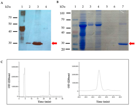
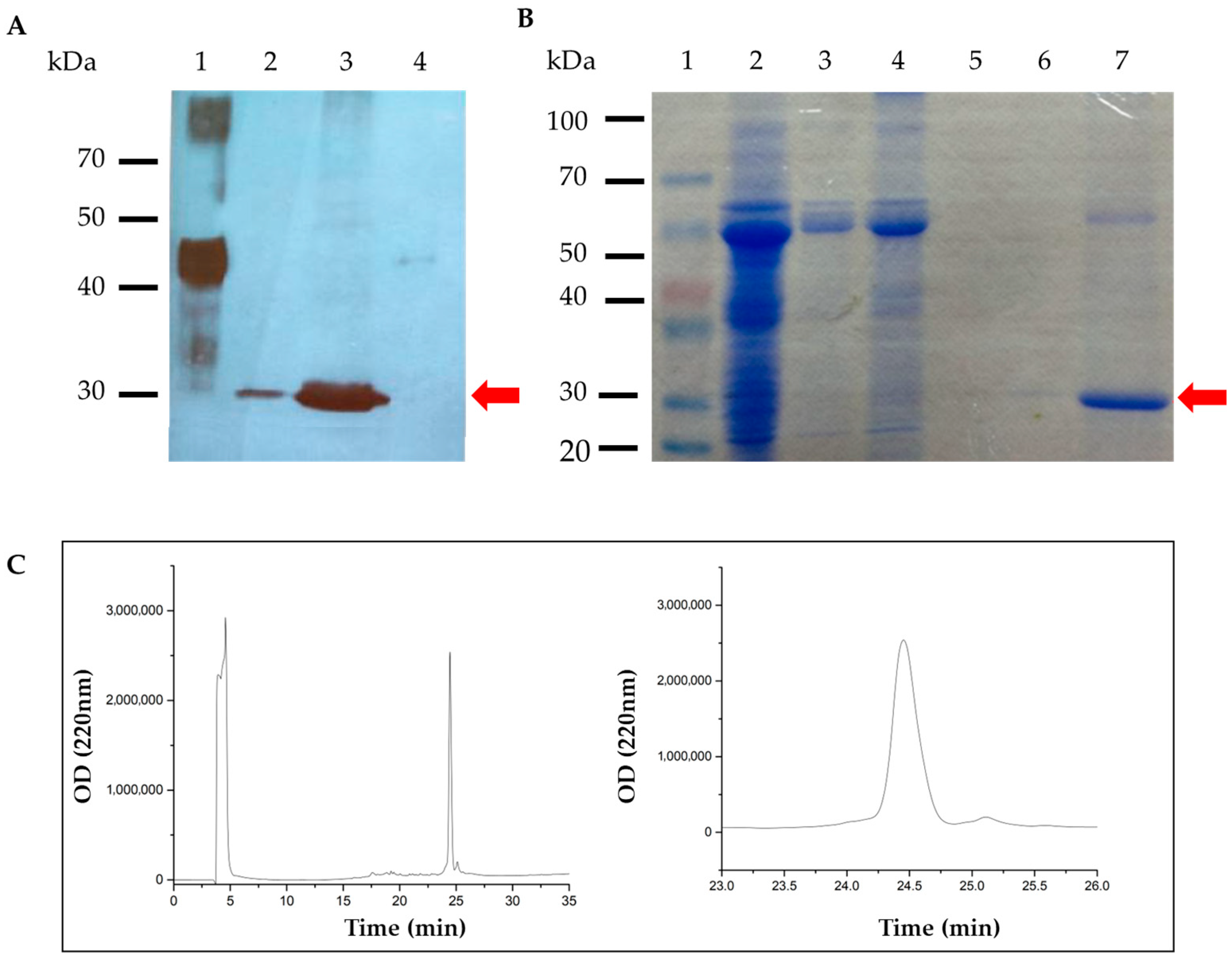
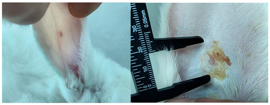
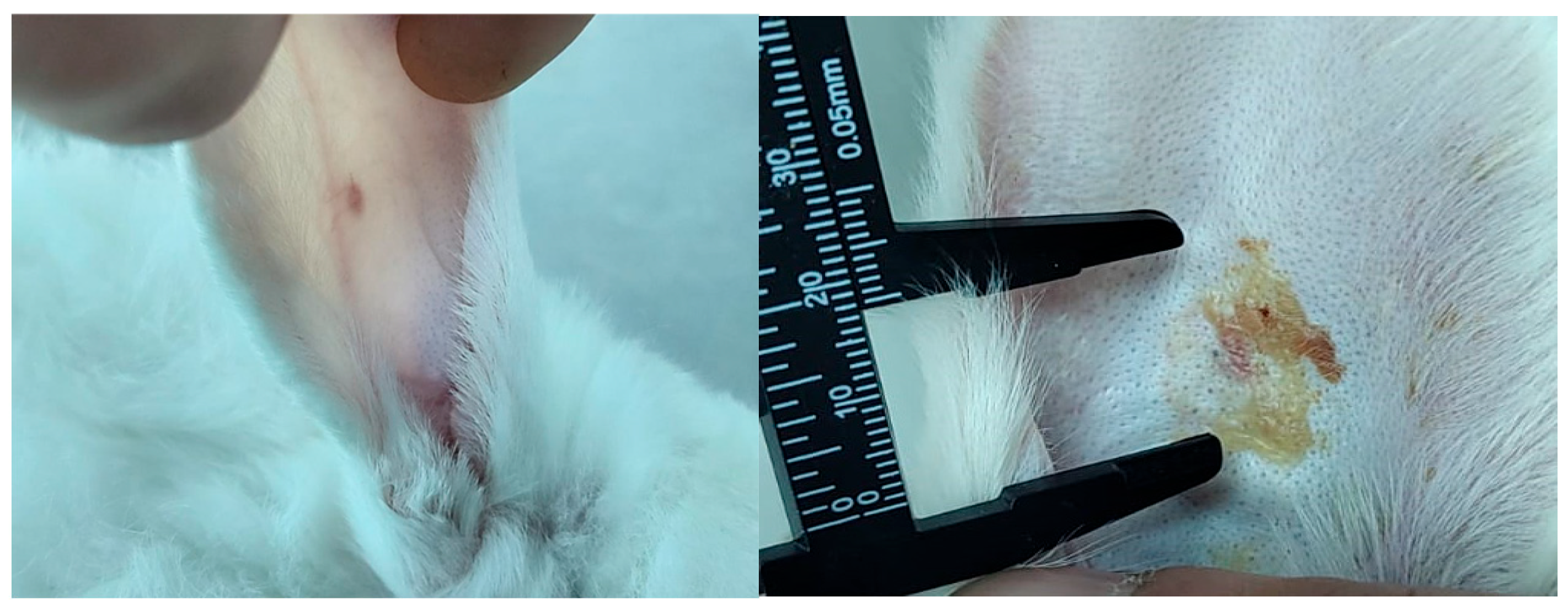
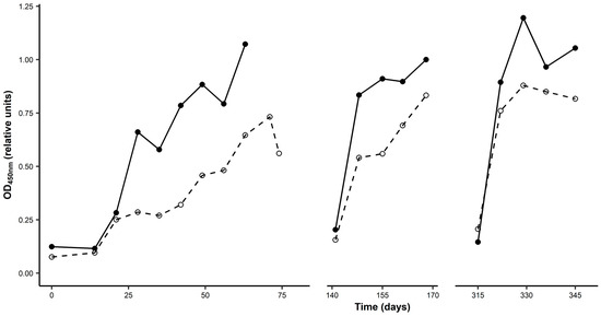
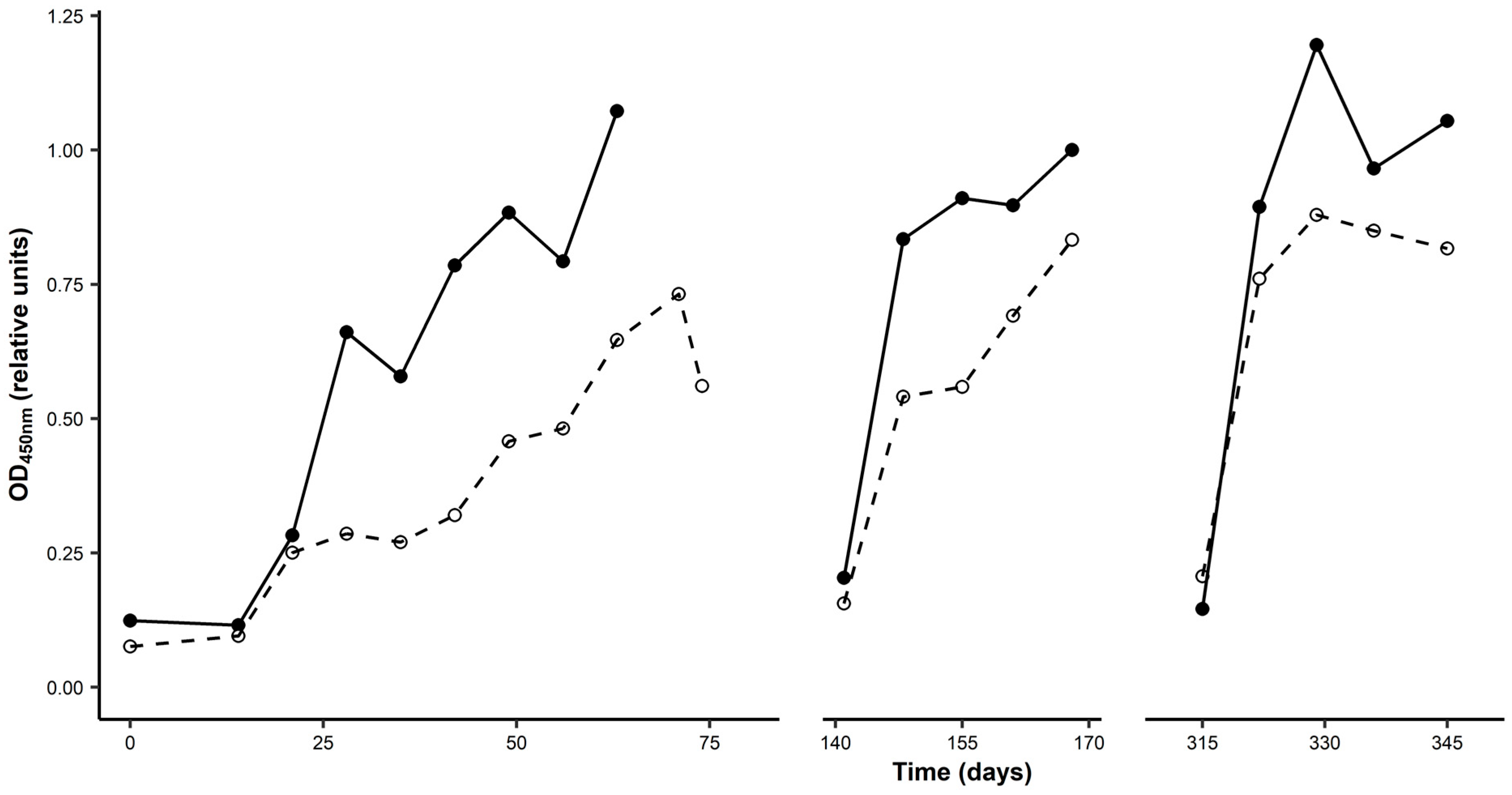
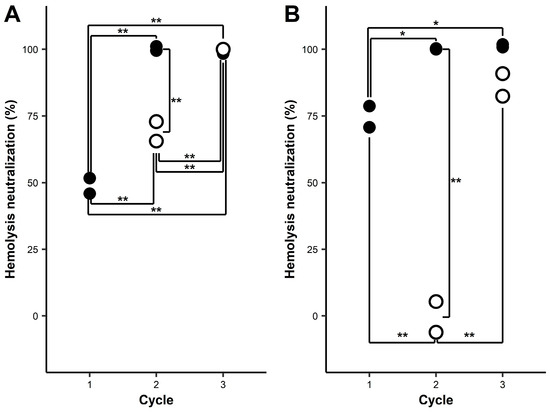
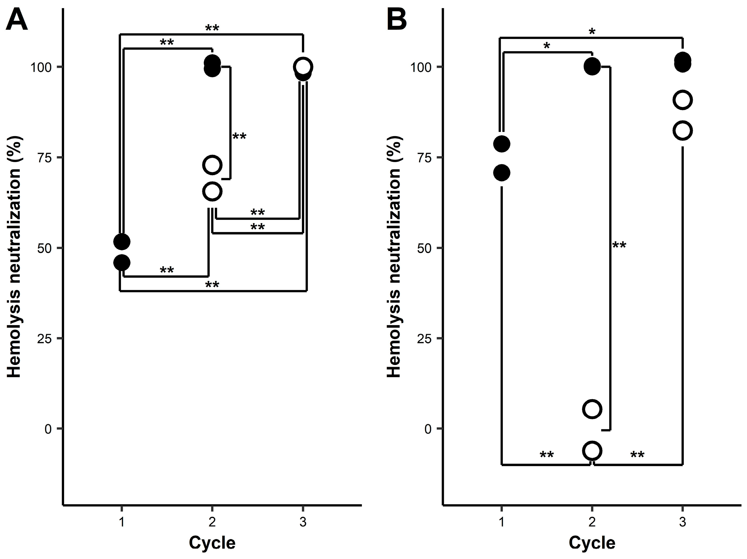
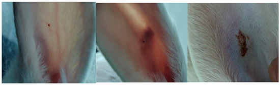
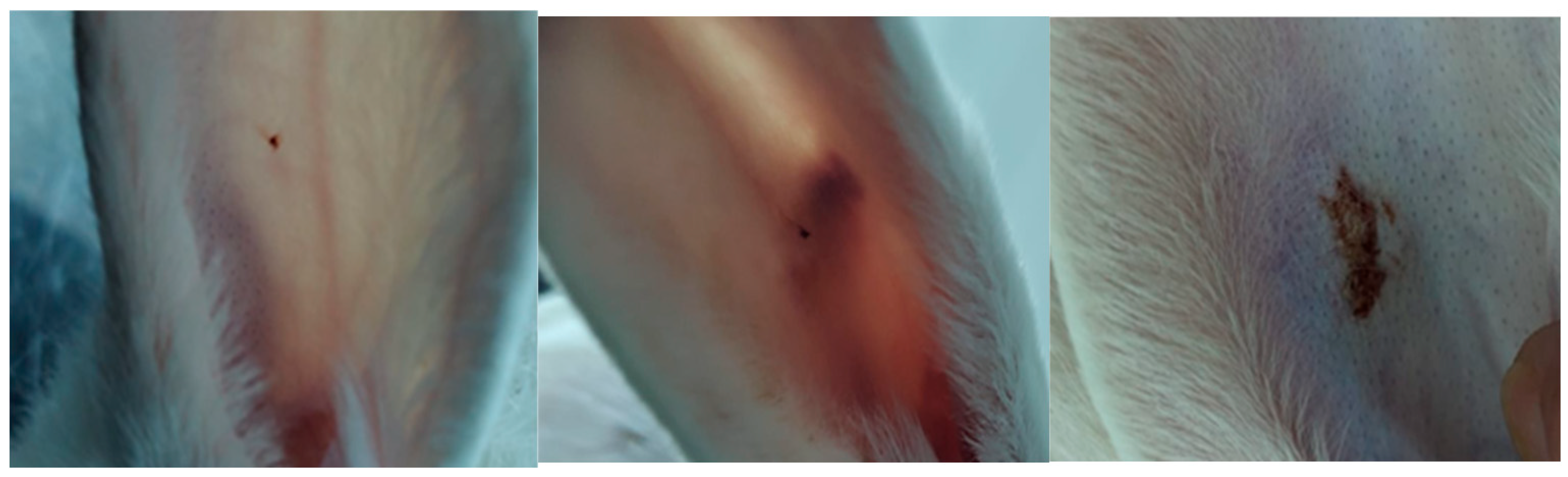

Disclaimer/Publisher’s Note: The statements, opinions and data contained in all publications are solely those of the individual author(s) and contributor(s) and not of MDPI and/or the editor(s). MDPI and/or the editor(s) disclaim responsibility for any injury to people or property resulting from any ideas, methods, instructions or products referred to in the content. |
© 2026 by the authors. Licensee MDPI, Basel, Switzerland. This article is an open access article distributed under the terms and conditions of the Creative Commons Attribution (CC BY) license.