
Article

Novel Citrus Fiber-Entrapped Curcuminoids Exhibit Improved Oral Bioavailability in Wistar Rats
1
Makams Industries Pvt. Ltd., Gurugram 122004, India
2
University Institute of Pharmaceutical Sciences, Panjab University, Chandigarh 160014, India
*
Author to whom correspondence should be addressed.
These authors contributed equally to this work.
J. Pharm. BioTech Ind. 2026, 3(2), 12; https://doi.org/10.3390/jpbi3020012
Submission received: 17 November 2025 / Revised: 3 April 2026 / Accepted: 15 April 2026 / Published: 21 May 2026

Abstract

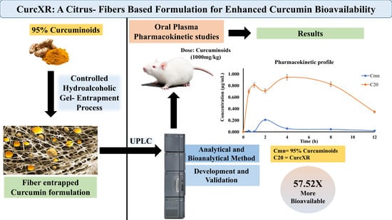
Curcuminoids (Cmn) are polyphenolic compounds from Curcuma longa that exhibit significant pharmacological activities but suffer from poor bioavailability due to low solubility and rapid metabolism. We have developed a novel formulation of Cmn entrapped within citrus fibers with the intent to improve its bioavailability. The physiological properties of citrus fibers improve aqueous dispersion and apparent solubilization of Cmn while protecting it from physiological degradation. Single-dose oral pharmacokinetics in Wistar rats revealed that citrus fiber-entrapped Cmn (CurcXR) exhibited a 57.52-fold increase in bioavailability compared to 95% standard Cmn. The maximum plasma concentration (Cmax) of 0.95 μg/mL at 4 h, and an area under the curve (AUC0−t) of 8.84 μg/mL·h was observed for CurcXR. These findings highlight that citrus fiber-based formulations are a simple, safe, and effective strategy to enhance the bioavailability of Cmn in nutraceuticals.

Graphical Abstract

1. Introduction

Curcuminoids (Cmn), polyphenolic compounds derived from the rhizome of Curcuma longa (Turmeric), have attracted considerable attention in recent years due to their wide-ranging pharmacological activities, including anti-inflammatory, antioxidant, anticancer, and antimicrobial effects [1]. Research has demonstrated that Cmn exhibits chemosensitizing properties and acts as a P-glycoprotein (P-gp) efflux inhibitor [2,3,4]. Cmn directly inhibits cyclooxygenase-2 (COX-2) and the transcription of the gene responsible for its production. COX catalyzes the synthesis of prostaglandins (PGs) from arachidonic acid [5]. Despite its promising therapeutic potential, the clinical application of Cmn is significantly limited by its poor bioavailability. This limitation arises primarily from its low water solubility, rapid metabolism, and poor permeability. Enzymes such as UDP-glucuronosyltransferase and CYP3A4 predominantly metabolize Cmn, further diminishing its bioavailability [6]. Consequently, enhancing its pharmacokinetic profile is an essential area of ongoing research [1].
The specific form of Cmn that exists within tissues and its active form at cellular and molecular levels remain unclear. Additionally, correlation of blood levels of Cmn along with its glucuronide and sulfate conjugates to the physiological effects, is not well understood. Given that Cmn is thought to be more physiologically active than its conjugated forms, it is assumed that higher levels of free Cmn in the blood indicate greater physiological activity [7,8,9,10]. To exert a physiological effect, Cmn must be adequately absorbed and presented in the form of free Cmn.
While various formulations have successfully increased total Cmn levels in the bloodstream, studies often fail to demonstrate a corresponding increase in free Cmn levels. With some exceptions [11,12,13,14,15,16,17], plasma samples are commonly subjected to enzymatic hydrolysis, to change the dominant sulfate and glucuronide conjugate metabolites of Cmn found in plasma back to free Cmn. This results in the measurement of total Cmn (including its metabolites) instead of free Cmn [18,19,20].
While numerous advanced platforms, such as nanoemulsions, micelles, phospholipid complexes, and chemical conjugates, have demonstrated improved Cmn bioavailability, these approaches often rely on synthetic, non-food-grade excipients, surfactants, or additives that may pose safety and regulatory hurdles, especially in food and nutraceutical contexts. For example, piperine-based enhancement is effective, but carries risks of enzyme inhibition and potential drug interactions, limiting its suitability for general consumer use or long-term supplementation regimes. Similarly, lipid or nanoparticle carriers can increase absorption, but are associated with increased production complexity, costs, and in certain cases, limited consumer acceptance due to their non-natural origin [21,22,23].
Citrus fibers, derived from citrus peel by-products, represent a promising excipient platform for enhancing the delivery of lipophilic bioactives such as Cmn. These fibers comprise both soluble (primarily pectin) and insoluble fractions (cellulose, hemicellulose, and lignin). The soluble pectin fraction provides key physicochemical functionalities, including gelling, emulsifying, and mucoadhesive properties, which support improved dispersion and stabilization of hydrophobic compounds [24,25,26]. In aqueous environments, pectin forms viscous gel networks that can prolong gastrointestinal residence time, modulate release kinetics, and protect labile compounds from gastric degradation [27]. Its fermentability by colonic microbiota further enables site-specific release and may contribute to sustained systemic availability [28]. The insoluble components provide structural integrity, facilitating entrapment of bioactives and contributing to their gradual release.
As food-grade sustainable ingredients derived directly from fruit sources, citrus fibers offer practical formulation and regulatory advantages. Their clean-label status supports broad dietary compatibility and consumer acceptance, while minimizing the regulatory complexity often associated with synthetic or chemically modified carriers [22]. Collectively, these attributes position citrus fibers as a scalable and safe platform for advanced oral delivery systems targeting improved Cmn bioavailability.
A novel proprietary Cmn formulation (patent application no. 202411074985) developed using food-grade materials and citrus fibers was evaluated for its primary objective of assessing its oral bioavailability relative to a standard 95% Cmn formulation. The study was conducted in healthy Wistar rats and was specifically designed to compare systemic exposure following equivalent administered Cmn doses.
The primary aim was to quantify and compare plasma concentrations of free Cmn over time, enabling the determination of key pharmacokinetic parameters such as Cmax, AUC, and relative bioavailability. Relative pharmacokinetic efficiency was calculated on a dose-normalized basis (milligrams of administered Cmn), allowing direct comparison between the citrus fiber-entrapped Cmn formulation (CurcXR) and pure 95% Cmn.

2. Materials and Method

2.1. Materials

Cmn was provided by Ms Makams Pvt. Ltd., Bhiwadi, Rajasthan, India. The citrus fibers used in the study were manufactured in-house at Makams Pvt. Ltd., Bhiwadi, Rajasthan, India. HPLC-grade methanol and ACN were purchased from Sigma Aldrich, Bangalore, India, HPLC grade water was produced by a Milli DI® system (Millipore, MA, USA), and K3 EDTA tubes were purchased from Microsidd, Gujrat, India. All other reagents used in the study were of analytical grade. Analytical studies were performed on Acquity UPLC, Waters, MA, USA.

2.2. Methods

2.2.1. Method of Formulation Preparation

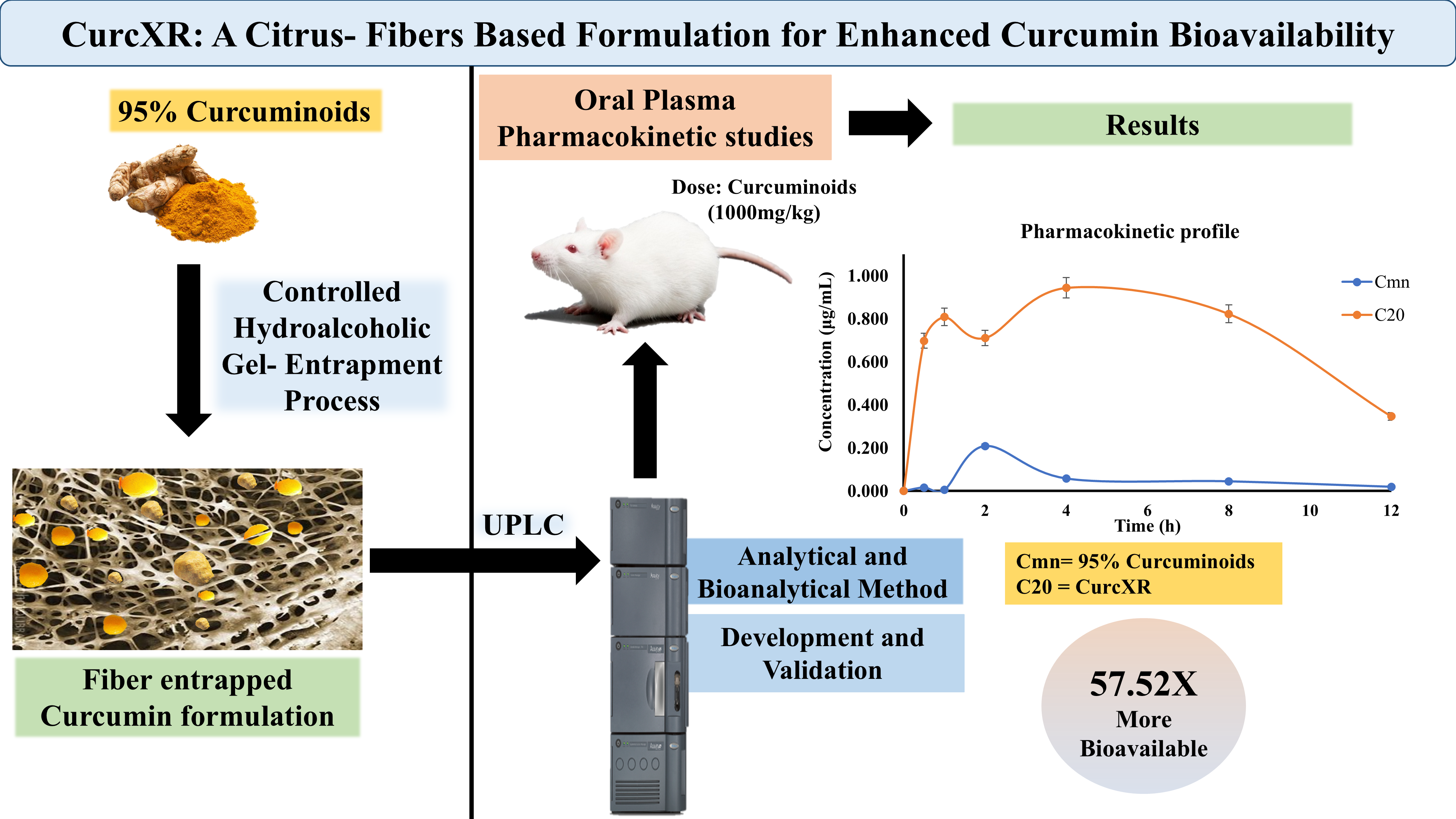
CurcXR was manufactured using a controlled hydroalcoholic gel-entrapment process designed to facilitate the dispersion and retention of Cmn within a citrus-fiber matrix. Citrus fibers, containing soluble and insoluble components in approximately a 1:5 ratio, were first dispersed in a required quantity of hydroalcoholic medium to enhance entrapment. The formulation ratio was fixed at 78.5% w/w citrus fiber and 21.5% w/w Cmn, so the final formulation had a 20% active Cmn concentration.
The solvent was transferred to a jacketed, temperature-regulated vessel equipped with a stirring mechanism. Citrus fibers were introduced gradually under continuous 20-60 RPM agitation to prevent clumping and to allow complete hydration. The mixture was stirred for 8 h at 25–35 °C temperature to allow full hydration and swelling of the fiber components. After stirring, the dispersion was left undisturbed for an additional 6 h to enable the formation of a cohesive gel network capable of entrapping hydrophobic actives. After gel formation, the required quantity of 95% Cmn was incorporated into it (Table 1) slowly under controlled stirring to promote uniform dispersion and maximize molecular entrapment within the fiber network. The mixing continued for 12 h to ensure complete embedding of Cmn into the hydrated fiber structure.
The resulting bioactive gel was then dried to remove the hydroalcoholic medium while preserving the integrity of the Cmn-fiber complex, preferably by vacuum drying at 60 °C for 48 h to achieve complete solvent removal. The dried mass was milled to obtain a fine, free-flowing powder, followed by sieving through a 60-mesh screen, which ensured a consistent particle size distribution crucial for improvised aqueous dispersion and stability of Cmn. The final CurcXR powder contained Cmn uniformly entrapped within a structured citrus-fiber matrix, yielding improved stability, dispersion behavior, and potential bioavailability, as represented in Figure 1.
The prepared formulation CurcXR was subjected to an accelerated stability study for 6 months at 40 °C/75% relative humidity (RH). The formulation was analyzed for any visual changes and for total curcuminoid content in CurcXR at 0, 1, 2, 3, and 6 months as provided in Supplementary Figure S3.

2.2.2. Animal Ethics and Approval

Wistar rats of either sex, obtained from the central animal house facility, Panjab University, Chandigarh, India, were placed in cages in groups of 4 rats/cage and had access to animal feed and water ad libitum. The procedures involving animal experimentation in the study were appropriately sanctioned by the Institutional Animal Ethics Committee (IAEC) at Panjab University, Chandigarh, India, under the approval number PU/45/99/CPCSEA/IAEC/2023/834.

2.2.3. Preparation of Sample for Oral Administration

Required amount of standard 95% Cmn was accurately weighed and added to a specific volume of water containing 1% carboxymethyl cellulose (CMC), which served as a suspending agent. The mixture was placed on a magnetic stirrer to facilitate thorough mixing and uniform dispersion of the formulation particles throughout the CMC solution. After allowing it to stir for 30 min, the suspension achieved a homogeneous consistency, indicating that the Cmn particles were effectively suspended and evenly distributed in the liquid medium [29,30]. This method ensured stability and uniformity in the suspension. Citrate buffer pH 4 was used instead of 1% CMC for CurcXR, because it contains soluble and insoluble fibers that significantly increases their viscosity in water, making it difficult to administer the formulation orally [31,32]. Table 1 contains the concentration of curcuminoids in the formulations prepared.

2.2.4. Sample Withdrawal and Preparation

In vivo pharmacokinetic studies were conducted using healthy Wistar rats of either sex, 8–12 weeks old, weighing 200–250 g. Animals were randomly divided into 2 groups containing 6 animals each for free Cmn and CurcXR administration, respectively. Simple randomization was performed using cage allocation, and dosing order was randomized. The rats were fed on the pre-study day and thereafter fasted overnight (for at least 10 h before dosing) as well as for 4 h after dosing. Water was not permitted 1 h before and 1 h after administration, but was allowed at all other times ad libitum. A 1000 mg/kg dose of Cmn and CurcXR formulation was administered orally. After dose administration, blood samples of rats were withdrawn via retro-orbital plexus into EDTA-coated tubes after intervals of 0 h, 1 h, 2 h, 4 h, 8 h, 12 h, 24 h, and 48 h, and centrifuged at 10,000 rpm for 10 min, and the plasma separated was collected into dry tubes [29]. All the samples were stored under refrigerated conditions before assay on the same day. Concentrations of Cmn in plasma were determined using the UPLC method.

2.2.5. UPLC Method Development

Plasma (0.25 mL) collected at each sampling time was transferred to dry Eppendorf tubes, Oldenburg, Germany. To each tube, 0.75 mL mixture of acetonitrile and methanol (1:1) was added, mixed thoroughly, and centrifuged at 10,000 rpm for 10 min. The organic layer (0.6 mL) was further transferred into dry UPLC vials, after filtration. Subsequently, 10 µL was injected into the column for UPLC analysis [33,34]. From the plasma concentration versus time data, various pharmacokinetic parameters, such as peak concentration (Cmax), time at which peak occurred (Tmax), area under the curve (AUC), biological half-life (t1/2), and bioavailability, were calculated in each case using standard methods.

2.2.6. Chromatographic Conditions

The determination of Cmn was carried out using a UPLC system (Waters, Acquity UPLC H class). A reversed-phase Thermo Fisher Scientific C18 UPLC column (4.6 × 150 mm, 5 µm; Accucore, Delhi, India) was used. ACN: water (75:25 v/v) was run as the mobile phase for Cmn. The elution was performed at a flow rate of 0.25 mL/min, and the analytical column was maintained at 30 °C. The detection was performed with a Waters eλ photodiode array detector (PDA) at a set wavelength of 425 nm for Cmn. The injection volume was 10.0 µL for all standards and samples. Cmn were eluted approximately at 5.6 min after injection. The method was validated according to ICH guidelines for parameters such as accuracy, precision, linearity, LOD, LOQ, and robustness [35,36,37,38].

2.2.7. Pharmacokinetic Analysis

Pharmacokinetic calculations were performed on individual datasets using the non-compartmental method in PK Solver software (Version 2.0), where key parameters were evaluated, including, (Cmax), the maximum concentration of the drug observed post-administration; (Tmax), the time taken to reach Cmax; AUC, representing the total drug exposure over time calculated using the trapezoidal rule; relative bioavailability, determined by comparing the AUC of the test formulation to that of a reference formulation; the elimination rate constant (Kel), derived from the slope of the terminal phase of the concentration-time curve; and mean residence time (MRT), calculated from AUC and total clearance, providing comprehensive insights into the drug’s pharmacokinetics and aiding in understanding its dosing, safety, and efficacy profiles [39,40].

2.2.8. Statistical Analysis

Pharmacokinetic parameters were evaluated using six animals per group (n = 6), and the results are presented as mean ± SD. An unpaired Student’s t-test was performed to evaluate the pharmacokinetic parameters, including Cmax, Tmax, T1/2, AUC0−t, AUC0–∞, and MRT, which were expressed as mean ± SD. The differences were considered statistically significant at p < 0.05. Results were analyzed using GraphPad Prism software version 6.01 (San Diego, CA, USA).

3. Results

The newly developed RP-UPLC method for the estimation of unmetabolized curcuminoids (Cmn) in pharmacokinetic studies was successfully validated and applied. “Unmetabolized curcuminoids” refers to the native (free) curcuminoid compounds, including curcumin, along with minor components such as demethoxycurcumin (DMC), and bisdemethoxycurcumin (BDMC). During method development, these compounds initially appeared as separate peaks; however, due to the low concentrations of DMC and BDMC (Supplementary Figure S4), the chromatographic conditions were optimized to obtain a single cumulative peak representing total curcuminoids for reliable quantification.
Furthermore, the term “unmetabolized” is specifically used because the reference standards employed were of native curcuminoids, and the detection wavelength (425 nm) selectively captures these free forms. Metabolized curcumin derivatives (e.g., glucuronides and sulfates) do not exhibit significant absorbance at this wavelength under the given conditions, thereby ensuring that only unmetabolized (free) curcuminoids are measured [6,9,10].
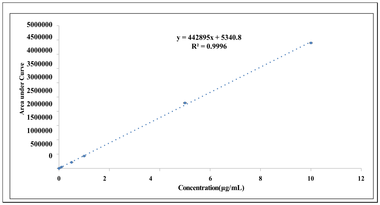
The calibration curve for the method showed excellent linearity with an R2 value of 0.9996 (Figure 2). Method validation confirmed that accuracy ranged from 99.33% to 104.97%, and precision was between 99.2% and 101.73%, with relative standard deviation (RSD) values of less than 2%, as depicted by Table 2, ensuring the method’s reliability for bioavailability studies. Further validation results are provided in the Supplementary File along with the UPLC chromatogram of Cmn in plasma; Supplementary Figure S1.
This validated method was used to evaluate the bioavailability of Cmn after oral administration to healthy Wistar rats. The rats, weighing between 200 and 300 g, were administered Cmn either in its standardized form (95%) or as CurcXR at a dose of 1000 mg/kg. Cmn is poorly water-soluble and rapidly sediments in aqueous media; therefore, 1% CMC was used in the control formulation only as a suspending agent to ensure uniform dispersion and consistent dosing, without affecting solubility or absorption. In contrast, CurcXR contains gel-forming fibers that swell quickly at neutral pH, resulting in high viscosity and handling difficulties. To prevent premature gelation and enable easy, reproducible dosing, citrate buffer (pH 4) was used, eliminating the need for an additional suspending agent such as CMC. Furthermore, following oral administration, both formulations were rapidly exposed to the gastric environment, where the physiological pH typically ranges from 1 to 3 under fed conditions. Therefore, despite the initial differences in vehicle pH (neutral vs. pH 4), both formulations are expected to experience a similar acidic environment shortly after administration, minimizing the impact of initial vehicle pH on overall in vivo exposure.
Plasma samples were collected and analyzed to obtain concentration-time profiles, from which pharmacokinetic parameters were calculated using PK Solver (Version 2.0). These values were then compared to assess the impact of citrus fiber entrapment on the bioavailability of Cmn.
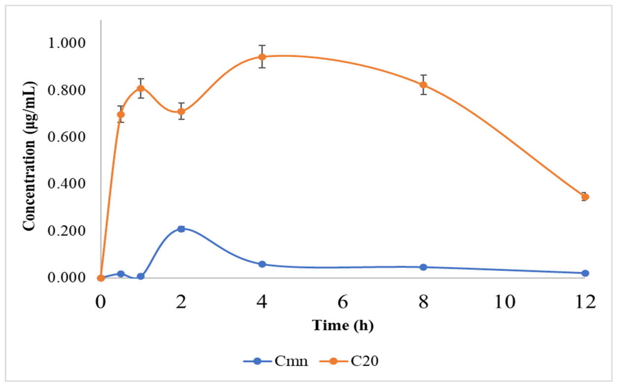
The pharmacokinetic data in Figure 3 and Table 3 revealed distinct differences in the bioavailability of free Cmn versus CurcXR. For the free Cmn formulation, the elimination rate constant (Kel) was calculated as 0.06 h−1, yielding a half-life (t1/2) of 5.14 h and a mean residence time (MRT) of 7.03 h [41,42,43]. These results confirm the expected pharmacokinetic behavior of free Cmn, which exhibits low bioavailability due to its rapid metabolism and poor solubility in the gastrointestinal tract. While the control formulation (Cmn) was administered as a suspension in 1% CMC, and CurcXR was dispersed in citrate buffer (pH 4), both preparations were administered immediately after preparation to minimize pre-administration degradation. The gastric environment in vivo is strongly acidic (pH ~ 1–3) and rapidly equilibrates with administered formulations, regardless of the initial vehicle pH. Therefore, the influence of the initial vehicle pH is expected to be transient under physiological conditions. However, Cmn exhibits pH-dependent degradation kinetics, and the possibility that vehicle composition may contribute to differences in apparent exposure cannot be completely excluded.
In comparison, the CurcXR formulation showed markedly improved pharmacokinetic parameters compared to the standardized Cmn. Although both formulations were dosed at 1000 mg/kg, the effective curcuminoid content differed (95% for standard curcumin vs. 20% for CurcXR). Therefore, AUC0–∞ values were corrected using a strength normalization factor (95/20.43 = 4.65), enabling comparison on an equivalent curcuminoid basis. This dose-normalization approach helps minimize the impact of potential non-linearity and ensures that the observed differences in bioavailability reflect formulation performance rather than differences in active dose.
The maximum plasma concentration (Cmax) for CurcXR was 0.95 μg/mL, significantly higher than the 0.21 μg/mL observed for standardized Cmn. The time to reach maximum concentration (Tmax) for CurcXR was 4 h, compared to 2 h for standardized Cmn, suggesting a delayed but sustained release of Cmn from the CurcXR formulation. More notably, the area under the curve (AUC0−t) for CurcXR was 8.84 μg/mL·h, compared to 0.73 μg/mL·h for standardized Cmn. The AUC0–∞ for CurcXR was 11.61 μg/mL·h, significantly higher than the 0.87 μg/mL·h for standardized Cmn. This represents a 57.52-fold increase in bioavailability for the CurcXR formulation, demonstrating the substantial impact of the citrus fiber entrapment on Cmn absorption and retention in the bloodstream. Furthermore, the pharmacokinetic parameters of both formulations revealed significant differences (p < 0.05), as confirmed by the t-test analysis.
The accelerated stability studies of the prepared CurcXR formulation (Supplementary Figure S3) at 40 °C/75%RH showed no significant difference in Cmn content over 6 months (21.04% to 20.01% Cmn).

4. Discussion

The fibers were derived from bergamot, lemon, orange, and green tangerine peels and comprised both soluble pectin and insoluble fiber cellulose-hemicellulose fractions. The optimized CurcXR system leverages the intrinsic structural and chemical properties of citrus fiber. Upon hydration, citrus fiber swells to form a three-dimensional gel network capable of entrapping hydrophobic actives. Within this matrix, curcumin (Cmn) may be retained through hydrophobic partitioning into swollen cellulose domains, hydrogen bonding between phenolic hydroxyl groups and pectin chains, and physical confinement within the hydrated microstructure [44].
The enhanced bioavailability observed with CurcXR likely relates to the complementary functions of soluble and insoluble fiber fractions in the gastrointestinal environment. Soluble components such as citrus pectin form viscous gels that can slow gastrointestinal transit and prolong small-intestinal residence time, potentially facilitating micelle-mediated absorption of Cmn. Insoluble fibers contribute to structural bulk and support luminal mixing, promoting balanced transit without stagnation. Together, these effects may create conditions favorable for improved dissolution and uptake [45,46,47].
Cmn is susceptible to hydrolysis, oxidation, and pH-dependent degradation under physiological conditions [6,32]. The hydrated fiber network may function as a diffusion barrier, limiting direct exposure to aqueous and enzymatic environments, while the structural matrix may support moderated release. Comparable protective effects of fiber-based systems for poorly soluble bioactives have been described previously [22,24].
A favorable gut environment, maintained by adequate fiber, supports the growth and activity of beneficial gut microbiota, fosters optimal pH, and supplies essential nutrients for microbial metabolism. Of relevance is the stimulation of bacterial populations such as Escherichia coli, Bacteroides, Clostridium, Lactobacillus, and Ruminococcus, which express β-glucuronidase. This enzyme hydrolyzes Cmn glucuronide conjugates that are excreted into the gut by the liver, thereby regenerating free, bioactive Cmn for reabsorption. The result is a higher pool of active Cmn available for physiological action, as opposed to rapid conversion to less bioactive derivatives [48,49]. Furthermore, recent clinical and preclinical studies demonstrate that dietary fiber-based delivery systems provide superior protection against premature degradation, maintain Cmn in its active form, and lead to higher plasma levels of unmetabolized curcuminoids, surpassing more complex synthetic or lipid-based delivery vehicles.
While the present study demonstrates a substantial enhancement in systemic exposure of free curcumin following oral administration of CurcXR, detailed in vitro physicochemical characterization of the formulation (e.g., entrapment efficiency, drug loading, dissolution profiling, swelling behavior, and rheological assessment) was not performed within the scope of this investigation. Therefore, the proposed mechanisms, such as gel-mediated protection, modulation of gastrointestinal residence time, and controlled release, should be interpreted as literature-supported hypotheses rather than experimentally validated mechanisms in this study. Future work will focus on comprehensive in vitro characterization under simulated gastrointestinal conditions to establish structure–performance relationships and mechanistically link formulation attributes to the observed pharmacokinetic enhancement. The present study has certain limitations. UPLC and HPLC (Supplementary Figure S5) were used to quantify total curcuminoids without individual component analysis. Detailed characterization of the citrus fiber matrix and formulation was not performed due to the scope of the study and proprietary constraints. Further studies on digestion resistance and dispersion are needed to strengthen the mechanistic understanding. Tissue distribution and safety evaluation will be undertaken in the future. Collectively, these findings highlight that citrus fiber-based carriers effectively leverage gut physiology and microbiota to maximize Cmn bioavailability, making them a safe, robust, and sustainable option for enhancing the health benefits of Cmn in nutraceutical products [23].

5. Conclusions

This study demonstrates that entrapment of curcuminoids within citrus fiber matrix substantially enhances their oral bioavailability by over 57-fold compared to free Cmn. The mechanism underlying this dramatic improvement involves the gel-forming and viscosity-enhancing properties of soluble citrus fibers, combined with the gastrointestinal motility-promoting effects of insoluble fibers. Together, these fibers optimize gut transit and mixing, protect Cmn from premature degradation, and create a favorable intestinal microenvironment conducive to absorption.
Importantly, the citrus fiber-based formulation not only increases total Cmn levels in plasma but also preserves a majority of the Cmn in its free, pharmacologically active form. This aspect is critical since free Cmn exhibits substantially higher therapeutic potency than its conjugated metabolites, which are often rapidly excreted or inactive. By safeguarding Cmn from early metabolism and possibly supporting microbial enzyme activity, key to regenerating free Cmn, citrus fibers ensure sustained delivery of this potent form to systemic circulation.
These findings position citrus fiber as a naturally derived, food-grade, and consumer-friendly carrier that overcomes major hurdles in Cmn delivery. The strategy offers significant potential for broader application in nutraceuticals and functional foods, aiming to maximize the health benefits of Cmn and similar bioactives with poor inherent bioavailability.

Supplementary Materials

The following supporting information can be downloaded at: https://www.mdpi.com/article/10.3390/jpbi3020012/s1, Figure S1: UPLC chromatogram overlay showing Cmn peak; Table S1: Data demonstrating system suitability of the developed bioanalytical method; Figure S2: UPLC chromatogram of blank plasma (Cmn) as a representative biological matrix; Figure S3: Stability data of CurcXR formulation; Figure S4: Unmetabolized Cmn showing three separate peaks of curcumin, demethoxycurcumin and bisdemethoxycurcumin; Figure S5: HPLC analysis of unmetabolized Cmn (curcumin, demethoxycurcumin and bisdemethoxycurcumin); Figure S6: COA of curcuminoids extract.

Author Contributions

A.A.: Conceptualization, Writing—original draft; S.M.B.P.: Investigation, Methodology, Formal analysis, Data curation, Writing—original draft; B.A.: Formal analysis, Validation, Writing–review and editing; S.N.: Formal analysis: Writing–original draft; J.S.: Methodology; I.P.K.: Conceptualization, Formal analysis, Project administration, Writing—review and editing, Supervision. All authors have read and agreed to the published version of the manuscript.

Funding

This research received no external funding.

Institutional Review Board Statement

The procedures involving animal experimentation in the study were appropriately approved by the Institutional Animal Ethics Committee (IAEC) at Panjab University, Chandigarh, India, on 28 August 2023, under the approval number PU/45/99/CPCSEA/IAEC/2023/834.

Data Availability Statement

The original contributions presented in this study are included in the article/Supplementary Material. Further inquiries can be directed to the corresponding author.

Conflicts of Interest

Shankh Mani Bhai Patel conducted this research work at Panjab University as part of his academic studies, before joining M/s Makams Industries Pvt. Ltd. Aakash Agarwal and Shankh Mani Bhai Patel are employed at M/s Makams Industries Pvt. Ltd. The authors confirm that these affiliations did not influence the study design, data collection, analysis, interpretation of results, or manuscript preparation. No financial or material support from Makams Industries was provided for this research. The remaining authors declare that the research was conducted in the absence of any commercial or financial relationships that could be construed as a potential conflict of interest.

References

  1. Sharma, R.A.; Gescher, A.J.; Steward, W.P. Curcumin: The Story so Far. Eur. J. Cancer 2005, 41, 1955–1968. [Google Scholar] [CrossRef] [Scilit] [PubMed]
  2. Chearwae, W.; Anuchapreeda, S.; Nandigama, K.; Ambudkar, S.V.; Limtrakul, P. Biochemical Mechanism of Modulation of Human P-Glycoprotein (ABCB1) by Curcumin I, II, and III Purified from Turmeric Powder. Biochem. Pharmacol. 2004, 68, 2043–2052. [Google Scholar] [CrossRef] [Scilit]
  3. Flory, S.; Männle, R.; Frank, J. The Inhibitory Activity of Curcumin on P-Glycoprotein and Its Uptake by and Efflux from LS180 Cells Is Not Affected by Its Galenic Formulation. Antioxidants 2021, 10, 1826. [Google Scholar] [CrossRef] [Scilit]
  4. Lopes-Rodrigues, V.; Sousa, E.; Vasconcelos, M.H. Curcumin as a Modulator of P-Glycoprotein in Cancer: Challenges and Perspectives. Pharmaceuticals 2016, 9, 71. [Google Scholar] [CrossRef] [Scilit]
  5. Antony, B. Composition to Enhance the Bioavailability of Curcumin. U.S. Patent US-9878040-B2, 30 January 2018. [Google Scholar]
  6. Metzler, M.; Pfeiffer, E.; Schulz, S.I.; Dempe, J.S. Curcumin Uptake and Metabolism. Biofactors 2013, 39, 14–20. [Google Scholar] [CrossRef] [Scilit]
  7. Douglass, B.J.; Clouatre, D.L. Beyond Yellow Curry: Assessing Commercial Curcumin Absorption Technologies. J. Am. Coll. Nutr. 2015, 34, 347–358. [Google Scholar] [CrossRef] [Scilit]
  8. Jankun, J.; Wyganowska-Świątkowska, M.; Dettlaff, K.; Jelińska, A.; Surdacka, A.; Wątróbska-Świetlikowska, D.; Skrzypczak-Jankun, E. Determining Whether Curcumin Degradation/Condensation Is Actually Bioactivation. Int. J. Mol. Med. 2016, 37, 1151–1158. [Google Scholar] [CrossRef] [Scilit] [PubMed]
  9. Prasad, S.; Tyagi, A.K.; Aggarwal, B.B. Recent Developments in Delivery, Bioavailability, Absorption and Metabolism of Curcumin: The Golden Pigment from Golden Spice. Cancer Res. Treat. 2014, 46, 2–18. [Google Scholar] [CrossRef] [Scilit] [PubMed]
  10. Pan, M.-H.; Huang, T.-M.; Lin, J.-K. Biotransformation of Curcumin through Reduction and Glucuronidation in Mice. Drug Metab. Dispos. 1999, 27, 486–494. [Google Scholar] [CrossRef] [Scilit]
  11. Sharma, R.A.; McLelland, H.R.; Hill, K.A.; Ireson, C.R.; Euden, S.A.; Manson, M.M.; Pirmohamed, M.; Marnett, L.J.; Gescher, A.J.; Steward, W.P. Pharmacodynamic and Pharmacokinetic Study of Oral Curcuma Extract in Patients with Colorectal Cancer. Clin. Cancer Res. 2001, 7, 1894–1900. [Google Scholar]
  12. Asher, G.N.; Xie, Y.; Moaddel, R.; Sanghvi, M.; Dossou, K.S.S.; Kashuba, A.D.M.; Sandler, R.S.; Hawke, R.L. Randomized Pharmacokinetic Crossover Study Comparing 2 Curcumin Preparations in Plasma and Rectal Tissue of Healthy Human Volunteers. J. Clin. Pharmacol. 2017, 57, 185–193. [Google Scholar] [CrossRef] [Scilit]
  13. Garcea, G.; Berry, D.P.; Jones, D.J.L.; Singh, R.; Dennison, A.R.; Farmer, P.B.; Sharma, R.A.; Steward, W.P.; Gescher, A.J. Consumption of the Putative Chemopreventive Agent Curcumin by Cancer Patients: Assessment of Curcumin Levels in the Colorectum and Their Pharmacodynamic Consequences. Cancer Epidemiol. Biomark. Prev. 2005, 14, 120–125. [Google Scholar] [CrossRef] [Scilit]
  14. Garcea, G.; Jones, D.J.L.; Singh, R.; Dennison, A.R.; Farmer, P.B.; Sharma, R.A.; Steward, W.P.; Gescher, A.J.; Berry, D.P. Detection of Curcumin and Its Metabolites in Hepatic Tissue and Portal Blood of Patients Following Oral Administration. Br. J. Cancer 2004, 90, 1011–1015. [Google Scholar] [CrossRef] [Scilit]
  15. Gota, V.S.; Maru, G.B.; Soni, T.G.; Gandhi, T.R.; Kochar, N.; Agarwal, M.G. Safety and Pharmacokinetics of a Solid Lipid Curcumin Particle Formulation in Osteosarcoma Patients and Healthy Volunteers. J. Agric. Food Chem. 2010, 58, 2095–2099. [Google Scholar] [CrossRef] [Scilit] [PubMed]
  16. Kanai, M.; Otsuka, Y.; Otsuka, K.; Sato, M.; Nishimura, T.; Mori, Y.; Kawaguchi, M.; Hatano, E.; Kodama, Y.; Matsumoto, S. A Phase I Study Investigating the Safety and Pharmacokinetics of Highly Bioavailable Curcumin (Theracurmin) in Cancer Patients. Cancer Chemother. Pharmacol. 2013, 71, 1521–1530. [Google Scholar] [CrossRef] [Scilit]
  17. Dinesh Kumar, D.K.; Jacob, D.; Subash, P.S.; Abhilash Maliakkal, A.M.; Johannah, N.M.; Ramadassan Kuttan, R.K.; Balu Maliakel, B.M.; Veera Konda, V.K.; Krishnakumar, I.M. Enhanced Bioavailability and Relative Distribution of Free (Unconjugated) Curcuminoids Following the Oral Administration of a Food-Grade Formulation with Fenugreek Dietary Fibre: A Randomised Double-Blind Crossover Study. J. Funct. Foods 2016, 22, 578–587. [Google Scholar] [CrossRef] [Scilit]
  18. Antony, B.; Merina, B.; Iyer, V.S.; Judy, N.; Lennertz, K.; Joyal, S. A Pilot Cross-over Study to Evaluate Human Oral Bioavailability of BCM-95® CG (BiocurcumaxTM), a Novel Bioenhanced Preparation of Curcumin. Indian J. Pharm. Sci. 2008, 70, 445–449. [Google Scholar] [CrossRef] [PubMed]
  19. Kanai, M.; Imaizumi, A.; Otsuka, Y.; Sasaki, H.; Hashiguchi, M.; Tsujiko, K.; Matsumoto, S.; Ishiguro, H.; Chiba, T. Dose-Escalation and Pharmacokinetic Study of Nanoparticle Curcumin, a Potential Anticancer Agent with Improved Bioavailability, in Healthy Human Volunteers. Cancer Chemother. Pharmacol. 2012, 69, 65–70. [Google Scholar] [CrossRef] [Scilit]
  20. Sasaki, H.; Sunagawa, Y.; Takahashi, K.; Imaizumi, A.; Fukuda, H.; Hashimoto, T.; Wada, H.; Katanasaka, Y.; Kakeya, H.; Fujita, M. Innovative Preparation of Curcumin for Improved Oral Bioavailability. Biol. Pharm. Bull. 2011, 34, 660–665. [Google Scholar] [CrossRef] [Scilit] [PubMed]
  21. Tabanelli, R.; Brogi, S.; Calderone, V. Improving Curcumin Bioavailability: Current Strategies and Future Perspectives. Pharmaceutics 2021, 13, 1715. [Google Scholar] [CrossRef] [Scilit]
  22. Mimica, B.; Bučević Popović, V.; Banjari, I.; Jeličić Kadić, A.; Puljak, L. Methods Used for Enhancing the Bioavailability of Oral Curcumin in Randomized Controlled Trials: A Meta-Research Study. Pharmaceuticals 2022, 15, 939. [Google Scholar] [CrossRef] [Scilit]
  23. Hegde, M.; Girisa, S.; BharathwajChetty, B.; Vishwa, R.; Kunnumakkara, A.B. Curcumin Formulations for Better Bioavailability: What We Learned from Clinical Trials Thus Far? ACS Omega 2023, 8, 10713–10746. [Google Scholar] [CrossRef] [Scilit]
  24. Huang, J.; Liao, J.; Qi, J.; Jiang, W.; Yang, X. Structural and Physicochemical Properties of Pectin-Rich Dietary Fiber Prepared from Citrus Peel. Food Hydrocoll. 2021, 110, 106140. [Google Scholar] [CrossRef] [Scilit]
  25. Wu, Q.; Zang, M.; Zhao, B.; Wang, S.; Zhang, S.; Zhu, N.; Liu, M.; Li, S.; Lv, G.; Liu, B. Effect of Citrus Fiber on the Phosphate-Mediated Gel Properties of Myofibrillar Protein and Partial Replacement of Phosphate. LWT 2023, 173, 114274. [Google Scholar] [CrossRef] [Scilit]
  26. Xiao, R.; Qi, J.; Liao, J.; Wei, H.; Zhuo, T. Preparation of Citrus Fiber from Lemon Peel Residue: Effects of Structure and Endogenous Pectin Components on Emulsifying Properties of Citrus Fiber. Int. J. Biol. Macromol. 2024, 283, 137679. [Google Scholar] [CrossRef] [Scilit]
  27. Rosales, T.K.O.; Fabi, J.P. Pectin-Based Nanoencapsulation Strategy to Improve the Bioavailability of Bioactive Compounds. Int. J. Biol. Macromol. 2023, 229, 11–21. [Google Scholar] [CrossRef] [Scilit] [PubMed]
  28. Jo, M.; Park, M.; Choi, Y.J. Citrus Peel Pectin and Alginate-Based Emulgel Particles for Small Intestine-Targeted Oral Delivery of Curcumin. Food Res. Int. 2024, 176, 113847. [Google Scholar] [CrossRef] [Scilit]
  29. Gupta, T.; Singh, J.; Kaur, S.; Sandhu, S.; Singh, G.; Kaur, I.P. Enhancing Bioavailability and Stability of Curcumin Using Solid Lipid Nanoparticles (CLEN): A Covenant for Its Effectiveness. Front. Bioeng. Biotechnol. 2020, 8, 879. [Google Scholar] [CrossRef] [Scilit]
  30. Sandhu, S.K.; Kumar, S.; Raut, J.; Singh, M.; Kaur, S.; Sharma, G.; Roldan, T.L.; Trehan, S.; Holloway, J.; Wahler, G. Systematic Development and Characterization of Novel, High Drug-Loaded, Photostable, Curcumin Solid Lipid Nanoparticle Hydrogel for Wound Healing. Antioxidants 2021, 10, 725. [Google Scholar] [CrossRef] [Scilit]
  31. Jiang, Z.; Zhang, M.; Huang, Y.; Ma, C.; Mu, S.; Li, H.; Liu, X.; Ma, Y.; Liu, Y.; Hou, J. Comparison and Characterization of the Structure and Physicochemical Properties of Three Citrus Fibers: Effect of Ball Milling Treatment. Foods 2022, 11, 2665. [Google Scholar] [CrossRef] [Scilit] [PubMed]
  32. Wang, L.; Xu, H.; Yuan, F.; Pan, Q.; Fan, R.; Gao, Y. Physicochemical Characterization of Five Types of Citrus Dietary Fibers. Biocatal. Agric. Biotechnol. 2015, 4, 250–258. [Google Scholar] [CrossRef] [Scilit]
  33. Kakkar, V.; Singh, S.; Singla, D.; Kaur, I.P. Exploring Solid Lipid Nanoparticles to Enhance the Oral Bioavailability of Curcumin. Mol. Nutr. Food Res. 2011, 55, 495–503. [Google Scholar] [CrossRef] [Scilit]
  34. Liu, H.; Xu, H.; Song, W.; Zhang, Y.; Yu, S.; Huang, X. Validated UPLC/MS/MS Assay for Quantitative Bioanalysis of Elbasvir in Rat Plasma and Application to Pharmacokinetic Study. J. Chromatogr. B 2016, 1015, 150–156. [Google Scholar] [CrossRef] [Scilit] [PubMed]
  35. ICH Q2 (R2) Validation of Analytical Procedures—Scientific Guideline; European Medicines Agency (EMA): Amsterdam, The Netherlands, 2024; Available online: https://www.ema.europa.eu/en/ich-q2r2-validation-analytical-procedures-scientific-guideline (accessed on 31 December 2024).
  36. ICH M10 Guideline on Bioanalytical Method Validation—Scientific Guideline; European Medicines Agency (EMA): Amsterdam, The Netherlands, 2019; Available online: https://www.ema.europa.eu/en/ich-m10-bioanalytical-method-validation-scientific-guideline (accessed on 31 December 2024).
  37. Bioanalytical Method Validation Guidance for Industry Biopharmaceutics Contains Nonbinding Recommendations; U.S. Food and Drug Administration (US-FDA): Silver Spring, MD, USA, 2018. Available online: https://www.fda.gov/regulatory-information/search-fda-guidance-documents/bioanalytical-method-validation-guidance-industry (accessed on 12 January 2025).
  38. Analytical Procedures and Methods Validation for Drugs and Biologics Guidance for Industry; U.S. Food and Drug Administration (US-FDA): Silver Spring, MD, USA, 2015. Available online: https://www.fda.gov/regulatory-information/search-fda-guidance-documents/analytical-procedures-and-methods-validation-drugs-and-biologics (accessed on 15 September 2024).
  39. Henderson, V.C.; Kimmelman, J.; Fergusson, D.; Grimshaw, J.M.; Hackam, D.G. Threats to Validity in the Design and Conduct of Preclinical Efficacy Studies: A Systematic Review of Guidelines for in Vivo Animal Experiments. PLoS Med. 2013, 10, e1001489. [Google Scholar] [CrossRef] [Scilit] [PubMed]
  40. Singh, J.; Elbarbry, F.; Lan, K.; Grabowski, T. Animal Pharmacokinetic/Pharmacodynamic Studies (APPS) Reporting Guidelines. Eur. J. Drug Metab. Pharmacokinet. 2018, 43, 483–494. [Google Scholar] [CrossRef] [Scilit] [PubMed]
  41. Bhatt, H.D.; McClain, S.A.; Lee, H.-M.; Zimmerman, T.; Deng, J.; Johnson, F.; Gu, Y.; Golub, L.M. The Maximum-Tolerated Dose and Pharmacokinetics of a Novel Chemically Modified Curcumin in Rats. J. Exp. Pharmacol. 2022, 14, 73–85. [Google Scholar] [CrossRef] [Scilit]
  42. Shoba, G.; Joy, D.; Joseph, T.; Majeed, M.; Rajendran, R.; Srinivas, P. Influence of Piperine on the Pharmacokinetics of Curcumin in Animals and Human Volunteers. Planta Med. 1998, 64, 353–356. [Google Scholar] [CrossRef] [Scilit]
  43. Yang, K.-Y.; Lin, L.-C.; Tseng, T.-Y.; Wang, S.-C.; Tsai, T.-H. Oral Bioavailability of Curcumin in Rat and the Herbal Analysis from Curcuma Longa by LC–MS/MS. J. Chromatogr. B 2007, 853, 183–189. [Google Scholar] [CrossRef] [Scilit]
  44. Buljeta, I.; Pichler, A.; Šimunović, J.; Kopjar, M. Polyphenols and Antioxidant Activity of Citrus Fiber/Blackberry Juice Complexes. Molecules 2021, 26, 4400. [Google Scholar] [CrossRef] [Scilit]
  45. El-Saadony, M.T.; Yang, T.; Korma, S.A.; Sitohy, M.; Abd El-Mageed, T.A.; Selim, S.; Al Jaouni, S.K.; Salem, H.M.; Mahmmod, Y.; Soliman, S.M.; et al. Impacts of Turmeric and Its Principal Bioactive Curcumin on Human Health: Pharmaceutical, Medicinal, and Food Applications: A Comprehensive Review. Front. Nutr. 2023, 9, 1040259. [Google Scholar] [CrossRef] [Scilit]
  46. El-Saadony, M.T.; Saad, A.M.; Mohammed, D.M.; Alkafaas, S.S.; Ghosh, S.; Negm, S.H.; Salem, H.M.; Fahmy, M.A.; Mosa, W.F.A.; Ibrahim, E.H.; et al. Curcumin, an Active Component of Turmeric: Biological Activities, Nutritional Aspects, Immunological, Bioavailability, and Human Health Benefits—A Comprehensive Review. Front. Immunol. 2025, 16, 1603018. [Google Scholar] [CrossRef] [Scilit]
  47. Wang, L.; Wang, J.; Wang, J.; Guo, Z.; Li, Z.; Qiu, J.; Wang, L. Soluble and Insoluble Dietary Fiber at Different Ratios: Hydration Characteristics, Rheological Properties, and Ameliorative Effects on Constipation. Food Chem. X 2024, 24, 101996. [Google Scholar] [CrossRef] [Scilit] [PubMed]
  48. Guo, K.; Yao, Z.; Yang, T. Intestinal Microbiota-Mediated Dietary Fiber Bioavailability. Front. Nutr. 2022, 9, 1003571. [Google Scholar] [CrossRef] [Scilit] [PubMed]
  49. Lattimer, J.M.; Haub, M.D. Effects of Dietary Fiber and Its Components on Metabolic Health. Nutrients 2010, 2, 1266–1289. [Google Scholar] [CrossRef] [Scilit] [PubMed]
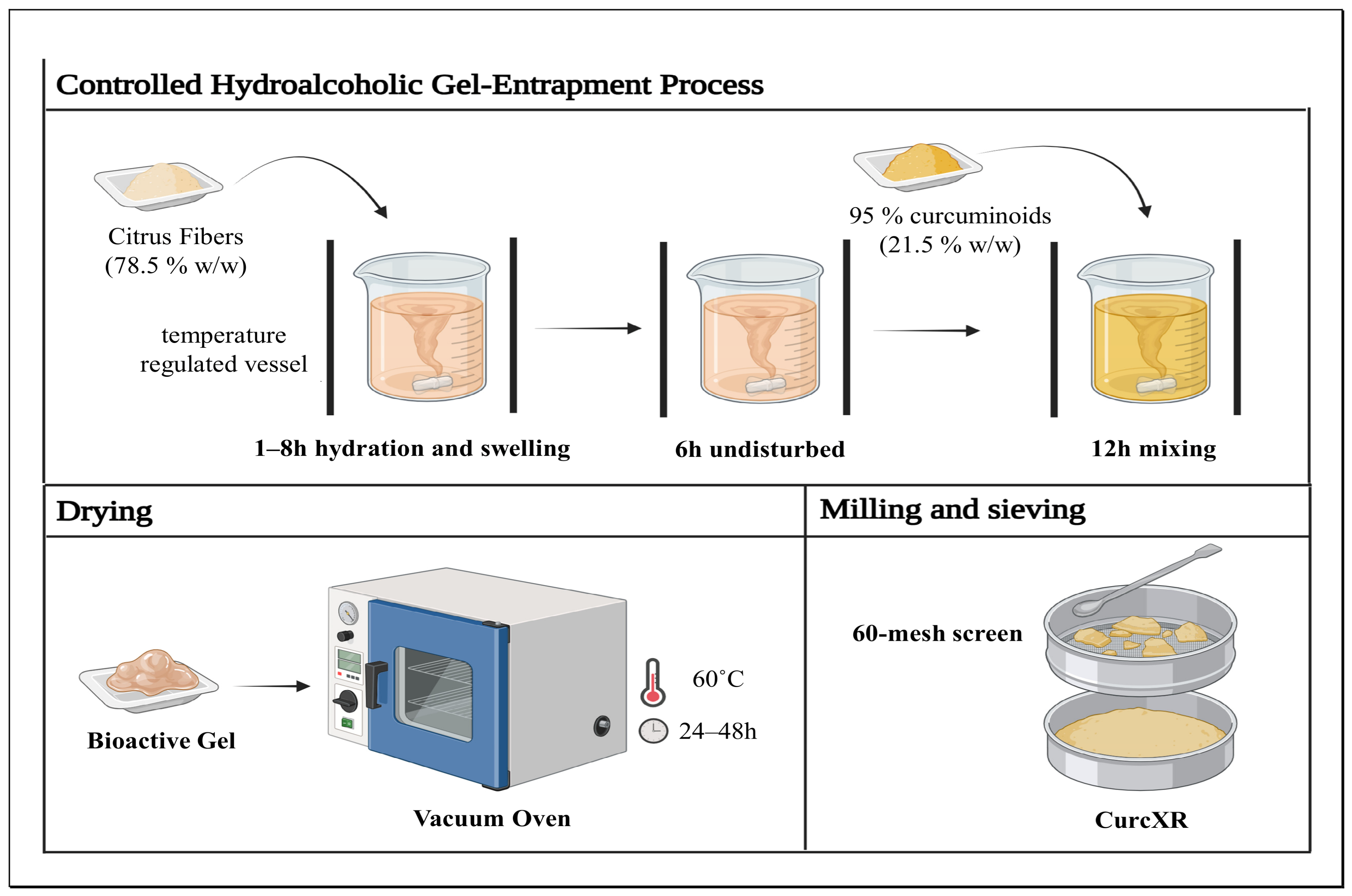
Figure 1. Preparation process of CurcXR using 95% standardized Cmn and citrus fibers.
Jpbi 03 00012 g001
Figure 2. Calibration curve for bioanalysis of Cmn in plasma sample.
Jpbi 03 00012 g002
Figure 3. The plasma concentration data of the prepared formulations i.e., Cmn = Curcuminoids 95%, C20 = CurcXR, up to 12 h of administration.
Jpbi 03 00012 g003
Table 1. Details about the formulation and preparation of the suspension.
FormulationConcentration of 95% CurcuminoidsDose (mg/kg)
Cmn100%1000
CurcXR20%1000
Table 2. Linearity data of the developed bioanalytical method for the determination of Cmn.
S. No.Concentration (µg/mL)Average Peak Area ± SD
1.00.00 ± 0.00
2.0.016147.50 ± 461.83
3.0.148,408.00 ± 888.59
4.0.5213,163.34 ± 619.74
5.1431,780.84 ± 1982.93
6.52,295,662.34 ± 9736.25
7.104,398,711.67 ± 15,451.82
Table 3. Pharmacokinetic parameters of CurcXR versus curcuminoids 95% (Cmn).
FormulationCmax (µg/mL)Tmax (h)T1/2 (h)AUC0−t (µg/mL·h)Comparative AUC0−t (µg/mL·h)Comparative Bioavailability
Cmn0.21 ± 0.052.00 ± 0.005.14 ± 1.060.73 ± 0.110.161
CurcXR0.95 ± 0.104.00 ± 2.025.54 ± 2.198.84 ± 1.678.8457.52
Disclaimer/Publisher’s Note: The statements, opinions and data contained in all publications are solely those of the individual author(s) and contributor(s) and not of MDPI and/or the editor(s). MDPI and/or the editor(s) disclaim responsibility for any injury to people or property resulting from any ideas, methods, instructions or products referred to in the content.