
Article

Oral Microbiota Shifts Associated with Tartar Removal in Dogs

1
Department of Veterinary Biomedical Sciences, Faculté de Médecine Vétérinaire, Université de Montréal, Saint-Hyacinthe, QC J2S 2M2, Canada
2
Biology Department, Faculty of Science and Technology, Vancouver Island University, Nanaimo, BC V9R 5S5, Canada
*
Author to whom correspondence should be addressed.
Submission received: 16 March 2026 / Revised: 27 April 2026 / Accepted: 7 May 2026 / Published: 12 May 2026

Abstract

Periodontal disease is one of the most common oral conditions in dogs. The oral microbiome plays a key role in maintaining oral health, yet the normal canine oral microbiota and the effects of dental cleaning remain understudied. This study investigated changes in the oral microbiota of healthy dogs after routine tartar removal. Fourteen healthy adult Beagles underwent dental cleaning under general anesthesia. Oral microbiota was sampled using swabs at D-03 (baseline), D0 (after tartar removal), D3, D7, D14, and D28. Microbiota composition was analyzed using 16S rRNA gene sequencing (V4 region). Alpha diversity analysis showed that tartar removal was associated with a significant decrease in richness (Kruskal–Wallis test, p < 0.001) but not with diversity (Shannon). Beta diversity analysis revealed significant differences (PERMANOVA; p < 0.05) across all sampling times compared with baseline. Dental tartar removal temporarily reduced several anaerobic taxa and increased aerotolerant bacteria, with partial recovery toward baseline within two weeks, indicating resilience of the oral microbiota. Porphyromonas dominated the oral microbiota but decreased following dental cleaning with concomitant increases in other bacterial species, notably Neisseria, Moraxella, and Pasteurella. These findings suggest that the canine oral microbiota demonstrates considerable resilience following mechanical disruption by dental cleaning. Future studies should focus on the importance of this microbial restructuring in the pathogenesis and clinical management of canine periodontitis and may inform the development of microbiota-targeted preventive or therapeutic strategies in veterinary dentistry.

1. Introduction

Periodontal disease is one of the most prevalent oral conditions in dogs, affecting approximately up to 18% of the population [1]. It begins with the adherence of salivary glycoproteins to the tooth surface, forming an acquired pellicle that facilitates bacterial attachment and biofilm formation. Over time, dental plaque may mineralize into calculus (tartar), providing a rough substrate that promotes further plaque accumulation [1]. The host’s immune response to this bacterial challenge can lead to gingivitis and progressive periodontal damage [2]. Controlling plaque formation is therefore fundamental for preventing periodontal disease. Multiple factors, including age, breed, diet, and oral hygiene, influence plaque development. Recent evidence highlights that the oral microbiome plays a central role in maintaining oral health and preventing disease [3]. A balanced microbial community supports colonization resistance against pathogens, whereas dysbiosis—characterized by a loss of beneficial taxa and an increase in pathogenic bacteria—has been associated with greater susceptibility to tartar accumulation and periodontal inflammation [4]. Longitudinal data indicate that shifts in microbial composition accompany the transition from oral health to disease [5]. Wallis et al. (2015) demonstrated that changes in the subgingival microbiota over time correlate with the severity of periodontitis in dogs, suggesting that the loss of health-associated bacteria may predispose to plaque accumulation [5].
High-throughput sequencing provides a more comprehensive view of microbial diversity compared with culture-based techniques, as most oral bacteria cannot be cultivated under standard laboratory conditions [6,7]. However, while the human oral microbiome has been extensively investigated [8], few studies have characterized the canine oral microbiota using DNA sequencing [9]. Yet, the microbial alterations associated with plaque formation differ between dogs and humans [10].
Only two studies have used sequencing technologies to evaluate the impact of tartar removal on the oral microbiota of dogs [7,10]. Flancman et al. (2018) [7] examined 30 clinically healthy beagles with no history of periodontal disease, while Holcombe et al. (2014) [10] studied 12 Labrador retrievers with pre-existing mild gingivitis. Both studies demonstrated marked temporal changes in oral microbial composition following dental cleaning, with subsequent recovery to baseline within 5 weeks.
This study aimed to investigate the changes in the oral microbiota of healthy dogs following tartar removal. Results add to current knowledge, providing a stronger foundation for new studies targeting therapeutic manipulation of the canine oral microbiome.

2. Materials and Methods

2.1. Animal Enrollment, Treatment and Sample Collection

This study followed the rules of the Canadian Council on Animal Care. It was approved by the University of Montreal’s Animal Care Committee (Rech-2184). Fourteen physically healthy female adult Beagles housed at the Faculty of Veterinary Medicine teaching colony, Saint-Hyacinthe, Quebec, Canada, were enrolled for a 31-day study. The dogs, aged 1–2 years, were fed a mixture of three commercial balanced adult dry foods (2 parts of Royal Canin Vet. Diet Canine Satiety Support, 1 part of Royal Canin Vet. Diet Canine Weight Control Medium Breed and 0.5 part of Royal Canin Vet. Diet Canine Dental Small Dog). None of the dogs had a history of periodontal disease, other oral or gastrointestinal diseases, or antimicrobial administration in the three preceding months.
All dogs underwent oral health evaluation using standard veterinary dental examination methods performed by an experienced veterinarian. Periodontal status assessments included routine inspection of plaque, gingival condition, and calculus accumulation. In addition, a plaque-disclosing solution was used to visualize and grade plaque levels before and after tartar removal.
At D0, all dogs underwent a manual dental cleaning and polishing procedure under general anesthesia performed by a veterinary dentist. All procedures were performed by the same professional, following a standard protocol that included an ultrasonic step, followed by mechanical tartar removal. To assess oral microbiota, sterile swabs (BD Liquid Amies Elution) were used to collect oral samples on days D-03 (Baseline), D0 (tartar removal), D03, D07, D14 and D28 by brushing the gums, tongue, teeth, and cheeks for 15 s. Swabs were kept at −20 °C until processing.

2.2. Oral Microbiota Analysis

DNA was extracted from the swabs using the DNeasy PowerSoil Pro DNA isolation kit (QIAGEN, Toronto, ON, Canada) according to the manufacturer’s instructions. DNA quantification and quality were assessed by spectrophotometry using the NanoDropTM 1000 (Thermo Fisher Scientific, Waltham, MA, USA). The V4 hypervariable region of the bacterial 16S ribosomal RNA gene was amplified by PCR with the primers 515F (GTGCCAGCMGCCGCGGTAA) and 806R (GGACTACHVGGGTWTCTAAT) in a dual-indexing sequencing strategy: the first PCR consisted of 2 min at 94 °C and 33 cycles of 30 s at 94 °C, 30 s at 58 °C and 30 s at 72 °C with a final 7 min at 72 °C. Illumina adapters were incorporated for 10 min at 95 °C, followed by 15 cycles of 15 s at 95 °C, 30 s at 60 °C, and 60 s at 72 °C, with a final period of 3 min at 72 °C. Sequencing was performed using an Illumina MiSeq IEMFile version 4 platform, using a reagent kit V2 (2 × 250 cycles) at the Genome Quebec Innovation Centre. Bioinformatic analyses were performed using the Mothur software version 1.48.3 [11]. Good quality reads were clustered into operational taxonomic units (OTUs) and combined within the same genus (phylotype approach). Reads were aligned with the SILVA reference database (version 138.2), and taxonomic classification was obtained using the Ribosomal Database Project (RDP). The software RStudio (version 2025.09.1+401) was used to calculate alpha diversity, evaluated using the number of observed species and the Shannon index, and beta diversity, evaluated using the Bray–Curtis index, which represents bacterial composition between samples (based on the abundance of each taxon). Alpha diversity indexes across sampling dates were visualized using boxplots, and community structure similarities were visualized using Principal Coordinates Analysis (PCoA) plots.

2.3. Statistical Analysis

Statistical analyses were performed in RStudio. A Kruskal–Wallis test was used to evaluate the effect of time on alpha diversity indices, and a PERMANOVA with 999 permutations (using the adonis package) to investigate the impact of tartar removal on microbial composition (beta-diversity). When the effect of the variable was significant, post hoc tests were applied, and p-values were corrected with the Benjamini–Hochberg method. Associations between sampling time and the relative abundance of bacterial taxa were evaluated using the multivariable statistical framework implemented in MaAsLin2 (Multivariate Association with Linear Models). This approach fits generalized linear models to identify taxa whose normalized abundances are significantly associated with explanatory variables while accounting for multiple testing. In this analysis, sampling time was included as the primary fixed effect to evaluate temporal changes in bacterial abundance following dental scaling. Relative abundance data were normalized and modelled according to the default MaAsLin2 workflow. The dogs were treated as a random effect (repeated measures) to increase the biological interpretability of the results. Statistical significance was evaluated using the Benjamini–Hochberg for False Discovery Rate (FDR) correction to account for multiple hypothesis testing across taxa. p-values of <0.05 were considered significant.

3. Results

3.1. Animals

Dogs were physically normal at the beginning and throughout the study, with no adverse events observed during general anesthesia and dental cleaning. All dogs presented with gingival and plaque indices equal to or below 2, and the plaque-disclosing solution test was negative for all dogs after tartar removal.

3.2. Oral Microbiota Analysis

One sample collected at D7 failed to amplify during PCR and was therefore not sequenced. A total of 1,986,184 good-quality sequences from 83 samples were used for the final analysis (mean: 23,929 reads per sample; median: 25,264, SD: 6382). A cut-off of 16,631 reads was used for subsampling, resulting in the exclusion of four samples (1 from D3, 2 from D7 and 1 from D14), remaining 79 for analysis.

3.2.1. Alpha Diversity

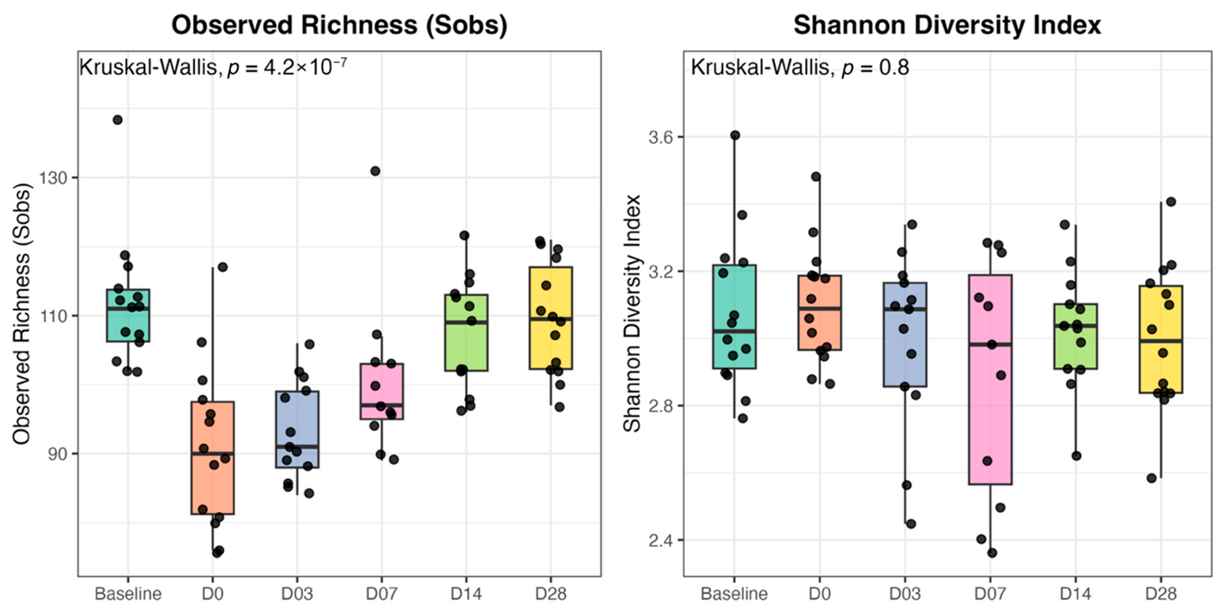
The total number of genera (richness) and the Shannon index (diversity) were used as measurements of alpha diversity. Figure 1 shows the variation in those indicators across different sampling times. Observed Richness (Sobs) refers to the number of different genera, and the Shannon index indicates how even the genera are distributed within a sample. Tartar removal was statistically associated with a decrease in richness (Kruskal–Wallis, p < 0.001), but not with diversity (p = 0.8).

3.2.2. Beta Diversity

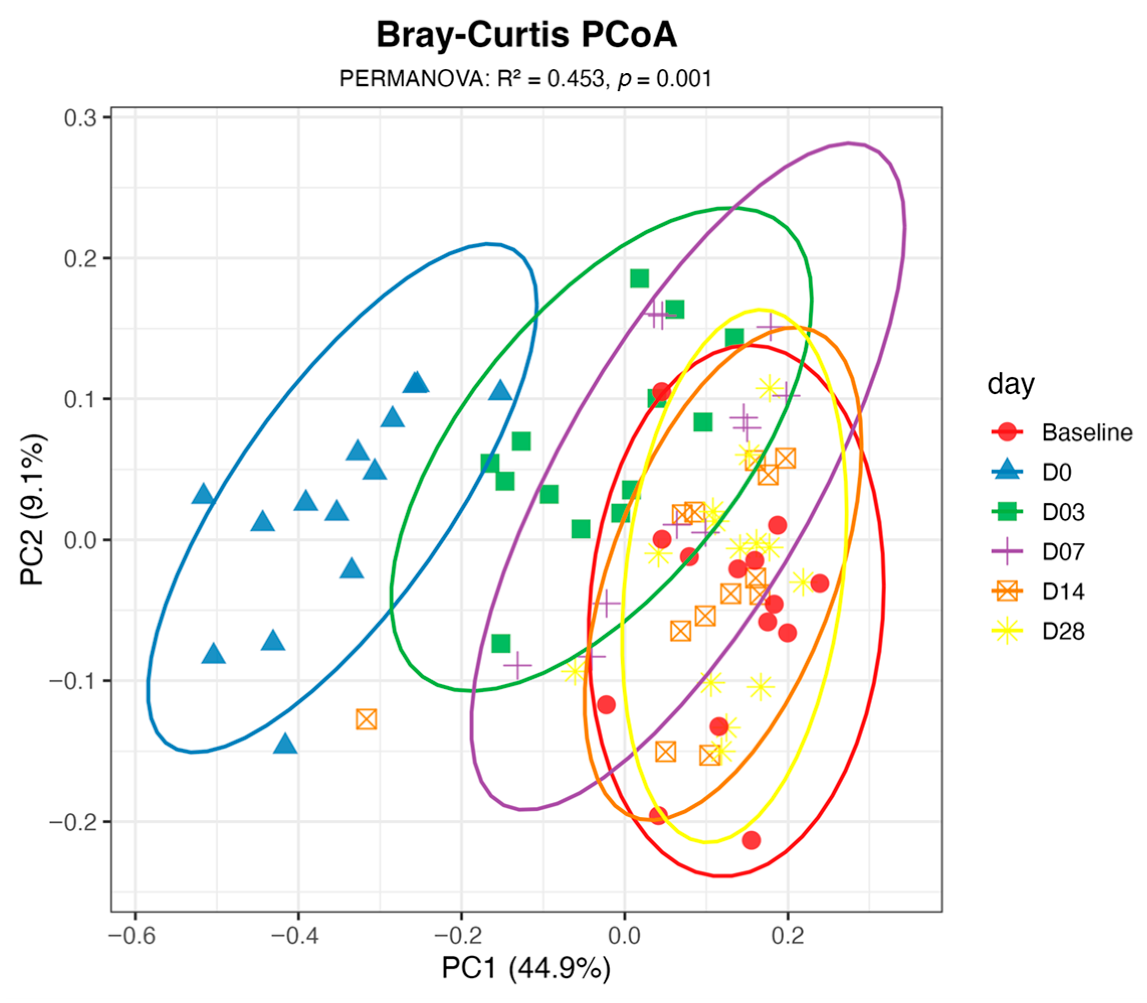
Beta-diversity analysis, which assesses the similarity of taxonomic composition among microbiota in each sample, revealed statistical differences across all sampling times relative to the baseline, as evidenced by pairwise PERMANOVA comparisons (R2 = 0.53, p < 0.001). The data visualization clearly shows a marked change in the composition of dogs’ oral microbiota immediately after tartar removal (Figure 2). Those changes are still evident on days 3 and 7, but samples collected at 14- and 28-day post-procedure overlap with baseline samples, suggesting that the oral microbiota underwent substantial disruption immediately following dental scaling and anesthesia, followed by progressive recovery toward a baseline-like composition within two weeks.

3.2.3. Relative Abundance

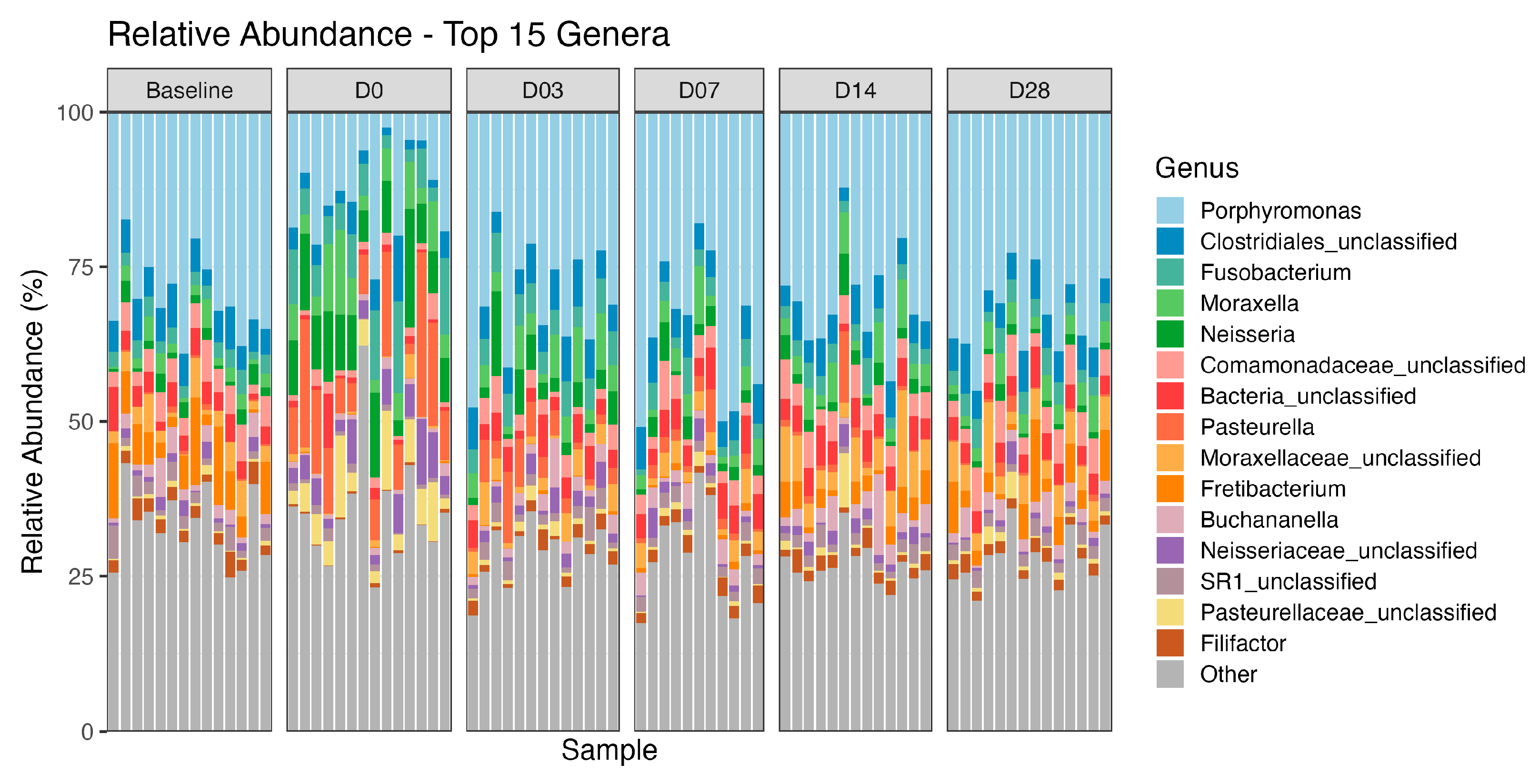
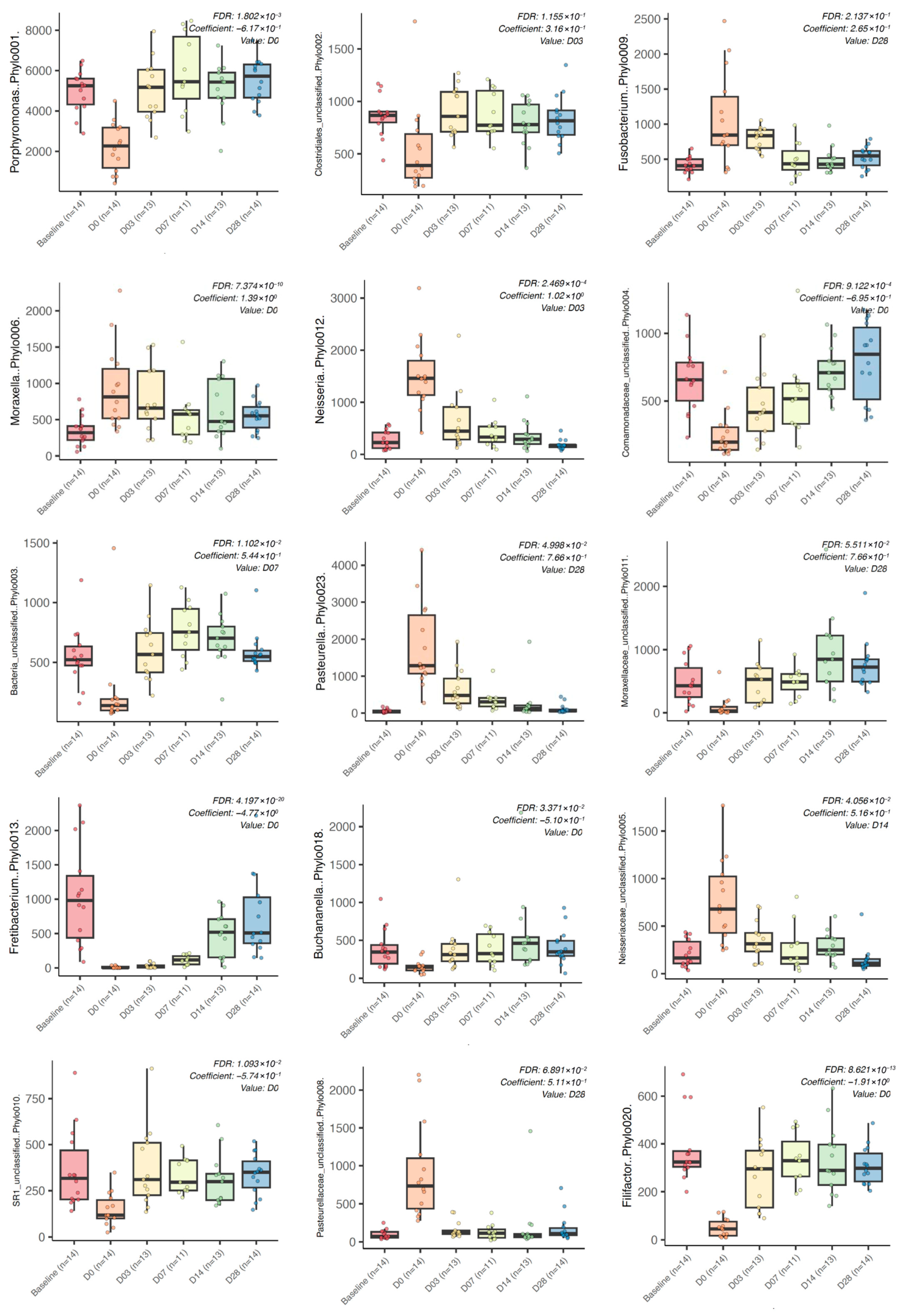
The composition of the oral microbiota of the studied dogs, as reflected by the 15 most abundant bacterial taxa, is shown in Figure 3. Porphyromonas dominated the oral environment, but its abundance was markedly decreased by the dental cleaning.
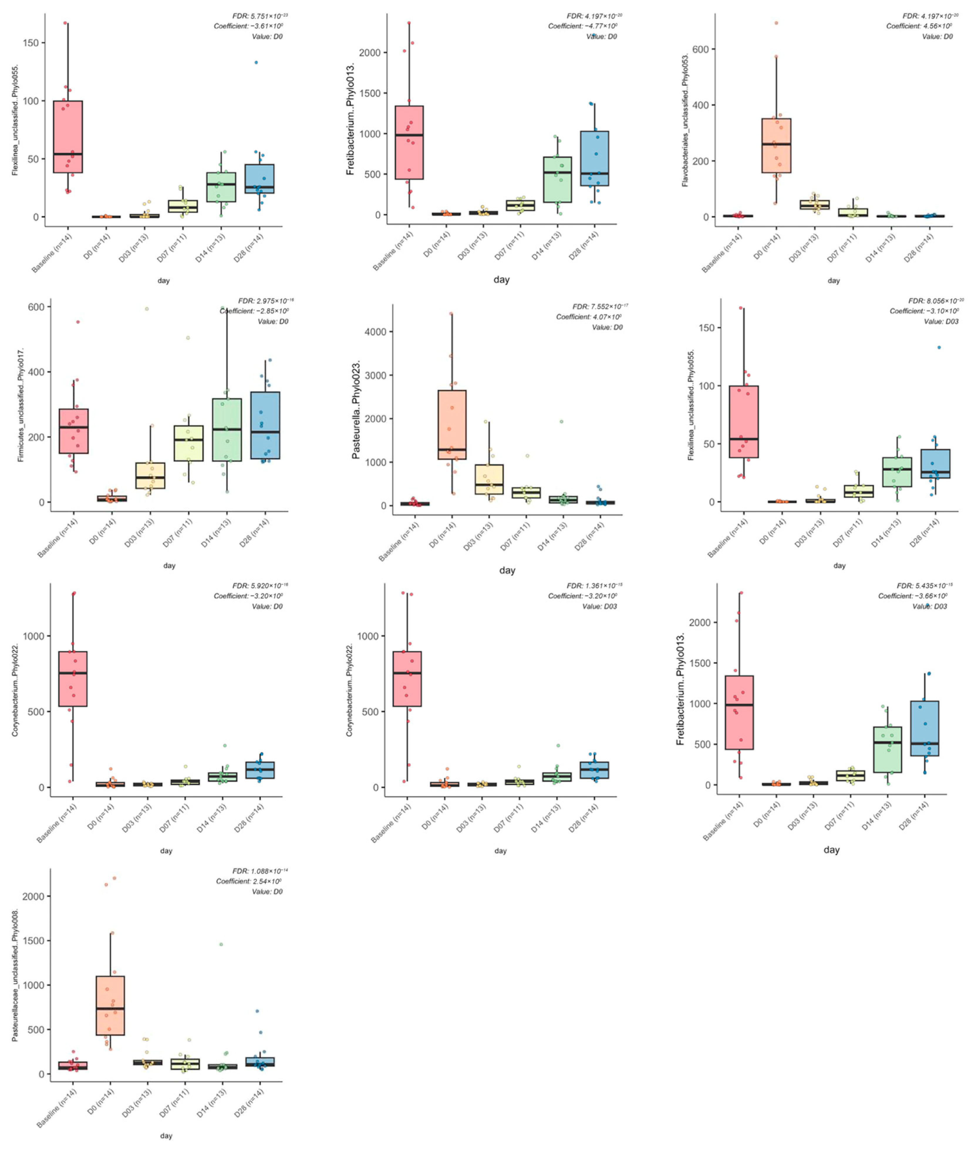
The Maaslim2 mixed-effects model was used to identify differences in the relative abundances of bacterial taxa across sampling times. Together, the coefficients and FDR values provide an estimate of how strongly each taxon’s abundance changes over time and whether those changes remain significant after correcting for multiple comparisons. The analysis identified 209 taxa that changed significantly at D0 relative to baseline. Figure 4 shows the taxa of greatest significance among the dogs studied. The strongest and most statistically robust alterations, ranked by statistical significance and effect size, included decreases in unclassified Flexilinea, Fretibacterium, unclassified Firmicutes, Corynebacterium, Desulfomicrobium, unclassified Ruminococcaceae, Filifactor, Desulfomicrobium, unclassified Bacteroidetes and Helcococcus, as well as increases in unclassified Flavobacteriales, Pasteurella, Frederiksenia, unclassified Pasteurellaceae, Gemella and Neisseria.
Figure 5 shows the 15 most abundant taxa across all samples, highlighting the dominant community members contributing to the temporal dynamics of the canine oral microbiota following tartar removal. Although not all comparisons were statistically significant (FDR < 0.05), the differences observed at day 0 are evident across all dominant taxa.

4. Discussion

The present study investigated the impact of professional dental cleaning under general anesthesia on the composition of the canine oral microbiota, assessed longitudinally from a pre-procedural baseline through 28 days of recovery. Taken together, the pattern emerging from alpha and beta diversity and differential abundance analyses indicates that dental decay is associated with acute and profound changes in the canine oral microbiota, characterized by the loss of core anaerobic community members and a transient bloom of aerotolerant bacteria, followed by microbiota recovery broadly resembling the baseline state within two weeks.
Only two studies have used similar technologies (i.e., DNA sequencing) to investigate the oral microbiota of dogs after tartar removal [7,10]. Flancman et al. (2018) [7] used 30 healthy Beagle dogs from a research colony to collect plaque and oral samples immediately before and one week after tartar removal. In addition, 10 of the dogs were sampled one month later [7]. Holcombe et al. (2014) characterized the microbiota of supragingival plaque from buccal tooth surfaces in 12 dogs with pre-existing mild gingivitis at 24- and 48-h following tartar removal [10].
In the present study, the beta diversity analysis revealed that the microbial composition of samples collected immediately following tartar removal was significantly different from that at all other time points. Interestingly, the inclusion of multiple sampling points after the procedure enabled accurate visualization of the recovery dynamics of the oral microbiota. Samples collected on the day of the procedure (D0) were the most distant from baseline. Then, the microbiota started to recover on days 3 and 7, finally clustering with baseline samples on days 14 and 28. Despite the microbial similarity observed after 14 days, the statistical analysis remained significantly different even at day 28, indicating either permanent changes or that more time is needed for the full recovery of microbiota composition. Similarly, Flancman et al. (2018) [7] observed a rapid transition back toward the pre-treatment microbiota composition by five weeks post-procedure. Both the plaque and oral microbiota returned to states similar to pre-treatment within this timeframe, with no persistent changes or evidence of altered equilibrium states beyond the initial one-week disruption [7]. This resilience pattern agrees with our findings, suggesting that while mechanical tartar removal profoundly disrupts the oral microbial ecosystem, the disruption is temporary in clinically healthy animals. The cause of those marked compositional changes in the oral microbiota of dogs immediately after tartar removal warrants further investigation, but it may be associated with exposure to antiseptic solutions, transient alterations in local oxygen tension, and local inflammation caused by gingival mechanical abrasion.
Regarding alpha diversity, dental decay reduced richness, which recovered to baseline levels after 14 days, but there was no difference in the Shannon index, indicating no change in diversity. Similarly to our study, Holcombe et al. (2014) reported that the overall Shannon index showed no significant difference between the two time points, indicating that, despite compositional changes associated with dental cleaning, the distribution (evenness) of bacteria remained stable during early biofilm establishment [10]. In contrast, Flancman et al. (2018) documented significant decreases in diversity and richness at one week post-prophylaxis [7]. This was mainly due to the dominance of opportunistic genera, such as Pseudomonas (up to 80%), leading to reduced evenness. Nevertheless, those indices increased back to baseline after 5 weeks [7].
Porphyromonas was found to be the most abundant bacterium in the dogs studied, a finding supported by several studies using similar methodologies [6,7,12,13], indicating that it is a native member fully adapted to the oral environment. The marked decrease in Porphyromonas abundance might be associated with increases in other bacterial species, notably Neisseria, Moraxella, and Pasteurella. Interestingly, Holcombe et al. (2014) found that the dominant taxa during this early recolonization period were Bergeyella zoohelcum (16.97%), Neisseria shayeganii (10.41%), and Moraxella spp. (9.31%) [10]. In addition, several other significant variations in bacterial abundances were reported over the 24-h interval between the two sampling time points (73 decreased, while 46 increased) [10]. Similarly, Flancman et al. (2018) documented profound shifts in both plaque and oral microbiota composition one week after dental prophylaxis [7]. The most dramatic change in oral swabs occurred in Psychrobacter, which completely disappeared one-week post-treatment, likely due to an increase in Pseudomonas as the dominant taxon, reaching 80% relative abundance [7]. In addition, the significant decrease in Treponema and increases in Moraxella and Neisseria in dental plaques were consistent with the Neisseria increases observed in the present study and by Holcombe et al. (2014) at 48 h [10].
Among the taxa with the strongest and most statistically robust alterations, Fretibacterium showed the largest decrease after tartar removal. Fretibacterium is a Gram-negative anaerobe belonging to the Synergistetes phylum and is consistently identified as a core constituent of the healthy oral microbiota of several species, where it participates in syntrophic amino acid catabolism [14,15]. Its marked depletion immediately after tartar removal is likely attributable to mechanical disruption of the subgingival biofilm during scaling, combined with exposure to antiseptic solutions and transient alterations in local oxygen tension. The subsequent recovery of Fretibacterium to near-baseline levels by D07 suggests the anaerobic niche is resilient once the gingival sulcus re-establishes its protective microenvironment. Members of the order Flavobacteriales are aerotolerant or strictly aerobic and Bacteroidetes are commonly found in mucosal surfaces exposed to oxygen [16,17]. Their expansion immediately post-scaling may reflect opportunistic colonization of the freshly cleaned, temporarily more aerobic tooth surface and exposed gingival tissue, before the re-establishment of the mature anaerobic biofilm. The decrease in the relative abundance of Flexilinea was the most statistically significant in the dataset. This bacterium belongs to the phylum Chloroflexota and represents a strictly anaerobic, filamentous organism [18]. Its prolonged depression relative to other anaerobes may indicate that it occupies a more structurally integrated niche within the mature biofilm scaffold, requiring a longer period to re-establish itself following complete biofilm removal. Its role in the canine oral environment remains to be elucidated.
The present study evaluated a population of clinically healthy dogs from a standardized population (i.e., teaching Beagles), which may limit extrapolation to a broader dog population. The study did not aim to draw major conclusions about the impact of the oral microbiota composition on the occurrence of oral diseases. Future clinical studies could substantially improve the understanding of canine periodontitis by combining longitudinal sampling with standardized dental examinations. The identification of microbial biomarkers associated with early-stage disease and treatment response would have the greatest clinical relevance for the field by enabling the development of diagnostic tests that could enable more personalized intervention. Previous cross-sectional work has shown that dogs with periodontal disease exhibit microbiota profiles distinct from those of clinically healthy dogs, supporting the value of more rigorous prospective clinical studies [19].
Importantly, the current study has limitations that warrant mention. The lack of absolute quantification inherent to DNA sequencing can be misinterpreted by most clinicians unfamiliar with microbiota studies. For example, the decrease in the relative abundance of Porphyromonas after dental cleaning might not reflect an absolute decrease but rather the proliferation of other bacteria, thereby decreasing the proportion of Porphyromonas spp. relative to the other bacteria. In addition, differences in methodologies across studies might preclude comparisons of results, especially at lower taxonomic levels. Those differences include different sequencing technologies, the use of older and different databases and protocols used for bioinformatic analyses, which explains the divergent dominant taxa. Finally, no time-control group of dogs undergoing repeated sampling without dental scaling was included, which limits the ability to fully exclude uncontrolled temporal variation or changes caused by general anesthesia. However, such effects are expected to be transient and less marked, and, along with microbiota changes consistent with previous studies, indicate that the observed changes were in fact caused by professional dental cleaning. Future studies incorporating functional approaches such as metabolomics would help clarify the biological consequences of these compositional shifts. In addition, dedicated longitudinal clinical studies comparing different prophylactic schedules could establish evidence-based recommendations for treatment frequency.
Despite those limitations, these results lay the groundwork for further investigation into its potential clinical implications. The transient depletion of putative periodontal pathogens, such as Fretibacterium and Desulfomicrobium, suggests that the window immediately following dental cleaning may represent an opportunity for adjunct therapeutic intervention, such as probiotic administration or oral antiseptic application, to delay the re-establishment of a dysbiotic biofilm. Future studies investigating clinical outcomes and associations with dental diseases are necessary.

5. Conclusions

The dental tartar removal procedure is associated with rapid and substantial perturbations of the oral microbial community of dogs. These changes are characterized by a temporary reduction in core anaerobic taxa and a concurrent expansion of aerotolerant bacteria, followed by a progressive recovery of the microbiota toward a composition broadly resembling the baseline state within two weeks.
These findings suggest that the canine oral microbiota demonstrates considerable resilience following mechanical disruption by dental cleaning. Future studies should focus on the importance of this microbial restructuring in the pathogenesis and clinical management of canine periodontitis and may inform the development of microbiota-targeted preventive or therapeutic strategies in veterinary dentistry.

Author Contributions

Conceptualization, M.C.C. and D.F.; methodology, S.A. and L.F.-G.; formal analysis, M.C.C., L.F.-G. and M.R.; writing—original draft preparation, L.F.-G. and M.R.; writing—review and editing, M.R., M.C.C. and D.F.; supervision, M.C.C. and D.F. All authors have read and agreed to the published version of the manuscript.

Funding

This research was funded by the Natural Sciences and Engineering Research Council of Canada (NSERC) through a Discovery Grant (#RGPIN/4514-2018).

Institutional Review Board Statement

The study was conducted in accordance with the rules of the Canadian Council on Animal Care. It was approved by the University of Montreal’s Animal Care Committee (Rech-2184) on 8 April 2022.

Informed Consent Statement

The adult Beagles involved in this study belong to the Faculty of Veterinary Medicine teaching colony, Saint-Hyacinthe, Quebec, Canada. There is no need for the dog's owner informed consent form.

Data Availability Statement

The original contributions presented in this study are included in the article. Further inquiries can be directed to the corresponding author.

Acknowledgments

We would like to thank Robert Chidiak for his help with dog handling and sample collection.

Conflicts of Interest

The authors declare no conflicts of interest.

Abbreviations

The following abbreviations are used in this manuscript:
SDStandard deviation
OTUOperational taxonomic unit
PCoAPrincipal Coordinate Analysis
FDRFalse Discovery Rate
SobsObserved Richness

References

  1. Wallis, C.; Holcombe, L.J. A review of the frequency and impact of periodontal disease in dogs. J. Small Anim. Pract. 2020, 61, 529–540. [Google Scholar] [CrossRef] [PubMed]
  2. Niemiec, B.A.; Gawor, J.; Tang, S.; Prem, A.; Krumbeck, J.A. The bacteriome of the oral cavity in healthy dogs and dogs with periodontal disease. Am. J. Vet. Res. 2022, 83, 50–58. [Google Scholar] [CrossRef] [PubMed]
  3. Pleskova, S.; Bezrukov, N. The Oral Microbiota, Its Evolution, and Aspects of Support for Oral Health. Mol. Oral Microbiol. 2026, 41, 1–25. [Google Scholar] [CrossRef] [PubMed]
  4. Alavi, S.E.; Ebrahimi Shahmabadi, H.; Sharma, L.A.; Sharma, A. The Role of the Oral Microbiome in Periodontal Disease: A Systematic Review of Microbial Associations and Therapeutic Implications. Curr. Microbiol. 2026, 83, 64. [Google Scholar] [CrossRef] [PubMed]
  5. Wallis, C.; Marshall, M.; Colyer, A.; O’Flynn, C.; Deusch, O.; Harris, S. A longitudinal assessment of changes in bacterial community composition associated with the development of periodontal disease in dogs. Vet. Microbiol. 2015, 181, 271–282. [Google Scholar] [CrossRef] [PubMed]
  6. Sturgeon, A.; Stull, J.W.; Costa, M.C.; Weese, J.S. Metagenomic analysis of the canine oral cavity as revealed by high-throughput pyrosequencing of the 16S rRNA gene. Vet. Microbiol. 2013, 162, 891–898. [Google Scholar] [CrossRef] [PubMed]
  7. Flancman, R.; Singh, A.; Weese, J.S. Evaluation of the impact of dental prophylaxis on the oral microbiota of dogs. PLoS ONE 2018, 13, e0199676. [Google Scholar] [CrossRef] [PubMed]
  8. Zhang, Y.; Wang, X.; Li, H.; Ni, C.; Du, Z.; Yan, F. Human oral microbiota and its modulation for oral health. Biomed. Pharmacother. 2018, 99, 883–893. [Google Scholar] [CrossRef] [PubMed]
  9. Davis, E.M.; Weese, J.S. Oral microbiome in dogs and cats: Dysbiosis and the utility of antimicrobial therapy in the treatment of periodontal disease. Vet. Clin. Small Anim. Pract. 2022, 52, 107–119. [Google Scholar] [CrossRef] [PubMed]
  10. Holcombe, L.J.; Patel, N.; Colyer, A.; Deusch, O.; O’Flynn, C.; Harris, S. Early canine plaque biofilms: Characterization of key bacterial interactions involved in initial colonization of enamel. PLoS ONE 2014, 9, e113744. [Google Scholar] [CrossRef] [PubMed]
  11. Schloss, P.D.; Westcott, S.L.; Ryabin, T.; Hall, J.R.; Hartmann, M.; Hollister, E.B.; Weber, C.F. Introducing mothur: Open-source, platform-independent, community-supported software for describing and comparing microbial communities. Appl. Environ. Microbiol. 2009, 75, 7537–7541. [Google Scholar] [CrossRef] [PubMed]
  12. Oh, C.; Lee, K.; Cheong, Y.; Lee, S.W.; Park, S.Y.; Song, C.S.; Lee, J.B. Comparison of the oral microbiomes of canines and their owners using next-generation sequencing. PLoS ONE 2015, 10, e0131468. [Google Scholar] [CrossRef]
  13. Elliott, D.R.; Wilson, M.; Buckley, C.M.; Spratt, D.A. Cultivable oral microbiota of domestic dogs. J. Clin. Microbiol. 2005, 43, 5470–5476. [Google Scholar] [CrossRef] [PubMed]
  14. Oba, P.M.; Swanson, O.R.; Pavlovsky, G.; Dupleix, C.R.; Sharping, S.C.; Swanson, K.S. Oral health indices and microbiota populations of adult cats consuming wet or dry diets. Front. Vet. Sci. 2025, 12, 1678016. [Google Scholar] [CrossRef] [PubMed]
  15. Liang, L.; Liu, P.; Song, D. Fretibacterium: Exploring Its Pathogenic Potential in Oral Infectious Diseases. Mol. Oral Microbiol. 2025, 40, 235–242. [Google Scholar] [CrossRef]
  16. Bernardet, J.F. Flavobacteriales ord. nov. In Bergey’s Manual of Systematics of Archaea and Bacteria; John Wiley & Sons: Hoboken, NJ, USA, 2015; pp. 1–2. [Google Scholar] [CrossRef]
  17. Pudlo, N.A.; Urs, K.; Crawford, R.; Pirani, A.; Atherly, T.; Jimenez, R.; Martens, E.C. Phenotypic and genomic diversification in complex carbohydrate-degrading human gut bacteria. MSystems 2022, 7, e00947-21. [Google Scholar] [CrossRef] [PubMed]
  18. Sun, L.; Toyonaga, M.; Ohashi, A.; Matsuura, N.; Tourlousse, D.M.; Meng, X.Y.; Sekiguchi, Y. Isolation and characterization of Flexilinea flocculi gen. nov., sp. nov., a filamentous, anaerobic bacterium belonging to the class Anaerolineae in the phylum Chloroflexi. Int. J. Syst. Evol. Microbiol. 2016, 66, 988–996. [Google Scholar] [CrossRef] [PubMed]
  19. Santibáñez, R.; Rodríguez-Salas, C.; Flores-Yáñez, C.; Garrido, D.; Thomson, P. Assessment of Changes in the Oral Microbiome That Occur in Dogs with Periodontal Disease. Vet. Sci. 2021, 8, 291. [Google Scholar] [CrossRef] [PubMed]
Figure 1. Boxplots indicating alpha diversity indices of the oral microbiota of dogs before (Baseline, n = 14), on the day of tartar removal (D0, n = 14), and at 3 (n = 13), 7 (n = 11), 14 (n = 13), and 28 (n = 14) days after the procedure. Bars depict the mean and standard deviation, and dots represent the data.
Pets 03 00020 g001
Figure 2. Principal Coordinate Analysis (PCoA) representing the similarity of the oral microbiota of dogs before (Baseline, n = 14), on the day of tartar removal (D0, n = 14), and at 3 (n = 13), 7 (n = 11), 14 (n = 13), and 28 (n = 14) days after the procedure. The numbers in brackets indicate that the Principal Coordinates 1 and 2 (PC1 and PC2, respectively) explained 55% of the variation in the data.
Pets 03 00020 g002
Figure 3. Relative abundance of the 15 most abundant bacterial taxa representing the canine oral microbiota. Samples were collected 3 days before tartar removal (Baseline, n = 14), on the day of the procedure (D0, n = 14), and at 3 (n = 13), 7 (n = 11), 14 (n = 13), and 28 (n = 14) days later.
Pets 03 00020 g003
Figure 4. Longitudinal variation in the 10 taxa most strongly associated with dental tartar removal across sampling times, assessed using the MaAsLin2 model. Each point in the figure represents the estimated model coefficient for the association between a given taxon and a sampling time relative to the reference time point (Baseline). The coefficient indicates both the direction and magnitude of the association: positive coefficients reflect an increase in the taxon’s abundance compared to the baseline, whereas negative coefficients indicate a decrease. Samples were collected 3 days before tartar removal (Baseline), on the day of the procedure (D0), and at 3, 7, 14, and 28 days later.
Pets 03 00020 g004
Figure 5. Results of the MaAsLin2 model illustrating the variation in the 15 most abundant taxa found in the oral cavity of dogs 3 days before tartar removal (Baseline), on the day of the procedure (D0), and at 3, 7, 14, and 28 days later. Each point in the figure represents the estimated model coefficient for the association between a given taxon and a sampling time relative to the reference time point (Baseline). The coefficient indicates both the direction and magnitude of the association: positive coefficients reflect an increase in the relative abundance of the taxon compared with baseline, whereas negative coefficients indicate a decrease.
Pets 03 00020 g005
Disclaimer/Publisher’s Note: The statements, opinions and data contained in all publications are solely those of the individual author(s) and contributor(s) and not of MDPI and/or the editor(s). MDPI and/or the editor(s) disclaim responsibility for any injury to people or property resulting from any ideas, methods, instructions or products referred to in the content.