Article

Molecular Epidemiology of mcr (1–5) and Other Critical Resistance Genes in Bacteria Isolated from Canine Otitis Externa in Ankara, Türkiye

1
Department of Pharmacology and Toxicology, Graduate School of Health Sciences, Ankara University, 06070 Ankara, Türkiye
2
Department of Pharmacology and Toxicology, Faculty of Veterinary Medicine, Ankara University, 06070 Ankara, Türkiye
*
Author to whom correspondence should be addressed.
Acta Microbiol. Hell. 2026, 71(2), 14; https://doi.org/10.3390/amh71020014
Submission received: 24 March 2026 / Revised: 12 May 2026 / Accepted: 15 May 2026 / Published: 16 May 2026

Abstract

Antimicrobial resistance in companion animals is a growing public health concern, yet data on last-resort resistance genes in clinical canine isolates remain scarce. This study characterizes the molecular distribution of critical resistance determinants, including mcr variants (1–5), in bacteria isolated from canine otitis externa in Ankara, Turkey. Using a combination of phenotypic disk diffusion and targeted quantitative polymerase chain reaction (qPCR), we identified Enterobacter spp., Staphylococcus spp., and Pseudomonas aeruginosa as the predominant pathogens. Notably, among Gram-negative isolates (n = 9), our results indicate preliminary evidence of mobile colistin resistance genes, particularly mcr-3 in 44.4% (4/9), and mcr-4 and mcr-5 each in 33.3% (3/9), marking the first report of these variants in canine ear infections within an urban environment. The observed notable discrepancies between genotypic carriage and phenotypic expression suggest the possible presence of silent resistance reservoirs that traditional diagnostics may overlook. These findings underscore the urgent need for molecular-integrated surveillance in veterinary clinical practice to prevent the zoonotic spread of last-resort resistance genes and to safeguard both animal and public health under the ‘One Health’ framework.

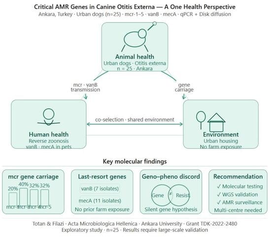
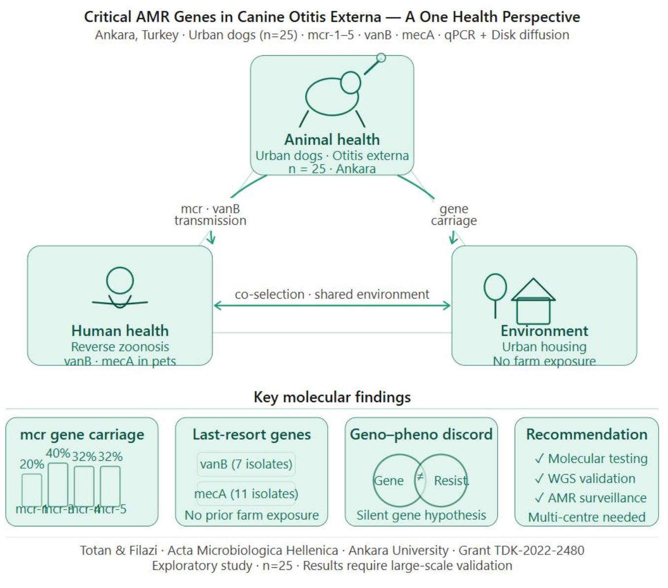
Graphical Abstract

1. Introduction

Dogs commonly develop otitis externa, often requiring antibiotic treatment due to the underlying bacterial infections associated with them. This specific disease model is very useful in any research study relating to the emergence and development of antimicrobial resistance (AMR) due to the high occurrence of otitis externa and the regularity of treatment. Certain bacterial pathogens that can be considered primary in canine otitis externa cases include Escherichia coli, Pseudomonas aeruginosa, and Staphylococcus pseudointermedius [1]. Even the variability of the frequency of bacterial genera cultured from cases of infection has been shown in various studies in different parts of the world. Variations in bacterial etiology and resistance profiles are primarily shaped by regional and local antimicrobial usage patterns [2,3,4,5].
Whereas P. aeruginosa is notoriously resistant to many classes of antibiotics including penicillins, β-lactams, cephalosporins, and trimethoprim-sulfamethoxazole, S. pseudointermedius generally shows moderate resistance to fluoroquinolones and gentamicin [3]. Together with the variability of bacterial resistance, these variations in AMR make treatment even more difficult and emphasize the need for cautious antibiotic use in veterinary medicine.
Companion animals, particularly dogs, are increasingly recognized as significant potential reservoirs for AMR. This risk is likely exacerbated by their close physical proximity to humans and the frequent administration of broad-spectrum antibiotics in veterinary practice [1,3,6]. However, due to incomplete recording of clinical cases, data on profiles of AMR in animals is rather scarce at the moment. In this context, since otitis externa is quite common in dogs and its treatment necessitates the use of antibiotics for bacterial infections, it becomes a very important condition through which resistance can be studied [4,7].
The most important among them is polymyxin resistance, and more specifically colistin resistance, because it is frequently the antimicrobial of last resort in human clinical practice against multidrug-resistant (MDR) bacteria. However, polymyxins are routinely employed in veterinary dermatology [8]. Specifically, they are common ingredients in topical ear drops used for managing canine otitis externa. This widespread application in pets creates a potential reservoir for resistance that directly threatens public health. Colistin resistance is plasmid-borne mcr gene-mediated; mcr-1 was originally detected in China in 2015 and has since been found globally in human, animal, and food isolates [9]. A total of ten mcr types (mcr-1 to mcr-10) have been identified [10,11]. The rapid global spread of these resistance genes from animals to humans, food, and the environment highlights the zoonotic risk, particularly as animals are estimated to play a crucial role in the transmission of these mobile genes due to shared environments [12]. Resistance to other last-resort antibiotics like vancomycin and imipenem (e.g., porin loss, efflux pumps) has also been observed [13]. The extensive use of antibiotics in animal treatment may facilitate the dissemination of resistance genes, posing a potential challenge to public health. Dispersion of resistant bacteria is more likely between human beings and animals sharing the same kind of environment [10]. Recent evidence has highlighted the emergence of last-resort resistance genes in urban settings, such as the identification of a transferable mcr-1 gene in a healthy dog in South Korea [14].
Since regional and environmental characteristics shape patterns of pathogens and resistance, local data generation is crucial for effective treatment regimen design and security of public health. The purpose of this study is therefore to determine the bacterial etiology of otitis externa in dogs, their pattern of resistance and resistance gene pattern to provide an evaluation of the risk of transmission to human health. The study was conducted within the “One Health” framework. This article presents in detail the techniques employed in the analysis of canine otitis externa, from collection of the sample to microbiological examination, AMR typing, and genetic resistance profiling.

2. Materials and Methods

2.1. Sample Collection

Ear swabs were collected from 25 dogs with otitis externa (50 samples) at clinics in Ankara, Turkey. Data on age, sex, breed, and weight were recorded with owner consent. Two gel-based and one standard swab were taken per dog; gels supported anaerobic growth. Genetic samples were stored in RNALater at −80 °C (DiaRex RL-0342, Diagen, Ankara, Türkiye), while bacteriological samples were transported at +4 °C on the same day. Genetic samples remained frozen until batch transfer.

2.2. Bacteriological Examination

Ear swabs were cultured on nutrient agar, 5% sheep blood agar, and MacConkey agar, then incubated at 37 °C for 24 h. Purified colonies were identified using Gram staining, catalase, and oxidase tests. Staphylococcus spp. were characterized as catalase-positive, oxidase-negative spheres, and Streptococcus spp. as negative in both tests. Among Gram-negative rods, Pseudomonas spp. were identified by green pigment on nutrient agar, and E. coli by typical colony characteristics on MacConkey and EMB agars [15]. In cases where mixed bacterial growth was observed or different species were isolated from the bilateral ear swabs of a single dog, the predominant microorganism for that patient was determined based on the highest relative colony density (heaviest growth) observed on the primary culture plates. In cases of polymicrobial growth, the predominant microorganism was defined as the species exhibiting the highest semi-quantitative colony-forming unit (CFU) count on primary culture media. While this selection focused on the primary clinical driver of the infection, we acknowledge the inherent limitation of excluding secondary pathogens that may harbor distinct resistance profiles. The selection of the predominant microorganism was a deliberate methodological choice to establish a standardized baseline for correlating specific genetic determinants with their corresponding phenotypic expressions. This approach focused on the primary clinical driver of the infection to ensure that the detected resistance genes could be more accurately attributed to the pathogen most likely responsible for the clinical presentation. To ensure culture purity, all isolates were subcultured at least twice on selective media prior to DNA extraction. Colony morphology, Gram staining results, and biochemical test outcomes were recorded for each subculture to confirm monoculture status. We acknowledge, however, that in the absence of 16S rRNA gene sequencing-based confirmation, the possibility of low-level contamination cannot be entirely excluded, particularly for the findings observed in polymicrobial samples. This represents a limitation of the current study and underscores the need for WGS-based validation of these results.

2.3. Antibiotic Susceptibility Tests

Bacterial susceptibility to antibiotics was determined by the Kirby-Bauer Disk Diffusion method, as recommended by The Clinical and Laboratory Standards Institute [16]. Forty antibacterial disks (shown in Table 1) were utilized. Agar plates were incubated at 37 °C for 18–24 h, and inhibition zone diameters were measured according to CLSI standards [16,17]. Selective media, including blood agar and mannitol-salt agar for Gram-positive bacteria and MacConkey agar for Gram-negative bacteria, were employed. While intermediate susceptibility results hold certain clinical relevance, this study exclusively reported ‘resistant’ outcomes. The primary rationale for this exclusion was to strictly highlight the isolates posing the highest risk of definitive therapeutic failure in clinical veterinary practice, thereby maintaining a sharp focus on multidrug resistance and the efficacy of ‘last-resort’ antibiotics within the One Health context. Colistin susceptibility testing was initially performed using the disk diffusion method; however, in accordance with current CLSI and EUCAST recommendations, this method is not considered reliable for polymyxins. Therefore, phenotypic colistin susceptibility results were excluded from further analysis and interpretation. Metronidazole was included in the initial panel; however, as all isolates in this study were aerobic or facultative bacteria, the results were excluded from further analysis due to intrinsic resistance.

2.4. Identification of Resistance Genes

Resistance genes were selected according to AMEG (Antimicrobial Advice Ad Hoc Expert Group) classification by the European Medicines Agency [18]. Target genes included ampC (cefepime; restricted to Gram-negative bacteria), mcr-1–5 (colistin; restricted to Gram-negative bacteria), blaIMP (imipenem), gyrA, gyrB (ciprofloxacin), and OXA-1 (amoxicillin-clavulanic acid). The penA gene, encoding a penicillin-binding protein specific to Neisseria spp., was excluded from the panel as it is not applicable to the bacterial species isolated in this study.
DNA was extracted from ear swab samples using the DiaRex® Genomic DNA Ex-traction Kit (Cat. No: SD-0323, Ankara, Turkey) following the manufacturer’s instructions, with minor modifications for swab samples [19]. In brief, 250 µL lysis solution was added to 200 µL of ear swab sample, followed by glass and zircon beads for mechanical disruption using a homogeniser. After homogenization, 25 µL Proteinase K was added and in-cubated at 56 °C for 60 min. Post-incubation, the sample was centrifuged (5000 g, 5 min), and the supernatant transferred. Next, 200 µL lysis buffer was added, incubated at 70 °C for 10 min, followed by the addition of 250 µL ethanol. The lysate was then passed through a column, washed, and DNA was eluted in 100 µL buffer after centrifugation (8000 g, 1 min).
Extracted DNA was analyzed using real-time qPCR (ROCHE LightCycler® 480, Roche Diagnostics GmbH, Mannheim, Germany). Primers were selected based on previous studies [9,10,20,21,22]. The qPCR protocol included an initial denaturation at 95 °C, followed by 40 cycles of denaturation (95 °C), annealing (55 °C), and extension (72 °C). Each gene was tested in separate monoplex reactions, and gene presence was assessed post-amplification (Table 2).
For primer validation, conventional PCR was also performed. Primers were designed using Primer3 software based on NCBI FASTA files. PCR was run in 20 µL reactions containing polymerase, Diagen PCR mix, genomic DNA, and Biomers primers. Cycling conditions included 95 °C for 10 min, followed by 40 cycles (95 °C for 30 s, 55 °C for 1 min, 72 °C for 1 min), and a final extension at 72 °C for 10 min.

2.5. Statistical Analysis

Descriptive statistics were used to summarize the prevalence of bacterial species and resistance genes. For key resistance determinants (mcr variants), 95% confidence intervals (CIs) were calculated using the Wilson score method to estimate the precision of the prevalence rates. All calculations were performed using SPSS software (version 20.0, IBM, Armonk, NY, USA).

3. Results

3.1. Isolates Detected and Resistance Genes of the Isolates

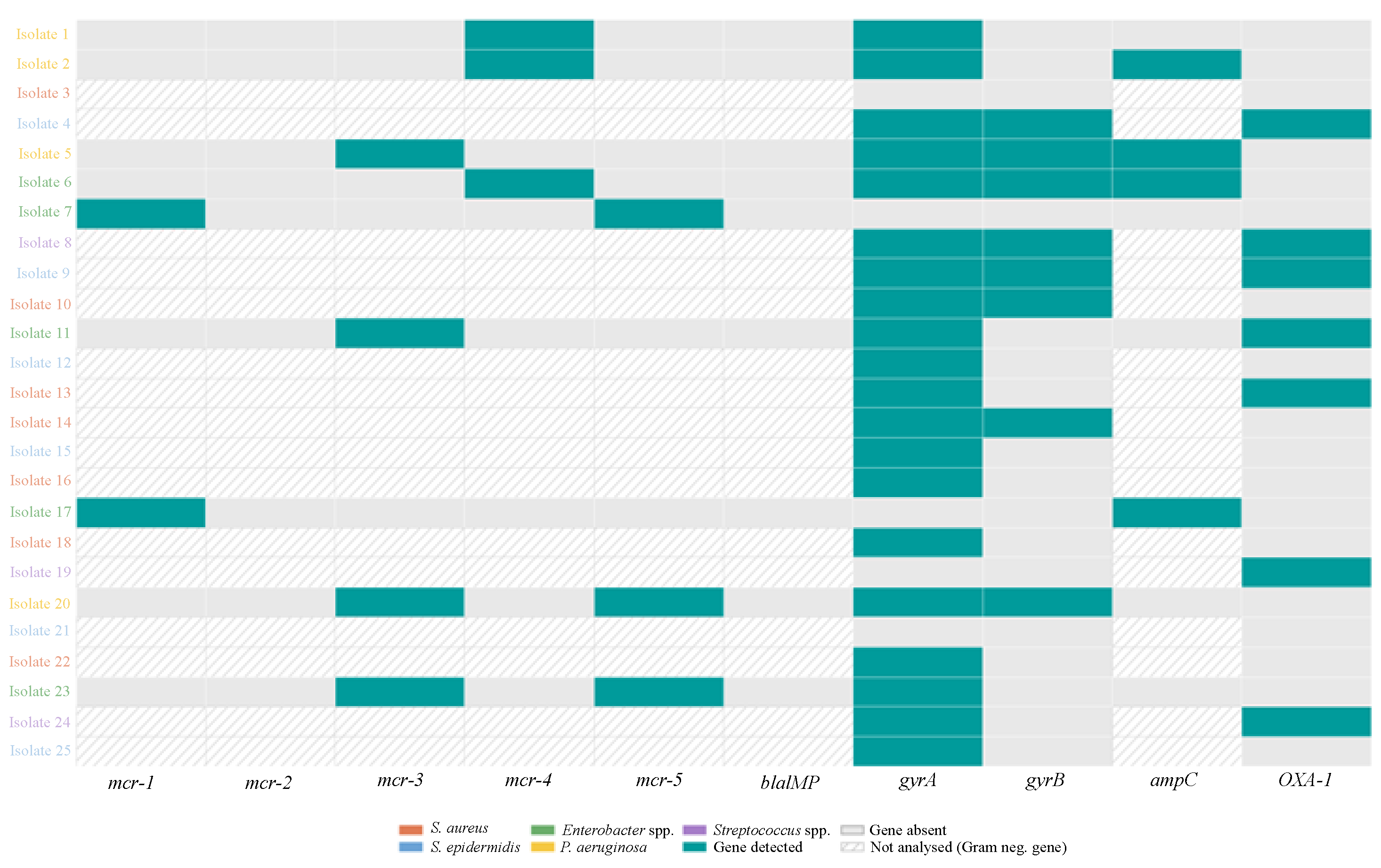
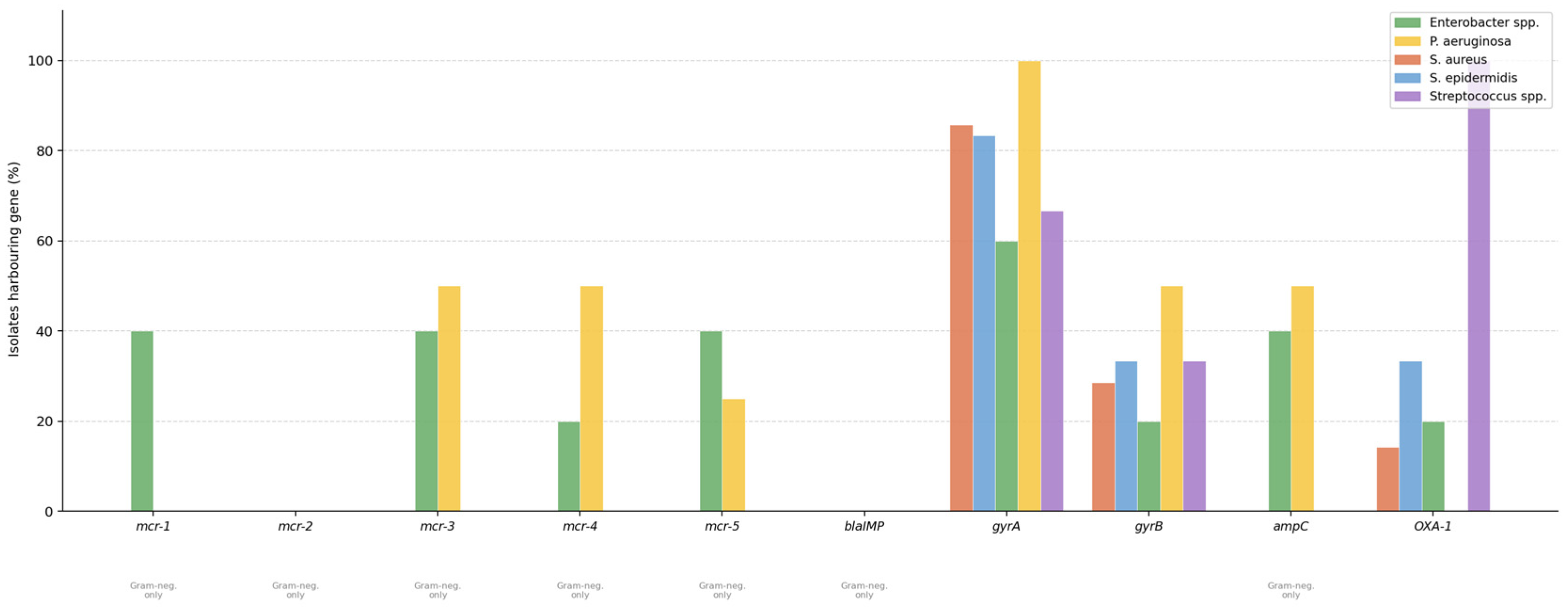
The dog breeds (age, weight and sex) with otitis externa included in the study, the dominant bacterial strain isolated from each dog, and the resistance genes detected in this bacterial strain are shown in Table 3. The distribution of resistance genes across all isolates is illustrated as a heatmap (Figure 1). Dominant bacteria isolated according to bacteriological analysis results: Staphylococcus aureus (28%), S. epidermidis (24%), Enterobacter spp. (20%), P. aeruginosa (16%) and Streptococcus spp. (12%) (Figure 2).

3.2. Antibiogram Results (40 Antibiotics)

It is important to explicitly state that all resistance rates and percentages presented in the following sections were calculated based on the 25 predominant bacterial isolates (i.e., exactly one characterized isolate per dog), rather than the total number of collected ear swabs (n = 50) or the total number of mixed colonies initially cultured. High resistance rates (80–96%) were observed across the aminoglycoside class, with the notable exception of gentamicin, which exhibited 84% susceptibility. Regarding cephalosporins, while all isolates were susceptible to cefepime and ceftriaxone, susceptibility to cefazolin and cefquinome was markedly low (4–8%). Almost all samples demonstrated resistance to beta-lactams such as amoxicillin, penicillin G, and ampicillin-cloxacillin, whereas ampicillin-sulbactam retained 36% susceptibility. In the quinolone group, marbofloxacin showed relatively high efficacy (76% susceptibility) compared to danofloxacin (44%). Susceptibility rates for other antibiotic classes were generally low, including phenicols (16–24%), macrolides (8–40%), and tetracyclines (12–20%). Additionally, fosfomycin showed 40% susceptibility. Notably, all samples were found to be resistant to sulfadiazine and sulfamethoxazole individually, although the trimethoprim-sulfamethoxazole combination displayed 28% susceptibility. All isolates exhibited intrinsic resistance to metronidazole, consistent with their aerobic or facultative nature; therefore, these results were not considered in the comparative resistance analysis.

3.3. Antimicrobial Resistance Distribution

Although mcr genes were detected in multiple isolates, phenotypic susceptibility to colistin was not evaluated due to methodological limitations. In contrast, all isolates remained susceptible to cefepime and ceftriaxone. For imipenem, resistance was observed in all Enterobacter spp. (5/5) and 75% of P. aeruginosa (3/4), consistent with intrinsic resistance mechanisms and/or carbapenemase activity. Phenotypic non-susceptibility was also recorded in 71% of S. aureus (5/7), 60% of Streptococcus spp. (3/5), and 33% of S. epidermidis (2/6); however, these findings in Gram-positive isolates should be interpreted with caution, as CLSI disk diffusion breakpoints for imipenem are not standardized for Staphylococcus or Streptococcus spp. in veterinary settings, and the clinical relevance of such profiles in these species remains uncertain. The blaIMP gene was not detected in any sample, suggesting the involvement of alternative resistance mechanisms or phenotypic misclassification in Gram-negative isolates, and questioning the biological significance of the observed non-susceptibility in Gram-positive strains. It should be noted that P. aeruginosa and Enterobacter spp. exhibit intrinsic resistance to amoxicillin-clavulanic acid and were therefore excluded from susceptibility interpretation for this agent; similarly, Streptococcus spp. are inherently susceptible to this agent and no acquired resistance was recorded. Among the interpretable species, resistance to amoxicillin-clavulanic acid was observed in 17% of S. epidermidis and 14% of S. aureus, with the OXA-1 gene identified in seven samples. Finally, ciprofloxacin resistance was found in 60% of Enterobacter spp., 50% of P. aeruginosa, 33% of both Streptococcus spp. and S. epidermidis, and 14% of S. aureus, with gyrA and/or gyrB genes detected in all resistant isolates.

3.4. Concordance Between Resistance Genes and Phenotypic Resistance

Analysis of resistance genes by PCR revealed notable discrepancies between genotype and phenotype. In some instances, resistance was observed in the absence of the targeted genes, whereas in others, isolates remained susceptible despite harboring resistance determinants. For example, among the interpretable species (Staphylococcus spp.), one of the two OXA-1-carrying isolates (S. epidermidis) remained susceptible to amoxicillin-clavulanic acid.

4. Discussion

The present study provides an evaluation of both phenotypic AMR and the underlying genotypic profiles in bacterial isolates from canine otitis externa. Our investigation yielded three core findings within the One Health framework. First, we observed notable rates of multidrug resistance within this small cohort among the predominant pathogens (e.g., Staphylococcus spp., Pseudomonas aeruginosa, and Enterobacter spp.), particularly against commonly prescribed veterinary antibiotics. Second, we detected multiple resistance genes associated with ‘last-resort’ human therapeutics—such as mcr variants (colistin)—in urban companion animals lacking agricultural exposure. Finally, our molecular analysis revealed discrepancies between genotypic carriage and phenotypic expression, suggesting the potential presence of transcriptionally inactive or “silent” genes as a working hypothesis, alongside other multifactorial resistance mechanisms. Together, these findings highlight the potential value of integrated molecular and phenotypic diagnostics to guide antimicrobial stewardship, as discussed in detail below.
The bacterial pathogens identified in this study are consistent with World Health Organization (WHO) priority classifications for AMR [23]. Specifically, S. aureus (28%), Enterobacter spp. (20%), P. aeruginosa (16%), and Streptococcus spp. (12%) represent high- and critical-priority organisms for which last-resort antibiotics are of particular concern. The detection of resistance determinants in these species therefore carries direct public health relevance within the One Health framework, and their therapeutic management should be guided by culture and sensitivity testing [24].
The genetic panel employed in this study was targeted toward specific, high-priority resistance determinants rather than providing a global resistome profile. Consequently, the absence of certain genes (e.g., blaIMP) does not preclude the existence of other resistance mechanisms, and our findings should be interpreted as a focused molecular snapshot. The discrepancies observed between phenotypic resistance patterns and the PCR panel—such as imipenem resistance in the absence of blaIMP—reflect the multifactorial nature of AMR in these isolates. Future studies employing broader diagnostic tools, such as whole genome sequencing (WGS), are therefore warranted.
Previous studies have reported mcr gene transfer predominantly in enteropathogenic E. coli and Salmonella spp. [25], with mcr-1 detected in companion animals in China [26] and South Korea [27]. All mcr variants except mcr-6 and mcr-7 have been identified in canine bacterial isolates [10,11]. In the present study, multiple mcr variants were detected among Gram-negative isolates (n = 9), with mcr-3 identified in 44.4% (95% CI:18.8–73.0), mcr-4 and mcr-5 each in 33.3% (95% CI: 12.1–64.6), and mcr-1 in 22.2% (95% CI: 6.3–54.7) of Gram-negative cases, while mcr-2 was absent. When calculated across the entire cohort (n = 25), these correspond to 16%, 12%, 12%, and 8%, respectively; however, as mcr genes are restricted to Gram-negative bacteria, prevalence estimates are most appropriately reported within this subpopulation. As phenotypic colistin susceptibility was not assessed due to the unreliability of disk diffusion for polymyxins, the presence of these genes should be interpreted as indicative of resistance potential rather than confirmed phenotypic expression. Notably, all dogs in this cohort resided in urban housing, consumed commercial dry food, and exercised in enclosed environments, suggesting that agricultural exposure is not a prerequisite for mcr gene carriage and that alternative urban transmission pathways warrant further investigation [14].
A notable finding in this study was the high-level imipenem resistance observed in all Enterobacter spp. and 75% of P. aeruginosa isolates, despite the absence of the blaIMP gene. This discrepancy suggests the involvement of alternative carbapenemase families not included in our PCR panel, such as blaKPC, blaNDM, or blaOXA-48-like enzymes, which have been increasingly reported in veterinary isolates [28,29]. Additionally, resistance in Enterobacter spp. may be driven by overproduction of intrinsic ampC beta-lactamases combined with downregulation of outer membrane porins [30]. These findings are consistent with reports of carbapenem non-susceptibility in 15–23% of P. aeruginosa isolates from companion animals [31], and further underscore the need for WGS-based approaches to comprehensively characterize carbapenem resistance mechanisms in canine isolates. The phenotypic non-susceptibility observed in Staphylococcus and Streptococcus spp. is not interpretable per current CLSI standards, as standardized breakpoints for imipenem disk diffusion are lacking for these species in veterinary microbiology.
All isolates remained susceptible to ceftriaxone, consistent with findings from comparable studies [32]. The penA gene was excluded from the analysis of all pathogens, as it encodes a penicillin-binding protein specific to Neisseria spp. and is not applicable to the bacterial species isolated in this study.
Ciprofloxacin resistance was detected in 36% of isolates overall, with the highest rates observed in Enterobacter spp. (60%) and P. aeruginosa (50%). Although gyrA and gyrB sequences were detected across isolates, phenotypic fluoroquinolone resistance arises from specific mutations within the quinolone resistance-determining regions (QRDRs) rather than mere gene presence, consistent with findings from a comparable South Korean study that identified novel QRDR mutations in P. aeruginosa from canine otitis externa [33]. These findings suggest that fluoroquinolone use in canine infections, particularly those involving P. aeruginosa, should be guided by susceptibility testing.
Methicillin resistance, assessed by disk diffusion using cefoxitin or oxacillin disks per CLSI guidelines, was observed in 77% of Staphylococcus spp. isolates.
All isolates in this study remained susceptible to cefepime, in contrast to a South Korean study reporting 44.4% cefepime resistance in P. aeruginosa from dogs and cats [34], possibly reflecting regional differences in antimicrobial usage patterns. The ampC gene was detected in four of 25 isolates (16%), with distribution restricted to Pseudomonas and Enterobacter spp. (Gram-negative organisms), consistent with the known host range of plasmid-mediated ampC beta-lactamases, and suggesting its relevance as a resistance determinant in canine otitis externa within this cohort.
Since P. aeruginosa is intrinsically resistant to amoxicillin-clavulanic acid, the disk diffusion results for this agent are not interpretable for this species and have been excluded from the susceptibility analysis. Accordingly, the apparent resistance reported for P. aeruginosa reflects intrinsic, not acquired, resistance and should not be attributed to the absence of the OXA-1 gene. Among the intrinsically susceptible species (Staphylococcus spp.), the OXA-1 gene was detected in seven isolates overall; however, among the interpretable species (Staphylococcus spp.), two of these isolates were OXA-1-positive, of which one remained phenotypically susceptible. This genotype-phenotype discrepancy is consistent with previous reports indicating that OXA-1-mediated resistance depends not only on gene presence but also on adequate enzyme expression, beta-lactamase activity, and the broader cellular environment including efflux pump efficiency and membrane permeability [35,36]. These findings further illustrate the multifactorial nature of resistance expression in this cohort, as discussed in detail in the following section.
The discrepancies between genotypic presence and phenotypic susceptibility observed for OXA-1-positive/amoxicillin-clavulanate-sensitive isolates may be attributable to several molecular and cellular mechanisms:
1. Gene Expression Regulation (Silent Genes): The mere presence of a resistance gene does not guarantee its transcription.
2. Protein Functional Defects: Even if transcribed, genetic mutations can impair the final protein’s function. Similarly, the OXA-1 gene might produce an enzyme with inadequate beta-lactamase activity due to point mutations or lack of concurrent synergistic resistance mechanisms [35].
3. Efflux Pumps and Membrane Permeability: Phenotypic resistance is often multifactorial. For genes like OXA-1, the resistance profile is influenced by the cellular environment, including efflux pump activity and outer membrane porin permeability. If antibiotic accumulation remains below the inhibitory threshold despite gene presence, a susceptible phenotype may be observed in standard testing [36].
4. The Role of Biofilms: In canine otitis externa, the discrepancy between in vitro susceptibility and clinical resistance may also be linked to biofilm formation. Standard disk diffusion tests assess planktonic bacteria; however, in vivo, gene-carrying strains may form biofilms in the ear canal that physically restrict antibiotic penetration and alter bacterial metabolism, potentially creating a more resistant phenotype clinically than laboratory testing suggests.
Limitations and Future Perspective: The present study has several limitations that should be considered when interpreting the findings. First, the small sample size (n = 25) from a single clinical setting in Ankara, Turkey, limits the generalizability of these results to broader canine or regional populations, and the findings should be regarded as preliminary and exploratory. Second, the targeted PCR panel covered specific high-priority resistance genes and does not represent a comprehensive resistome profile; the absence of detected genes does not exclude other resistance mechanisms. Third, gene expression levels and regulatory controls were not assessed, meaning that genotype-phenotype discrepancies remain mechanistically unresolved and the ‘silent gene’ interpretation should be regarded as a working hypothesis requiring transcriptional validation. Fourth, the selection of a single predominant isolate per case may have introduced selection bias, as secondary pathogens with distinct resistance profiles may have been overlooked. The detection of OXA-1 in Staphylococcus spp. is atypical, as this gene is predominantly associated with Gram-negative Enterobacterales. While primer cross-reactivity or low-level contamination cannot be entirely excluded, the consistent phenotypic correlation in some isolates supports genuine detection. Whole genome sequencing is warranted to confirm this unexpected finding. Future studies incorporating larger, multi-center cohorts, whole genome sequencing, transcriptional assays, and metagenomic approaches are warranted to address these limitations comprehensively.

5. Conclusions

Our preliminary findings suggest the complexity of AMR in bacterial isolates from canine otitis externa. These results further emphasize the importance of comprehensive analysis of antibiograms in antibiotic selection. Importantly, a lack of agreement between the presence of resistance genes (mcr variants and OXA-1) and actual phenotype was observed. This suggests that an approach including both molecular and phenotypic testing is required. This is necessary to accurately assess resistance profiles and guide effective therapeutic strategies. This is particularly important in the context of the global threat posed by AMR and the WHO’s classification of critical-priority pathogens.

Author Contributions

F.E.T.: Writing—original draft, Investigation, Formal analysis, Data curation, Conceptualization, A.F.: Supervision, Investigation, Formal analysis, Data curation, Conceptualization. All authors have read and agreed to the published version of the manuscript.

Funding

This study was funded by Ankara University Scientific Research Projects (Grant number TDK-2022-2480).

Institutional Review Board Statement

The animal study protocol was approved by the Central Animal Experiments Ethics Committee of the Ministry of Agriculture and Forestry, Turkey (Approval No: E-26137614-280.01.01-4665419; Date: 25 February 2022).

Informed Consent Statement

Permission to participate was obtained from the owners of all dogs included in the study.

Data Availability Statement

Data will be made available on request.

Acknowledgments

We would like to thank Pet Hospital for their assistance in collecting the samples.

Conflicts of Interest

The authors declare no conflicts of interest.

References

  1. Tesin, N.; Stojanovic, D.; Stancic, L.; Kladar, N.; Ružíc, Z. Prevalence of the microbiological causes of canine otitis externa and the antibiotic susceptibility of the isolated bacterial strains. Pol. J. Vet. Sci. 2023, 26, 449–459. [Google Scholar] [CrossRef]
  2. Awad, A.; Hassoon, S.; Ali, H.; Mohammad, S. Cultural and biochemical identification of antibi-otic-resistant bacteria in the ears of dogs and patients with otitis externa. Arch. Vet. Med. 2025, 18, 51–70. [Google Scholar] [CrossRef]
  3. De Martino, L.; Nocera, F.P.; Mallardo, K.; Nizza, S.; Masturzo, E.; Fiorito, F.; Iovane, G.; Catalanotti, P. An update on microbiological causes of canine otitis externa in Campania Region, Italy. Asian Pac. J. Trop. Biomed. 2016, 6, 384–389. [Google Scholar] [CrossRef]
  4. Li, Y.; Fernández, R.; Durán, I.; Molina-López, R.A.; Darwich, L. Antimicrobial resistance in bacteria isolated from cats and dogs from the Iberian Peninsula. Front. Microbiol. 2021, 11, 3628. [Google Scholar] [CrossRef] [PubMed]
  5. Parasana, D.K.; Makwana, P.M.; Shobha, K.; Prakruti, J.P.; Irshadullakhan, H.K. Isolation and Identification of Corynebacterium Species from cases of skin and ear infection of dogs. Int. J. Vet. Sci. Anim. Husb. 2023, 8, 31–33. [Google Scholar]
  6. Beaudoin, A.L.; Bollig, E.R.; Burgess, B.A.; Cohn, L.A.; Cole, S.D.; Dear, J.D.; Fellman, C.L.; Frey, E.; Goggs, R.; Johnston, A.; et al. Prevalence of antibiotic use for dogs and cats in United States veterinary teaching hospitals. J. Vet. Intern. Med. 2023, 37, 1864–1875. [Google Scholar] [CrossRef]
  7. Chan, W.Y.; Hobi, S.; Ferguson, A.; Elsohaby, I. Canine Pyoderma and Otitis Externa: A Retrospective Analysis of Multidrug-Resistant Bacterial Carriage in Hong Kong. Antibiotics 2025, 14, 685. [Google Scholar] [CrossRef]
  8. Schwarz, S.; Johnson, A.P. Transferable resistance to colistin: A new but old threat. J. Antimicrob. Chemother. 2016, 71, 2066–2070. [Google Scholar] [CrossRef]
  9. Liu, Y.Y.; Wang, Y.; Walsh, T.R.; Yi, L.X.; Zhang, R.; Spencer, J.; Doi, Y.; Tian, G.; Dong, B.; Huang, X. Emergence of plasmid-mediated colistin resistance mechanism mcr-1 in animals and human beings in China: A microbiological and molecular biological study. Lancet Infect. Dis. 2016, 16, 161–168. [Google Scholar] [CrossRef] [PubMed]
  10. Borowiak, M.; Baumann, B.; Fischer, J.; Thomas, K.; Deneke, C.; Hammerl, J.A.; Szabo, I.; Malorny, B. Development of a novel mcr-6 to mcr-9 multiplex PCR and assessment of mcr-1 to mcr-9 occurrence in colistin-resistant Salmonella enterica isolates from environment, feed, animals and food (2011–2018) in Germany. Front. Microbiol. 2020, 11, 80. [Google Scholar] [CrossRef]
  11. Wang, G.; Liu, H.; Feng, Y.; Zhang, Z.; Hu, H.; Liu, J.; Qiu, L.; Guo, Z.; Huang, J.; Qiu, J.; et al. Colistin-resistance mcr genes in Klebsiella pneumoniae from companion animals. J. Glob. Antimicrob. Resist. 2021, 25, 35–36. [Google Scholar] [CrossRef] [PubMed]
  12. Hamame, A.; Davoust, B.; Hasnaoui, B.; Mwenebitu, D.L.; Rolain, J.M.; Diene, S.M. Screening of colistin-resistant bacteria in livestock animals from France. Vet. Res. 2022, 53, 96. [Google Scholar] [CrossRef]
  13. Kwon, J.; Ko, H.J.; Yang, M.H.; Park, C.; Park, S.C. Antibiotic Resistance and Species Profile of Enterococcus Species in Dogs with Chronic Otitis Externa. Vet. Sci. 2022, 9, 592. [Google Scholar] [CrossRef]
  14. Oh, J.Y.; Kwak, S.M.; Kim, J.Y.; Ro, W.B.; Lee, K.J.; Chae, J.C. Case Report: Transferable IncX4 plasmid carrying mcr-1 in colistin-resistant Escherichia coli from a healthy pet dog in South Korea. Front. Vet. Sci. 2026, 12, 1746399. [Google Scholar] [CrossRef]
  15. Agnihotri, D.; Charaya, G.; Chabbra, R.; Kumar, T.; Jain, V.K. Antibiogram of bacteria isolated from dogs suffering from otitis externa. Indian J. Comp. Microbiol. Immunol. Infect. Dis. 2019, 40, 15–20. [Google Scholar] [CrossRef]
  16. CLSI. Performance Standards for Antimicrobial Susceptibility Testing, 29th ed.; Clinical and Laboratory Standards Institute: Wayne, PA, USA, 2019. [Google Scholar]
  17. CLSI. Performance Standards for Antimicrobial Disk and Dilution Susceptibility Test for Bacteria Isolated from Animals. In Approved Standard Document, 5th ed.; Clinical and Laboratory Standards Institute: Wayne, PA, USA, 2020. [Google Scholar]
  18. European Medicines Agency. Categorisation of Antibiotics Used in Animals Promotes Responsible Use to Protect Public and Animal Health. 2020. Available online: https://www.ema.europa.eu/en/news/categorisation-antibiotics-used-animals-promotes-responsible-use-protect-public-animal-health (accessed on 20 July 2025).
  19. DiaRex. Genomic DNA Extraction Kit User Manual; Diagen Biotechnological Systems: Ankara, Turkey, 2024. [Google Scholar]
  20. Monteiro, J.; Widen, R.H.; Pignatari, A.C.; Kubasek, C.; Silbert, S. Rapid detection of carbapenemase genes by multiplex real-time PCR. J. Antimicrob. Chemother. 2012, 67, 906–909. [Google Scholar] [CrossRef] [PubMed]
  21. Hopkins, K.L.; Davies, R.H.; Threlfall, E.J. Mechanisms of quinolone resistance in Escherichia coli and Salmonella: Recent developments. Int. J. Antimicrob. Agents 2005, 25, 358–373. [Google Scholar] [CrossRef] [PubMed]
  22. Pérez-Pérez, F.J.; Hanson, N.D. Detection of plasmid-mediated AmpC β-lactamase genes in clinical isolates by using multiplex PCR. J. Clin. Microbiol. 2002, 40, 2153–2162. [Google Scholar] [CrossRef]
  23. World Health Organisation. Publishes List of Bacteria for Which New Antibiotics Are Urgently Needed. 2017. Available online: https://www.who.int/news/item/27-02-2017-who-publishes-list-of-bacteria-for-which-new-antibiotics-are-urgently-needed (accessed on 20 July 2025).
  24. World Organisation for Animal Health. WOAH List of Antimicrobials of Veterinary Importance. 2021. Available online: https://www.woah.org/app/uploads/2021/06/a-oie-list-antimicrobials-june2021.pdf (accessed on 20 July 2025).
  25. Kempf, I.; Fleury, M.A.; Drider, D.; Bruneau, M.; Sanders, P.; Chauvin, C.; Haenni, M.; Jouy, E. What do we know about resistance to colistin in Enterobacteriaceae in avian and pig production in Europe? Int. J. Antimicrob. Agents 2013, 42, 379–383. [Google Scholar] [CrossRef]
  26. Lei, L.; Wang, Y.; Schwarz, S.; Walsh, T.R.; Ou, Y.; Wu, Y.; Li, M.; Shen, Z. mcr-1 in Enterobacteriaceae from Companion Animals, Beijing, China. Emerg. Infect. Dis. 2017, 23, 710. [Google Scholar] [CrossRef] [PubMed]
  27. Moon, D.C.; Kim, S.J.; Mechesso, A.F.; Kang, H.Y.; Song, H.J.; Choi, J.-H.; Yoon, S.-S.; Lim, S.-K. Mobile colistin resistance gene mcr-1 detected on an IncI2 plasmid in Salmonella typhimurium sequence type 19 from a healthy pig in South Korea. Microorganisms 2021, 9, 398. [Google Scholar] [CrossRef]
  28. Khosravi, Y.; Tay, S.T.; Vadivelu, J. Metallo-β-lactamase-producing imipenem-resistant Pseudomonas aeruginosa clinical isolates in a university teaching hospital in Malaysia: Detection of IMP-7 and first identification of IMP-4, VIM-2, and VIM-11. Diagn. Microbiol. Infect. Dis. 2010, 67, 294–296. [Google Scholar] [CrossRef]
  29. Ocak, M.; Özer, B.; İnci, M.; Duran, N. Antibiotic Resistance and Investigation of IMP-1, IMP-2, VIM-1 and VIM-2 Metallo-β-Lactamases in Acinetobacter Strains Isolated From Clinical Samples. Klimik J. 2015, 28, 23. [Google Scholar] [CrossRef]
  30. Wang, Y.; Wang, X.; Schwarz, S.; Zhang, R.; Lei, L.; Liu, X.; Shen, J. IMP-45-producing multidrug-resistant Pseudomonas aeruginosa of canine origin. J. Antimicrob. Chemother. 2014, 69, 2579–2581. [Google Scholar] [CrossRef]
  31. KuKanich, K.S.; Bagladi-Swanson, M.; KuKanich, B. Pseudomonas aeruginosa susceptibility, antibiogram and clinical interpretation, and antimicrobial prescribing behaviors for dogs with otitis in the Midwestern United States. J. Vet. Pharmacol. Ther. 2022, 45, 440–449. [Google Scholar] [CrossRef] [PubMed]
  32. Zamankhan Malayeri, H.; Jamshidi, S.; Zahraei Salehi, T. Identification and antimicrobial susceptibility patterns of bacteria causing otitis externa in dogs. Vet. Res. Commun. 2010, 34, 435–444. [Google Scholar] [CrossRef]
  33. Park, Y.; Oh, J.; Park, S.; Sum, S.; Song, W.; Chae, J.; Park, H. Antimicrobial resistance and novel mutations detected in the gyrA and parC genes of Pseudomonas aeruginosa strains isolated from companion dogs. BMC Vet. Res. 2020, 16, 111. [Google Scholar] [CrossRef]
  34. Cho, J.K.; Kim, J.M.; Kim, K.H.; Lim, H.S.; Yang, C.R. Antimicrobial resistance of Pseudomonas aeruginosa isolated from dogs and cats. Korean J. Vet. Serv. 2021, 44, 21–26. [Google Scholar]
  35. Livermore, D.M.; Day, M.; Cleary, P.; Hopkins, K.L.; Toleman, M.A.; Wareham, D.W.; Wiuff, C.; Doumith, M.; Woodford, N. OXA-1 β-lactamase and non-susceptibility to penicillin/β-lactamase inhibitor combinations among ESBL-producing Escherichia coli. J. Antimicrob. Chemother. 2019, 74, 326–333. [Google Scholar] [CrossRef] [PubMed]
  36. Oteo, J.; González-López, J.J.; Ortega, A.; Quintero-Zárate, J.N.; Bou, G.; Cercenado, E.; Conejo, M.C.; Martínez-Martínez, L.; Navarro, F.; Oliver, A.; et al. Inhibitor-resistant TEM-and OXA-1-producing Escherichia coli isolates resistant to amoxicillin-clavulanate are more clonal and possess lower virulence gene content than susceptible clinical isolates. Antimicrob. Agents Chemother. 2014, 58, 3874–3881. [Google Scholar] [CrossRef] [PubMed][Green Version]
Figure 1. Heatmap of resistance gene distribution across 25 canine otitis externa isolates. Teal cells indicate gene detection; gray cells indicate absence. Isolate labels on the y-axis are color-coded by bacterial species (see legend).
Amh 71 00014 g001
Figure 2. Prevalence of resistance genes by bacterial species in canine otitis externa isolates (n = 25). Bar height represents the percentage of isolates within each species harboring the respective gene. Gram-negative-specific genes (mcr variants, ampC, blaIMP) are shown only for Gram-negative species.
Amh 71 00014 g002
Table 1. Antibiotic disks and concentrations selected to determine the susceptibility of bacteria isolated from external ear infections of dogs by disk diffusion method.
AntibioticSymbol/ConcentrationAntibioticSymbol/Concentration
AminosidinAN-60 µgFlorfenicolFFC-30 µg
AmoxicillinAML-20 µgFosfomycinFF-30 µg
Amoxicillin/clavulanic acidAMC-30 µgImipenemIMP-10 µg
Ampicillin/cloxacillinAPX 30 µgLincomycinL-15 µg
Ampicillin/sulbactamSAM-20 µgMarbofloxacinMAR-5 µg
CefazolinCZ-30 µgMethicillinME-5 µg
CefepimeFEP-10 µgMetronidazoleMET-10 µg
CefoperazoneCEP-75 µgNeomycinN-30 µg
CefquinomeCEQ-30 µgOxytetracyclineT-30 µg
CeftriaxoneCRO-30 µgPenicillin GP-10 U
CephalexinCL-30 µgSpectinomycinSPT-25 µg
ChloramphenicolC-30 µgSpiramycinSP-30 µg
CiprofloxacinCIP-10 µgStreptomycinS-25 µg
ClarithromycinCLR-15 µgSulfadiazineSD-300 µg
ClindamycinDA-10 µgSulphamethoxazoleSMZ-100 µg
CloxacillinCX-30 µgTetracyclineTE-30 µg
ColistinCT-25 µgTrimethoprimTMP-30 µg
DanofloxacinDAN-5 µgTrimethoprim/sulphamethoxazoleSXT-25 µg
DoxycyclineDO-30 µgTulathromycinTUL-30 µg
ErythromycinE-15 µgVancomycinVA-10 µg
Table 2. Primers used to produce real-time qPCR standards.
Target GenPrimer NamePrimer Sequence (5′ → 3′)
mcr-1colistin_mcr_1-FGTCCGTTTGTTCTTGTGG
colistin_mcr_1-RGTCTGTAGGGCATTTTGG
mcr-2colistin_mcr_2-FGTATTCTGTGCCGTGTATG
colistin_mcr_2-RGTATTGTTGGTTGCTGATTT
mcr-3colistin_mcr_3-FGCCTCATTTTGATTGGTTTC
colistin_mcr_3-RTAAGTTTGGTTTCGCCATTT
mcr-4colistin_mcr_4-FCCCGAACACTAAACCTAAC
colistin_mcr_4-RAAACATACAGGGTAGAGACA
mcr-5colistin_mcr_5-FACTGATTCTGCTTGCTGT
colistin_mcr_5-RTCATTACCGCTTGTTTCC
blaIMPImipenem-FGAGTGGCTTAATTCTCRATC
Imipenem-RAACTAYCCAATAYRTAAC
gyrAgyrA-FTGTCCGAGATGGCCTGAAGC
gyrA-RTACCGTCATAGTTATCCACG
gyrBgyrB-FTCGGCGTCGTTGTTGTCATA
gyrB-RGCGGTGGGTTTCAAAATCTG
ampCCefepime_ampC-FTTCTTGTCTACTTTTATCCCC
Cefepime_ampC-RACTGCTATTTACGGCTTTTT
OXA-1Amoxicillin + clavulanic acid_OXA1-FTTTTCTGTTGTTTGGGTTTC
Amoxicillin + clavulanic acid_OXA1-RCTATGGTGTTTTCTATGGCT
Table 3. Dog breeds sampled, characteristics, dominant bacterial species, and detected genetic determinants 1.
No.Dog BreedAge-Weight-SexDominant MicroorganismDetected Resistance Genes/Determinants 2Phenotypic Resistance Profile
1Belgian Sheepdog4 years old-35 kg-femalePseudomonas aeruginosamcr-4, gyrACiprofloxacin, Sulfonamides
2Beagle3 years old-17 kg-malePseudomonas aeruginosamcr-4, ampC, gyrACiprofloxacin, Beta-lactams
3Yorkshire Terrier5 years old-3 kg-maleStaphylococcus aureusBeta-lactams, Sulfonamides
4Basset Hound10 years old-36 kg-maleStaphylococcus epidermidisgyrA, gyrB, OXA-1Ciprofloxacin, Amoxicillin-Clav, Beta-lactams
5Golden Retriever10 years old-33 kg-malePseudomonas aeruginosamcr-3, ampC, gyrA, gyrBCiprofloxacin, Beta-lactams
6German Shepherd7 years old-30 kg-maleEnterobacter spp.mcr-4, ampC, gyrA, gyrBImipenem, Ciprofloxacin
7Pug4 years old-8 kg-femaleEnterobacter spp.mcr-1, mcr-5Imipenem, Sulfonamides
8Akita1 year old-36 kg-maleStreptococcus spp.gyrA, gyrB, OXA-1Ciprofloxacin, Beta-lactams
9English Cocker Spaniel7 years old-10 kg-maleStaphylococcus epidermidisgyrA, gyrB, OXA-1Ciprofloxacin
10Golden Retriever13 years old-28 kg-femaleStaphylococcus aureusgyrA, gyrBCiprofloxacin, Beta-lactams
11Maltese Terrier4 years old-2.5 kg-femaleEnterobacter spp.mcr-3, gyrA, OXA-1Imipenem, Ciprofloxacin
12English Cocker Spaniel11 years old-9 kg-maleStaphylococcus epidermidisgyrACiprofloxacin, Beta-lactams
13Bernese Mountain Dog6 years old-50 kg-femaleStaphylococcus aureusOXA-1, gyrACiprofloxacin, Amoxicillin-Clav
14Chihuahua7 years old-1.5 kg-maleStaphylococcus aureusgyrA, gyrBCiprofloxacin, Beta-lactams, Sulfonamides
15Golden Retriever3 years old-28 kg-maleStaphylococcus epidermidisgyrACiprofloxacin
16Golden Retriever3 years old-33 kg-femaleStaphylococcus aureusgyrACiprofloxacin
17Cavalier King Charles Spaniel10 years old-15 kg-maleEnterobacter spp.mcr-1, ampCImipenem, Colistin, Beta-lactams
18Yorkshire Terrier7 years old-2 kg-male Staphylococcus aureusgyrACiprofloxacin
19Golden Retriever3 years old-25 kg-femaleStreptococcus spp.OXA-1Beta-lactams
20Golden Retriever10 years old-33 kg-femalePseudomonas aeruginosamcr-3, mcr-5, gyrA, gyrBCiprofloxacin, Beta-lactams
21Hybrid Dog2 years old-23 kg-femaleStaphylococcus epidermidisBeta-lactams
22Golden Retriever13 years old-40 kg-femaleStaphylococcus aureusgyrACiprofloxacin, Beta-lactams
23Kangal Shepherd Dog1 year old-45 kg-maleEnterobacter spp.mcr-3, mcr-5, gyrAImipenem, Ciprofloxacin
24Golden Retriever9 years old-35 kg-femaleStreptococcus spp.gyrA, OXA-1Ciprofloxacin
25English Cocker Spaniel5 years old-11 kg-femaleStaphylococcus epidermidisgyrACiprofloxacin, Beta-lactams
1 Colistin susceptibility was not assessed phenotypically due to methodological limitations; only the genotypic (mcr) presence was reported. 2 The presence of gyrA and gyrB refers to the detection of target gene sequences; phenotypic resistance is associated with specific mutations within these regions.
Disclaimer/Publisher’s Note: The statements, opinions and data contained in all publications are solely those of the individual author(s) and contributor(s) and not of MDPI and/or the editor(s). MDPI and/or the editor(s) disclaim responsibility for any injury to people or property resulting from any ideas, methods, instructions or products referred to in the content.