Article

Trichomonas vaginalis in Vaginal Samples from Symptomatic Women in Greece: Assessment of Test Performance and Prevalence Rate, and Comparison with European Prevalence Estimates

by
Lazaros Tsoukalas
1,†,
Constantine M. Vassalos
2,*,†,
Nikos Gkitsakis
3,
Panagiota Gkotzamani
1,
Eleni Gkoumalatsou
1,
Konstantia Bakalianou
4,
Eleftheria Palla
1,
Stavroula Baka
5,
Constantina Skanavis
2 and
Evdokia Vassalou
2
1
Microbiology Laboratory, Biochemistry-Biopathology Department, General Hospital of Nea Ionia “Konstantopouleio-Patision”, 142 33 Athens, Greece
2
Department of Public and Community Health, School of Public Health, University of West Attica, 122 43 Athens, Greece
3
School of Engineering, University of Thessaly, 382 21 Volos, Greece
4
Gynaecology Clinic, General Hospital of Nea Ionia “Konstantopouleio-Patision”, 142 33 Athens, Greece
5
Microbiology Laboratory, Aretaieio Hospital, Medical School, National and Kapodistrian University of Athens, 115 27 Athens, Greece
*
Author to whom correspondence should be addressed.
These authors contributed equally to this work.
Acta Microbiol. Hell. 2025, 70(3), 29; https://doi.org/10.3390/amh70030029
Submission received: 6 May 2025 / Revised: 10 June 2025 / Accepted: 30 June 2025 / Published: 11 July 2025

Abstract

Trichomonas vaginalis infection (TVI) is the most common curable sexually transmitted infection (STI). In this study, we aimed to assess the performances of different tests for TVI diagnosis in symptomatic Greek women, evaluating the TVI prevalence rate (PR) in Greece and comparing the latter with TVI-PR estimates from Europe. A laboratory-based cross-sectional analysis and a meta-analysis were conducted. Of 399 symptomatic Greek women, 17 had TVI, corresponding to a TVI-PR of 4.3%. The commercial nucleic acid amplification test (NAAT) achieved a sensitivity of 94.1%, which was 6% higher than the sensitivity of the culture method, 35% higher than that of the wet mount test, and 59% higher than that of the Giemsa stain test. The wet mount test achieved the lowest positive predictive value of 76.9%. All the tests had high specificity levels and negative predictive values. Data from 34 European TVI-PR studies in symptomatic women were pooled. The TVI-PR established in our study was similar to the TVI-PR estimates of 4.8% in Europe and 4.5% in Greece, with the second being higher than those of 2.1% in Northwestern Europe and 1.5% in Southern Europe but closer to that of 6.7% in Türkiye. In Greece, a European country with a relatively high TVI-PR among symptomatic women, the highly sensitive and specific, automated, point-of-care NAAT would facilitate rapid, accurate TVI diagnosis and the treatment of this target population to meet the WHO’s goal of ending STI epidemics by 2030.

1. Introduction

Trichomonas vaginalis is an aerotolerant anaerobic flagellated protozoan living in the urogenital tracts of both sexes worldwide. It is thought to exist in the infective stage of a trophozoite; however, cyst-like structures have been seen recently. The 7–30 μm long pear-shaped, motile T. vaginalis trophozoite has a single nucleus, four flagella, an undulating membrane enhancing its motility, and a mastigont system [1]. Being adapted to oxygen-poor environments, T. vaginalis possesses highly modified mitochondria called hydrogenosomes. This protozoan parasite is transmitted from an infected person to others through sexual intercourse, more often during vaginal sex; non-sexual transmission might also occur.
Vaginal trichomonads (T. vaginalis), the causative agents of trichomoniasis, are the only pathogenic species of human trichomonads. The lesions that they cause are usually limited to the genitourinary organs. In the genitourinary system, trichomonads cause inflammation, but trichomonas infection is often asymptomatic. Sometimes, especially in men, the parasites die immediately or after a short period of time (transient carriage). The frequency of trichomonad carriage ranges from 2% to 41%. The causes of carriage have not been established, but there are several theories of self-healing, such as the theory that pathogens that have not had time to become attached to epithelial cells with urine are mechanically removed. Most individuals who are infected with T. vaginalis are asymptomatic due to the low pathogenicity of the majority of its strains. In women, however, some strains of T. vaginalis can cause vaginal symptoms, including foul-smelling vaginal discharge and irritation. It is of note that an acute infection can turn chronic, with a lessening in the severity of symptoms. Trichomonas vaginalis infection may be responsible for long-term conditions, such as cervicitis, in women. In addition, T. vaginalis infection has been associated with an increase in the risk of acquiring other sexually transmitted infections (STIs) and a greater likelihood of adverse pregnancy outcomes. Untreated T. vaginalis infection may also lead to pelvic inflammatory disease. By contrast, infection with T. vaginalis is typically symptomless or presents with milder symptoms in men.
When a diagnosis of T. vaginalis infection is received, it can usually be cured with antiprotozoal agents, primarily metronidazole, which are given orally for a week, although the occurrence of resistant strains has been noticed. In such cases, secnidazole, which has recently been approved by the FDA for oral treatment against T. vaginalis infection, may be a useful therapeutic option. Persistent and recurrent infections can often be present due to re-infection by an untreatable sexual partner or an infection by a new partner who is infected with T. vaginalis.
The diagnosis cannot be made based on clinical features alone, since the clinical symptoms may be a manifestation of other diseases of the genitourinary tract. If trichomoniasis is diagnosed based only on clinical data, the disease is not detected in more than 85% of infected women, and vice versa, in almost 30% of patients who are not infected with trichomonas, this diagnosis is made erroneously. Based on the above, the need to use laboratory methods for diagnosing trichomoniasis becomes clear. In many areas around the world, especially those with limited resources such as low- and middle-income countries with high prevalence rates of T. vaginalis infection, the detection of the protozoan continues to rely on direct microscopy of vaginal wet smears—a cost-effective method—for the recognition of T. vaginalis in vaginal secretions The Giemsa stain has been used sporadically. Culture enables the detection of low numbers of T. vaginalis parasites [2]. Despite not being widely used, culture has still been considered the gold standard method for the diagnosis of T. vaginalis infection. Molecular methods have gained favour in recent years, and the rapid nucleic acid amplification test (NAAT) has recently been recommended frequently as an alternative gold standard for the detection of T. vaginalis [3], especially in areas such as high-income countries, where it is commercially available. The NAAT method can identify the genetic material (DNA or RNA) of T. vaginalis in cervical or vaginal swabs and urine samples. The NAAT is considered to be better at detecting T. vaginalis infection than culture and wet mount microscopy and can be used in symptomatic or asymptomatic people, regardless of their sex. Current commercial NAATs that are based on real-time PCR, transcription-mediated assay, strand displacement assay, and helicase-dependent amplification show high sensitivity and specificity. They can also be performed using the same instrumentation platforms as NAATs for other STIs and used as point-of-care tests. Loop-mediated isothermal amplification has been studied recently as a good candidate for the development of a point-of-care NAAT for T. vaginalis infection [3,4].
The problem of diagnosing sexually transmitted diseases continues to attract the attention of doctors. Urogenital trichomoniasis is one of the most common diseases of the urogenital tract and ranks first in the world among STIs. According to the WHO, 10% of the world’s population is affected by trichomonas infection, and 170 million people become ill with trichomoniasis every year. Infection due to the parasite T. vaginalis is a non-viral, treatable STI like those caused by the bacteria Chlamydia trachomatis, Neisseria gonorrhoeae, and Treponema pallidum [4]. In 2020, the World Health Organization (WHO) reported 374 million new cases with one of these four curable STIs globally. Trichomonas vaginalis infection was estimated to be responsible for the majority (156/374 million; 42%) of those cases [5]. However, the prevalence of T. vaginalis infection is likely to be underestimated, as it is not currently included in most established screening programmes for STIs [6]. The fact that the prevalence of T. vaginalis infection varies worldwide may be attributed to differences between geographical regions, ethnicities, and age groups, as well as different habits and risky sexual behaviours [7]. Due to the relatively unknown epidemiology of T. vaginalis infection, symptomatic women remain the key target group for interventions, especially considering that the infection can easily be treated with inexpensive drugs [8].
In the European region, according to the WHO and based on studies conducted in different population groups, the prevalence of T. vaginalis infection was recently estimated to be 1.6% for women [9]. There is a need to collect more data at the country level to provide an initial baseline estimate in order to monitor the effectiveness of interventions as part of the organisation’s strategies to end the worldwide epidemics of STIs by 2030 [10].
In Greece, infections with T. vaginalis are not mandatorily reported [11]; thus, there is currently no operational national programme for controlling the spread of this treatable infection. Data on the prevalence of T. vaginalis infection in the country are scarce, and more recent data are required. Wet mount microscopy remains the most common method for the diagnosis of T. vaginalis infection in the country, despite commercial NAATs now being available.
The aim of the present study was to assess the performance of four diagnostic methods (wet mount microscopy, Giemsa-stained smear microscopy, culture, and NAAT) for the detection of T. vaginalis in vaginal samples from symptomatic Greek women and the current prevalence rate of T. vaginalis infection in women with vaginal symptoms in Greece. We also aimed to compare the latter with the estimated prevalence rates of T. vaginalis infection in symptomatic European women.

2. Materials and Methods

2.1. Laboratory Investigation of T. vaginalis Infection in Symptomatic Greek Women

2.1.1. Study Design

This was a laboratory-based cross-sectional study for the detection of T. vaginalis in vaginal samples from symptomatic women, conducted in Athens, Greece, between 1 January and 31 December 2023. Women meeting our research inclusion criteria for vaginal secretion collection had developed non-specific symptoms such as vaginal discharge, odour, and vulvar irritation, which are considered suggestive of T. vaginalis infection. This study was conducted at the General Hospital of Nea Ionia “Konstantopouleio-Patision”, which services an adult population in the northeast part of Greater Athens, Greece.

2.1.2. Sampling

Routine swabs were taken at the gynaecology clinic and sent to the adjacent clinical laboratory for microbiological testing. Two additional vaginal swabs were also collected for the detection of T. vaginalis: one for culture and the other for testing with a commercially available real-time PCR kit. Samples were anonymised and decoded.

2.1.3. Wet Mount Microscopy

A saline wet mount slide was promptly prepared from a vaginal swab and directly examined under an optical microscope with a 40-fold objective to look for the presence of actively motile pear-shaped trophozoites, which was considered a positive result.

2.1.4. Giemsa-Stained Smear Microscopy

Another vaginal swab was rolled over a glass slide to perform a Giemsa-stained thin smear. The stained slide was viewed using a light microscope, delivering a 1000-fold magnification of the vaginal swab-based sample to detect the presence of violet, oval, or pyriform trophozoites with a darker nucleus (and characteristic morphological features—axostyle, flagella, and undulating membrane—which were not always observable), which was considered a positive result.

2.1.5. Culture

The vaginal swab was inoculated into Oxoid Trichomonas Medium No.2 (Thermo Fisher Scientific Inc., Waltham, MA, USA) and incubated at 37 °C. Culture medium from the bottom of the bijou bottle was examined daily for the presence of motile T. vaginalis parasites, which was considered a positive result. Samples were incubated until day 7, after which, unless trophozoites were observed, they were deemed negative for T. vaginalis.

2.1.6. Commercial NAAT

The vaginal swab was collected using the Xpert® Swab Collection kit (Cepheid, Sunnyvale, CA, USA) and tested with the Cepheid Xpert® TV assay according to the manufacturer’s specifications. The NAAT was performed on the Cepheid GeneXpert® 4-module system based on a patented, single-use, self-contained cartridge technology, minimising the risk of cross-contamination. This rapid, automated real-time PCR-based test detected T. vaginalis genomic DNA, providing qualitative results such as “TV detected” and “TV not detected” (where TV is T. vaginalis) in 1 h or less. For positive results (“TV detected”), the assay could be in 42 min. Samples providing indeterminate results were re-tested using a new cartridge.

2.1.7. Test Performance and Prevalence Calculation for T. vaginalis

We assessed the performance of the four diagnostic tests, namely, vaginal wet mount, Giemsa staining, culture in a specialised medium, and a commercial NAAT, for the detection of T. vaginalis in vaginal samples from symptomatic Greek women. Their sensitivity (Se) and specificity (Sp) were computed and compared. The status of T. vaginalis infection was determined as positive if the results of either or both gold standards, that is, culture and/or NAAT, were T. vaginalis-positive; thus, the prevalence rate (PR) of T. vaginalis infection could be calculated in the study population. The positive predictive values (PPVs) and negative predictive values (NPVs) were recorded. The percentage (%) agreement and kappa coefficient (κ) were used to measure the agreement between tests.

2.2. Estimation of Pooled T. vaginalis Prevalence in European Symptomatic Women

2.2.1. Study Design

This was a systematic review with meta-analysis, conducted in line with the Preferred Reporting Items for Systematic and Meta-analysis (PRISMA) protocol [12] to estimate the prevalence of T. vaginalis infection in symptomatic European women.

2.2.2. Database Search

Two of our researchers searched the PubMed, Science Direct, and Scopus databases using the following keywords or combined sets: “Epidemiology”, “Europe”, “European country”, “European region”, “European women”, “Prevalence”, “Sexually transmitted diseases”, “STD”, “Sexually transmitted infections”, “STI”, “Symptomatic women”, “Trichomonas”, “Trichomonas vaginalis”, “T. vaginalis”, “Trichomoniasis”, “Vaginal discharge”, “Vaginal infections”, “Vaginal irritation”, “Vaginal symptoms”, and “Vaginitis”. The “AND” and/or “OR” Boolean operators were used. Databases were last searched on 7 August 2024. All retrieved full texts were reviewed to identify additional studies.

2.2.3. Eligibility Criteria

The inclusion criteria set for this systematic review and meta-analysis were as follows:
  • Peer-reviewed original articles covering countries in the European region;
  • Laboratory-based cross-sectional studies on the prevalence of T. vaginalis among women with vaginal symptoms;
  • Studies identifying T. vaginalis in vaginal samples;
  • Papers published from 1 January 2008 to 31 December 2023;
  • Research studies with accessible abstracts and full texts in English;
  • Articles with English abstracts, even if their full texts were not in English;
  • Studies providing the exact number of study participants and positive cases.
The exclusion criteria corresponded to our established eligibility criteria and were as follows:
  • Editorials;
  • Letters;
  • Reviews;
  • Papers referring to other geographical regions;
  • Articles without a specified total sample size and number of positives;
  • Surveys using electronic data collection;
  • Research studies based on screening or routine testing;
  • Laboratory-based studies testing samples other than vaginal ones;
  • Prevalence studies on men, children, female sex workers, pregnant women, and asymptomatic women.

2.2.4. Selection Process

Duplicate records were removed after datasets were extracted to comma-separated values files, and all digital objective identifiers were compared using Microsoft Excel. The management and review of references were performed using the 2.119.0 version of the Mendeley reference manager software (Elsevier B.V., Amsterdam, The Netherlands). Through this entire process, two additional researchers independently screened articles based on title and abstract and further evaluated the obtained full texts to decide separately whether these records were eligible or not. Another expert was involved to resolve any discrepancies or uncertainties.
The data extraction from each study was conducted using the version 2405 version of the Microsoft Excel spreadsheet software programme (Microsoft Corporation, Redmond, WA, USA). The relevant data extracted were author(s); publication year; country where the study was conducted; study duration; aim of the study; basic demographic data; population and sample size; sample types; analysis method; and basic data and results from the analyses.

2.2.5. Data Synthesis

For each study, the prevalence ratio (PR) of T. vaginalis at a 95% CI was computed. Summary estimates were obtained by applying the random effects model. Heterogeneity was judged by looking at forest plots; the study weight was determined based on the size of the square; the crossed lines demonstrated the specific 95% CI; and the diamond shape represented pooled study results. Statistical heterogeneity was examined using the I2 statistic. If at least 10 studies were included in the meta-analysis, publication bias was assessed by drawing funnel plots and tested through Egger’s regression-based asymmetry test. High p-values suggested less evidence of publication bias. If heterogeneity was observed between studies, subgroup analyses would be undertaken. Subtotal estimates between subgroups would be compared using the Q statistic. If less than three studies were available, exploration would be carried out descriptively.

2.3. Statistical Analysis

All statistical analyses were carried out with the 29.0 version of IBM SPSS Statistics (International Business Machines Corporation, Armonk, NY, USA). The χ2-test was used for the comparison of proportions. The 95% confidence intervals (CIs) were calculated. A p-value less than 0.05 was considered statistically significant.

3. Results

3.1. Results from Laboratory Investigation of T. vaginalis Infection in Symptomatic Greek Women

3.1.1. Study Samples

Vaginal samples from 402 adult symptomatic women were submitted for testing for the presence of T. vaginalis. All the vaginal specimens collected from 399 (99.3%) women at the clinic were included in this laboratory-based study. Follow-up samples from two (0.7%) patients were excluded from the final dataset.

3.1.2. Detection of T. vaginalis in Vaginal Samples

The T. vaginalis-positive results of the four diagnostic methods are provided in Supplementary Table S1. The overlap and differences in T. vaginalis-positive results among the four methods are visualised in Figure 1.
The NAAT yielded valid results for 98.3% (392/399) of the collected vaginal samples on the first attempt. The seven indeterminate cases were re-tested. In the six cases with inconclusive results due to technical errors, the assay successfully produced results upon repeat testing. Thus, successful testing was accomplished in 398 cases, representing an overall assay success rate of 99.8%. T. vaginalis was detected in 16 (4%) of the 399 vaginal samples. Τhe sample that remained invalid after repeat processing was found positive for T. vaginalis using culture (Supplementary Figure S1).
In total, 15 (3.8%) of the 399 samples yielded a T. vaginalis-positive culture result (Figure 1). Of these, 14 [93.3%; 14/399 (3.5%)] were also found to be positive when using the NAAT. In addition, 2 (0.5%) of the 384 samples that tested negative when using culture tested positive for T. vaginalis using the NAAT.
There was a total of 13 T. vaginalis-positive results based on the microscopic examination. All 13/399 (3.3%) samples were found to be positive for T. vaginalis by means of wet mount microscopy (Figure 1), while 9/13 (69.2%) tested positive by means of culture and the NAAT, and 1/13 (7.7%) were positive based only on the NAAT. Finally, 3/9 (23.1%) tested negative for T. vaginalis based on the other available testing methods. Trichomonas vaginalis parasites were microscopically detected in Giemsa-stained smears of 6/399 (1.5%) of the vaginal samples (Figure 2); all six samples were positive based on wet mount microscopy (6/13; 46.2%), culture (6/15; 40%), and NAAT (6/16; 37.5%).
The 5 (1.3%) of the 386 samples that were deemed negative with microscopic examination were found to be positive by means of both culture and the NAAT, while 1 (0.3%) was positive based on culture and 1 (0.3%) was positive using the NAAT.

3.1.3. Comparative Test Performance and Prevalence for T. vaginalis

Culture yielded nine, six, and one more T. vaginalis-positive results than the Giemsa stain, the vaginal wet smear, and the NAAT, respectively. The NAAT allowed for T. vaginalis detection in ten, six, and two additional vaginal samples compared with Giemsa staining, wet mount slide examination, and culture, respectively. Wet mount microscopy detected T. vaginalis in six additional vaginal samples in comparison with Giemsa stain smear microscopy. The yield did not differ statistically significantly among the tests, except for those of culture and NAAT, which exceeded 2.5% [(95% CI: 0.2–5.0%); χ2 = 4.66; d.f. = 1; p = 0.03] and 2.3% [(95% CI: 0.02–4.8%); χ2 = 4.09; d.f. = 1; p = 0.04] of that of Giemsa staining.
The sensitivity and specificity of the employed tests were defined relative to the gold standard (culture) as follows: for the NAAT, Se = 93.3% and Sp = 95.5%; for wet mount microscopy, Se = 60% and Sp = 99%; and for Giemsa stain, Se = 40% and Sp = 99.2%. These results are demonstrated in Table 1.
The sensitivity and specificity of the tests were also defined relative to the alternative gold standard (NAAT) as follows: for culture, Se = 85.5% and Sp = 99.7%; for wet mount microscopy, Se = 62.5% and Sp = 99.2%; and for Giemsa stain, Se = 37.5% and Sp = 100%. These results are shown in Table 2.
The five samples that were found to be negative using the wet mount method but positive based on culture were determined as true positives using the NAAT. Culture and NAAT also detected three additional false positives compared with wet mount microscopy. The Giemsa stain was 46.2% (95% CI: 19.2–74.9%) sensitive and 100% (95% CI: 99.1–100%) specific compared with the wet mount slide examination.
In our analysis, the 14 NAAT-positives and culture-positive, two NAAT-positives, and one culture-positive samples were deemed to be true T. vaginalis positives, reflecting that T. vaginalis infection was present in 17/399 of the symptomatic cases; that is, a T. vaginalis infection prevalence of 4.3% was found among women with vaginal symptoms in Greece. Hence, relative to the determined patient infection status, the sensitivity, specificity, positive predictive value, and negative predictive value of the four diagnostic tests were defined as follows: for the NAAT, Se = 94.1%, Sp = 100%, PPV = 100%, and NPV = 99.7%; for culture, Se = 88.2%, Sp = 100%, PPV = 100%, and NPV = 99.5%; for wet mount microscopy, Se = 58.8%, Sp = 99.2%, PPV = 76.9%, and NPV = 98.2%; for Giemsa stain, Se = 35.3%, Sp = 100%, PPV = 100%, and NPV = 97.2%. These results are shown in Table 3.
The sensitivity of the NAAT was 6% higher [(95% CI: 2–10%); χ2 = 8.60; d.f. = 1; p = 0.003] than that of culture; 35% [(95% CI: 30–41%); χ2 = 137; d.f. = 1; p < 0.001] higher than that of wet mount; and 59% [(95% CI: 53–64%); χ2 = 302; d.f. = 1; p < 0.001] higher than that of Giemsa staining. There were no differences in specificity between the tests. The predictive values of the NAAT, culture, and Giemsa staining were 23% higher [(95% CI: 19–28%); χ2 = 104; d.f. = 1; p < 0.001] than that of wet mount. No differences in the negative predictive values of the NAAT, culture, and wet mount test were shown. The negative predictive values of the NAAT and culture were 2.5% [95% CI: 0.8–4.6%); χ2 = 8.16; d.f. = 1; p = 0.004] and 2.3% [(95% CI: 0.5–4.5%); χ2 = 6.50; d.f. = 1; p = 0.001] higher, respectively, than that of Giemsa staining.
The percentage agreement with the NAAT was the highest with culture (396/399 samples; 99.2%), followed by wet mount microscopy (390/399 samples; 97.7%) and Giemsa stain (389/399 samples; 97.5%). A 97.5% agreement (389/399 samples) was noted between the results of culture and wet mount smear. The culture and Giemsa staining results were in agreement in 97.7% (389/399) of the samples. The wet mount prep and Giemsa stain were in agreement in 392/399 of the samples (98.2%). The κ coefficient was 0.90 (95% CI: 0.79–1.00) between the NAAT and culture, 0.68 (95% CI: 0.48–0.88) between the automated NAAT and wet mount microscopy, and 0.54 (95% CI: 0.28–0.79) between the NAAT and Giemsa staining. A κ coefficient of 0.68 (95% CI: 0.48–0.88) was found between culture and vaginal wet smear, while one of 0.56 (95% CI: 0.31–0.82) was determined between the culture and Giemsa stain. The κ value between wet mount and smear microscopy was 0.63 (95% CI: 0.37–0.88).

3.2. Results from Systematic Review and Meta-Analyses

3.2.1. Systematic Review

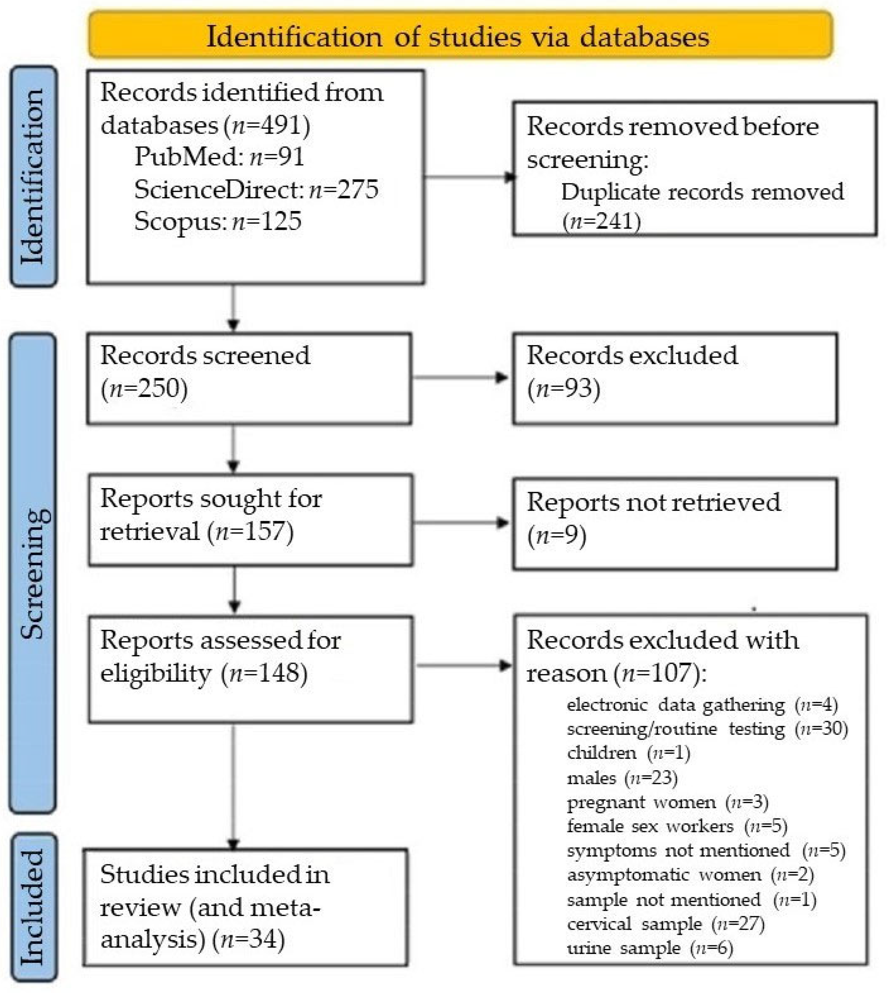
Following database exploration in accordance with the PRISMA guidelines from 1 January 2008 to 31 December 2023, a total number of 491 articles relating to the prevalence of T. vaginalis among European women with symptoms of vaginitis were retrieved initially (Figure 3).
Finally, 34 eligible papers were selected for further meta-analysis (Supplementary Table S2). The 34 studies analysing the prevalence of T. vaginalis comprised a sample population of 101,212 symptomatic women (median, 255; interquartile range, 143–625; range, 25–40,730) from 34 study populations, were conducted from 1996 to 2021, and were published from 2008 to 2023. On average, articles were published five years after the year that the corresponding study was started. The included studies were performed in 10/55 (18.2%) European states. About half of the contributions were from Türkiye (18/34; 52.9%; n = 4153), followed by Italy (3/34; 8.8%%; n = 68,458), Greece (3/34; 8.8%%; n = 9520), and the United Kingdom (3/34; 8.8%; n = 5397). Of the 34 studies, 26 (76.5%) were carried out in five countries in Southern Europe, namely, Bosnia and Herzegovina, Greece, Italy, Spain, and Türkiye, and 8 (23.5%) were carried out in five countries in Northwestern Europe, namely, the Netherlands, Lithuania, Poland, Sweden, and the United Kingdom (England, Scotland), enrolling 87,301 and 13,911 adult female patients, respectively (Table 4). A prevalence of T. vaginalis infection of 4.5% (95% CI: 1.6–7.3%) was estimated in Greece (Supplementary Figure S2). A T. vaginalis infection prevalence of 4.8% (95% CI: 3.2–6.3%) was estimated in Europe (Supplementary Figure S3). Regarding the studies from Greece and Europe, we identified a considerable between-study heterogeneity of 94% and 100%, respectively. Concerning the studies from Europe as a whole, we found a high p-value of 0.28 using Egger’s test (Supplementary Figure S4).

3.2.2. Subgroup Analyses Based on Geographical Location

The results from the subgroup analyses regarding geographic variations in prevalence estimates among symptomatic women in Europe are summarised in Table 4.
Studies from Europe were split into subsets based on their study location to explore the between-study heterogeneity. The estimate for the pooled prevalence of T. vaginalis infection in Northwestern Europe—The Netherlands, Lithuania, Poland, Sweden, and the United Kingdom (England, Scotland)—differed statistically significantly from that in Southern Europe, which included Bosnia and Herzegovina, Greece, Italy, Spain, and Türkiye (estimated PR = 2.1% vs. estimated PR = 5.7%, respectively), as shown in Supplementary Figure S5. There was a highly statistically significant difference between the overall results for Türkiye and Northwestern Europe, which included the Netherlands, Lithuania, Poland, Sweden, and the United Kingdom (England, Scotland) (estimated PR = 6.7% vs. estimated PR = 2.1%, respectively), as demonstrated in Supplementary Figure S6. A highly statistically significant difference was also found between the overall results for Türkiye and the rest of Southern Europe, i.e., Bosnia and Herzegovina, Greece, Italy, and Spain (estimated PR = 6.7% vs. estimated PR = 3.2%, respectively), as shown in Supplementary Figure S7. The estimated prevalence in Greece did not differ statistically significantly from that in Northwestern Europe, i.e., the Netherlands, Lithuania, Poland, Sweden, and the United Kingdom (England, Scotland) (estimated PR = 4.5% vs. estimated PR = 2.1%, respectively), as demonstrated in Supplementary Figure S8. Additionally, it did not differ statistically significantly from that in the rest of Southern Europe excluding Türkiye, i.e., Bosnia and Herzegovina, Italy, and Spain (estimated PR = 4.5% vs. estimated PR = 1.5%, respectively), as shown in Supplementary Figure S9. No statistically significant difference was found between the T. vaginalis prevalence estimates for Greece and Türkiye either (estimated PR = 4.5% vs. estimated PR = 6.7%, respectively), as demonstrated in Supplementary Figure S10.

4. Discussion

This is the first laboratory-based cross-sectional study to assess the performance of diagnostic tests, including a commercial automated NAAT, to identify the best option for the detection of T. vaginalis in vaginal samples from symptomatic women in Greece. The fact that the NAAT was found to detect T. vaginalis infections in Greek women with vaginal symptoms more effectively than culture did is in agreement with the findings from other studies [13,14]. The failure of the NAAT in one culture-positive sample may perhaps be attributed to the presence of PCR inhibitors, according to method’s specifications. However, the two methods did not differ statistically significantly in their diagnostic yields. Vaginal wet mount examination failed to detect all the samples that tested positive by means of culture [15,16] and/or the NAAT [17], and, as expected, Giemsa staining had the lowest yield of all the diagnostic tests that were used in our study [18]. The wet mount preparation test, irrespective of the chosen referent, was found to be less sensitive than both the automated NAAT and culture and only outperformed the Giemsa stain. Our findings were similar to previously reported results [19].
In our symptomatic adult female population, we determined the prevalence of infection with T. vaginalis to be 4.3% by using the results from both the conventional and the alternative gold standard: culture and the NAAT. It is worth noting that in our study, only very few vaginal samples were deemed positive for T. vaginalis based on all the methods, including the least sensitive Giemsa stain; we hypothesise that this finding could reflect a low rate of symptomatic women who are heavily parasitised by T. vaginalis in Greece [20]. The positivity rate of the NAAT was very high (94.1%), outperforming those of all the other tests. The specificity ranged between 99.2% and 100% and did not differ among tests. The positive predictive values of the NAAT and culture were high, corresponding to at least four out of five positive results for T. vaginalis being true positives. Wet mount had the lowest positive predictive value of 76.9%; the very wide confidence intervals of the positive predictive value did not enable precise population estimates [21]. All tests had high negative predictive values.
The culture and NAAT results were in almost perfect agreement (99.2%; κ = 0.90), which is in accordance with those from previous studies [13,14].
To sum up, in order to detect T. vaginalis in vaginal samples from symptomatic Greek women, culture and the NAAT have been proven to perform better than the wet mount method (the most commonly used method for the detection of T. vaginalis in Greece) and the Giemsa stain. The automated NAAT outperformed culture in a specialised medium in terms of sensitivity, specificity, positive predictive value, and negative predictive value [14]. Hence, the NAAT should be preferred to culture for the detection of T. vaginalis, considering that specialised molecular equipment is now available in Greek laboratory facilities, despite not currently being the most cost-effective option [22]. The automated NAAT also proved to be a diagnostic modality that ensures rapid turnaround times for results, thus enabling a reduction in the time to diagnosis and treatment of T. vaginalis infection in symptomatic women seeking medical attention in a Greek clinical setting such as ours. In contrast, culture in a specialised medium was unsurprisingly shown in our study to be a labour-intensive and time-consuming process [23]. This finding could have prevented culture from being routinely implemented in clinical laboratories throughout Greece.
Infections with T. vaginalis have not been given sufficient medical attention in Greece. The available data on the prevalence of T. vaginalis infection come from only a limited number of studies in mixed female populations, published over a period of almost 40 years. A Greek study, published in 1986, used wet mount microscopy and found that the prevalence of T. vaginalis infection reached nearly 21% in women with vaginal discharge, while showing a significant association between the infection and “women’s professions”—perhaps a modest reference of the era to those providing sex work—and educational level [24]. In a four-year study that was published in 1988 but started in 1982, T. vaginalis was microscopically detected in 11% of vaginal samples collected from women with lower urinary tract symptoms at a Greek university’s obstetrics and gynaecology department [25]. Twenty years later, a four-year study was published in 2008; it was conducted in a Greek private obstetrics and gynaecology hospital from 2003 to 2007 and detected T. vaginalis in 6.2% of vaginal samples from women with vaginitis using light microscopy [26]. A study, run in a major Greek gynaecology hospital in 2006–2007 and published in 2010, used wet mount, antigen testing, culture and in-house PCR for the detection of T. vaginalis in vaginal samples and found prevalences of 6.7% and 2.4% in symptomatic and asymptomatic Greek and immigrant women, respectively [27]. In 2017, two studies were published. The first was a three-year study that had started in 2010 and microscopically detected T. vaginalis in vaginal samples from 1.3% of women presenting with vaginitis at an outpatient gynaecology clinic in Greece [28]. The second study, conducted in a Greek university hospital in 2015, found no cases of T. vaginalis infection among females of different ages using wet mount slide observation [29]. Two additional studies, both published in 2018, found zero cases of T. vaginalis infection. The two-year study, conducted from 2014 to 2016, did not detect T. vaginalis in the vaginal secretions of asymptomatic women by means of vaginal smear microscopy [30]. The other study, conducted in 2015–2016, used a molecular syndromic panel but failed to detect T. vaginalis in cervical samples [31]. In all but one study, wet mount microscopy was used to detect T. vaginalis in vaginal samples.
Since 2008, three studies have succeeded in detecting T. vaginalis in vaginal samples from women with vaginal symptoms in Greece and were, therefore, included in our meta-analysis [26,27,28]. The results from the pooled analyses of Greek and European studies showed that the prevalence estimates of T. vaginalis infection in Greece and Europe were very similar. An estimated 4.5% and 4.8% of symptomatic women were infected with T. vaginalis in Greece and Europe, respectively. That the prevalence estimates were much higher than that reported by the WHO [9] came as no surprise, as we selected studies on T. vaginalis infection in symptomatic women and not in the general population, which also includes asymptomatic women and men. A similar prevalence of 4.3%, set based on our laboratory-based analysis, fell within the 95% confidence intervals of the pooled estimates for T. vaginalis prevalence in Greece and Europe.
Concerning the studies from Greece that were included in our meta-analysis, a bias due to their small number (n = 3) cannot be excluded [32].
Considering the studies from Europe, there was no indication of publication bias. Therefore, the heterogeneity observed between the studies, which was analysed through subgroup analyses based on geographical settings, can be regarded as valid [33]. The results from these subgroup analyses supported our notion that there were geographical variations in T. vaginalis prevalence across Europe [34]. This might explain, at least partially, the heterogeneity that was observed between studies from Europe on the T. vaginalis prevalence in symptomatic women. The estimated prevalence of T. vaginalis infection in Southern Europe was nearly two times higher than that in Northwestern Europe. The estimate in Türkiye was three times as high as that in Northwestern Europe and two times as high as that in Southern Europe, including Greece. In Greece, the prevalence estimate for T. vaginalis infection was two times as high as that in Northwestern Europe and three times as high as that in the rest of Southern Europe without Türkiye, while it was two-thirds that in Türkiye.
To summarise our meta-analysis findings, the prevalence of T. vaginalis infection in Greece is similar to that in Europe as a whole. However, it was shown to be higher than those in Northwestern and Southern Europe were but closer to that in Türkiye, where the highest prevalence rate in Europe has been observed and where cultural reasons have prevented women from being tested for T. vaginalis [35,36]. It is possible that Greek women also underutilise medical services [37], as symptoms of T. vaginalis infection have been seen as troubling but not serious, as opposed to those of other STIs. In addition, social stigma related to STIs remains a barrier to testing [38] and seeking treatment for T. vaginalis infection, even though it can be quickly and easily cured. Nevertheless, countries are advised to meet the WHO’s goal of ending the worldwide epidemics of STIs, including infections caused by T. vaginalis, by 2030 [39]. For more than a decade, European countries, such as those in the United Kingdom with a T. vaginalis infection prevalence of around 3–4% [13,17,21], have already been establishing guidelines and implemented programmes targeting women with vaginal symptoms in order to prevent and control this treatable STI. Despite being a European country with a relatively high T. vaginalis infection prevalence, Greece currently remains behind this target; thus, there is an urgent need to set national guidelines outlining testing, treating, and monitoring T. vaginalis infection in key target populations such as symptomatic women in order to plan interventions against the spread of T. vaginalis infections.
Our findings should be interpreted taking five limitations into consideration. First, the laboratory-based study did not also assess other commercial kits for the detection of T. vaginalis in vaginal samples. However, immunochromatography (lateral flow immunoassay) tests, albeit available, are not routinely used in Greek clinical laboratories, and other molecular point-of-care devices or NAAT techniques, such as those utilising chemiluminescent probe hybridisation, that are used in other European countries [13,17,21] are currently unavailable in the Greek market. Second, as our study focused on adult women with vaginal symptoms, asymptomatic women and at-risk groups such as female sex workers were excluded from our analysis; thus, our study findings cannot be applied to the broader female population or the general population, including males. Third, specifically designed studies on the prevalence of T. vaginalis were lacking, partially limiting the generalisability of the current meta-analysis conclusions, partly because T. vaginalis infection has not usually been considered harmful, apart from Türkiye, where the infection has not been considered a minor public health issue, which partly explains the over-representation of Turkish studies in the meta-analysis. Fourth, the within-study prevalence rate heterogeneity might be partitioned due to the geographical distribution of the included studies but also due to time-dependent prevalence differentiations, as studies conducted in different decades tend to raise questions about the validity of meta-analysis results. Fifth, most of the included studies (19 out of 34; 56%) did not provide details regarding their laboratory testing methods. Consequently, conducting a subgroup meta-analysis that compares studies employing various diagnostic methods lacked the capacity to adequately account for the variability in T. vaginalis prevalence. There was not sufficient information to determine whether the prevalence rates reported in our meta-analyses were influenced by the measurement methods that were utilised. Nevertheless, we did evaluate the sensitivity and specificity of different techniques in our laboratory-based cross-sectional study.
Despite these limitations, our study has presented new data on the performance of diagnostic tests, including an automated rapid NAAT that can be used as a point-of-care test [40], for the detection of T. vaginalis in vaginal samples from symptomatic women in Greece. We also provided recent data on the relatively high prevalence of T. vaginalis infection in this target population to promote testing and control this treatable STI in the country.

5. Conclusions

Our findings highlight the significance of testing T. vaginalis infection in women with vaginal symptoms in Greece. We suggest that Greek health authorities urgently undertake measures towards reducing the prevalence of T. vaginalis infection, the most frequent of the four curable STIs, to reach the WHO’s goal of ending the hidden epidemics of STIs globally. NAATs, such as the rapid molecular test that we employed, may be the preferred modality for the diagnosis of T. vaginalis infection in Greek clinical settings. Also considered a point-of-care test, the rapid automated PCR-based NAAT would be an asset in facilitating the detection and, thus, the fast treatment of T. vaginalis infection in symptomatic women in Greece.

Supplementary Materials

The following supporting information can be downloaded at https://www.mdpi.com/article/10.3390/amh70030029/s1, Figure S1: Results from Xpert TV® assay. a1. and a2. Trichomonas vaginalis positive Sample #4 tested negative using all other methods; b1. and b2. Trichomonas vaginalis-negative Sample #19 tested positive with the wet mount method. All samples were anonymised and decoded; Figure S2: Pooled prevalence of Trichomonas vaginalis infection in Greece, 2008–2023; Figure S3: Pooled prevalence of Trichomonas vaginalis infection in Europe, 2008–2023; Figure S4: Publication bias of studies on the prevalence of Trichomonas vaginalis infection in Europe, 2008–2023; Figure S5: Pooled prevalence of Trichomonas vaginalis infection in Northwestern Europe—the Netherlands, Lithuania, Poland, Sweden, and the United Kingdom (England, Scotland)—and Southern Europe—Bosnia and Herzegovina, Greece, Italy, Spain, and Türkiye—in 2008–2023; Figure S6: Pooled prevalence of Trichomonas vaginalis infection in Türkiye and Northwestern Europe (the Netherlands, Lithuania, Poland, Sweden, and the United Kingdom (England, Scotland)), 2008–2023; Figure S7: Pooled prevalence of Trichomonas vaginalis infection in Türkiye and the rest of Southern Europe, i.e., Bosnia and Herzegovina, Greece, Italy, and Spain, 2008–2023; Figure S8: Pooled prevalence of Trichomonas vaginalis infection in Greece and Northwestern Europe (the Netherlands, Lithuania, Poland, Sweden, and the United Kingdom (England, Scotland)), 2008–2023; Figure S9: Pooled prevalence of Trichomonas vaginalis infection in Greece and the rest of Southern Europe, i.e., Bosnia and Herzegovina, Italy, Spain, and Türkiye, 2008–2023; Figure S10: Pooled prevalence of Trichomonas vaginalis infection in Greece and Türkiye, 2008–2023; Table S1: Vaginal samples that tested Trichomonas vaginalis-positive by means of at least one method. All samples were anonymised and decoded; Table S2: Eligible studies reporting Trichomonas vaginalis prevalence among symptomatic women in Europe, 2008–2023 [41,42,43,44,45,46,47,48,49,50,51,52,53,54,55,56,57,58,59,60,61,62,63,64,65,66].

Author Contributions

E.V., C.M.V. and L.T. were involved in the conception of this study. L.T. and C.M.V. wrote the manuscript. K.B. was involved in the clinical sample acquisition. L.T. and P.G. contributed to the clinical laboratory data acquisition and methodology. L.T. and E.G. contributed to the acquisition of data for the review. C.M.V., N.G. and E.V. contributed to the review and meta-analysis methodology. C.M.V. and N.G. checked and approved the authenticity of the raw data. N.G. analysed the data. C.M.V. and E.V. reviewed and edited the manuscript. E.P. coordinated the transport of clinical samples. E.V., C.S. and S.B. were involved in the supervision of this study. L.T. conducted this study as part of his research towards a Ph.D. degree at the Department of Public and Community Health, School of Public Health, University of West Attica, Athens, Greece. All authors have read and agreed to the published version of the manuscript.

Funding

This research received no external funding.

Institutional Review Board Statement

The Scientific Board of the General Hospital of Nea Ionia “Konstantopouleio-Patision”, Athens, Greece, approved this study following a thorough review by the hospital’s Ethics Committee (SBO No 20059/20 July 2021).

Informed Consent Statement

A waiver of signed consent was granted due to the anonymous analysis of clinical data, minimal risk of harm, and potential social stigma. Samples were anonymised and decoded.

Data Availability Statement

The data are contained within the article.

Acknowledgments

We are thankful to the laboratory staff of the hospital. The opinions presented in this article are those of the authors and do not necessarily represent those of their institutions.

Conflicts of Interest

The authors declare no conflicts of interest.

References

  1. Shiratori, M.; Patel, A.; Gerhold, R.W.; Sullivan, S.A.; Carlton, J.M. Persistent Trichomonas vaginalis infections and the pseudocyst form. Trends Parasitol. 2023, 39, 1023–1031. [Google Scholar] [CrossRef] [PubMed]
  2. Rosales-Rimache, J.; Inolopú, J.L.; Soncco-Llulluy, F.C.; Medina-Ciprian, L. Comparison of three methods for diagnosing trichomoniasis in female patients with sexual activity attended at a hospital in Peru. J. Parasitol. Res. 2023, 2023, 9528942. [Google Scholar] [CrossRef] [PubMed]
  3. Dadwal, R.; Sharma, N.; Kanaujia, R.; Malhorta, S.; Chaundry, H.; Rathore, S.; Saini, A.; Bagga, R.; Mewara, A.; Khurana, S.; et al. Prevalence of Trichomonas vaginalis by polymerase chain reaction-based molecular method among symptomatic women from Northern India. Indian J. Sex. Transm. Dis. AIDS 2023, 44, 40–44. [Google Scholar] [CrossRef] [PubMed]
  4. Workowski, K.A.; Bola, G.A. Sexually transmitted diseases treatment guidelines, 2025. MMWR Recomm. Rep. 2015, 64, 1–137. [Google Scholar]
  5. Sinka, K. The global burden of sexually transmitted infections. Clin. Dermatol. 2024, 42, 110–118. [Google Scholar] [CrossRef]
  6. Sherrard, J.; Pitt, R.; Russel Hobbs, K.; Maynard, M.; Cochrane, E.; Wilson, J.; Tipple, C. British Association for Sexual Health and HIV (BASHH) United Kingdom national guideline on the management of Trichomonas vaginalis 2021. Int. J. STD AIDS 2022, 33, 740–750. [Google Scholar] [CrossRef]
  7. Wayal, S.; Aicken, C.R.H.; Griffiths, C.; Blomquist, P.B.; Hughes, G.; Mercer, C.H. Understanding the burden of bacterial sexually transmitted infections and Trichomonas vaginalis among black Caribbeans in the United Kingdom: Findings from a systematic review. PLoS ONE 2018, 13, e0208315. [Google Scholar] [CrossRef]
  8. Meites, E.; Gaydos, C.A.; Hobbs, M.M.; Kissinger, P.; Nyirjesy, P.; Schwebke, J.R.; Secor, W.E.; Sobel, J.D.; Workowski, K.A. A review of evidence-based care of symptomatic trichomoniasis and asymptomatic Trichomonas vaginalis infections. Clin. Infect. Dis. 2015, 61 (Suppl. S8), S837–S848. [Google Scholar] [CrossRef]
  9. Rowley, J.; Vander Hoorn, S.; Korenromp, E.; Low, N.; Unemo, M.; AbuRaddad, L.J.; Chico, R.M.; Smolak, A.; Newman, L.; Gottlieb, S.; et al. Chlamydia, gonorrhoea, trichomoniasis and syphilis: Global prevalence and incidence estimates, 2016. Bull. World Health Organ. 2019, 97, 548–562. [Google Scholar] [CrossRef]
  10. World Health Organization. Global Lobal Health Sector Strategies on, Respectively, HIV, Viral Hepatitis and Sexually Transmitted Infections for the Period 2022–2030; World Health Organization: Geneva, Switzerland, 2022; ISBN 978-92-4-005377-9. [Google Scholar]
  11. National Health Organization. Epidemiological and Laboratory Surveillance of Sexually Transmitted Diseases. 2023. Available online: www.eody.gov.gr/smn_dedomena_2023 (accessed on 4 May 2025). (In Greek)
  12. Moher, D.; Shamseer, L.; Clarke, M.; Ghersi, D.; Liberati, A.; Petticrew, M.; Shekelle, P.; Stewart, L.A. Preferred Reporting Items for Systematic Review and Meta-Analysis Protocols (PRISMA-P) 2015 statement. Syst. Rev. 2015, 4, 1. [Google Scholar] [CrossRef]
  13. Hathorn, E.; Ng, A.; Page, M.; Hodson, J.; Gaydos, C.; Ross, J.D. A service evaluation of the Gen-Probe APTIMA nucleic acid amplification test for Trichomonas vaginalis: Should it change whom we screen for infection? Sex Transm Infect. 2015, 91, 81–86. [Google Scholar] [CrossRef] [PubMed]
  14. Schwebke, J.R.; Gaydos, C.A.; Davis, T.; Marrazzo, J.; Furgerson, D.; Taylor, S.N.; Smith, B.; Bachmann, L.H.; Ackerman, R.; Spurrell, T.; et al. Clinical evaluation of the Cepheid Xpert TV assay for detection of Trichomonas vaginalis with prospectively collected specimens from men and women. J. Clin. Microbiol. 2018, 56, e01091-17. [Google Scholar] [CrossRef]
  15. Çulha, G.; Günoren, A.; Demir, C.; Hakverdi, A.; Duran, N. Detection of Trichomonas vaginalis in vaginal specimens from women by wet mount, culture and PCR. J. Clin. Anal. Med. 2015, 6, 537–554. [Google Scholar]
  16. Sönmez, C.; Usluca, S. Cases with urogenital discharge: Trichomonas vaginalis rates? Flora J. Infect. Dis. Clin. Microbiol. 2018, 23, 79–83. [Google Scholar] [CrossRef]
  17. Shone, J.; Winter, A.; Jones, B.L.; Butt, A.; Brawley, D.; Cunningham, C.; Paterson, J.; McAllister, G.; Alexander, C.L. A Scottish multi-centre service evaluation examining the prevalence and diagnosis of Trichomonas vaginalis in symptomatic women attending sexual health clinics. Int. J. STD AIDS 2016, 27, 1066–1070. [Google Scholar] [CrossRef] [PubMed]
  18. Radonjic, I.V.; Dzamic, A.M.; Mitrovic, S.M.; Arsenijevic, V.S.A.; Popadic, D.M.; Zec, I.F.K. Diagnosis of Trichomonas vaginalis infection: The sensitivities and specificities of microscopy, culture and PCR assay. Eur. J. Obstet. Gynecol. Repro. Biol. 2006, 126, 116–120. [Google Scholar] [CrossRef] [PubMed]
  19. Gomes Cardoso, F.; Freitas, M.D.; Tasca, T.; Vargas Rigo, G. From wet mount to nucleic acid amplification techniques: Current diagnostic methods and future perspectives based on patenting of new assays, stains, and diagnostic images for Trichomonas vaginalis detection. Venereology 2024, 3, 35–50. [Google Scholar] [CrossRef]
  20. Mahmoud, A.; Sherif, N.A.; Abdella, R.; El-genedy, A.R.; Ek Kateb, A.Y.; Askalani, A.N.H. Prevalence of Trichomonas vaginalis infection among Egyptian women using culture and Latex agglutination: Cross-sectional study. BMC Women’s Health 2015, 15, 7. [Google Scholar] [CrossRef]
  21. Nathan, B.; Appiah, J.; Saunders, P.; Heron, D.; Nichols, T.; Brum, R.; Alexander, S.; Baraitser, P.; Ison, C. Microscopy outperformed in a comparison of five methods for detecting Trichomonas vaginalis in symptomatic women. Int. J. STD AIDS 2014, 26, 251–256. [Google Scholar] [CrossRef]
  22. Nicholls, J.E.; Turner, K.M.E.; North, P.; Ferguson, R.; May, M.T.; Gough, K.; Macleod, J.; Muir, P.; Horner, P.J. Cross-sectional study to evaluate Trichomonas vaginalis positivity in women tested for Neisseria gonorrhoeae and Chlamydia trachomatis, attending genitourinary medicine and primary care clinics in Bristol, South West England. Sex. Transm. Infect. 2018, 94, 93–99. [Google Scholar] [CrossRef]
  23. Hobbs, M.M.; Seña, A.C. Modern diagnosis of Trichomonas vaginalis infection. Sex. Transm. Infect. 2013, 89, 434–438. [Google Scholar] [CrossRef] [PubMed]
  24. Papapetropoulou, M.; Legakis, N.J.; Detorakis, J.; Kalambokas, E.; Lymberopoulou, T. Frequency and epidemiologic associations of different types of vaginitis in symptomatic women in Greece. Eur. J. Clin. Microbiol. 1986, 5, 447–449. [Google Scholar] [CrossRef]
  25. Vitoratos, N.; Gregoriou, O.; Papadias, C.; Liapis, A.; Zourlas, P.A. Sexually transmittes diseses in women with urethral syndrome. Int. J. Gynecol. Obstet. 1988, 27, 177–180. [Google Scholar] [CrossRef] [PubMed]
  26. Iavazzo, C.; Vogiatzi, C.; Falagas, M.E. A retrospective analysis of isolates from patients with vaginitis in a private Greek obstetric/gynecological hospital (2003–2006). Med. Sci. Monit. 2008, 14, CR228-31. [Google Scholar] [PubMed]
  27. Piperaki, E.T.; Theodora, M.; Mendris, M.; Barbitsa, L.; Pitiriga, V.; Antsaklis, A.; Tsakris, A. Prevalence of Trichomonas vaginalis infection in women attending a major gynaecological hospital in Greece: A cross-sectional study. J. Clin. Pathol. 2010, 63, 249–253. [Google Scholar] [CrossRef]
  28. Ioannidis, A.; Papaioannou, P.; Magiorkinis, E.; Magana, M.; Ioannidou, V.; Tzanetou, K.; Burriel, A.R.; Tsironi, M.; Chatzipanagiotou, S. Detecting the diversity of Mycoplasma and Ureaplasma endosymbionts hosted by Trichomonas vaginalis isolates. Front. Microbiol. 2017, 28, 1188. [Google Scholar] [CrossRef]
  29. Sianou, A.; Galyfos, G.; Moragianni, D.; Baka, S. Prevalence of vaginitis in different age groups among females in Greece. J. Obstet. Gynaecol. 2017, 37, 790–794. [Google Scholar] [CrossRef]
  30. Florou, Z.; Pantelidi, K.; Fountas, S.; Skoulakis, A.; Messini, C.; Lachanas, V.; Petinaki, E. High prevalence of sexually transmitted infections (STIs) in asymptomatic Greek women. Arch. Clin. Microbiol. 2018, 9, 78. [Google Scholar]
  31. Parthenis, C.; Panagopoulos, P.; Margari, N.; Kottaridi, C.; Spathis, A.; Pouliakis, A.; Konstantoudakis, S.; Chrelias, G.; Chrelias, C.; Papantoniou, N.; et al. The association between sexually transmitted infections, human papillomavirus, and cervical cytology abnormalities among women in Greece. Int. J. Infect. Dis. 2018, 73, 72–77. [Google Scholar] [CrossRef]
  32. von Hippel, P.T. The hererogeneity statistic I2 can be biases in small meta-analyses. BMC Med. Res. Methodol. 2015, 15, 35. [Google Scholar] [CrossRef]
  33. Augusteijn, H.E.M.; van Aert, R.C.; van Assen, M.A.L.M. The effect of publication bias on the Q test and assessment of heterogeneity. Psychol. Methods 2010, 24, 116–134. [Google Scholar] [CrossRef] [PubMed]
  34. Spineli, L.M.; Pandis, N. Exploring heterogeneity in meta-analysis: Subgroup analysis. Part 1. Am. J. Orthod. Dentofacial Orthop. 2020, 158, 302–304. [Google Scholar] [CrossRef] [PubMed]
  35. Özarmagan, G.; Bingham, J.S. Sexually transmitted infections in Turkey. Int. J. STD AIDS 2001, 12, 824–828. [Google Scholar] [CrossRef] [PubMed]
  36. Polat, F.; Kaya Şenol, D. Examining the correlation between sexual and reproductive health stigmatization level and gender perception: A case of a university in Turkey—A descriptive cross-sectional study. Sao Paulo Med. J. 2022, 141, 146–153. [Google Scholar] [CrossRef]
  37. Kalavrezou, N. Gender and access to healthcare in Greece. Soc. Cohes. Dev. 2009, 4, 205–216. [Google Scholar] [CrossRef]
  38. Garcia, P.J.; Espinosa Miranda, A.; Gupta, S.; Garland, S.M.; Escobar, M.E.; Fortenberry, J.D.; The International Union Against Sexually Transmitted Infections. The role of sexually transmitted infections (STI) prevention and control programs in reducing gender, sexual and STI-related stigma. eClinicalMedicine 2021, 33, 100764. [Google Scholar] [CrossRef]
  39. European Centre for Disease Prevention and Control. Progress Towards Reaching the Sustainable Development Goals Related to HIV, Viral Hepatitis, Sexually Transmitted Infections and Tuberculosis in the EU/EEA: 2024 Progress Report; ECDC: Stockholm, Sweden, 2025. [Google Scholar]
  40. Jacobsson, S.; Boiko, I.; Golparian, D.; Blondeel, K.; Kiarie, J.; Toskin, I.; Peeling, R.W.; Unemo, M. WHO laboratory validation of Xpert® CT/NG and Xpert® TV on the GeneXpert system verifies high performances. APMIS 2018, 126, 907–912. [Google Scholar] [CrossRef]
  41. Sönmez Tamer, G.; Dündar, D.; Çalışkan, Ş.; Doğer, E. Comparison of direct microscopy and in–vitro cultures in detection of Trichomonas vaginalis. Turk. Bull. Hyg. Exp. Biol. 2008, 65, 75–80. [Google Scholar]
  42. Sönmez Tamer, G.; Keçeli Ozcan, S.; Yücesoy, G.; Gacar, G. The relation between trichomoniasis and contraceptive methods. Turk. Parazitol. Derg. 2009, 33, 266–269. (In Turkish) [Google Scholar]
  43. Tibaldi, C.; Cappello, N.; Latino, M.A.; Masuelli, G.; Marini, S.; Benedetto, C. Vaginal and endocervical microorganisms in symptomatic and asymptomatic non-pregnant females: Risk factors and rates of occurrence. Clin. Microbiol. Infect. 2009, 15, 670–679. [Google Scholar] [CrossRef]
  44. Akdemir, C.; Keskin, N.; Çoksüer, H. A survey of prevalence of Trichomonas vaginalis in cases with vaginal discharge in Kütahya by classic miscroscopy and DNA hybridization. Türk. Hij. Den. Biyol. Derg. 2010, 67, 161–166. [Google Scholar]
  45. Casari, E.; Ferrario, A.; Morenghi, E.; Montanelli, A. Gardnerella, Trichomonas vaginalis, Candida, Chlamydia trachomatis, Mycoplasma hominis and Ureaplasma urealyticum in the genital discharge of symptomatic fertile and asymptomatic infertile women. New Microbiol. 2010, 33, 69–76. [Google Scholar] [PubMed]
  46. Keleştemur, N.; Kaplan, M. The prevalence of T. vaginalıs in women with vaginitis in Elazığ. Med. Sci. 2010, 5, 1–5. [Google Scholar]
  47. Çetinkaya, Ü.; Yazar, S.; Serin, S.; Hamamci, B.; Kuk, S. Trichomonas vaginalis positivity according to type of vaginal discharge in women. Turk. Klin. Tip Bilim. Derg. 2011, 31, 1094–1099. [Google Scholar] [CrossRef]
  48. Değerli, S.; Şalk, S.; Malatyalı, E. Incidence in Sivas of Trichomonas vaginalis in patients with vaginitis. Turk. Parazitol. Derg. 2011, 35, 145–147. (In Turkish) [Google Scholar] [CrossRef]
  49. Polat, E.; Sirekbasan, S.; Yıldırım, Z.; Bağdatlı, Y.; Çepni, I.; Çift, T.; Baltalı, N.D. Comparing the occurrence of Trichomonas vaginalis infections today to ten years ago among women prostitutes and gynecology and obstetrics patients in Istanbul. Turk. Parazitol. Derg. 2011, 35, 68–71. (In Turkish) [Google Scholar] [CrossRef]
  50. Keleştemur, N.; Kaplan, M.; Özdemir, E.; Erensoy, A. The frequency of Trichomonas vaginalis, Gardnerella vaginalis and Candida spp. among infertile men and women with vaginitis. Kafkas Unv. Vet. Fak. Derg. 2012, 18, 47–52. [Google Scholar]
  51. Keşli, R.; Pektaş, B.; Ozdemir, M.; Günenc, O.; Coşkun, E.; Baykan, M.; Baysal, B. Microscopic examination of vaginal discharge specimens for Trichomonas vaginalis and other micro-organisms in 18–45 age group women. Turk. Parazitol. Derg. 2012, 36, 182–184. (In Turkish) [Google Scholar] [CrossRef]
  52. Jahic, M.; Mulavdic, M.; Nurkic, J.; Jahic, E.; Nurkic, M. Clinical characteristics of aerobic vaginitis and its association to vaginal candidiasis, trichomonas vaginitis and bacterial vaginosis. Med. Arch. 2013, 67, 428–430. [Google Scholar] [CrossRef]
  53. Aycan-Kaya, Ö.; Silfeler, D.B.; Kurt, R.K.; Gözükarad, I.; Yengil, E.; Bayramoǧlu, N. Investigation of the presence of Trichomonas vaginalis in infertile Turkish women. Asian Biomed. 2015, 9, 659–663. [Google Scholar] [CrossRef]
  54. Pellrud, H.; Golparian, D.; Nilsson, C.S.; Falk, M.; Fredlund, H.; Unemo, M. Trichomonas vaginalis infections are rare among young patients attending an STI clinic in Sweden. Acta Derm. Venereol. 2015, 95, 343–344. [Google Scholar] [CrossRef] [PubMed]
  55. Salfa, M.C.; Suligoi, B.; Italian STI Laboratory-based surveillance working group. prevalence of Chlamydia trachomatis, Trichomonas vaginalis and Neisseria gonorrhoeae based on data collected by a network of clinical microbiology laboratories, in Italy. Adv. Exp. Med. Biol. 2016, 901, 47–57. [Google Scholar] [PubMed]
  56. Carrillo-Ávila, J.A.; Serrano-García, M.L.; Fernández-Parra, J.; Sorlózano-Puerto, A.; Navarro-Marí, J.M.; Stensvold, C.R.; Gutiérrez-Fernández, J. Prevalence and genetic diversity of Trichomonas vaginalis in the general population of Granada and co-infections with Gardnerella vaginalis and Candida species. J. Med. Microbiol. 2017, 66, 1436–1442. [Google Scholar] [CrossRef] [PubMed]
  57. Korycińska, J.; Dzika, E.; Waśniewski, T.; Lepczyńska, M.; Kubiak, K. The prevalence of Trichomonas vaginalis infections in the population of Warmińsko-Mazurskie voivodeship (North-Eastern Poland). Przegl. Epidemiol. 2017, 71, 547–554. [Google Scholar]
  58. Akyıldız, F.; Özçelik, S.; Özpınar, N.; Karakuş, S. Comparison of three different culture methods in the diagnosis and investigation of frequency of Trichomonas vaginalis in women with the pre–diagnosis of vaginitis. Turk. Bull. Hyg. Exp. Biol. 2018, 75, 43–52. [Google Scholar] [CrossRef]
  59. Sonmez, C.; Usluca, S.; Hakki Usluca, I.; Kalipci, I.; Sezen, F.; Resat Atalay, C.; Kilic, S. Evaluation of symptomatic patients with resistant discharge. Acta Dermatovenerol. Croat. 2018, 26, 1–7. [Google Scholar]
  60. Dogan, N.; Gitmez, F. Investigation of Trichomonas vaginalis frequency by different methods in women in Eskişehir province and evaluation of its relation with various social variables. Osman. J. Med. 2019, 41, 46–57. [Google Scholar] [CrossRef]
  61. Nijhuis, R.H.T.; Duinsbergen, R.G.; Pol, A.; Godschalk, P.C.R. Prevalence of Chlamydia trachomatis, Neisseria gonorrhoeae, Mycoplasma genitalium and Trichomonas vaginalis including relevant resistance-associated mutations in a single center in the Netherlands. Eur. J. Clin. Microbiol. Infect. Dis. 2021, 40, 591–595. [Google Scholar] [CrossRef]
  62. Türkmen Albayrak, H.; Albayrak, A.M.; Bakır, A.; Şahin, İ. Comparison of vulvovaginal infection diagnostic methods and effects of predisposing factors vulvovaginal infections. J. DU Health Sci. Inst. 2020, 10, 52–57. [Google Scholar]
  63. Yazısız, H.; Koyuncu Özyurt, Ö.; Öztürk Eryiğit, F.; Özhak, B.; Öngüt, G.; Özekinci, M.; Dönmez, L.; Çolak, D.; Gümüş, S.; Öğünç, D. Evaluation of microscopic examination, culture and polymerase chain reaction tests in the diagnosis of Trichomonas vaginalis infection. Mikrobiyol. Bul. 2020, 54, 135–143. (In Turkish) [Google Scholar] [CrossRef]
  64. Ertabaklar, H.; Malatyali, E.; Özün Özbay, E.P.; Yildiz, İ.; Sinecen, M.; Ertuğ, S.; Bozdoğan, B.; Güçlü, Ö. Microsatellite-Based Genotyping, analysis of population structure, presence of Trichomonas vaginalis Virus (TVV) and Mycoplasma hominis in T. vaginalis isolates from Southwest of Turkey. Iran J. Parasitol. 2021, 16, 81–90. [Google Scholar] [CrossRef] [PubMed]
  65. Opolskiene, G.; Bumbuliene, Z.; Kiveryte, S.; Bartkeviciute, A.; Ramasauskaite, D.; Bartkeviciene, D. The use of vaginal wet smear: Can we predict Mycoplasmas/Ureaplasmas? Arch. Gynecol. Obstet. 2021, 304, 157–162. [Google Scholar] [CrossRef] [PubMed]
  66. Yusuf, E.; Mertens, K.; van Lisdonk, N.; Houwen, C.; Thai, K.T.D. Epidemiology of Mycoplasma genitalium and Trichomonas vaginalis in the primary health care setting in the Netherlands. Epidemiol. Infect. 2023, 5, 151.e79. [Google Scholar] [CrossRef] [PubMed]
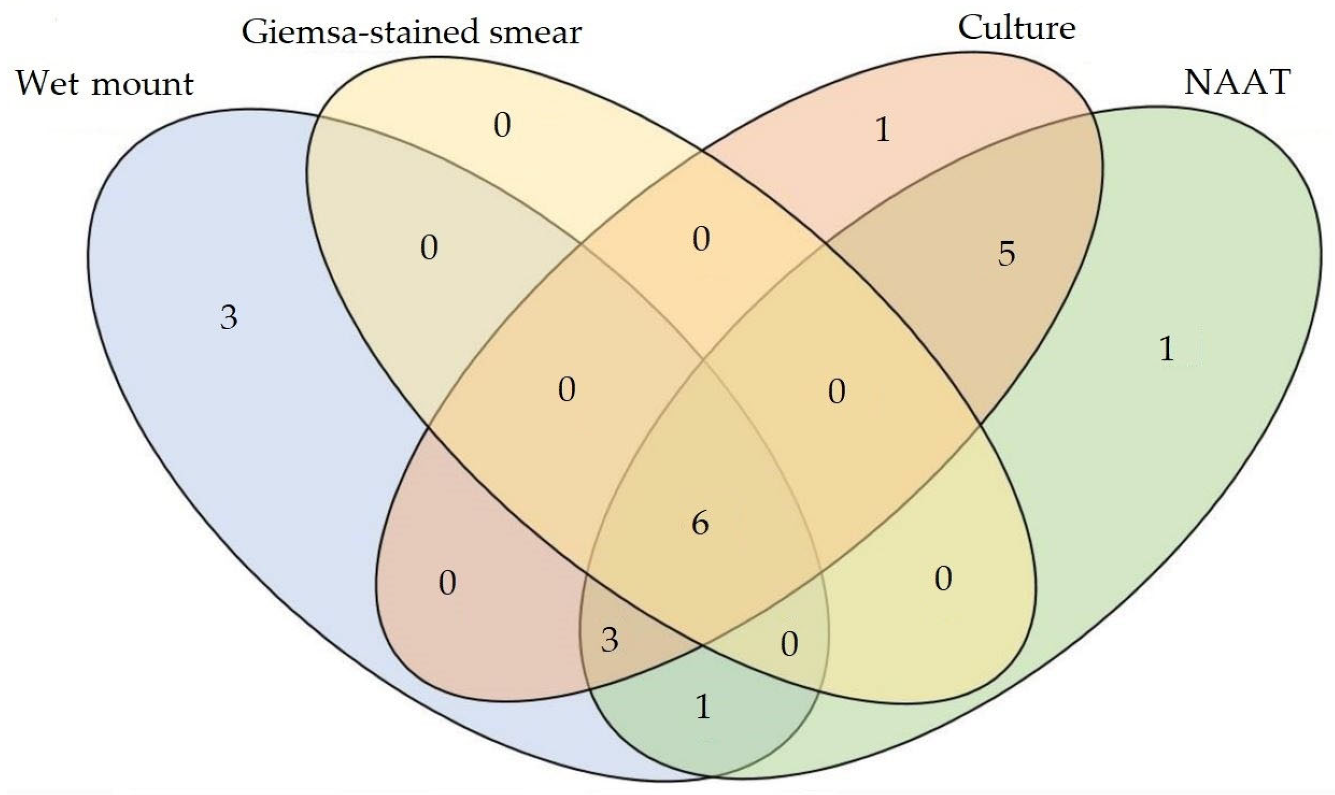
Figure 1. Venn diagram of the four methods for diagnosing Trichomonas vaginalis infection, visualising the overlap and differences in T. vaginalis-positive results between the methods.
Amh 70 00029 g001
Figure 2. Results from the wet mount, Giemsa staining, and culture in Oxoid Trichomonas Medium No.2. (a) Sample #16: a motile T. vaginalis trophozoite (arrow), observed in a wet mount (×400). (b) Sample #14: a violet, oval T. vaginalis trophozoite with a single darker nucleus (nu), a barely visible axostyle (ax), and a not well-outlined undulating membrane (um), detected using the Giemsa stain method (×1000). (c,d) Sample #9: T. vaginalis trophozoites from culture, observed by means of (c) wet mount examination (×400) and (d) Giemsa staining (×1000). All samples were anonymised and decoded.
Amh 70 00029 g002
Figure 3. Flow chart for the selection of studies on the Trichomonas vaginalis infection prevalence of systematic women in Europe.
Amh 70 00029 g003
Table 1. Performance of the wet mount, Giemsa stain, and nucleic acid amplification test (NAAT) for the detection of Trichomonas vaginalis, compared with culture as the gold standard.
Se aSp a
Method%(95% CI)%(95% CI)
Wet mount6032.3–83.79997.4–99.7
Giemsa stain4016.3–67.799.297.8–99.8
NAAT b93.368.1–99.895.598.1–99.9
a Se, sensitivity; Sp, specificity; b NAAT, nucleic acid amplification test.
Table 2. Performance of the wet mount, Giemsa stain, and culture for the detection of Trichomonas vaginalis, compared with the nucleic acid amplification test (NAAT) as the alternative gold standard.
Se aSp a
Method%(95% CI)%(95% CI)
Wet mount62.535.4–84.899.297.7–99.8
Giemsa stain37.515.2–64.610099.0–100
Culture85.561.6–98.599.798.6–99.9
a Se, sensitivity; Sp, specificity.
Table 3. Performance of the wet mount, Giemsa staining, culture, and nucleic acid amplification test (NAAT) for the detection of Trichomonas vaginalis in 399 vaginal samples from symptomatic women in Greece.
Se aSp aPPV aNPV a
Method%(95% CI)%(95% CI)%(95% CI)(%)(95% CI)
Wet mount58.832.9–81.699.297.7–99.876.950.2–91.798.296.8–99.0
Giemsa stain35.314.2–61.710099.0–10010054.1–10097.295.1–98.6
Culture88.263.6–98.510099.0–10010078.2–10099.598.1–99.9
NAAT b94.171.3–99.910099.0–10010079.4–10099.798.3–99.9
a Se, sensitivity; Sp, specificity; PPV, positive predictive value; NPV, negative predictive value; b NAAT, nucleic acid amplification test. N.B.: Calculations were carried out relative to the determined patient infection status.
Table 4. Estimated prevalence of Trichomonas vaginalis infection in European symptomatic women based on geographic location.
EuropeIncluded StudiesEstimated Prevalence of T. vaginalis Infection
Geograhic LocationCountries aNumberSample Size(%)(95% CI)Qd.f.p
Northwestern EuropeLt, Nl, Pl, Se, UK813,9112.11.1–3.19.831<0.001
Southern EuropeBa, Es, Gr, It, Tr2687,3015.73.7–7.6
Türkiye Tr1841536.74.0–9.310.11<0.001
Northwestern EuropeLt, Nl, Pl, Se, UK813,9112.11.1–3.1
TürkiyeTr1841536.74.0–9.35.571<0.001
Southern Europe (rest)Ba, Es, Gr, It883,1483.21.3–5.1
GreeceGr393874.51.6–7.32.3710.12
Northwestern EuropeLt, Nl, Pl, Se, UK813,9112.11.1–3.1
GreeceGr393874.51.6–7.33.9210.05
Southern Europe (rest)Ba, Es, It573,7611.50.8–2.2
GreeceGr393874.51.6–7.31.2210.27
TürkiyeTr1841536.74.0–9.3
a Ba, Bosnia and Herzegovina; Es, Spain; Gr, Greece; It, Italy; Lt, Lithuania; Nl, The Netherlands; Pl, Poland; Se, Sweden; Tr, Türkiye; UK, the United Kingdom (England, Scotland).
Disclaimer/Publisher’s Note: The statements, opinions and data contained in all publications are solely those of the individual author(s) and contributor(s) and not of MDPI and/or the editor(s). MDPI and/or the editor(s) disclaim responsibility for any injury to people or property resulting from any ideas, methods, instructions or products referred to in the content.