Article

Molecular Detection of SARS-CoV-2 Viral Particles in Exhaled Breath Condensate via Engineered Face Masks

by
Hannes Dörfler
1,*,
John Daniels
2,
Shekhar Wadekar
2,
Quentin Pagneux
3,
Dennis Ladage
4,
Georg Greiner
5,
Ojan Assadian
6,
Rabah Boukherroub
7 and
Sabine Szunerits
1,7
1
Laboratory for Life Sciences and Technology (LiST), Danube Private University, A-3500 Krems an der Donau, Austria
2
DiagMetrics Inc., 30 Renees Way, Madison, CT 06443, USA
3
e-DiagMetrics GmbH, Parkring 2, A-1010 Vienna, Austria
4
Department of Medicine, Danube Private University, A-3500 Krems an der Donau, Austria
5
Ludwig Boltzmann Institute for Hematology & Oncology, Medical University of Vienna, A-1090 Vienna, Austria
6
Department of Medicine, University Hospital Wiener Neustadt, A-2700 Wiener Neustadt, Austria
7
Univ. Lille, CNRS, Univ. Polytechnique Hauts-de-France, UMR 8520—IEMN, F-59000 Lille, France
*
Author to whom correspondence should be addressed.
LabMed 2024, 1(1), 22-32; https://doi.org/10.3390/labmed1010005
Submission received: 28 August 2024 / Revised: 24 September 2024 / Accepted: 28 October 2024 / Published: 12 November 2024
(This article belongs to the Collection Feature Papers in Laboratory Medicine)

Abstract

:
In this study, we present a novel face mask engineered for the collection of exhaled breath condensate (EBC) and its application and performance in a clinical study of COVID-19 infection status assessment versus the gold standard polymerase chain reaction (PCR) nasopharyngeal swab testing. EBC was collected within a clinical trial of COVID-19-infected and non-infected patients and analyzed by reverse transcription quantitative (RT-q) PCR, with the results being compared with nasopharyngeal sampling of the same patient. The cycle threshold (Ct) values of the nasopharyngeal samples were generally lower than those of EBC, with viral loads in EBC ranging from 1.2 × 10⁴ to 5 × 10⁸ viral particles mL−1 with 5 min of breathing. From the 60 clinical patients’ samples collected, 30 showed a confirmed SARS-CoV-2 infection. Of these 30 individuals, 22 (73%) had Ct values < 40 (representing the threshold for SARS-CoV-2 infectivity) using both amplification of ORF1a/b and the E-gene. The 30 EBC samples from non-infected participants were all identified as negative, indicating a 100% specificity. These first results encourage the use of the face mask as a noninvasive sampling method for patients with lung-related diseases, especially with a view to equipping the face mask with miniaturized sensing devices, representing a true point-of-care solution in the future.

1. Introduction

While globally the number of new cases due to infection with severe acute respiratory syndrome coronavirus 2 (SARS-CoV-2) is decreasing compared with the last few years, with a clear decline in mortality rate, the virus remains present and is mutating. A sub-lineage of the BA.2.86 Omicron variant was designated at the end of December 2023 as a variant of interest due to its rapid increase in prevalence in the last weeks [1].
As the virus is spread through respiratory droplets expelled from infected individuals during coughing, sneezing and speaking, as well as by simple breathing [2,3], face masks are not only an effective means to reduce the airborne transmission risk but are also useable for sample collection, specifically in the form of exhaled breath condensate (EBC). With approximately 102 EBC droplets per liter exhaled by healthy patients [4,5] and a breathing rate of 10 L min−1, about 103 EBC droplets are expelled every minute. This is comparable to coughing (~3 × 10³ droplets), with sneezing releasing about ten times more droplets (~4 × 10⁴ droplets) [6].
While breath has been used as diagnostic tool for the detection of a COVID-19 infection before [7], the analysis of EBC is particularly expected to provide valuable insights into the disease progression of respiratory conditions, including, but not limited to, COVID-19 [8,9], asthma [10,11], lung cancer [12], tuberculosis [13], and chronic obstructive pulmonary disease (COPD) [14]. Challenges related to sample collection and clinical viability have so far impeded the wider adoption of EBC for non-volatile biomarker analysis. Clinically, EBC is currently collected using condenser tubes [15], a rather expensive approach that limits the implementation for personalized use. However, Ryan et al. [15] demonstrated a 93.5% concordance between the RT-qPCR results of nasopharyngeal and EBC samples using R-Tube condensers for EBC collection and a four gene (S-gene, E-gene, N-gene, ORF1a/b) RT-qPCR analysis, which decreased to 68% when using two genes (E-gene and S-gene).
Next to commercial EBC collection systems such as the R-tube condensers, several engineered EBC-collection systems have been reported in the literature [16,17,18,19,20]. Wagner and co-workers recently proposed an innovative EBC collection device in which EBC is condensed inside stainless steel tubes with a collection efficiency of 500 µL min−1 [21]. Several face mask-based EBC collection systems have been proposed in parallel since the start of the COVID-19 pandemic [19,20,22,23]. Duan et al. have previously demonstrated the effectiveness of using EBC as a probe for the detection of SARS-CoV-2 infections [24].
An alternative to R-Tube condensers for EBC collection are engineered face masks. The interest in using mask-based collection systems is that they are cheap alternatives and allow the integration of sensing devices in addition to achieving point-of-care testing [23]. Some of us have designed an engineered face mask with an integrated cooling trap made out of a hydrophobic Teflon layer which allowed for the collection of about 400 µL EBC in 5 min. Targeting the S1 spike protein via an aptamer-modified electrochemical sensor allowed to sense cultured patient-derived SARS-CoV-2 viral particles [9]. However, a systemic analysis of the presence of the amounts of viral RNA in EBC compared with nasopharyngeal species using face mask-based EBC collection is still pending.
Here, we present our results from a clinical trial including 60 samples being collected by these smart engineered face masks in the context of differentiation between patients infected or not infected with COVID-19. The findings are encouraging and reveal that EBC potentially presents a future alternative probe for lung-related diseases. Compared with nasopharyngeal specimens, EBC has shown to have lower amounts of viral RNA. Diagnostic sensitivity using PCR was slightly lower using EBC compared with nasopharyngeal samples. The specificity was high, based on an evaluation of 30 negative specimens. The noninvasive sampling process with face masks as outlined in this study is a promising means for the future large-scale testing of the population due to the use of low-cost materials and the possibility of rapid upscaling. This study indicates, however, that although the detection of viral RNA can often be performed using these breathing masks, the low amounts of viral RNA obtained impose challenges for analytical methods. In the future, the mask may be improved with the implementation of, e.g., sensing devices to achieve actual point-of-care testing, omitting the costly need for a sample readout by a dedicated lab.

2. Materials and Methods

2.1. EBC Collection Device

The smart EBC collection mask comprises a vacuum-formed EBC collector retrofitted into a commercially available face mask. The mask is chilled before use to allow for efficient EBC formation in about 5 min under tidal breathing and use of the collected EBC droplets for analysis by RT-PCR. The EBC-collecting face masks were made in-house, as reported previously [9], and include a vacuum-formed EBC collector which is retrofitted into a pre-existing, commercially available KF94 face mask (Kyungin Flax, Gyeonggi, Republic of Korea), (Figure 1).
The EBC collectors were produced in computer numerical control machined molds using vacuum forming and heat pressing equipment (Vacuum Former: Formech USA Ltd., Middleton, WI, USA; Heat Press: Geo Knight & Co., Inc., Brockton, MA, USA). A lamination stack was first formed comprising a 0.002-inch sheet of Teflon which adhered using a low surface energy pressure sensitive adhesive (LSE PSA) film to a first 0.004-inch PET thermobonding sheet. Via vacuum, using a cold roll laminator (Vevor, Cucamonga, CA, USA), the lamination stack was molded (Formech 300SQ, Formech USA Ltd., Middleton, WI, USA) to form a front condensation surface (the Teflon sheet) and a back thermobonding surface (the first thermobonding sheet). The vacuum forming process was designed to yield two thermal mass pockets in the lamination stack for each EBC collector. The thermal mass pockets were filled with a thermal mass material comprising water and super absorbent polymer (SAP) using a liquid dispensing machine (Vevor liquid filing machine). A second 0.004-inch thermobonding PET sheet was placed over the lamination stack sandwiching the thermal mass in the pockets between the two thermobonding PET sheets. A heat press operation was performed to weld together the thermal bonding sheets and seal in the water/SAP thermal mass. To ensure forming an EBC collector blank with a strong and secure bond, a cold press operation was immediately performed after the heat press operation using a cold press (Vevor, Cucamonga, CA, USA). A collection pool formed from a folded strip of the Teflon sheet was applied by hand and sealed with the LSE PSA to the EBC collector blank. Another PSA sheet was added to the back side of the second thermobonding sheet and the EBC collector blank was cut using scissors to fit into the facemask. The PSA sheet attached the EBC collector to the inside of the face mask in such a way that the EBC collector had the Teflon condensation surface facing towards the mouth and nose of the test subject (Figure 1). Before use, the breathing mask was placed for 30 min into a freezer at −20 °C and was then stored in a cooling box with dry ice to ensure a low-temperature environment for optimized breath condensation before being placed on the patient. Following the provision of informed consent, qualified medical staff explained the use of the breathing masks to the patient. Patients were asked to breathe with mouth open for 5 min into the mask. This allowed for the collection of EBC in the range of 100–500 µL per patient. The EBC collected in the condensation pool of the mask was transferred to a 2 mL plastic syringe, placed into a plastic bag, and immediately analyzed by RT-qPCR in a certified biosafety level 2 environment. The masks were disposed of as infectious medical waste after EBC collection.

2.2. Study Design and Ethical Considerations

Sixty patients were recruited between 29 December 2022 and 10 March 2023, of which, 30 were hospitalized participants with RT-qPCR-confirmed presence of SARS-CoV-2 in nasopharyngeal swab samples. Eligible patients were informed about the study and written consent was obtained prior to nasal swab and EBC sampling from both COVID-negative as well as from COVID-positive patients. Patients were chosen according to the following criteria: All patients were seen in the hospital’s emergency department and selected for SARS-CoV-2 testing due to possible exposure to SARS-CoV-2-infected individuals. Patients were aged between 18 and 70 years, were either male or female, with the ability to sit or stand upright as required by the sample collection process. Selected patients were both asymptotic as well as symptomatic for a SARS-CoV-2 infection. Patients were excluded from the study if they had difficulty breathing or were short of breath. Since this was an initial proof-of-concept study, data were not collected regarding subject co-morbidities, medical background, SARS-CoV-2 vaccination status, or other conditions. Signed informed consent was obtained from all patients prior to study participation. Subject-specific characteristics were identified using a questionnaire that comprised age, sex, current symptom, main symptom, and time in days since the occurrence of the first symptoms. The date of symptom onset was defined as the date on which the subject showed at least one of the symptoms of COVID-19 as defined by the World Health Organization (WHO) [25]. The symptoms exhibited comprised fever, fatigue, cough, cough and sputum, rhinitis (head cold), dyspnea (shortness of breath), angina pectoris (angina), myalgia (muscle pain), diarrhea, cephalalgia (headache), pharyngitis, and nausea. The study aimed to achieve disease state diagnostics using exhaled breath condensate collected for 5 min using engineered face masks. The validity of the approach was determined by comparing SARS-CoV-2 PCR results in nasopharyngeal swab samples to those in EBC collected from the same patient using one or two genes (ORF1a/b and/or E-gene). Patients were coded and all personal and identifiable information was removed.

2.3. Sample Collection and Quantification of SARS-CoV-2 RNA

All patients were sampled both via SARS-CoV-2 PCR nasopharyngeal swabs and in parallel via EBC, which was collected through modified face masks. Real-Time PCR was performed using the cobas SARS-CoV-2 assay on a Roche cobas 6800 system according to the suggestions of the manufacturer. For nasal swabs, samples were diluted to 2 mL with sodium chloride as sample buffer before PCR; EBC samples were diluted to 1 mL with sodium chloride before PCR. Amplification of E-gene and ORF1a/b was performed on a 7.500 Real-Time PCR System (Applied Biosystems, Carlsbad, CA, USA). Calibration curves of RT-qPCR Ct values with viral RNA copies were obtained using the EDX SARS-CoV-2 standard (Exact Diagnostics) with a Ct value of 28 corresponding to 288,487 copies mL−1.

2.4. Biological Experiments

Vero E6 cells (ATCC CRL-1586) were cultured in Dulbecco’s modified Eagle medium (DMEM) supplemented with 10% fetal bovine serum (FBS), 1% L-glutamine, 1% antibiotics (100 U mL−1 penicillin and 100 μL mL−1 streptomycin), in a humidified atmosphere of 5% CO2 at 37 °C. For virus titration, Vero E6 cells were plated in 96-well plates (2.5 × 10⁵ cells/well) 24 h before performing the virus titration. A clinical isolate, obtained from a SARS-CoV-2 positive specimen, was cultured on Vero E6 cells. Infected cell culture supernatant was centrifuged for 10 min at 1500 rpm at 4 °C to obtain a virus suspension. The virus suspension was used undiluted and in serial ten-fold dilutions (10−1 to 10−9). Virus suspensions were distributed in six wells in DMEM supplemented with 10% FBS (Fetal Bovine Serum) to Vero E6 cells, 1% antibiotics (100 U mL−1 penicillin and 100 μL mL−1 streptomycin), and 1% L-glutamine. The plates were incubated for 6 days in 5% CO2 atmosphere at 37 °C. The plates were examined daily using an inverted microscope (ZEISS Primovert) to evaluate the extent of the virus-induced cytopathic effect in cell culture. Calculation of estimated virus concentration was carried out by the Spearman and Karber method and expressed as TCID50/mL (50% tissue culture infectious dose). TCID50/mL values were transformed to PFU (plaque forming unit)/mL by using the formula PFU/mL = TCID50/mL × 0.7.

2.5. Statistical Methods
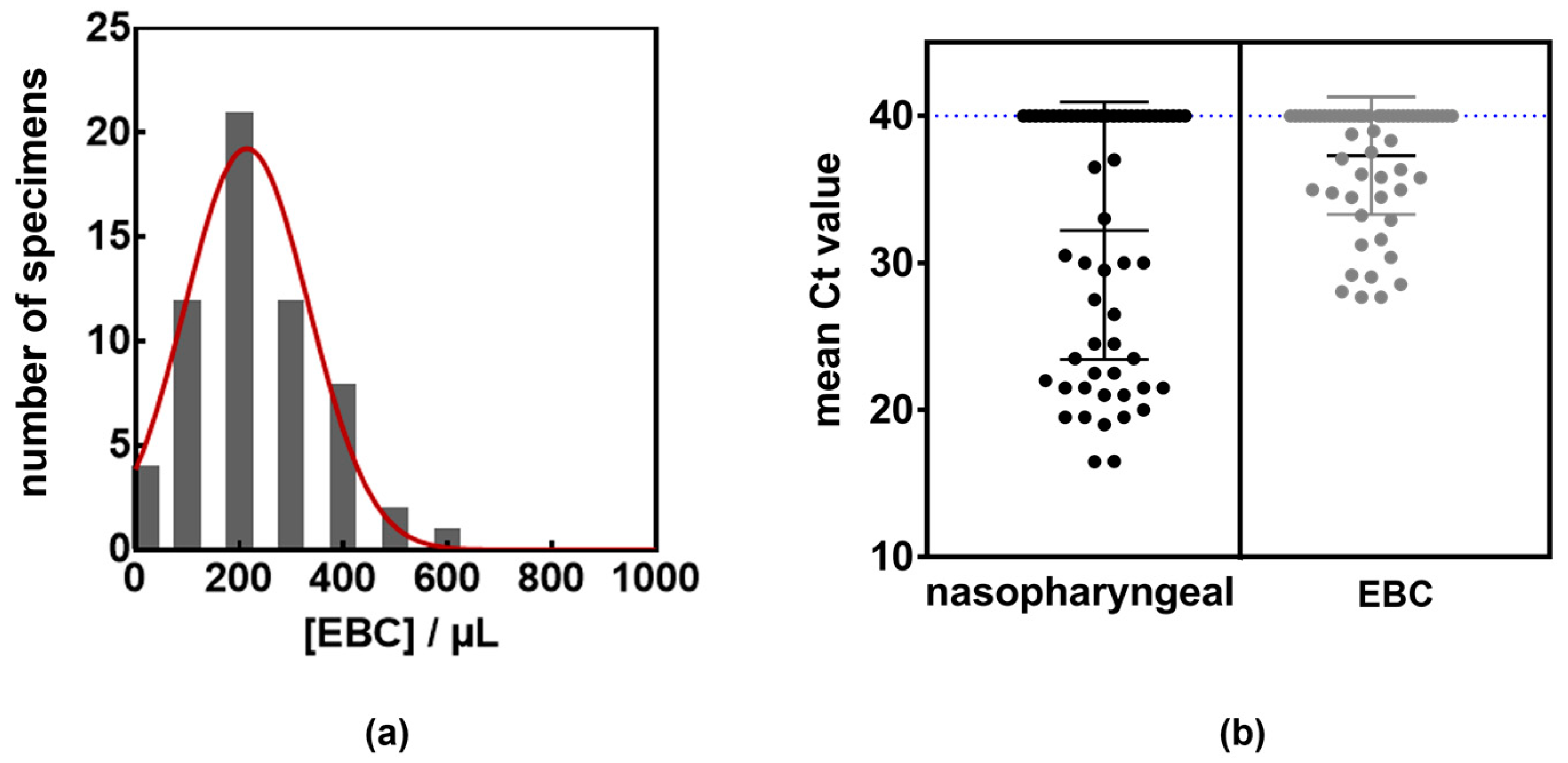
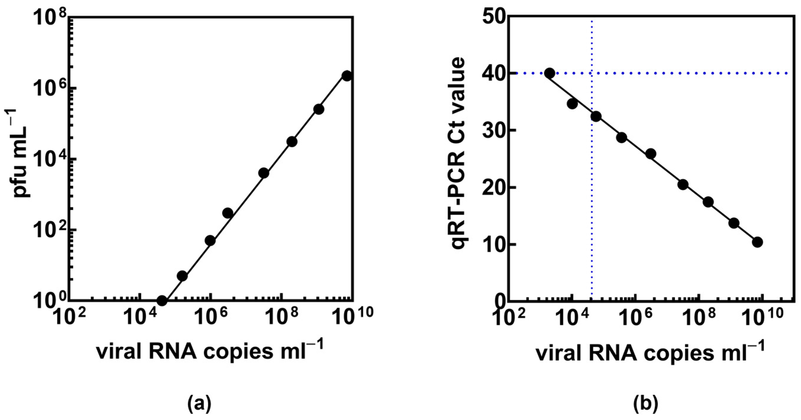
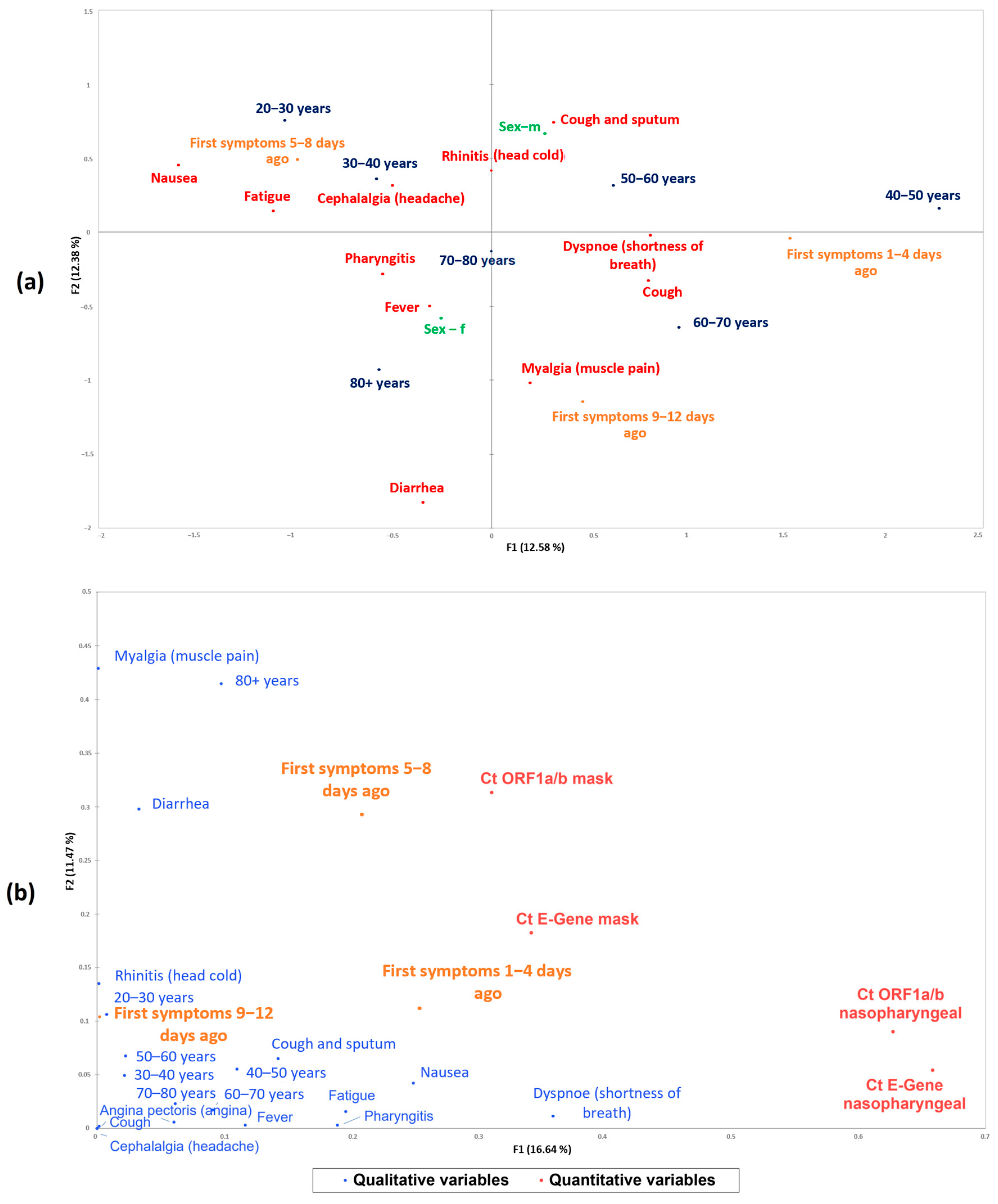
Multivariate statistics were conducted in XLSTAT v2021.3.1/1191 (Addinsoft, Paris, France). For multiple correspondence analysis, the settings were as follows: MCA type disjunctive table, filtering of factors 1/p, no additional data, with variable table output at 5% significance. For mixed principal component analysis (PCAmix), factors were filtered to a maximum of 5. Qualitative data for both methods were the questionnaire symptoms and sex, while patient age was segmented into 20–30, 30–40, 40–50, 60–70, 70–80, and 80+ years (no patients below age 20), and occurrence of first symptoms relative to testing was grouped into 1–4, 5–8, and 9–12 days. Quantitative data for PCAmix were the original Ct values from PCR analysis.

3. Results

Sixty individuals were recruited in this study, 30 with a RT-qPCR-confirmed SARS-CoV-2 infection in nasopharyngeal samples and 30 noninfected patients (Table 1).
Table 2 depicts the Ct values of both the nasopharyngeal and EBC samples of the hospitalized patients (Table 2).
The 60 patients comprised 32 females and 28 males, with a median age of 27 years and an average age of 45.7 years. Thirty of them were hospitalized with a SARS-CoV-2 infection, including 16 females and 14 males, with a median age of 71 years and average age of 66.2 years. On average, 5 min of tidal breathing yielded 200 ± 140 µL of EBC (Figure 2a), which represented sufficient volume for a follow up RT-qPCR analysis using two-gene (ORF1a/b and the E-gene) or one-gene analysis. The 30 EBC samples from non-infected participants were all identified as negative in this study, indicating a 100% specificity. From the 30 hospitalized patients, all nasopharyngeal samples were found to have Ct values < 40 in both ORF1a/b and E-gene. In total, 22 of these 30 patients (73%) had Ct values < 40 in both ORF1a/b and E-gene in EBC, while 25 of these patients had Ct values < 40 in either ORF1a/b or E-gene in EBC (Figure 2b).
The other eight patients were identified as negative for a SARS-CoV-2 infection as per RT-qPCR in EBC in at least one target. In fact, all eight patients were among the highest-aged participants in this study, with an average age of 82.6 years compared with an overall average age of 66.2 years of the hospitalized group. This might point to insufficient sampling of EBC in the elderly proportion in the positive group due to challenges with breathing into the mask. The correlation of the number of plaque-forming units with 1 PFU mL−1 = TCID50 mL−1 × 0.7, with TCID50 being the 50% tissue culture infective dose, set the limit of infectivity to about 4.2 × 10⁴ copies mL−1 (Figure 3), yielding a Ct of about 33.5. The EBC sample of one out of the eight patients identified as SARS-CoV-2 negative was in line with the limit of infectivity. The other seven patients, however, had lower Ct values ranging from 19.5 to 28.2 and were truly false EBC negatives.

4. Discussion

EBC contains rich molecular information about the state of an individual’s health [26,27,28] and larger efforts have been devoted to gaining a better understanding both of the content of EBC [29,30] and its diagnostic value [9,15,23]. The Ct values determined using EBC were generally higher compared with those of the nasopharyngeal sample. Indeed, it is expected that less viral particles might be collected in EBC. When respiratory fluid is aerosolized and travels up from the lower airways to the mouth, it is diluted with droplets of water vapor, which is not the case in nasopharyngeal samples. Another dilution factor of EBC is the inclusion of anatomic dead space air, the air from the upper respiratory tract (nose, mouth) that does not participate in gas exchange, in the condensed exhaled air samples. Compared with the work of Duan et al., where the employed immunosensor, based on high density conductive nanowires, demonstrated viral particle detection down to 7 pfu mL−1 from coronavirus aerosol mimics in only 5 min [24], the current study shows an LoD of about 104 copies mL−1 correlating to roughly 100 pfu mL−1. Using only the Ct value of the ORF1a/b gene as a criterium for infection, 23 (76%) samples were identified correctly. Using only the E-gene, 24 (80%) samples agreed with nasopharyngeal swab samples.
In order to identify latent correlations and patterns in the data pertaining to infectivity status, as determined by nasopharyngeal and EBC-PCR testing in relation to patient characteristics, multivariate analysis was performed. First, the portion of qualitative data of the data set (patient symptoms and sex, with age and occurrence of first symptoms being segmented into categorial values) were subjected to multiple correspondence analysis (MCA), which is a type of correspondence analysis that enables the analysis of relationships of several dependent categorial variables. MCA revealed a clustering of the data into patient groups according to their age (20–30, 30–40, 40–50, 50–60, 60–70, 70–80, 80+ years), their main symptoms, as well as the time point of occurrence of their first symptoms (Figure 4a). The heterogeneity in the symptoms and presentation of COVID disease is evident. Earlier large-scale population studies have demonstrated that early symptoms reported by participants from different age groups vary significantly and can be clustered [31]. Overall, the risk of developing dangerous symptoms increases with age, with patients aged 85 and older being at the highest risk of developing serious symptoms. The existing heterogeneity of the immune system within individuals increases during aging and may also explain differences in susceptibility to COVID-19. Reasons for these differences are the aging immune system undergoing immune-senescence, T-cell diversity alterations, and chronic activation of the innate immune system, known as inflammaging [32]. Additionally, multivariate analyses of both qualitative (patient parameters) and quantitative variables (Ct values of nasopharyngeal and EBC PCR testing) in a factorial analysis of mixed data (mixed principal component analysis, PCAmix) were conducted. The correlation of infection status as determined by Ct values with patient characteristics are shown in Figure 4b. The Ct values from EBC RT-qPCR testing are spatially closer to the actual symptoms and could indicate a better expressiveness of EBC as a sample type for a SARS-CoV-2 infection than nasopharyngeal testing, as the lung might represent a more up-to-date source of biological material than lingering mucus from the nasopharyngeal tract.

5. Conclusions

This study aimed to validate EBC as an alternative, noninvasive biological sample for laboratory-based testing for a COVID-19 infection. Even though the Ct values detected in EBC were higher compared with those in nasopharyngeal swab testing, which is in line with the general view that the viral load is several orders of magnitude lower in EBC, the mask sampling method showed a good specificity concerning the detection of infection status. Thirty individuals could be clearly identified as COVID-19 positive using nasopharyngeal swab samples. Using two-gene (E-gene, ORF1a/b) RT-qPCR analysis, 22 EBC samples (73%) had Ct values < 40. Three patients showed Ct values > 40 in either E-gene or ORF1a/b and five showed Ct values > 40 in both targets. Using RT-qPCR as a diagnostic platform targeting the E-gene only, the best results were obtained with a sensitivity of 80% (positive percent agreement) and 100% selectivity (negative percent agreement) compared with the gold standard nasopharyngeal sampling. This study lays the foundation for further techniques using EBC as a noninvasive sample type for the detection of diseases. For future clinical trials, the face mask will be further engineered toward improved EBC collection efficiency through material optimization and ease of use for the patient to overcome some of the current analytical limitations. As the first rapid point-of-care EBC analysis strategy adapted toward symptomatic SARS-CoV-2-positive individuals, it may pave the way for future developments in fast and easy-to-use screening tools, be it at home, in schools, at airports, or other epidemiologically important hotspots.

Author Contributions

Conceptualization, H.D. and S.S.; methodology, S.S., J.D, S.W. and Q.P.; software, H.D.; validation, G.G., D.L. and O.A.; formal analysis, G.G.; investigation, H.D.; resources, J.D. and S.W.; data curation, D.L.; writing—original draft preparation, S.S.; writing—review and editing, H.D.; visualization, H.D.; supervision, D.L. and R.B.; project administration, S.S.; funding acquisition, H.D. All authors have read and agreed to the published version of the manuscript.

Funding

This research received no external funding.

Institutional Review Board Statement

This proof-of-concept study was approved by the ethics committee of the Danube Private University (DPU), Krems, Austria (EC-No.: DPU-EK/016, stipulated 22 February 2022). The Danube Private University (DPU), Krems, Austria supports transparent reporting of health data.

Informed Consent Statement

Informed consent was obtained from all subjects involved in this study. Written informed consent has been obtained from the patient(s) to publish this paper.

Data Availability Statement

The data presented in this study are available on request from the corresponding author due to ethical restrictions concerning patient rights.

Conflicts of Interest

Authors J.D. and S.W. were employed by the company DiagMetrics. Author Q.P. was employed by the company e-DiagMetrics. The remaining authors declare that the research was conducted in the absence of any commercial or financial relationships that could be construed as a potential conflict of interest.

References

  1. Meo, S.A.; Meo, A.S.; Klonoff, D.C. Omicron new variant BA.2.86 (Pirola): Epidemiological, biological, and clinical characteristics—A global data-based analysis. Eur. Rev. Med. Pharmacol. Sci. 2023, 27, 9470–9476. [Google Scholar] [CrossRef] [PubMed]
  2. Duval, D.; Palmer, J.C.; Tudge, I.; Pearce-Smith, N.; O’Connell, E.; Bennett, A.; Clark, R. Long distance airborne transmission of SARS-CoV-2: Rapid systematic review. BMJ 2022, 377, e068743. [Google Scholar] [CrossRef] [PubMed]
  3. Prather, K.A.; Marr, L.C.; Schooley, R.T.; McDiarmid, M.A.; Wilson, M.E.; Milton, D.K. Airborne transmission of SARS-CoV-2. Science 2020, 370, 303–304. [Google Scholar] [CrossRef] [PubMed]
  4. Almstrand, A.C.; Ljungström, E.; Lausmaa, J.; Bake, B.; Sjövall, P.; Olin, A.-C. Airway monitoring by collection and mass spectrometric analysis of exhaled particles. Anal. Chem. 2009, 81, 662–668. [Google Scholar] [CrossRef]
  5. Dbouk, T.; Drikakis, D. On respiratory droplets and face masks. Phys. Fluid. 2020, 32, 063303. [Google Scholar] [CrossRef]
  6. Dhang, R.; Li, J. Coughs and Sneezes: Their Role in Transmission of Respiratory Viral Infections, Including SARS-CoV-2. Am. J. Respir. Crit. Care Med. 2020, 202, 651–659. [Google Scholar]
  7. Soto, F.; Ozen, M.O.; Guimaraes, C.F.; Wang, J.; Hokanson, K.; Ahmed, R.; Reis, R.L.; Paulmurugan, R.; Demirci, U. Wearable Collector for Noninvasive Sampling of SARS-CoV-2 from Exhaled Breath for Rapid Detection. ACS Appl. Mater. Interfaces 2021, 13, 41445–41453. [Google Scholar] [CrossRef]
  8. Nwanochie, E.; Linnes, J.C. Review of non-invasive detection of SARS-CoV-2 and other respiratory pathogens in exhaled breath condensate. J. Breath Res. 2022, 16, 024002. [Google Scholar] [CrossRef]
  9. Daniels, J.; Wadekar, S.; DeCubellis, K.; Jackson, G.W.; Chiu, A.S.; Pagneux, P.; Saada, H.; Engelmann, I.; Ogiez, J.; Loze-Warot, D.; et al. A mask-based diagnostic platform for point-of-care screening of COVID-19. Biosens. Bioelectron. 2021, 192, 113486. [Google Scholar] [CrossRef]
  10. Cavaleiro Rufo, J.; Paciencia, I.; Mendes, F.C.; Farraia, M.; Rodolfo, A.; Silva, D.; de Oliveira Fernandes, E.; Delgado, L.; Moreira, A. Exhaled breath condensate volatilome allows sensitive diagnosis of persistent asthma. Allergy 2019, 74, 527–534. [Google Scholar] [CrossRef]
  11. Cruickshank-Quinn, C.; Armstrong, M.; Powell, R.; Gomez, J.; Elie, M.; Reisdorph, N. Determining the presence of asthma-related molecules and salivary contamination in exhaled breath condensate. Respir. Res. 2017, 18, 57. [Google Scholar] [CrossRef] [PubMed]
  12. Kazeminasab, S.; Ghanbari, R.; Emamalizadeh, B.; Jouyban-Gharamaleki, V.; Taghizadieh, A.; Jouyban, A.; Khoubnasabjafari, M. Exhaled breath condensate efficacy to identify mutations in patients with lung cancer: A pilot study. Nucleosides Nucleotides Nucleic Acids 2022, 41, 370–383. [Google Scholar] [CrossRef] [PubMed]
  13. Guzman-Beltran, S.; Carreto-Binaghi, L.E.; Carranza, C.; Torres, M.; Gonzalez, Y.; Munoz-Torrico, M.; Juarez, E. Oxidative Stress and Inflammatory Mediators in Exhaled Breath Condensate of Patients with Pulmonary Tuberculosis. A Pilot Study with a Biomarker Perspective. Antioxidants 2021, 10, 1572. [Google Scholar] [CrossRef] [PubMed]
  14. Patsiris, S.; Papanikolaou, I.; Stelios, G.; Exarchos, T.P.; Vlamos, P. Exhaled Breath Condensate and Dyspnea in COPD. Adv. Exp. Med. Biol. 2021, 1337, 339–344. [Google Scholar] [CrossRef]
  15. Ryan, D.J.; Toomey, S.; Madden, S.F.; Casey, M.; Breathnach, O.S.; Morris, P.G.; Grogan, L.; Branagan, P.; Costello, R.W.; De Barra, E. Use of exhaled breath condensate (EBC) in the diagnosis of SARS-CoV-2 (COVID-19). Thorax 2021, 76, 86–88. [Google Scholar] [CrossRef]
  16. Ilhamsyah, R.; Dimandja, J.-M.D.; Hesketh, P.J. Design and Analysis of Exhaled Breath Condenser System for Rapid Collection of Breath Condensate. J. Electrochem. Soc. 2021, 168, 107503. [Google Scholar] [CrossRef]
  17. Sol, J.A.; Quindry, J.C. Application of a Novel Collection of Exhaled Breath Condensate to Exercise Settings. Int. J. Environ. Res. Public Health 2022, 19, 3948. [Google Scholar] [CrossRef]
  18. Szunerits, S.; Dӧrfler, H.; Pagneux, Q.; Daniel, J.; Wadekar, S.; Woitrain, E.; Ladage, D.; Montaigne, D.; Boukherroub, R. Exhaled breath condensate as bioanalyte: From collection considerations to biomarker sensing. Anal. Bioanal. Chem. 2023, 415, 27–34. [Google Scholar] [CrossRef]
  19. Kim, H.S.; Lee, H.; Park, J.; Abbas, N.; Kang, S.; Hyun, H.; Seong, H.; Yoon, J.G.; Noh, J.Y.; Kim, W.J.; et al. Collection and detection of SARS-CoV-2 in exhaled breath using face mask. PLoS ONE 2022, 17, e0270765. [Google Scholar] [CrossRef]
  20. Xue, Q.; Kan, X.; Pan, Z.; Li, Z.; Pan, W.; Zhou, F.; Duan, X. An intelligent face mask integrated with high density conductive nanowire array for directly exhaled coronavirus aerosols screening. Biosens. Bioelectron. 2021, 186, 113286. [Google Scholar] [CrossRef]
  21. Khorshid, M.; Bakhshi Sichani, S.; Barbosa Estrada, D.L.; Neefs, W.; Clement, A.; Pohlmann, G.; Epaud, R.; Lanone, S.; Wagner, P. An Efficient Low-Cost Device for Sampling Exhaled Breath Condensate EBC. Adv. Sens. Res. 2024, 3, 2400020. [Google Scholar] [CrossRef]
  22. Williams, C.M.; Cheah, E.S.; Malkin, J.; Patel, H.; Otu, J.; Mlaga, K.; Sutherland, J.S.; Antonio, M.; Perera, N.; Woltmann, G.; et al. Face mask sampling for the detection of Mycobacterium tuberculosis in expelled aerosols. PLoS ONE 2014, 9, e104921. [Google Scholar] [CrossRef] [PubMed]
  23. Nguyen, P.Q.; Soenksen, L.R.; Donghia, N.M.; Angenent-Mari, N.M.; de Puig, H.; Huang, A.; Lee, R.; Slomovic, S.; Galbersanini, T.; Lansberry, G.; et al. Wearable materials with embedded synthetic biology sensors for biomolecule detection. Nat. Biotechnol. 2021, 39, 1366–1374. [Google Scholar] [CrossRef]
  24. Duan, C.; Buerer, L.; Wang, J.; Kaplan, S.; Sabalewski, G.; Jay, G.D.; Monaghan, S.F.; Arena, A.E.; Fairbrother, W.G. Efficient Detection of Severe Acute Respiratory Syndrome Coronavirus 2 (SARS-CoV-2) from Exhaled Breath. J. Mol. Diagn. 2021, 23, 1661–1670. [Google Scholar] [CrossRef]
  25. Tanno, L.K.; Casale, T.; Demoly, P. Coronavirus Disease (COVID)-19: World Health Organization Definitions and Coding to Support the Allergy Community and Health Professionals. J. Allergy Clin. Immunol. Pract. 2020, 8, 2144–2148. [Google Scholar] [CrossRef]
  26. Hayes, S.A.; Haefliger, S.; Harris, B.; Pavlakis, N.; Clarke, S.J.; Molloy, M.P.; Howell, V.M. Exhaled breath condensate for lung cancer protein analysis: A review of methods and biomarkers. J. Breath Res. 2016, 10, 034001. [Google Scholar] [CrossRef]
  27. Lin, X.F.; Zhang, L.; Shi, S.Y.; Fan, Y.C.; Wu, Z.L.; Zhang, X.; Sun, D.Q. Expression of surfactant protein-A in exhaled breath condensate of patients with chronic obstructive pulmonary disease. Mol. Med. Rep. 2016, 13, 1667–1672. [Google Scholar] [CrossRef] [PubMed]
  28. Muccilli, V.; Saletti, R.; Cunsolo, V.; Ho, J.; Gili, E.; Conte, E.; Sichili, S.; Vancheri, C.; Foti, S. Protein profile of exhaled breath condensate determined by high resolution mass spectrometry. J. Pharm. Biomed. Anal. 2015, 105, 134–149. [Google Scholar] [CrossRef]
  29. Beccaria, M.; Mellors, T.R.; Petion, J.S.; Rees, C.A.; Nasir, M.; Systrom, H.K.; Sairistil, J.W.; Jean-Juste, M.-A.; Rivera, V.; Lavoile, K.; et al. Preliminary investigation of human exhaled breath for tuberculosis diagnosis by multidimensional gas chromatography—Time of flight mass spectrometry and machine learning. J. Chromatogr. B Analyt. Technol. Biomed. Life Sci. 2018, 1074–1075, 46–50. [Google Scholar] [CrossRef]
  30. Yuan, Z.-C.; Li, W.; Wu, L.; Huang, D.; Wu, M.; Hu, B. Solid-Phase Microextraction Fiber in Face Mask for In Vivo Sampling and Direct Mass Spectrometry Analysis of Exhaled Breath Aerosol. Anal. Chem. 2020, 92, 11543–11547. [Google Scholar] [CrossRef]
  31. Canas, L.S.; Sudre, C.H.; Capdevila Pujol, J.; Polidori, L.; Murray, B.; Molteni, E.; Graham, M.S.; Klaser, K.; Antonelli, M.; Berry, S.; et al. Early detection of COVID-19 in the UK using self-reported symptoms: A large-scale, prospective, epidemiological surveillance study. Lancet Digit. Health 2021, 3, e587–e598. [Google Scholar] [CrossRef] [PubMed]
  32. Rea, I.M.; Alexander, H.D. Triple jeopardy in ageing: COVID-19, co-morbidities and inflamm-ageing. Ageing Res. Rev. 2022, 73, 101494. [Google Scholar] [CrossRef] [PubMed]
Figure 1. Smart mask engineering. (a) Formation process of the proposed exhaled breath condensate (EBC) collection system to be retrofitted into commercial face masks. (b) Photos of mask used to collect EBC together with EBC droplets formed and collected.
Labmed 01 00005 g001
Figure 2. Efficacy of EBC compared with nasopharyngeal swab samples for the identification of individuals with suspicion of a SARS-CoV-2 infection. (a) EBC volume distribution after 5 min tidal breathing into chilled smart face masks. (b) Results of RT-qPCR 2 gene (ORF1a/b and E-gene) analysis: Ct values determined in nasopharyngeal swab samples (black) and EBC samples (grey). Ct cutoff for negative result was set at 40 (dashed line).
Labmed 01 00005 g002
Figure 3. Correlation of viral RNA copies mL−1 with Ct values and plaque forming units. (a) Correlation of viral RNA copies mL−1 with plaque-forming units of SARS-CoV-2 as a measure of infectivity. (b) Correlation of qRT-PCR Ct counts and viral RNA copies mL−1 of SARS-CoV-2. The horizontal dashed line correlates to the Ct cutoff for negative results, while the vertical one shows the limit of infectivity derived from (a).
Labmed 01 00005 g003
Figure 4. Multiple correspondence analysis (MCA) and factorial analysis of patient data. (a) Positively correlated variables are found in spatial proximity by MCA, with negative correlations being located on the opposite sides of the axes in the plot. In red: main symptom; in orange: occurrence of first symptoms; in blue: age range; in green: sex. A proximity of patients groups per age to the first occurring symptoms can be observed: Patients aged 20–30 and 30–40 were heavily correlated with nausea, fatigue, cephalalgia, and an onset of symptoms 5–8 days before testing. The group of patients aged 40–50 and 50–60 exhibited dyspnea and cough and sputum and had first symptoms very close to being tested (first symptoms 1–4 days ago). Patients of 60–70 years of age showed similar symptoms as those of 50–60 years with the addition of cough and a later onset of first symptoms. Older patients (70–80 and 80+ years) had symptoms occurring 9 to 12 days prior to testing with a stronger correlation to diarrhea and myalgia. Axes F1 and F2 are the main separators in this plot, explaining 24.96% of the total variance in the data of the 30 hospitalized patients. (b) Factorial analysis of mixed data (PCAmix). In blue: main symptom; in orange: occurrence of first symptoms; in red: Ct values. In this plot, the squared loadings represent the coordinates and also the absolute contributions to the variance of the components, depicting the correlation of the Ct values of both nasopharyngeal and EBC PCR testing with the symptoms displayed by the hospitalized patients. It is visible that the Ct values of EBC obtained by mask sampling correlate closer to the onset of the symptoms in the earlier days than those obtained by nasopharyngeal sampling. At the time point of testing, the symptoms displayed are typical for an acute infection, characterized by dyspnea (shortness of breath), pharyngitis, cough and sputum, nausea, fatigue, and fever, all in the first 1–4 days.
Labmed 01 00005 g004
Table 1. Summary of patient characteristics, clinical features, and laboratory analysis.
Patient CharacteristicsTotal Cohort (n = 60)Presence of Viral RNA in Nasopharyngeal SamplesPresence of Viral RNA in EBC
Sex32 females, 28 males30/30 (16 females, 14 males)25 */30 (16 females, 14 males)
Age (years)
Median277165
Mean45.766.263.4
Average time from COVID-19 symptoms onset to test (days)6.76.74.7
Nasopharyngeal samplingEBC/mask sampling
ORF1a/bE-geneORF1a/bE-gene
Sensitivity 110.90.8
Specifity 1111
Positive predictive value 1111
Negative predictive value 110.910.83
Negative likelihood ratio 000.10.2
Accuracy 110.950.9
* 22 patients with Ct values < 40 using 2 genes (ORF1a/b, E-gene) and 25 patients with a confirmed infection by at least one target.
Table 2. Ct values of hospitalized patients from RT-qPCR testing from nasopharyngeal and EBC mask sampling.
Hospitalized
Patient No.
Age (Years)Ct ORF1a/b
Nasopharyngeal
Ct E-Gene
Nasopharyngeal
Ct ORF1a/b
EBC (Mask)
Ct E-Gene
EBC (Mask)
12130.430.536.338.8
28316.216.830.731.8
33625.026.028.729.4
48322.022.6--
55819.119.632.434.0
67724.625.427.628.5
77519.620.734.737.0
88322.823.4-37.5
98919.520.3--
107136.137.9--
116222.223.128.129.0
127129.832.034.035.5
138829.230.936.6-
144216.417.129.129.2
158030.531.9--
165721.122.227.328.1
177422.222.734.335.6
186621.722.433.135.8
193821.121.231.032.2
209219.119.235.838.4
216426.627.434.235.8
226121.821.827.427.9
232419.920.332.433.5
245320.120.430.330.4
256323.824.234.034.9
268229.630.536.036.7
277524.925.434.937.2
285139.934.734.736.9
297732.734.5--
309027.428.3-37.9
Disclaimer/Publisher’s Note: The statements, opinions and data contained in all publications are solely those of the individual author(s) and contributor(s) and not of MDPI and/or the editor(s). MDPI and/or the editor(s) disclaim responsibility for any injury to people or property resulting from any ideas, methods, instructions or products referred to in the content.

