Article

Ciraparantag Does Not Remove Anticoagulant Activities In Vitro, but DOAC-Stop™ May Mitigate Ciraparantag-Associated Interferences in Coagulation Testing

by
James V. Harte
1,2,* and
Gavin T. Buckley
1,2
1
EOLAS Research Group, Cork University Hospital, Wilton, T12 DC4A Cork, Ireland
2
Department of Haematology, Cork University Hospital, Wilton, T12 DC4A Cork, Ireland
*
Author to whom correspondence should be addressed.
LabMed 2024, 1(1), 33-42; https://doi.org/10.3390/labmed1010006
Submission received: 27 August 2024 / Revised: 19 September 2024 / Accepted: 22 October 2024 / Published: 18 November 2024
(This article belongs to the Collection Feature Papers in Laboratory Medicine)

Abstract

:
Anticoagulants can complicate the interpretation of routine and specialised coagulation assays. Several methodologies have been developed to minimise or eliminate anticoagulant-associated interferences; however, no ‘universal methodology’ that encompasses different anticoagulant classes is currently available. Ciraparantag is a promising reversal agent that can bind both direct oral anticoagulants (DOACs) and heparin-like anticoagulants. As such, we aimed to investigate whether ciraparantag could be employed as a ‘universal’ anticoagulant chelator in vitro. Human plasma was spiked with ascending concentrations of ciraparantag, with or without DOACs or heparin, and assayed for routine coagulation parameters. Ciraparantag had minimal effects on coagulation testing when added to human plasma at concentrations similar to pharmacokinetic maxima; however, ciraparantag did not remove DOAC- or heparin-associated activities in vitro, which was likely due to the preferential chelation of anionic substances in the coagulation reagents. In contrast, DOAC-Stop™, a commercial activated charcoal-based adsorbent, efficiently removed both DOAC- and ciraparantag-associated interferences. In conclusion, although ciraparantag is not effective as a ‘universal’ anticoagulant chelator in vitro, we report that activated charcoal-based adsorbents may be clinically useful in situations where laboratory investigations are complicated by the presence of DOACs and/or ciraparantag.

1. Introduction

The development of direct oral anticoagulants (DOACs) revolutionised the prevention and treatment of thrombosis by offering superior pharmacokinetic and pharmacodynamic profiles compared to contemporary coumarin-based vitamin K antagonists [1,2]. However, the presence of these DOACs in patient-derived plasma samples is associated with significant interferences in both clotting and chromogenic coagulation assays, due to the inhibition of either factor IIa or factor Xa activities, respectively [3].
As a result, clinical laboratories require supplementary methodologies to prevent the misinterpretation of routine and specialised coagulation assays due to the presence of DOACs ex vivo.
We have previously reported that the addition of raw activated charcoal (AC) to plasma is a cost-effective and efficient method that removes DOAC-associated interferences in coagulation assays [4,5]. There are also two commercially available AC-based adsorbents—DOAC-Stop® (Haematex Research) and DOAC-Remove® (5-Diagnostics)—that have been consistently shown to facilitate the correct interpretation of various coagulation parameters [6], and are recommended in the latest guidelines of the British Society for Haematology [7] and the International Society on Thrombosis and Haemostasis [8,9].
However, AC-based removal agents only adsorb low molecular weight substances and, therefore, are ineffective against larger heparin-like anticoagulants [10]. Similar to DOACs, the removal of heparin-associated interferences is sometimes required in laboratory investigations [11]; however, methodologies are heparin-exclusive and no gold standard has been established [6].
As such, no ‘universal methodology’ that encompasses the different anticoagulant classes is currently available.
Ciraparantag, a small cationic molecule composed of two arginine residues coupled by a piperazine ring, is a promising anticoagulant reversal agent currently under investigation in clinical trial [12]. Dynamic light scattering experiments have shown that ciraparantag directly associates with DOACs and heparins through non-covalent, charge-charge interactions [13]. In pre-clinical models of DOAC- and heparin-associated bleeding, a single dose of ciraparantag significantly reduced blood loss when administered either prophylactically or therapeutically [13]. Furthermore, in phase I and phase II clinical trials, disruption of the interaction between both DOACs and heparins with intended coagulation factors effectively reversed anticoagulant-associated activities in treated patients [14,15,16].
Interestingly, a previous in vitro investigation with ciraparantag suggested a complete concentration-dependent removal of rivaroxaban- and apixaban-associated interferences in human plasma by the in vitro addition of the anticoagulant reversal agent [17].
As ciraparantag can bind DOACs and heparins, we aimed to determine whether ciraparantag could be employed as a ‘universal methodology’ to remove anticoagulant-associated activities in routine and specialised coagulation assays.

2. Materials and Methods

2.1. Preparation of Human Plasma with and Without Anticoagulants

Commercial human plasma (catalogue no.: 10446238 ORKL17) was obtained from Siemens Healthineers (Erlangen, Germany) and was reconstituted with distilled water according to the manufacturer’s specifications.
Dabigatran (catalogue no.: HY-10163), apixaban (catalogue no.: HY-50667), edoxaban (catalogue no.: HY-10264), and rivaroxaban (catalogue no.: HY-50903) were obtained from MedChemExpress (Sollentuna, Sweden). Stock solutions were prepared by dissolving each anticoagulant in either hydrochloric acid (HCl) or dimethyl sulfoxide (DMSO), as per the manufacturer’s specifications, to a concentration of 1 mg/mL. Anticoagulants were further diluted in phosphate-buffered saline to 100 µg/mL before spiking into 1 mL of human plasma at the time of analysis at a concentration of 500 ng/mL.
Human plasma without dabigatran, apixaban, edoxaban, or rivaroxaban was spiked with an equal volume of either HCl-phosphate-buffered saline or DMSO-phosphate-buffered saline, as appropriate, as a vehicle control. The addition of vehicle controls did not affect the routine coagulation parameters of human plasma (Table S1).
Enoxaparin (catalogue no.: HY-109509) was obtained from MedChemExpress (Sollentuna, Sweden). Stock solutions were prepared by dissolving in distilled water, as per the manufacturer’s specifications, to a concentration of 100 mg/mL. Enoxaparin was further diluted in distilled water to 1 mg/mL before spiking into standard human plasma. Enoxaparin was then assayed with a chromogenic anti-factor Xa BIOPHEN™ Heparin LRT assay (HYPHEN Biomed, Neuville-sur-Oise, France); the concentration of enoxaparin was adjusted to 0.7 IU/mL by the addition of either human plasma or by the addition of human plasma spiked with higher concentrations of enoxaparin.
Human plasma without enoxaparin was spiked with an equal volume of distilled water as a vehicle control. The addition of the vehicle control did not affect the routine coagulation parameters of human plasma (Table S1).

2.2. Removal of Anticoagulant-Associated Activities from Human Plasma

Two methods were compared to remove anticoagulant-associated activities from plasma: anticoagulant chelation with ciraparantag and anticoagulant adsorption with DOAC-Stop™.
Ciraparantag (catalogue no.: HY-18660) was obtained from MedChemExpress (Sollentuna, Sweden). Stock solutions were prepared by dissolution in distilled water, as per the manufacturer’s specifications, to a concentration of 10 mg/mL. The stock solution was used neat or further diluted in distilled water to intermediate concentrations before spiking into human plasma at the time of analysis at a concentration between 100 ng/mL and 100,000 ng/mL. Briefly, ciraparantag was added to 1 mL of human plasma—with or without anticoagulants, or vehicle control—and incubated for various time points at ambient temperature with gentle agitation. Then, the plasma was analysed without further processing.
DOAC-Stop™ was generously gifted by Haematex Research (Hornsby, Australia) and used according to the manufacturer’s specifications. Briefly, one DOAC-Stop™ tablet was added to 1 mL of human plasma—with or without direct oral anticoagulants, and/or ciraparantag—and incubated for 5 min at ambient temperature with gentle agitation. The plasma was then centrifuged at 2000× g for 5 min to remove the activated charcoal, and the clarified plasma was analysed without further processing.

2.3. Routine and Specialised Coagulation Testing of Human Plasma

The prothrombin time (PT), activated partial thromboplastin time (APTT), and fibrinogen assay were performed using reagents from Siemens Healthineers (Erlangen, Germany): Dade® Innovin®, Dade® Actin® FS, and Dade® Thrombin, respectively. Calcium chloride and Dade® Owren’s Buffer were used as assay supplements (Siemens Healthineers, Erlangen, Germany).
Routine coagulation assays were performed on a CS-5100 analyser (Sysmex, Kobe, Japan), which was routinely monitored and maintained according to the manufacturer’s specifications.
Dabigatran was measured by a dilute thrombin time assay using Thromboclotin® (Siemens Healthineers, Erlangen, Germany). Apixaban, edoxaban, rivaroxaban, and enoxaparin were quantitatively measured using a chromogenic anti-factor Xa BIOPHEN™ Heparin LRT assay (HYPHEN Biomed, Neuville-sur-Oise, France). Assays were calibrated against known standard concentrations, and the lower limits of quantification (LLoQ) for dabigatran, apixaban, edoxaban, rivaroxaban, and enoxaparin were 20 ng/mL, 10 ng/mL, 15 ng/mL, 5 ng/mL, and 0.08 IU/mL, respectively.
Specialised assays were performed on a CS-2500 analyser (Sysmex, Kobe, Japan), which was routinely monitored and maintained according to the manufacturer’s specifications.

2.4. Statistical Analysis

Data were curated using Microsoft Excel (version 2018; Microsoft Corporation, Redmond, WA, USA). All statistical analyses were performed using GraphPad Prism (version 10.1.1; GraphPad Software, Boston, MA, USA).
Data were compared using one-way ANOVA, as appropriate, with the post hoc Dunnett’s test for multiple comparisons. A p-value of less than 0.05 was considered statistically significant (*, p-value < 0.05; **, p-value < 0.01; ***, p-value < 0.001; ****, p-value < 0.0001).

3. Results

3.1. Ciraparantag Has Minimal Impact on Routine Coagulation Parameters In Vitro

Anticoagulant removal agents for use in coagulation testing should minimally impact coagulation parameters. As such, we assayed commercially available human plasma before and after the in vitro addition of ascending concentrations of ciraparantag to determine any ciraparantag-associated interferences in routine testing. A concentration range of 100 ng/mL to 10,000 ng/mL was chosen for our investigations based on the maximum ciraparantag concentrations reported in recent pharmacokinetic trials [13,18].
The addition of ciraparantag to human plasma had minimal impact on coagulation parameters between 100 ng/mL and 1000 ng/mL (Table 1); however, at the highest concentration, a mild prolongation of the APTT clotting time (26.2 ± 0.60 s versus 30.9 ± 1.43 s; p-value < 0.001) was observed compared to baseline (Table 1).
Statistically significant differences were not observed between the different ascending concentrations of ciraparantag, suggesting an additive concentration-dependent effect in human plasma. Moreover, no impact was observed over time for ascending concentrations of ciraparantag between 100 ng/mL and 10,000 ng/mL up to 8 h post-addition, suggesting an immediate concentration-dependent effect in human plasma (Table S2).

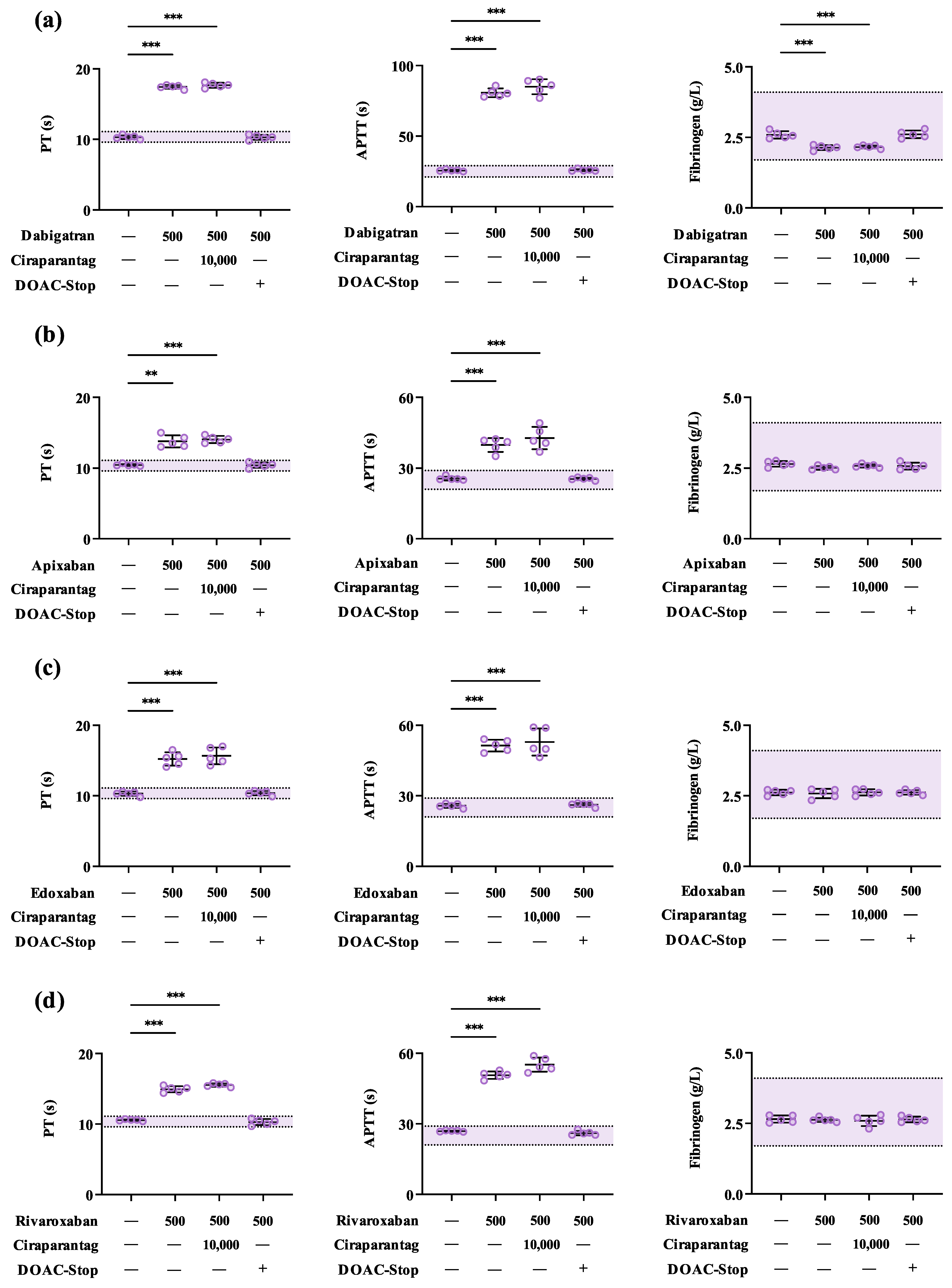
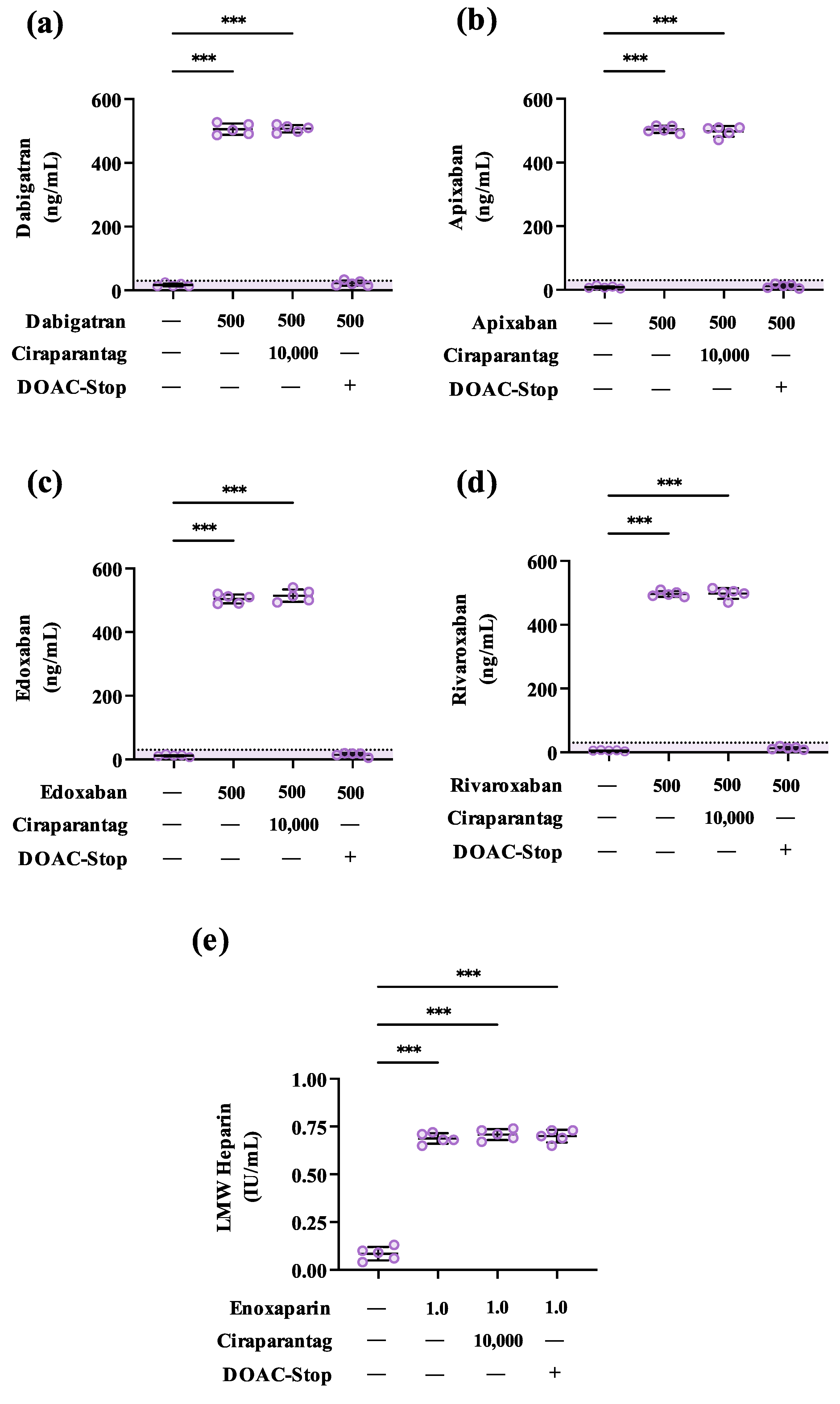
3.2. Ciraparantag Does Not Remove Anticoagulant-Associated Activities in Routine and Specialised Coagulation Assays

To determine whether ciraparantag could chelate DOACs or heparins in vitro, leading to the removal of anticoagulant-associated activities, we assayed commercial human plasma with and without direct factor IIa or factor Xa inhibitors or enoxaparin, as well as after the in vitro addition of the anticoagulant reversal agent. A concentration of each DOAC slightly higher than expected in patient-derived plasma [19], and a concentration of enoxaparin representative of anti-factor Xa levels for once and twice-daily dosing [20,21], were chosen for our investigations to ensure anticoagulant-associated activities were observed in relevant coagulation assays.
Preliminary experiments with 1000 ng/mL of ciraparantag failed to remove anticoagulant-associated activities (Figure S1); therefore, a ciraparantag concentration of 10,000 ng/mL was chosen to maximise the chance of removing anticoagulant-associated activities, despite the prolongation observed in the APTT-based assay (Figure 1).
In human plasma spiked with 500 ng/mL of each respective DOAC, significant prolongations of the PT- and APTT-based clotting times were observed for dabigatran, apixaban, edoxaban, and rivaroxaban at 500 ng/mL (Figure 1a–d), and an apparent reduction in fibrinogen concentrations was also observed for dabigatran (Figure 1a). However, the subsequent addition of 10,000 ng/mL of ciraparantag did not remove DOAC-associated activities in PT-, APTT-, or fibrinogen-based assays (Figure 2). Furthermore, ciraparantag did not remove DOAC-associated activities in either factor IIa- or factor Xa-specific assays calibrated for dabigatran, apixaban, edoxaban, and rivaroxaban, respectively (Figure 2a–d).
In human plasma spiked with 0.7 IU/mL of enoxaparin, significant anti-factor Xa activity was observed (Figure 2e); however, similar to human plasma spiked with DOACs, the subsequent addition of 10,000 ng/mL of ciraparantag did not remove heparin-associated activity in factor Xa-specific assays calibrated for low molecular weight heparins (Figure 2e).
In contrast to the addition of ciraparantag to human plasma, all DOAC-associated activities and interferences—but not heparin-associated activities—were efficiently removed when human plasma was treated with DOAC-Stop™ (Figure 2).

3.3. DOAC-Stop™ May Mitigate Ciraparantag-Associated Interferences in Coagulation Testing

As ciraparantag was observed to prolong APTT-based clotting time in vitro, we next investigated whether DOAC-Stop™ could remove ciraparantag from human plasma. Although unlikely, ciraparantag removal ex vivo may be necessary in situations where DOACs and/or ciraparantag are present in patient-derived plasma requiring laboratory investigation.
Treatment of ciraparantag-containing plasma with DOAC-Stop™ mitigated ciraparantag-associated prolongations of the APTT (Table 2), suggesting adsorption by the activated charcoal. Moreover, no unexpected differences were observed for PT-based clotting times or fibrinogen concentrations after treatment with DOAC-Stop™.
It should be noted that at a supratherapeutic concentration of 100,000 ng/mL—10-fold greater than concentrations reported in recent pharmacokinetic trials [13,18]—slight ciraparantag-associated interferences were observed in PT-based clotting times and fibrinogen concentrations, which were also mitigated by treatment with DOAC-Stop™ (Table S3).

4. Discussion

Anticoagulant-associated activities are a considerable source of diagnostic error in coagulation testing. Although methodologies have been developed to independently minimise or eliminate DOAC- and heparin-associated interferences in vitro, no ‘universal methodology’ is currently available.
Given that ciraparantag can bind both DOACs and heparins, and a previous investigation suggested the removal of DOAC-associated interferences upon supplementation of human plasma in vitro [17], we investigated whether the addition of the anticoagulant reversal agent to human plasma would remove anticoagulant-associated activities and interferences in a series of routine and specialised coagulation assays.
Ciraparantag exhibited minimal impact on coagulation parameters when added to human plasma at clinically relevant concentrations, with only a slight prolongation of the APTT observed at the highest concentration tested (10,000 ng/mL). However, even at this concentration, we could not replicate the previous in vitro findings in our plasma-based assays [17]: the addition of ciraparantag to DOAC- and heparin-spiked plasma did not remove anticoagulant-associated activities in PT-, APTT-, or fibrinogen-based assays.
Higher concentrations of ciraparantag were associated with concentration-dependent interferences in routine coagulation assays and were therefore not suitable for further experimentation.
The inability of ciraparantag to remove anticoagulant-associated activities may be due to the inherent mechanism of action of ciraparantag as an anticoagulant reversal agent. It was noted initially in preclinical models that inconsistent and variable results were obtained for routine coagulation parameters when ciraparantag was administered as an anticoagulant reversal agent [13]. Ciraparantag failed to correct APTT-based clotting times and anti-factor Xa activities despite quantitative reductions in anticoagulant-associated blood loss [13]. It was subsequently demonstrated that ciraparantag preferentially chelates anionic substances available in standard blood collection tubes and plasma-based assays, leading to the release of functional anticoagulant ex vivo [13]. Given this understanding of the mechanism of action of ciraparantag, we believe that the chelation of anionic substances in our coagulation reagents may render ciraparantag ineffective as an anticoagulant removal agent in vitro.
Furthermore, the binding of anionic substances necessary for routine coagulation testing may also explain the interferences observed in the presence of ciraparantag.
It is important to note that these observations do not rule out ciraparantag as a viable anticoagulant reversal agent in vivo, as clinical trial data from phase I and phase II trials remain very promising [23]. The data presented herein are specific only to the in vitro addition of ciraparantag as an anticoagulant removal agent.
Furthermore, our study of ciraparantag in vitro is limited by performing our investigations in commercially available human plasma and by the limited range of assays evaluated. Commercial human plasma may not completely replicate fresh or fresh-frozen patient-derived plasma samples under anticoagulation therapy. However, we have previously employed commercial human plasma for the evaluation of AC-based removal agents and have observed similar efficacy to patient-derived plasma. Moreover, it has been reported that in vitro approaches to investigating anticoagulant removal are suitable mimetics for in vivo conditions, as both produce consistent results [24,25,26,27]. It is also noteworthy that ciraparantag-associated interferences may be pronounced in alternative routine or specialised coagulation assays, even in similar assays from different manufacturers, depending on composition. A greater understanding of the effects of ciraparantag in the evaluation of patient-derived plasma samples is necessary.
Pharmacokinetic data suggest that maximum serum ciraparantag concentrations are reached within several minutes of administration [13,15,16], and that ciraparantag is rapidly cleared by the kidneys and urinary bladder within 24 h [13]. Therefore, ciraparantag-associated interferences may not be significant in patient-derived plasma samples. However, the presence of both anticoagulant- and ciraparantag-associated activities in patient-derived plasma following the administration of ciraparantag may occur and may, in turn, complicate the interpretation of routine and specialised coagulation assays—an area that has not been fully explored herein.
Given that the molecular weight of ciraparantag is similar to other APTT-prolonging agents adsorbed by AC-based removal agents [10], our findings suggest that it is possible that DOAC-Stop™ may be a simple method to evaluate underlying haemostatic measurands in rare cases of both anticoagulant- and ciraparantag-associated activities. However, as we could not measure the effectiveness of ciraparantag—nor its precise concentration—with the assays described herein, these findings would require validation with alternative systems in patient-derived plasma.

5. Conclusions

In conclusion, although ciraparantag universally reverses anticoagulation therapy in vivo, the effect does not appear to be reproducible in vitro due to the inherent mechanism of action of the anticoagulant reversal agent itself. Anticoagulant-associated activities were consistently observed in plasma spiked with clinically relevant concentrations of both DOACs and heparins, regardless of ascending concentrations of ciraparantag. However, DOAC-Stop™ was shown to remove both DOAC- and ciraparantag-associated activities, and may be suitable for the laboratory investigation of haemostatic measurands in situations where DOACs and/or ciraparantag are present in human-derived plasma.

Supplementary Materials

The following supporting information can be downloaded at: https://www.mdpi.com/article/10.3390/labmed1010006/s1, Table S1: Absolute mean values (standard deviation) of coagulation parameters in human plasma spiked with vehicle controls; Table S2: Time-dependent interference of ciraparantag on coagulation parameters; Table S3: DOAC-Stop™ mitigates supratherapeutic ciraparantag-associated interferences in coagulation testing; Figure S1: Ciraparantag does not resolve DOAC-associated interferences in routine coagulation assays at 1000 ng/mL.

Author Contributions

Conceptualization, J.V.H.; methodology, G.T.B. and J.V.H.; investigation, G.T.B. and J.V.H.; validation, G.T.B. and J.V.H.; formal analysis, G.T.B. and J.V.H.; visualization, J.V.H.; writing—original draft preparation, G.T.B. and J.V.H.; writing—review and editing, J.V.H.; supervision, J.V.H. All authors have read and agreed to the published version of the manuscript.

Funding

This research received no external funding.

Institutional Review Board Statement

Not applicable.

Informed Consent Statement

Not applicable.

Data Availability Statement

The original contributions presented in the study are included in the article; further inquiries can be directed to the corresponding author.

Acknowledgments

The authors would like to kindly thank the Medical Scientists in the Department of Haematology, Cork University Hospital, for their support. The authors would also like to graciously acknowledge Haematex Research (Hornsby, Australia) for the provision of DOAC-Stop™.

Conflicts of Interest

The authors have received no funding, reimbursement, or other consideration from Haematex Research (Hornsby, Australia) or its stakeholders, and no instructions or directions of any kind from Haematex Research (Hornsby, Australia) or its stakeholders. No other competing interests exist.

Correction Statement

This article has been republished with a minor correction to the Conflicts of Interest Statement. This change does not affect the scientific content of the article.

References

  1. Wheelock, K.M.; Ross, J.S.; Murugiah, K.; Lin, Z.; Krumholz, H.M.; Khera, R. Clinician Trends in Prescribing Direct Oral Anticoagulants for US Medicare Beneficiaries. JAMA Netw. Open 2021, 4, e2137288. [Google Scholar] [CrossRef] [PubMed]
  2. Iyer, G.S.; Tesfaye, H.; Khan, N.F.; Zakoul, H.; Bykov, K. Trends in the Use of Oral Anticoagulants for Adults With Venous Thromboembolism in the US, 2010–2020. JAMA Netw. Open 2023, 6, e234059. [Google Scholar] [CrossRef] [PubMed]
  3. Moser, K.A.; Smock, K.J. Direct Oral Anticoagulant (DOAC) Interference in Hemostasis Assays. Hematol. Am. Soc. Hematol. Educ. Program 2021, 2021, 129–133. [Google Scholar] [CrossRef] [PubMed]
  4. Buckley, G.T.; Murphy, B.; Fleming, N.; Crowley, M.P.; Harte, J.V. Removing Direct Oral Factor Xa Inhibitor Interferences from Routine and Specialised Coagulation Assays Using a Raw Activated Charcoal Product. Clin. Chim. Acta 2023, 550, 117565. [Google Scholar] [CrossRef]
  5. Buckley, G.T.; Bowen, A.; Crowley, M.P.; Harte, J.V. Precision-Weighing of Raw Activated Charcoal Using a 3D-Printed Micromeasure for the Removal of Direct Oral Anticoagulant-Associated Interferences in Coagulation Testing. Int. J. Lab. Hematol. 2024, 46, 405–407. [Google Scholar] [CrossRef]
  6. Frackiewicz, A.; Kalaska, B.; Miklosz, J.; Mogielnicki, A. The Methods for Removal of Direct Oral Anticoagulants and Heparins to Improve the Monitoring of Hemostasis: A Narrative Literature Review. Thromb. J. 2023, 21, 58. [Google Scholar] [CrossRef]
  7. Baker, S.A.; Jin, J.; Pfaffroth, C.; Vu, T.; Zehnder, J.L. DOAC-Stop in Lupus Anticoagulant Testing: Direct Oral Anticoagulant Interference Removed in Most Samples. Res. Pract. Thromb. Haemost. 2021, 5, 314–325. [Google Scholar] [CrossRef]
  8. Devreese, K.M.J.; de Groot, P.G.; de Laat, B.; Erkan, D.; Favaloro, E.J.; Mackie, I.; Martinuzzo, M.; Ortel, T.L.; Pengo, V.; Rand, J.H.; et al. Guidance from the Scientific and Standardization Committee for Lupus Anticoagulant/Antiphospholipid Antibodies of the International Society on Thrombosis and Haemostasis: Update of the Guidelines for Lupus Anticoagulant Detection and Interpretation. J. Thromb. Haemost. 2020, 18, 2828–2839. [Google Scholar] [CrossRef] [PubMed]
  9. Tripodi, A.; Cohen, H.; Devreese, K.M.J. Lupus Anticoagulant Detection in Anticoagulated Patients. Guidance from the Scientific and Standardization Committee for Lupus Anticoagulant/Antiphospholipid Antibodies of the International Society on Thrombosis and Haemostasis. J. Thromb. Haemost. 2020, 18, 1569–1575. [Google Scholar] [CrossRef]
  10. Exner, T.; Ahuja, M.; Ellwood, L. Effect of an Activated Charcoal Product (DOAC StopTM) Intended for Extracting DOACs on Various Other APTT-Prolonging Anticoagulants. Clin. Chem. Lab. Med. 2019, 57, 690–696. [Google Scholar] [CrossRef]
  11. Favaloro, E.J.; (Adcock) Funk, D.M.; Lippi, G. Pre-Analytical Variables in Coagulation Testing Associated with Diagnostic Errors in Hemostasis. Lab. Med. 2012, 43, 1–10. [Google Scholar] [CrossRef]
  12. Lindhoff-Last, E.; Ansell, J. Reversal of Direct Oral Anticoagulants: Meeting a Need & Filling a Gap. Med. Res. Arch. 2023, 11, 1–17. [Google Scholar] [CrossRef]
  13. Ansell, J.; Laulicht, B.E.; Bakhru, S.H.; Burnett, A.; Jiang, X.; Chen, L.; Baker, C.; Villano, S.; Steiner, S. Ciraparantag, an Anticoagulant Reversal Drug: Mechanism of Action, Pharmacokinetics, and Reversal of Anticoagulants. Blood 2021, 137, 115–125. [Google Scholar] [CrossRef] [PubMed]
  14. Ansell, J.E.; Bakhru, S.H.; Laulicht, B.E.; Steiner, S.S.; Grosso, M.; Brown, K.; Dishy, V.; Noveck, R.J.; Costin, J.C. Use of PER977 to Reverse the Anticoagulant Effect of Edoxaban. N. Engl. J. Med. 2014, 371, 2141–2142. [Google Scholar] [CrossRef] [PubMed]
  15. Ansell, J.E.; Laulicht, B.E.; Bakhru, S.H.; Hoffman, M.; Steiner, S.S.; Costin, J.C. Ciraparantag Safely and Completely Reverses the Anticoagulant Effects of Low Molecular Weight Heparin. Thromb. Res. 2016, 146, 113–118. [Google Scholar] [CrossRef]
  16. Ansell, J.; Bakhru, S.; Laulicht, B.E.; Tracey, G.; Villano, S.; Freedman, D. Ciraparantag Reverses the Anticoagulant Activity of Apixaban and Rivaroxaban in Healthy Elderly Subjects. Eur. Heart J. 2022, 43, 985–992. [Google Scholar] [CrossRef]
  17. Laulicht, B.; Bakhru, S.; Bakhru, S.; Lee, C.; Baker, C.; Jiang, X.; Mathiowitz, E.; Costin, J.; Steiner, S. Small Molecule Antidote for Anticoagulants. Circulation 2012, 126, A11395. [Google Scholar]
  18. Ansell, J.E.; Bakhru, S.H.; Laulicht, B.E.; Steiner, S.S.; Grosso, M.A.; Brown, K.; Dishy, V.; Lanz, H.J.; Mercuri, M.F.; Noveck, R.J.; et al. Single-Dose Ciraparantag Safely and Completely Reverses Anticoagulant Effects of Edoxaban. Thromb. Haemost. 2017, 117, 238–245. [Google Scholar] [CrossRef]
  19. Exner, T.; Dangol, M.; Favaloro, E.J. Simplified Method for Removing Direct Oral Anticoagulant Interference in Mechanical Coagulation Test Systems—A Proof of Concept. J. Clin. Med. 2024, 13, 1042. [Google Scholar] [CrossRef]
  20. Hirsh, J.; Warkentin, T.E.; Shaughnessy, S.G.; Anand, S.S.; Halperin, J.L.; Raschke, R.; Granger, C.; Ohman, E.M.; Dalen, J.E. Heparin and Low-Molecular-Weight Heparin: Mechanisms of Action, Pharmacokinetics, Dosing, Monitoring, Efficacy, and Safety. Chest 2001, 119, 64S–94S. [Google Scholar] [CrossRef]
  21. Boneu, B.; de Moerloose, P. How and When to Monitor a Patient Treated with Low Molecular Weight Heparin. Semin. Thromb. Hemost. 2001, 27, 519–522. [Google Scholar] [CrossRef] [PubMed]
  22. Zhang, H.; Liu, Z.; Mu, G.; Wang, Z.; Zhou, S.; Xie, Q.; Ma, L.; Wang, Z.; Hu, K.; Gong, Y.; et al. Diagnostic Performance of Coagulation Indices for Direct Oral Anticoagulant Concentration. Thromb. Res. 2020, 195, 171–179. [Google Scholar] [CrossRef] [PubMed]
  23. Leentjens, J.; Middeldorp, S.; Jung, C. A Short Review of Ciraparantag in Perspective of the Currently Available Anticoagulant Reversal Agents. Drug Discov. Today 2022, 27, 103332. [Google Scholar] [CrossRef] [PubMed]
  24. Hillarp, A.; Baghaei, F.; Blixter, I.F.; Gustafsson, K.M.; Stigendal, L.; Sten-Linder, M.; Strandberg, K.; Lindahl, T.L. Effects of the Oral, Direct Factor Xa Inhibitor Rivaroxaban on Commonly Used Coagulation Assays. J. Thromb. Haemost. 2011, 9, 133–139. [Google Scholar] [CrossRef] [PubMed]
  25. Lindahl, T.L.; Baghaei, F.; Blixter, I.F.; Gustafsson, K.M.; Stigendal, L.; Sten-Linder, M.; Strandberg, K.; Hillarp, A.; Expert Group on Coagulation of the External Quality Assurance in Laboratory Medicine in Sweden. Effects of the Oral, Direct Thrombin Inhibitor Dabigatran on Five Common Coagulation Assays. Thromb. Haemost. 2011, 105, 371–378. [Google Scholar] [CrossRef]
  26. Hillarp, A.; Gustafsson, K.M.; Faxälv, L.; Strandberg, K.; Baghaei, F.; Fagerberg Blixter, I.; Berndtsson, M.; Lindahl, T.L. Effects of the Oral, Direct Factor Xa Inhibitor Apixaban on Routine Coagulation Assays and Anti-FXa Assays. J. Thromb. Haemost. 2014, 12, 1545–1553. [Google Scholar] [CrossRef]
  27. Hillarp, A.; Strandberg, K.; Baghaei, F.; Fagerberg Blixter, I.; Gustafsson, K.M.; Lindahl, T.L. Effects of the Oral, Direct Factor Xa Inhibitor Edoxaban on Routine Coagulation Assays, Lupus Anticoagulant and Anti-Xa Assays. Scand. J. Clin. Lab. Investig. 2018, 78, 575–583. [Google Scholar] [CrossRef]
Figure 1. Ciraparantag does not resolve DOAC-associated interferences in routine coagulation assays. The means (standard deviations) are shown for the PT, APTT, and fibrinogen for standard human plasma containing (a) dabigatran (500 ng/mL), (b) apixaban (500 ng/mL), (c) edoxaban (500 ng/mL), or (d) rivaroxaban (500 ng/mL) before and after the addition of ciraparantag (10,000 ng/mL) or before and after treatment with DOAC-Stop™. Statistically significant differences were determined by one-way ANOVA with the post hoc Dunnett’s test for multiple comparisons; a p-value < 0.05 was considered significant (**, p-value < 0.01; ***, p-value < 0.001). For each anticoagulant, five independent replicates of reconstituted standard human plasma, respectively, were assayed (n = 5). Horizontal dashed lines represent clinically insignificant ranges (DOAC: <30 ng/mL [22]).
Labmed 01 00006 g001
Figure 2. Ciraparantag does not remove the activity of DOACs or heparin-like anticoagulants in anticoagulant-specific assays. The means (standard deviations) are shown for the concentration of anticoagulants in standard human plasma containing (a) dabigatran (500 ng/mL), (b) apixaban (500 ng/mL), (c) edoxaban (500 ng/mL), (d) rivaroxaban (500 ng/mL), or (e) low molecular weight heparin (0.7 IU/mL), as measured by the dilute thrombin time or anti-factor Xa assay, respectively, before and after the addition of ciraparantag (10,000 ng/mL) or before and after treatment with DOAC-Stop™. Statistically significant differences were determined by one-way ANOVA with post hoc Dunnett’s test for multiple comparisons; a p-value < 0.05 was considered significant (***, p-value < 0.001). For each anticoagulant, five independent replicates of reconstituted standard human plasma were assayed (n = 5). Horizontal dashed lines represent clinically insignificant ranges, where applicable (DOAC: <30 ng/mL [22]).
Labmed 01 00006 g002
Table 1. Concentration-dependent interference of ciraparantag on coagulation parameters.
ParameterPT (s)APTT (s)Fibrinogen (g/L)
Reference Range9.5–11.121.0–29.01.7–4.1
N 555
Baseline10.5 (0.97)26.2 (0.60)2.44 (0.15)
+100 ng/mL Ciraparantag10.6 (0.51)25.6 (0.80)2.51 (0.13)
+250 ng/mL Ciraparantag10.9 (0.58)24.8 (0.89)2.42 (0.08)
+500 ng/mL Ciraparantag10.5 (0.47)25.5 (0.30)2.54 (0.12)
+1000 ng/mL Ciraparantag11.0 (0.56)26.1 (1.22)2.58 (0.12)
+10,000 ng/mL Ciraparantag11.7 (0.87)30.9 (1.43) ***2.36 (0.12)
Data are presented as mean (standard deviation). Note: Data in bold indicate statistically significant differences compared to baseline without ciraparantag. Statistically significant differences were determined by one-way ANOVA with the post hoc Dunnett’s test for multiple comparisons; a p-value < 0.05 was considered significant (***, p < 0.0001). For each sample (N), five aliquots of reconstituted standard human plasma were repeatedly assayed for each concentration of ciraparantag (n = 5). Abbreviations: APTT, activated partial thromboplastin time; PT, prothrombin time.
Table 2. DOAC-Stop™ mitigates ciraparantag-associated interferences in coagulation testing.
Parameter PT (s)APTT (s)Fibrinogen (g/L)
Reference Range 9.5–11.121.0–29.01.7–4.1
N DOAC-Stop™555
Baseline10.4 (0.44)26.4 (0.84)2.49 (0.08)
+ 10,000 ng/mL Ciraparantag10.4 (0.41)30.4 (0.91) **2.41 (0.09)
+ 10,000 ng/mL Ciraparantag+10.6 (0.32)26.9 (1.00)2.58 (0.09)
Data are presented as mean (standard deviation). Note: Data in bold indicate statistically significant differences compared to baseline without ciraparantag. Statistically significant differences were determined by one-way ANOVA with post hoc Dunnett’s test for multiple comparisons; a p-value < 0.05 was considered significant (**, p-value < 0.01). For each sample (N), five aliquots of reconstituted standard human plasma were repeatedly assayed for each concentration of ciraparantag (n = 5). Abbreviations: APTT, activated partial thromboplastin time; PT, prothrombin time.
Disclaimer/Publisher’s Note: The statements, opinions and data contained in all publications are solely those of the individual author(s) and contributor(s) and not of MDPI and/or the editor(s). MDPI and/or the editor(s) disclaim responsibility for any injury to people or property resulting from any ideas, methods, instructions or products referred to in the content.

